HAL Id: hal-02994322
https://hal.archives-ouvertes.fr/hal-02994322
Submitted on 11 Nov 2020HAL is a multi-disciplinary open access archive for the deposit and dissemination of sci-entific research documents, whether they are pub-lished or not. The documents may come from teaching and research institutions in France or abroad, or from public or private research centers.
L’archive ouverte pluridisciplinaire HAL, est destinée au dépôt et à la diffusion de documents scientifiques de niveau recherche, publiés ou non, émanant des établissements d’enseignement et de recherche français ou étrangers, des laboratoires publics ou privés.
Comparative masticatory myology in anteaters and its
implications for interpreting morphological convergence
in myrmecophagous placentals
Sérgio Ferreira-Cardoso, Pierre-Henri Fabre, Benoit de Thoisy, Frédéric
Delsuc, Lionel Hautier
To cite this version:
Sérgio Ferreira-Cardoso, Pierre-Henri Fabre, Benoit de Thoisy, Frédéric Delsuc, Lionel Hautier. Com-parative masticatory myology in anteaters and its implications for interpreting morphological conver-gence in myrmecophagous placentals. PeerJ, PeerJ, 2020, 8, pp.e9690. �10.7717/peerj.9690�. �hal-02994322�
Submitted2 April 2020 Accepted 19 July 2020 Published3 September 2020 Corresponding authors Sérgio Ferreira-Cardoso, [email protected] Lionel Hautier, [email protected] Academic editor Virginia Abdala
Additional Information and Declarations can be found on page 38
DOI 10.7717/peerj.9690 Copyright
2020 Ferreira-Cardoso et al. Distributed under
Creative Commons CC-BY 4.0 OPEN ACCESS
Comparative masticatory myology
in anteaters and its implications for
interpreting morphological convergence
in myrmecophagous placentals
Sérgio Ferreira-Cardoso1, Pierre-Henri Fabre1,2, Benoit de Thoisy3,4, Frédéric Delsuc1and Lionel Hautier1,2
1CNRS, IRD, EPHE, Université de Montpellier, Institut des Sciences de l’Evolution de Montpellier (ISEM), Montpellier, France
2Mammal Section, Life Sciences, Vertebrate Division, The Natural History Museum, London, United Kingdom
3Institut Pasteur de la Guyane, Cayenne, French Guiana, France 4Kwata NGO, Cayenne, French Guiana, France
ABSTRACT
Background. Ecological adaptations of mammals are reflected in the morphological diversity of their feeding apparatus, which includes differences in tooth crown morphologies, variation in snout size, or changes in muscles of the feeding appara-tus. The adaptability of their feeding apparatus allowed them to optimize resource exploitation in a wide range of habitats. The combination of computer-assisted X-ray microtomography (µ-CT) with contrast-enhancing staining protocols has bolstered
the reconstruction of three-dimensional (3D) models of muscles. This new approach allows for accurate descriptions of muscular anatomy, as well as the quick measurement of muscle volumes and fiber orientation. Ant- and termite-eating (myrmecophagy) represents a case of extreme feeding specialization, which is usually accompanied by tooth reduction or complete tooth loss, snout elongation, acquisition of a long vermiform tongue, and loss of the zygomatic arch. Many of these traits evolved independently in distantly-related mammalian lineages. Previous reports on South American anteaters (Vermilingua) have shown major changes in the masticatory, intermandibular, and lingual muscular apparatus. These changes have been related to a functional shift in the role of upper and lower jaws in the evolutionary context of their complete loss of teeth and masticatory ability.
Methods. We used an iodine staining solution (I2KI) to perform contrast-enhanced
µ-CT scanning on heads of the pygmy (Cyclopes didactylus), collared (Tamandua tetradactyla) and giant (Myrmecophaga tridactyla) anteaters. We reconstructed the musculature of the feeding apparatus of the three extant anteater genera using 3D re-constructions complemented with classical dissections of the specimens. We performed a description of the musculature of the feeding apparatus in the two morphologically divergent vermilinguan families (Myrmecophagidae and Cyclopedidae) and compared it to the association of morphological features found in other myrmecophagous placentals.
Results. We found that pygmy anteaters (Cyclopes) present a relatively larger and architecturally complex temporal musculature than that of collared (Tamandua) and giant (Myrmecophaga) anteaters, but shows a reduced masseter musculature, including
the loss of the deep masseter. The loss of this muscle concurs with the loss of the jugal bone in Cyclopedidae. We show that anteaters, pangolins, and aardvarks present distinct anatomies despite morphological and ecological convergences.
SubjectsEvolutionary Studies, Zoology, Anatomy and Physiology
Keywords Masticatory apparatus, Anteaters, Myrmecophagy, Myology, Comparative anatomy, Convergence
INTRODUCTION
The Cretaceous terrestrial revolution and the Cretaceous-Paleogene (K-Pg) mass extinction event are often viewed as milestones in placental mammal evolution (Meredith et al., 2011). These events promoted the opening of terrestrial ecological niches available to placentals, contributing to their morphological diversification (Romer, 1974;Alroy, 1999;Halliday et al., 2019). The adaptation of the placental feeding apparatus likely contributed to this radiation (Price et al., 2012), as mechanical processing of food items is essential to ensure a better energetic intake (Hiiemae, 2000). In mammals, food processing essentially occurs via mastication, which mainly consists of mandibular adduction/abduction (sagittal and coronal planes motion; e.g.,Herring & Scapino, 1973). Mandibular elevation during adduction is performed by the temporal and the masseter, while transverse movements involve mainly the internal pterygoid (Kendall et al., 1993;Hylander, 2006). Despite the homogeneity of the main complexes of the masticatory apparatus among placental mammals, muscular architecture and proportions vary largely (e.g., Parsons, 1896;Toldt, 1905;Turnbull, 1970). This suggests a wide range of functional disparity associated with both phylogenetic constraints and ecological specialization (Samuels, 2009;Hautier, Lebrun & Cox, 2012;Fabre et al., 2017;Ginot, Claude & Hautier, 2018;Kohli & Rowe, 2019).
Among ecological factors, dietary specialization is often considered to be a major driver of cranial morphological specialization (Varrela, 1990; Barlow, Jones & Barratt, 1997;Nogueira, Peracchi & Monteiro, 2009;Hautier, Lebrun & Cox, 2012;Klaczko, Sherratt & Setz, 2016;Maestri et al., 2016). In placental mammals, the evolution of myrmecophagy (ant- and termite-eating) is a textbook example of morphological convergence driven by diet (McGhee, 2011). It evolved in three of the four major placental clades (e.g., Springer et al., 2013), including Laurasiatheria (pangolins and aardwolf), Afrotheria (aardvark), and Xenarthra (anteaters and giant armadillo). Morphological convergence associated with myrmecophagy is such that early classifications grouped pangolins, aardvarks, and xenarthrans in the monophyletic Edentata (toothless;Vicq-d’Azyr, 1742;Cuvier, 1798). In these taxa, convergent cranial traits related to ant- and termite-eating include tooth reduction or complete loss, extreme snout elongation, and long extensible tongues (Rose & Emry, 1993;Davit-Béal, Tucker & Sire, 2009;Ferreira-Cardoso, Delsuc & Hautier, 2019;
Gaudin et al., 2020). Additionally, myrmecophagy led to the loss of the ability to chew in anteaters, pangolins, and giant armadillos (Naples, 1999;Davit-Béal, Tucker & Sire, 2009;
Anteaters are a good example of the morphofunctional adaptation to myrmecophagy, with their specialized feeding apparatus musculature (Reiss, 1997;Naples, 1999;Endo et al., 2007;Endo et al., 2017) associated with unique skeletal features such as edentulous jaws, unfused mandibular symphysis, and extremely reduced coronoid and angular processes (with the exception of Cyclopes). South American anteaters (Vermilingua, Xenarthra) consist of ten currently recognized extant species (Reeve, 1940;Wetzel, 1985;
Hayssen, 2011;Navarrete & Ortega, 2011;Hayssen, Miranda & Pasch, 2012;Gaudin, Hicks & Di Blanco, 2018;Miranda et al., 2018) that split from their sloth sister-group around 58 million years ago (Gibb et al., 2016). The monogeneric Cyclopedidae comprises the pygmy anteaters (Cyclopes spp.), a group of small arboreal recently described cryptic species (Miranda et al., 2018) feeding solely on ants (Montgomery, 1985;Redford, 1987;
Hayssen, Miranda & Pasch, 2012). The Myrmecophagidae include two ant- and termite-eating genera (Montgomery, 1985;Hayssen, 2011): the semi-arboreal collared anteater (Tamandua tetradactyla) and northern tamandua (Tamandua mexicana), and the terrestrial giant anteater (Myrmecophaga tridactyla;Wetzel, 1985;Gaudin & Branham, 1998;Gibb et al., 2016). Despite their similar diets and prey capture strategies (Montgomery, 1983;
Montgomery, 1985), the extreme elongation of the myrmecophagid rostrum and the loss of the jugal bone in cyclopedids are illustrative examples of morphological differences between the two families. Moreover, the Cyclopedidae present several peculiar morphological features such as the strongly curved basicranial/basifacial axis or the plesiomorphic unfused pterygoid bones (Gaudin & Branham, 1998). Yet, the masticatory musculature of the putatively most diversified anteater family (Miranda et al., 2018) has only been briefly explored (Reiss, 1997). A comparative study of the muscles involved in mastication would be key to assess if the adaptation to myrmecophagy constrained the degree of morphofunctional disparity within the Vermilingua.
The unique morphology of anteaters has intrigued early anatomists. Rapp (1852)
provided the first description of the myology of the collared anteater (T. tetradactyla), but did not include the head musculature.Owen (1856)andPouchet (1874)described the limb and head muscles of the giant anteater. Galton (1869),Humphry (1869), andMacalister (1875)studied the myology of the pygmy anteater (C. didactylus), but once again did not consider the head muscles. More recently,Naples (1985a)provided a detailed description of the superficial musculature of the head for all pilosans, including the three anteater genera. However,Reiss (1997)was the first to provide a comprehensive description of the head musculature of the northern tamandua, using the pygmy and giant anteaters mostly for comparisons.Naples (1999)andEndo et al. (2007)provided a thorough description of the masticatory musculatures of the giant anteater. Both authors suggested that the reduced masticatory muscles reflect a functional shift from the typical adduction/abduction cycle towards a predominantly hemimandibular rotation about the anteroposterior axis (roll).
Endo et al. (2017)described the masticatory muscles of the collared anteater, highlighting their similarities with those of the giant anteater. The studies listed above concurred on two main points: (i) the masticatory musculature is reduced in all anteaters, when compared to their sloth sister-group or to other placental mammals (Naples, 1985b;Naples, 1999),
and (ii) the modified hyolingual apparatus (protruding elongated tongue) coincides with a functional shift of the masticatory apparatus (roll-dominated mandibular movements). Here, we describe the masticatory, facial-masticatory, and intermandibular muscles in the three anteater genera Cyclopes, Tamandua, and Myrmecophaga (Gaudin & Branham, 1998). We used a combination of traditional and virtual dissections to accurately measure muscular mass and volumes, while reconstructing 3D surfaces based on iodine-enhancedµCT-scanning (e.g., Gignac & Kley, 2014;Ginot, Claude & Hautier, 2018). Our
study aims to provide the first comprehensive description of the masticatory apparatus of the three anteater genera. Finally, we compare our results to existing data from other myrmecophagous placentals (pangolins and the aardvark). We hypothesize that the convergent reduction/loss of mastication linked to myrmecophagy was accompanied with similar muscular morphologies.
MATERIALS AND METHODS
Biological sampling
We dissected specimens from the three extant anteater genera: Cyclopes didactylus (n = 2); Tamandua tetradactyla(n = 3); Myrmecophaga tridactyla (n = 1). C. didactylus specimens (M1525_JAG, M1571_JAG) and one specimen of T. tetradactyla (UM-778-N) were alcohol-preserved collection specimens previously fixed in a 10% formaldehyde solution. T. tetradactyla (M3074_JAG) and M. tridactyla (M3023_JAG) were frozen collection specimens. T. tetradactyla specimens correspond to wild roadkills while M. tridactyla was a zoo specimen (M3023_JAG). M3075_JAG (T. tetradactyla) was immediately dissected after collection along the road. All heads were extracted and, when possible, the complete sternum and the tongue musculature were also detached (M1525_JAG, M3075_JAG). Frozen and fresh heads were then fixed in a 10% formaldehyde solution to allow for long term storage. All specimens were stored in 70% ethanol. All wild specimens were collected in French Guiana and were stored in the collections of the Association Kwata (JAGUARS collection, Cayenne, France) and the Université de Montpellier (UM; Montpellier, France).
Conventional dissections
For each specimen, only one side was dissected. The areas of insertion and origin were described and each muscle was then stored separately in a 70% ethanol solution. All muscles were posteriorly removed from the ethanol solution and weighted with a Sartorius A 120 S precision weighing scale (precision = 0.01 mg). Individual wet muscle masses are provided as Supplemental Tables. Muscular volume was calculated for the three stained specimens based on mass and a density of 1.06 g cm−3(Murphy & Beardsley, 1974). These estimations
were then compared to the volumes obtained with the digital segmentations. All dissected specimens were re-stored in a 70% ethanol solution for a period no longer than two weeks, prior to staining (see below).
Iodine-enhanced CT-scanning
For each species, the most complete and well-preserved specimen (Figs. S1A–S1C) was selected to be stained. Contrast-enhancedµCT-scans result in an increase of density of the
soft tissues and thus the contrast between muscles and bone is lost (Cox & Jeffery, 2011). Therefore, the specimens wereµCT-scanned prior to staining, so that the bone tissue
could be easily reconstructed. A second scan was performed after staining (see below). High-resolution microtomography (µCT) was performed at Montpellier Rio Imaging
(MRI; Microtomograph RX EasyTom 150, X-ray source 40–150 kV) platform. Original voxel sizes were 35.0µm for C. didactylus (M1571_JAG), 76.0µm for T. tetradactyla
(M3075_JAG), and 112.1µm for M. tridactyla (M3023_JAG).
The contrast enhancement protocol was adapted from Cox & Jeffery (2011). All specimens were removed from the 70% ethanol solution and directly transferred to a solution of iodine (5% I2KI) for a period of two to eight weeks, depending on size. This
concentration represents a trade-off between observed staining efficiency and the soft-tissue shrinkage associated with iodine staining, even if incubation period seems to have a limited effect in soft-tissue shrinkage, after the first two days (Vickerton, Jarvis & Jeffery, 2013). In T. tetradactylaand M. tridactyla, small volumes of I2KI solution were directly injected into
the muscles, as the large size of the specimens hinders an efficient passive diffusion of the contrasting agent.
The contrast-enhanced scans were imported to Fiji (Schindelin et al., 2012) and 2-fold binning was performed in order to allow for a better handling of the three-dimensional (3D) volumes. 3D volumes of each muscle were generated using Avizo 9.7.0 (Thermo Fisher Scientific). We generated surfaces for the skull and muscles separately and then used the function ‘‘register’’ in Avizo 9.7.0 to align these reconstructions. Most tendons and aponeuroses were not stained by the iodine solution, and were therefore not reconstructed. Some muscles may thus appear artificially detached from the skull (e.g., M. masseter superficialisin myrmecophagids).
Nomenclature
We used the muscular nomenclature for the masticatory apparatus of xenarthrans defined byNaples (1985a),Naples (1985b) andNaples (1999). More recent descriptions of the masticatory apparatuses of M. tridactyla and T. tetradactyla adopted the English version of the same terminology (Endo et al., 2007;Endo et al., 2017). All muscle names are fully written in Latin. We followNaples (1999)in using the term ‘pars’ to address myologically distinct units with developmentally common origins (e.g., M. buccinatorius pars externa vs M. buccinatorius pars interna), while the term ‘pars reflexa’ is used here to characterize a part of a myological unit which wraps around a bone structure (e.g., Cox & Jeffery, 2015). Muscle abbreviations are provided inTable 1.
RESULTS
Measurements of the muscles involved in mastication are summarized inTables 2and 3. Volume measurements were performed on the segmented muscles of the contrast-enhanced specimens. Mass measurements of all dissected specimens are provided in Table S1. Volumes estimated from muscle weights are correlated with those obtained from the 3D-reconstructions for the three specimens (all p < 0.05; Table S2). In C. didactylusand T. tetradactyla the volumes estimated from the mass were smaller than those
Table 1 Abbreviations of the illustrated muscles.This list includes masticatory, facial-masticatory, intermandibular, and hyoid muscles.
Muscle Abbreviation Muscle Abbreviation
M. masseter profundus M.m.p. M. pterygoideus internus pars anterior pa-M.p.i.
M.masseter superficialis M.m.s. M. pterygoideus internus pars posterior pp-M.p.i.
M. masseter superficialis pars anterior pa-M.m.s. M. mandibuloauricularis M.ma.
M. masseter superficialis pars posterior pp-M.m.s. M. buccinatorius pars externa pe-M.b.
M. temporalis superficialis M.t.s. M. buccinatorius pars interna pi-M.b.
M. temporalis superficialis pars zygomatica pz-M.t.s. M. mylohyoideus pars posterior pp-M.mh.
M. temporalis profundus pars lateralis pl-M.t.p. M. intermandibularis anterior M.i.a.
M. temporalis profundus pars medialis pm-M.t.p. M. mylohyoideus pars anterior pa-M.mh.
M. pterygoideus externus pars superior ps-M.p.e. M. interstylohyoideus M.ish.
M. pterygoideus externus pars inferior pi-M.p.e. M. geniohyoideus M.gh.
M. pterygoideus internus M.p.i. M. mastostyloideus M.mst.
Table 2 Masticatory muscle volumes (mm3; left column) and percentages (right column) obtained
from the 3D models of the contrast-enhanced specimens segmentation.
Volume in mm3/Masticatory volume (%)
Muscles C. didactylus T. tetradactyla M. tridactyla
M.t.s. 139.8 42.2 575.2 20.9 1718.1 13.5 pz-M.t.s. 8.0 2.4 153.9 5.6 1198.0 9.4 pm-M.t.p. 19.5 5.9 120.2 4.4 510.2 4.0 pl-M.t.p. 33.6 10.1 128.0 4.6 594.1 4.7 M.m.p. – – 307.1 11.1 1592.6 12.5 M.m.s. 60.4 18.3 806.1 29.2 3951.6 31.0 ps-M.p.e. 9.4 2.8 135.3 4.9 1013.6 7.9 pi-M.p.e. 14.0 4.2 59.0 2.1 489.5 3.8 pa-M.p.i. 227.9 8.3 851.8 6.7 pp-M.p.i. 46.5 14.0 245.1 8.9 842.8 6.6 Total 332.3 100 2757.8 100 12762.2 100 Notes.
M.t.s., M. temporalis superficialis; pz-M.t.s., M. temporalis superficialis pars zygomatica; pm-M.t.p., M. temporalis
profun-dus pars medialis; pl-M.t.p., M. temporalis profundus pars lateralis; M.m.p., M. masseter profundus; M.m.s., M. masseter
su-perficialis; ps-M.p.e., M. pterygoideus externus pars superior; pi-M.p.e., M. pterygoideus externus pars inferior; pa-M.p.i., M.
pterygoideus internus pars anterior; pp-M.p.i., M. pterygoideus internus pars posterior.
obtained from 3D-reconstructions, while in M. tridactyla they were larger, possibly due to soft tissue shrinking caused by the long period of staining of the latter (eight weeks;
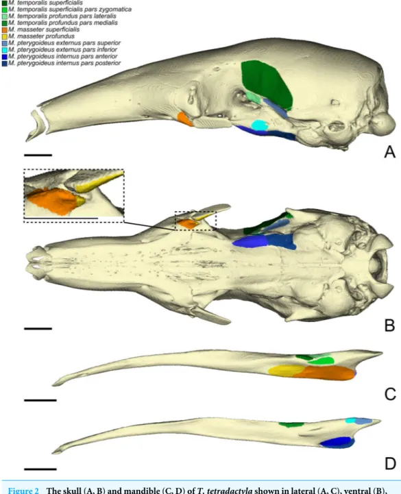
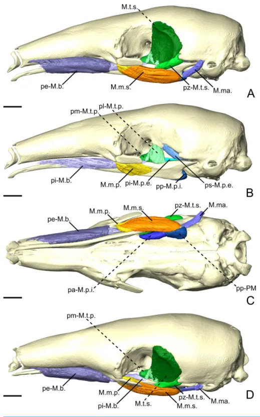
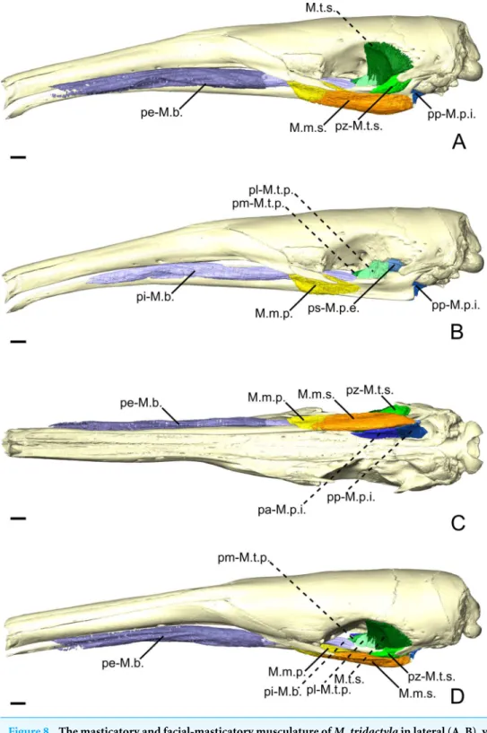
Hedrick et al., 2018). Below, we provide an anatomical description of the musculature of each of the three anteater species. Anatomical structures of the skull and mandible that are relevant to the description are depicted inFig. 1. The origins and insertions of the masticatory muscles are figured for one species (i.e., Tamandua tetradactyla) (Fig. 2) to serve as a reference and to complement the 3D reconstructions. Three-dimensional surface models of the illustrated specimens (Fig. S1) are freely available at MorphoMuseumM (http://www.morphomuseum.com;Ferreira-Cardoso et al., 2020).
Table 3 Facial-masticatory muscle volumes (mm3; left column) and percentages (right column)
ob-tained from the 3D models of the segmentation of the contrast-enhanced specimens.
Volume in mm3/facial-mast. volume (%)
Muscles C. didactylus T. tetradactyla M. tridactyla
pe-M.b. 17.9 25.7 371.9 27.4 990.4 15.1
pi-M.b. 49.9 71.6 911.1 67.2 5589.4 85.0
M.ma. 1.94 2.8 73.7 5.4 NA –
Total 69.7 100 1356.7 100 6579.8 100 Notes.
pe-M.b., M. buccinatorius pars externa; pi-M.b., M. buccinatorius pars interna; M.ma., M. mandibuloauricularis. Anatomical description
Cyclopes didactylus
Masticatory apparatus
M. masseter superficialis. The M. masseter superficialis (M.m.s.;Figs. 3A,Figs. 3CandFigs.
3D) is the only muscle of the masseter muscle complex present in C. didactylus. The M.m.s. is anteroposteriorly elongated and originates from the lateral surface of the zygomatic process of the maxilla (Fig. 1B). The jugal bone is absent in C. didactylus. The origin of the M.m.s. consists of a long and strong posteroventrally projecting tendon that covers the most anterior half of the M.m.s. The muscle fibers of this anterior part are slightly obliquely oriented and compose the pars anterior of the M.m.s. (pa-M.m.s.;Fig. 3). The pa-M.m.s. inserts laterally from the posterior part of the dentary pad (Fig. 1C) to the anterior margin of the condyle (Fig. 3B). The pa-M.m.s. presents a pars reflexa inserting on the ventromedial margin of the ascending ramus of the mandible extending anteroposteriorly the level of the anterior margin of the coronoid process to the level of the mandibular canal. This part is covered laterally by the tendon from which it originates. Posteriorly, the M.m.s. presents a distinct pars posterior (pp-M.m.s.;Fig. 3;Fig. S2A) with anteroposteriorly oriented fibers. The pp-M.m.s. shares the origin with the pa-M.m.s. The former covers the pa-M.m.s. posteriorly to the coronoid process and inserts on the angular process of the mandible (Fig. 3). Its pars reflexa is continuous with the pars reflexa of the pa-M.m.s. and almost reaches the most posterior point of the angular process.
M. masseter profundus The M. masseter profundus is absent in C. didactylus.
M. temporalis superficialis. The M. temporalis superficialis (M.t.s.;Figs. 3A,3B and3C)
is the largest of the four muscles of the temporal complex (Table 2). It is a fan-shaped muscle that originates from a scar along the dorsal edge of the temporal fossa (Fig. 1A, area in green). The temporal crest runs from the posterior end of the orbital ridge to the anterior surface of the root of the zygomatic process of the squamosal. A thick tendinous layer stretches from the origin of the M.t.s. and covers the posterodorsal part of the muscle. The M.t.s. is thinner at its origin and thicker at its insertion. The insertion is muscular on the dorsal tip and the dorsal part of the posterior margin of the coronoid process. An aponeurosis runs dorsoventrally along the anterior surface of the M.t.s. and completely covers the lateral and anterior surfaces of the coronoid process. The fiber fascicles of the
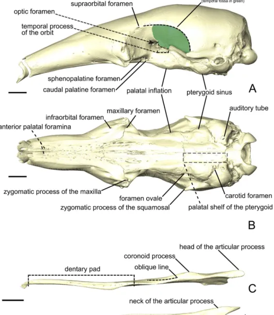
Figure 1 The skull (A, B) and mandible (C-E) of Tamandua tetradactyla shown in lateral (A) and ven-tral (B) views.The area in green delimits the temporal fossa. The mandible is shown in dorsal (C), medial (D), and lateral (E) views. Anterior is to the left. Scale bar 10 mm.
Figure 2 The skull (A, B) and mandible (C, D) of T. tetradactyla shown in lateral (A, C), ventral (B), and medial (D) views.The colored areas represent the origin (A, B) and insertions (C, D) of the mastica-tory muscles. A color–coded legend is provided.
Full-size DOI: 10.7717/peerj.9690/fig-2
M.t.s. are organized in a bipennate structure (Fig. S2B). Deep fibers are dorsomedially oriented while superficial ones are dorsolaterally oriented. In cross-section, the insertion angle of medial fibers with the axis of pennation is about 26◦, while lateral fibers present
an angle of around 12◦.
M. temporalis superficialis pars zygomatica. The M. temporalis superficialis pars zygomatica
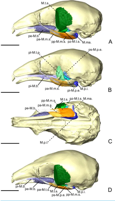
Figure 3 The masticatory and facial-masticatory musculature of C. didactylus in lateral (A, B), ventral (C), and dorsolateral (D) views. Scale bar 10 mm. The more superficial muscles were removed in B. Mus-cle abbreviations as inTable 1.
M.t.s.. It originates from the ventromedial part of the zygomatic process of the squamosal and broadens ventrally to end on an anteroposteriorly elongated muscular insertion. The insertion occupies the lateral part of the mandibular notch. The pz-M.t.s. is wider dorsally and thinner ventrally, with fibers presenting an oblique orientation.
M. temporalis profundus pars lateralis. The temporal complex includes a deep component
divided in two parts, the M. temporalis profundus pars lateralis pl-M.t.p.; (Figs. 3B,4Aand 4B) being the largest. The pl-M.t.p. takes its origin on a pseudo-elliptical area that extends from the posteroventral part of the orbital contribution of the frontal to the anteroventral part of the temporal fossa. The insertion of the pl-M.t.p. covers most of the posterolateral surface of the coronoid process, and narrows posteriorly along the mandibular notch. Contrary to the M.t.s., the pl-M.t.p. does not present a pennate structure, with fibers roughly vertically oriented.
M. temporalis profundus pars medialis. The M. temporalis profundus pars medialis
(pm-M.t.p.;Figs. 4AandFigs. 4B) consists of the inner part of the M.t.p. that takes its origin from the orbit, between the ventral edge of the temporal fossa and the optic foramen. The pm-M.t.p. and the pl-M.t.p. are clearly separated posteriorly on the insertion, with the posterior tip of the pm-M.t.p. occupying a more ventromedial position at the level of the mandibular foramen. Fiber orientation and shape of the pm-M.t.p. is similar to that of pl-M.t.p., but the former’s volume is about two thirds that of the latter. However, both muscles are anastomosed anteriorly.
M. pterygoideus externus pars superior. The M. pterygoideus externus pars superior
(ps-M.p.e.;Figs. 3B,4Aand4B) is a small anteroposteriorly elongated muscle. The ps-M.p.e. arises from a fossa that extends from the ventral part of the parietal, at the lower limit of the temporal fossa, into the glenoid fossa. It is the only part of the pterygoid muscle complex that takes its origin outside the pterygoid fossa. The muscle is mediolaterally compressed and obliquely oriented. Its posterior part presents a small torsion anterior to its ventrolateral projection towards the mandible. The insertion of the ps-M.p.e. consists of a small concavity just medioventral to the head of the articular condyle.
M. pterygoideus externus pars inferior. The pars inferior of the M.p.e. (pi-M.p.e.;Figs. 3B,
4A and4B) consists of a short and fleshy muscle strap. The pi-M.p.e. originates from a small area on the sphenoid, laterally to the foramen rotundum, and dorsally adjacent to the origin of the M. pterygoideus internus. The muscle is mediolaterally wide and presents a more horizontal orientation than the ps-M.p.e. The pi-BE projects posterolaterally to insert on the anterior margin of the articular condyle, at mid-height. The medial part of the pi-M.p.e. projects posteriorly, inserting below the insertion area of the ps-M.p.e., reaching the mid-length of the head of the condyle.
M. pterygoideus internus. The M. pterygoideus internus (M.p.i.;Figs. 3, 4A,4B and5)
arises from the pterygoid fossa and consists of a fleshy block that originates from the posterolateral part of the palatine to the level of the anterior margin of the ectotympanic
Figure 4 The M. pterygoideus and M. temporalis profundus muscle complexes of C. didactylus (A, B), T. tetradactyla (C, D), and M. tridactyla (E, F) in lateral (A, C, E) and dorsal (B, D, F).E and F are zoomed on the ascending ramus. Scale bar 10 mm. Muscle abbreviations as inTable 1.
Figure 5 The intermandibular musculature, M. geniohyoideus, and M. pterygoideus internus of
C. didactylus in lateral (A), ventral (B), and posteromedial (C) view.Only the left half of the M.
intermandibularisanterior is illustrated. A small vestige of the M. interstylohyoideus is also depicted. Scale bar 10 mm. Muscle abbreviations as inTable 1.
(Fig. 3Aand3B). Its fibers run anteroposteriorly with an oblique orientation and insert medially on the angular process of the mandible, from the level of the anterior margin of the head of the articular condyle to its posterior margin. In the most posterior part of their insertion, the fibers have a more posteroventral direction and form a small pars reflexa that wraps the posteriormost tip of the angular process. A dense connective tissue lies dorsal to the insertion of the M.p.i., posterior to the opening of the mandibular canal.
Facial-masticatory musculature
M. buccinatorius pars externa. The M. buccinatorius pars externa (pe-M.b.;Figs. 3A,3C
and3D) is distinguishable from the internal part of this muscle. It is a sheet-like muscle that envelopes the external surface of the M. buccinatorius pars interna, as well as the buccal salivary glands. Its origin stretches along the ventral edge of the maxilla and the palatine, from anteriorly to the inferior orbital foramen until the anterior part of the insertion of the M. pterygoideus internus. The ventral part of the pe-M.b. wraps the ventral portion of the M. buccinatorius pars interna (and the salivary glands, anteriorly) and attaches on a broad insertion area on the lateral surface of the mandible. The fibers have a dorsoventral orientation.
M. buccinatorius pars interna. The pars interna of the M. buccinatorius muscle (pi-M.b.;
Fig. 3BandFig. 3D) is more voluminous when compared to the pars interna. The pi-M.b. originates from a thin fiber bundle posterior to the buccal commissure and is covered by the pe-M.b. just posteriorly. The pi-M.b. is bordered by the salivary glands, ventrally and laterally, anterior to the level of the sphenopalatine foramen. The pi-M.b. is a long muscle that reaches as far posteriorly as the level of the coronoid process. It is characterized by a buccal projection that sits between the upper and lower jaws (Fig. 3D). The lateral part of the pi-M.b. contacts the pe-M.b. and does not attach to any bone surface. Posteriorly, the pi-M.b. inserts on the dorsomedial surface of the mandible, along the fossa located between the posterior part of the dentary pad and the coronoid process. Its insertion ends posterior to the coronoid process where it contacts the M. temporalis profundus pars medialis and the anterior part of the M. pterygoideus internus. The fibers of the pi-M.b. are anteroposteriorly oriented.
M. mandibuloauricularis. The M. mandibuloauricularis (M.ma.; Figs. 3Aand3C) is a
strap-like bundle that takes its origin on the anteroventral part of the auricular cartilage. The M.ma. projects ventromedially to insert on the posterodorsal edge of the angular process of the mandible. The insertion is small and is located between the posterior parts of the masseteric and pterygoid fossae of the mandible. The M.ma. fibers presents a mediolateral orientation with a strong ventral component.
Intermandibular musculature
M. intermandibularis anterior. The M. intermandibularis anterior (M.i.a.;Fig. 5) is a
thin, dorsolaterally wide, and elongated muscle.Naples (1999)described this muscle as the anterior part of the M. mylohyoideus pars anterior. The M.i.a. takes its origin on the
cartilage of the unfused mandibular symphysis. The muscle has two insertions on the ventrolateral margin of both hemimandibles, wrapping around their ventral edges. In ventral view (Fig. 5B), it covers the anterior part of the base of the tongue and the anterior part of the geniohyoideus (Fig. 5). The M.i.a. extends posteriorly for about half the length of the mandible, its posterior end being clearly separated from the anterior margin of the M. mylohyoideus pars anterior (see below). Its fibers are transversely oriented and are continuous between mandibles, with this muscle consisting of one single element.
M. mylohyoideus pars anterior. The M. mylohyoideus pars anterior (pa-M.mh.;Fig. 5)
consists of a fibrous sheet that originates ventrally to the dentary pad, on the medial surface of the mandible. This muscle is homologous to the pars medius of the M. mylohyoideus described byNaples (1999). The origin area stretches from the widest point of the dentary pad to its posteriormost point. Posteriorly, its origin shifts from the mandible to the ventromedial surface of the M. pterygoideus internus (M.p.i.). At the posterior end of the M.p.i. the origin changes again, creating a dorsolateral gap separating the anterior and the posterior fibers. We consider this to be the posterior limit of the pa-M.mh., with the posterior part being considered the M. mylohyoideus pars posterior. The fibers are transversely oriented ventrally and insert along a fibrous midline raphe that connects the left and right pa-M.mh.s (as inFig. S2C).
M. mylohyoideus pars posterior. The M. mylohyoideus pars posterior (pp-M.mh.;Fig. 5) is
continuous with the pa-M.mh. The division between the two parts is set by the difference of the origin. The pp-M.mh. takes its origin on the ventromedial surface of the tympanic bulla, parallel to the auditory tube. The fibers display the same orientation as in the pars anterior and insert on a fibrous midline raphe. However, near the posterior end of the hard palate, the left and right muscles appear to anastomose in the midline, with the intertonguing contact becoming less spaced. As the M. interstylohyoideus (Fig. 5) and the posterior part of the M. mylohyoideus pars posterior were not preserved in our specimens of C. didactylus, the attachment of the pp-M.mh. to the hyoid system is not visible.
Tamandua tetradactyla
Masticatory apparatus
M. masseter superficialis. The M.m.s. (Fig. 6A, Fig. 6C and Fig. 6D) is a fleshy,
anteroposteriorly long muscle; its anterior and posterior ends are angular in shape in lateral view. The fibers of the M.m.s. are slightly oblique and take their origin on the lateral surface of the zygomatic process of the maxilla through a strong tendon. The M.m.s. inserts on the shallow masseteric fossa of the mandible. It covers most of the lateral surface of the ascending ramus, including most of the more anterior M. masseter profundus (see below). The M.m.s. is thicker posteriorly, and thins down anteriorly as it overlies the M. masseter profundus. The tendon of the M.m.s. was not visible in the contrast-enhanced specimen. The M.m.s. presents a pars reflexa that runs from the level of the posterior part of the jugal to the posterior tip of the angular process of the mandible. Anteriorly, the M.m.s. presents a small projection towards the zygomatic process of the mandible.
Figure 6 The masticatory and facial-masticatory musculature of T. tetradactyla in lateral (A, B), ven-tral (C), and dorsolateral (D) views. Scale bar 10 mm.The more superficial muscles were removed in B. Muscle abbreviations as inTable 1.
M. masseter profundus. The M. masseter profundus (M.m.p.;Fig. 6B–D) is smaller than its superficial counterpart (M.m.s.). It takes its origin on the anterior part of the ventromedial surface of the jugal bone. Anteriorly, its origin area includes the most posteroventral surface of the zygomatic process of the maxilla. The fibers of the M.m.p. run obliquely to insert posteroventrally on the lateral surface of the mandible. The fibers are more vertical than those of the M.m.s. The muscle presents components with slight lateral and anterior orientations. The insertion area on the mandible stretches from the coronoid process to the level of the oblique line. Contrary to the M.m.s., the M.m.p. is thicker at its origin than at its insertion.
M. temporalis superficialis. The M.t.s. (Fig. 6AandFig. 6D) is one of the three muscles
that forms the temporal complex. It is also the largest, arising from a relatively large surface between the dorsal edge of the temporal fossa and the origin of the ps-M.p.e. (Fig. 6). It is wide and broad in lateral view, and transversely compressed. It presents a fan-like shape, the fibers converging ventrally towards the small and flat coronoid process. The M.t.s. is medial to a large lacrimal gland, which fills most of the temporal fossa. The lateral surface of the M.t.s. is covered by a thin tendinous layer. Ventrally, the M.t.s. inserts on the dorsomedial surface of the coronoid process via a large aponeurosis. The M.t.s. muscle fibers are oriented vertically in the anterior part of the muscle, and are more oblique posteriorly.
M. temporalis superficialis pars zygomatica. The pars zygomatica of the M.t.s. (pz-M.t.s.;
Figs. 6A,Figs. 6CandFigs. 6D) is a small fleshy strip on the ventral margin of the M.t.s. Unlike the M.t.s., the pz-M.t.s. originates on a small area limited to the ventral surface of the zygomatic process of the squamosal (Fig. 6). Its obliquely oriented fibers insert on the dorsolateral surface of the mandibular notch. While the insertion area and orientation of the fibers are distinct from the anterior part of the M.t.s., both muscles are anastomosed posteriorly to their mid-length.
M. temporalis profundus pars lateralis. The M.t.p. (Figs. 4C,4Dand6B) is divided into
two distinct parts. The pars lateralis (pl-M.t.p.;Figs. 4C,Figs. 4Dand6B) is a small fleshy block deep to the larger M.t.s. The pl-M.t.p. takes its origin from the crest formed between the anteroventral border of the temporal fossa and the groove for the ophthalmic vein and the oculomotor nerve (III) (orbital process). The pl-M.t.p. transversely widens from its origin to its insertion. Fiber orientation is similar to that of the anterior part of the M.t.s., although slightly more oblique in coronal view. The insertion of the pl-M.t.p. is short and extends from the mid-length of the mandibular notch to the anterior part of the coronoid process. It covers most of the dorsal surface of the mandible in width. While the insertion is mostly muscular, the pl-M.t.p. shares the aponeurosis with the M.t.s. anteriorly.
M. temporalis profundus pars medialis. The pm-M.t.p. (Figs. 4C,Figs. 4D,6Band6D) is
the smallest part of the temporal muscle complex. It has no insertion, as it anastomoses with the pl-M.t.p. posterolaterally, but both parts could be easily separated during dissection. The fibers of the pm-M.t.p. are vertically oriented. Their insertion is medial to that of the
pl-M.t.p. and extends from the level of the anterior tip of the pi-M.p.e. to the anterior margin of the optic foramen. The medialmost part of the pm-M.t.p. wraps the mandible medially to insert on its dorsomedial surface; it contacts the dorsal part of the pa-M.mh. (see ‘Intermandibular musculature’).
M. pterygoideus externus pars superior. The ps-M.p.e. (Figs. 4B,4C,4Dand 6B) is a
strap-like muscle that arises from an elongated fossa along the ventral limit of the temporal fossa. Its obliquely oriented fibers run posteriorly to medially wrap around the head of the articular condyle of the mandible (Figs. 4Cand4D). The insertion extends from the anterior part to the posterior tip of the blunt articular condyle. The ps-M.p.e. overlies the insertion of the pi-M.p.e. (see below).
M. pterygoideus externus pars inferior. Similarly to the ps-M.p.e., the pi-M.p.e. (Figs. 4C
and4D) has a strap-like shape. In contrast with its upper counterpart, the pi-M.p.e. takes its origin on the pterygoid fossa. Specifically, the origin of the pi-M.p.e. is a small flattened area on the lateral surface of the palatal inflation. Its fibers are obliquely oriented and insert dorsally on the neck of the condylar process of the mandible.
M. pterygoideus internus pars anterior. The M.p.i. is divided into two distinct parts. The
pars anterior(pa-M.p.i.;Figs. 4C,4Dand6C) takes its origin on the lateral and ventrolateral surfaces of the palatine sinus. The origin is muscular and spans from level of the caudal palatine foramen to an area just posterior to the origin of the pi-M.p.e., near the posterior limit of the palatal inflation. The fibers are more oblique anteriorly than posteriorly, and insert on the dorsal part of the pterygoid fossa of the mandibular ascending ramus. The posterior part of the pa-M.p.i. is thinner than the anterior part. The thick portion of the pa-M.p.i. serves as an attachment area for a small anterior projection of the pp-M.mh. (see ‘Intermandibular musculature’).
M. pterygoideus internus pars posterior. The pp-M.p.i. (Figs. 4C,4D,6Band6C) consists
of a fleshy block that takes its origin on an area located between the posterior part of the palatal inflation and the small fossa anterior to the pterygoid sinus. A coronal section shows that the fibers are obliquely oriented (Fig. S2D). The pp-M.p.i. presents a very small pars reflexathat extends from the anterior- to the posteriormost part of the pterygoid fossa of the ascending ramus, wrapping around the margin of the small angular process (Figs. 4C andFigs. 4D).
Facial-masticatory musculature
M. buccinatorius pars externa. The pe-M.b. (Fig. 6A,6Cand6D) is a thin sheet of obliquely
oriented muscle fibers that envelops the pi-M.b. and the buccal salivary glands. The muscle takes its narrow and anteroposteriorly elongated origin on the maxilla. Its posterior limit attaches just anteroventral to the zygomatic process of the maxilla. Its anterior part consists of a thin strap on the lateral surface of the maxilla, close to the lateral limit of the nasal cavity. The muscle wraps around the pi-M.b. and reflects medially to insert along the dorsal part of the lateral surface of the mandible. Its insertion is shorter than its origin, extending
from the level of the infraorbital foramen for the posterior two thirds of the length of the horizontal ramus.
M. buccinatorius pars interna. The pi-M.b. (Fig. 6B) is an elongated and fleshy muscle that
takes its origin just posterior to the buccal commissure on the ventral part of the lateral surface of the maxilla. The anterior part of the pi-M.b. has a thin projection of its dorsal part that wraps around the lateral border of the dentary pad, to project into the space between the upper and lower jaws. This part of the muscle contacts the salivary glands ventrolaterally. The pi-M.b. lateral surface is enveloped by the pe-M.b. anterior to the zygomatic process of the maxilla. The muscle fibers are horizontally oriented. Posteriorly, the pi-M.b. inserts on the dorsal surface of the mandible, at the level of the optic foramen. The insertion is laterally adjacent to that of the pm-M.t.p. It extends anteriorly to reach the level of the maxillary foramen. The orbital part of the pi-M.b. is flattened due to the presence of the large lacrimal gland, dorsally. Madially, it is limited by the presence of the pm-M.t.p.
M. mandibuloauricularis. The M.ma. (Figs. 6A,6C, and6D) is a small fleshy muscle with
a pseudocylindrical shape. It takes its origin on the anteroventral part of the auricular cartilage. The M.ma. narrows ventrally towards its insertion on a small area of the posterodorsal margin of the angular process of the mandible, between the insertions of the M.m.s. and the M.p.i. The M.ma. presents dorsoventrally directed fibers with a slight medial component.
Intermandibular musculature
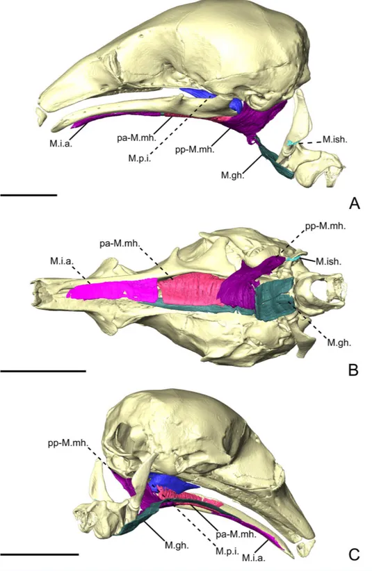
M. intermandibularis anterior. The M.i.a. (Fig. 7; pa-M.mh. sensuNaples, 1999) is a
sheet-like muscle that arises from the symphysial cartilage. The M.i.a. fibers are transversely oriented. They insert on both hemimandibles, covering the base of the tongue and the tendon of the geniohyoideus in ventral view (Fig. 7). The M.i.a. is, therefore, a single muscle with no bilateral counterpart (Fig. S2E). It wraps around the ventral margin of the mandible to insert just dorsal to it, on the lateral surface. The M.i.a. extends posteriorly for slightly more than half the length of the horizontal ramus of the mandible. Posteriorly, it is adjacent to the anterior margin of the pa-M.mh.
M. mylohyoideus pars anterior. The pa-M.mh. (Fig. 7) is a sheet-like muscle with
transversely oriented fibers, and covers the base of the tongue and the long tendon of the geniohyoideus (M.gh., not described). Its morphological similarities with the M.i.a. caused previous studies to describe the latter as a distinct part of the mylohyoideus complex (Naples, 1999). In contrast to the M.i.a., the pa-M.mh. insertion takes its origin on the ventral part of the medial surface of the mandible, between the widest point of the dentary pad and the pterygoid fossa posteriorly (Fig. 7). In addition to a different insertion, the pa-M.mh. is a bilaterally symmetric element, with both counterparts united medially by a small layer of conjunctive tissue (Fig. S2C). The pa-M.mh. is slightly thicker than the M.i.a. Posteriorly, the pa-M.mh. anastomoses with the pp-M.mh., the two parts being
Figure 7 The intermandibular musculature, M. geniohyoideus, M. interstylohyoideus, and M.
mas-tostyloideus of T. tetradactyla in lateral (A), ventral (B), and posteromedial (C) view. Only the left half of the M. intermandibularis anterior is illustrated. Scale bar 10 mm. Muscle abbreviations as inTable 1.
continuous. In coronal view, the division between the two muscles is characterized by the passage of the sublingual artery (Evans & De Lahunta, 2013), ventral to the pa-M.p.i. (Fig. S2F).
M. mylohyoideus pars posterior. The pars posterior of the M. mylohyoideus (pp-M.mh.;
Fig. 7) is broader than pa-M.mh. At the level of the orbital fissure, the sublingual artery (Evans & De Lahunta, 2013) splits the insertions of the pa-M.mh. and the pp-M.mh. While the pa-M.mh. inserts on the mandibular ramus, the insertion of the pp-M.mh. extends along the medial surface of the palatine inflation, then along the ventromedial surface of the pterygoid sinus to continue posteriorly to the level of the auditory tube (Fig. 7). Additionally, a thin muscular projection inserts on the medial surface of the pa-M.p.i. Posterior to the hard palate, the pp-M.mh. inserts on the soft palate, keeping its shape until it reaches the anterior part of the M. stylopharyngeus (not described), where it bifurcates. A fleshy fiber extension projects posteriorly to attach on a small area of the anterior surface of the stylohyal, just dorsal to its suture with the epihyal. On the other hand, a ventral sheet-like projection attaches to the tendon of the M. interstylohyoideus (M.ish., not described;Fig. 7). As in other cases, the tendon could not be segmented. Nevertheless, the presence of muscular fibers of the M.ish. confirm the position of the insertion of the pp-M.mh. described in previous studies (Reiss, 1997).
Myrmecophaga tridactyla
Masticatory apparatus
M. masseter superficialis. In M. tridactyla, the M.m.s. (Figs. 8A,8Cand8D) is a fleshy and
anteroposteriorly elongated muscle. The M.m.s. originates from the ventrolateral margin of the zygomatic process of the maxilla. A strong tendon connects the origin to the almost horizontally oriented muscular fibers. The M.m.s. is thin at the origin, as it overlies the posterior part of the M.m.p. It thickens posteriorly, as it extends anteriorly to the lacrimal foramen and the posterior part of the masseteric fossa. The M.m.s. presents a pars reflexa throughout most of its length (Fig. 8C). The pars reflexa wraps around the ventral edge of the mandible and becomes larger posteriorly, covering only the very posteroventral tip of the small angular process (Figs. 8Aand8C).
M. masseter profundus. The M.m.p. (Figs. 8B,8Cand8D) takes its origin on the anterior
part of the ventrolateral surface of the zygomatic arch. Its area of origin includes the small jugal bone and the posteroventral surface of the zygomatic process of the maxilla. The M.m.p. is in contact with the posterior part of the pi-M.b., medially (Fig. 8D). The M.m.p. is obliquely oriented; it inserts ventrally on the mandible and presents a small pars reflexa. The muscle is thick at its origin but thins down posteriorly, where it is overlain by the M.m.s. The M.m.p. is half the length of the M.m.s., with its insertion area stretching from the most anterior part of the masseteric fossa to near the level of the coronoid process.
M. temporalis superficialis. The M.t.s. (Figs. 8Aand8D) is a flat muscle covered almost
Figure 8 The masticatory and facial-masticatory musculature of M. tridactyla in lateral (A, B), ventral (C), and dorsolateral (D) views. Scale bar 10 mm. The more superficial muscles were removed in B. Mus-cle abbreviations as inTable 1.
fossa extending from the level of the optic foramen to the root of the zygomatic process of the squamosal. The lateral surface of the M.t.s. is covered by a tendinous layer that thickens ventrally near the insertion of the muscle on the small coronoid process. While the ventrally converging fibers of the M.t.s. reach the coronoid process posteriorly, the anterior part of the muscle inserts on the mandible uniquely via its tendinous layer (Fig. 8A). The M.t.s. is well-separated from the pars zygomatica, posteriorly, due to the very distinct orientation of the muscular fibers.
M. temporalis superficialis pars zygomatica. The pars zygomatica of the M.t.s. (pz-M.t.s.;
Figs. 8A,8C, and8D) is a fleshy and thick part of the M.t.s. complex. It arises from the medial and posteroventral surfaces of the zygomatic process of the squamosal and extends anteroventrally with an oblique orientation. The pz-M.t.s. displays a medial portion that extends along the anterior margin of the neck of the mandibular articular process and inserts on the posterior surface of the blunt coronoid process. The lateral part of the pz-M.t.s. is larger and extends along the surface lateral to the mandibular notch. The most ventral part of the pz-M.t.s. is slightly overlain by the dorsal margin of the M.m.s. The pz-M.t.s. is easily distinguishable from its larger counterpart due to the different orientation angle of its fibers.
M. temporalis profundus pars lateralis. The M.t.p. (Figs. 4E,4F,8B and8D) is divided
into medial and lateral parts. The pars lateralis (pl-M.t.p.) is a blocky-shaped muscle arising from the ventral limit of the temporal fossa between the anterior tip of the orbital process and the insertion of the ps-M.p.e. The posterior part of the pl-M.t.p. presents a quadrangular shape in lateral view, with the anterior part tapering in near the pi-M.b. The muscular fibers are dorsoventrally oriented with an oblique transversal component. The pl-M.t.p. inserts on the dorsal surface of the ascending ramus deep to the insertion of the M.t.s. While the M.t.p. is well separated from the M.t.s. during the classical dissection, the incomplete staining of the former makes it sometimes hard to delimit. Anteriorly, the insertion of the pl-M.t.p. extends until the level of the anterior margin of the optic foramen (Fig. 8B).
M. temporalis profundus pars medialis. The pm-M.t.p. (Figs. 4E, 4F, 8B and 8D) in
M. tridactylais a medioventrally extending projection of the pl-M.t.p. Both parts are anastomosed posteriorly, sharing the medial part of the M.t.p. origin. The pm-M.t.p. arises from the ventral surface of the orbital process lateral to the orbital fissure and the foramen rotundum. Slightly anterior to its origin, the pm-M.t.p. extends ventrally on the lateral surface of the ascending ramus (Figs. 4Eand4F). Anterior to this point, the two parts of the M.t.p. are distinguished by different insertion areas (Figs. 4EandFigs. 4F), with pm-M.t.p. reflecting medially. The insertion of the pm-M.t.p. is broad and extends ventrally almost until the level of the mandibular canal. It is limited posteriorly by the mandibular canal. The pm-M.t.p. tapers anteriorly to its contact with the posterior part of the pi-M.b. at the orbit mid-length. Fiber orientation in the pm-M.t.p. is similar to that of the pl-M.t.p.
M. pterygoideus externus pars superior. The ps-M.p.e. (Figs. 4E,4Fand8B) is a broad and wide fleshy sheet muscle arising from the large fossa extending from the anteroventral part of the squamosal to the ventral part of the temporal fossa. Its fibers are obliquely oriented and extend posteroventrally to insert on the mandible just anterior to the jaw joint. The posteroventral part of the ps-M.p.e. is characterized by a large pars reflexa that wraps around the medial edge of the articular process. The pars reflexa of the ps-M.p.e. overlays the posterior part of the pars inferior of the M.p.e.
M. pterygoideus externus pars inferior. The pi-M.p.e. (Figs. 4Eand4F) is a strap-shaped
muscle that originates from the anterior part of the pterygoid fossa, at the level of the optic foramen. In contrast with the ps-M.p.e., the pi-M.p.e. is narrow and elongated. Its origin is thin and lies medial to the pm-M.t.p. The anterior part of the pi-M.p.e. is in tight contact with the pa-M.p.i. The pi-M.p.e. slightly thickens up posteriorly, assuming a circular cross-section. The muscular fibers are horizontally oriented, with an oblique component as they insert posterolaterally on the anterior part of the neck of the articular process (Figs. 4Eand4F). The insertion of the pi-M.p.e. reaches about half the length of the neck and is overlain laterally by the pars reflexa of the ps-M.p.e.. The pi-M.p.e. merges with the pars reflexa of the ps-M.p.e. by a thick band of connective tissue.
M. pterygoideus internus pars anterior. The pars anterior (pa-M.p.i.;Figs. 4E,4Fand8C)
is the larger of the two parts of the M.p.i. It takes its origin from the small crest formed by the lateral edge of the palatine. In lateral view, the pa-M.p.i. presents a pseudorectangular shape. Anteriorly, the muscle narrows down (Figs. 4Eand4F). The most anterior fibers originate just anterior to the level of the optic foramen. The fibers extend ventrally to insert on a lateral prominence of the mandibular ascending ramus, ventral to the passage of the inferior alveolar nerve and artery. Posteriorly, the fibers are dorsoventrally oriented, with an oblique transverse component. Both origin and insertion of the pa-M.p.i. end roughly at the level of the pterygopalatine suture.
M. pterygoideus internus pars posterior. The smallest component of the M.p.i. is a fleshy
pseudorectangular band in lateral view (Figs. 4Eand4F). The origin of the pp-M.p.i. (Figs. 4E,4Fand8C) is very thin and extends from near the palatine-pterygoid suture to the pterygoid sinus at the level of the posterior limit of the jaw joint. The pp-M.p.i. is the continuation of the pa-M.p.i. until the tip of the angular process, where it reaches the insertion area of the M.ma. In lateral view, the fibers are vertically oriented, with a transversal component of about 21◦relative to the sagittal axis of the skull. Posteriorly, the
pa-M.p.i. becomes thicker but it tapers off abruptly at the level of the pterygoid sinus. Facial-masticatory musculature
M. buccinatorius pars externa. The pe-M.b. (Figs. 8A,8C, and8D) is an extremely thin
sheet enveloping the much thicker pars interna (see below) and the buccal salivary glands. The fibers of the pe-M.b. have an oblique orientation, arising from the long and extremely narrow origin on the maxilla. The origin extends from the level of the most posterior
mental foramen to the anterior edge of the zygomatic process of the maxilla. The pe-M.b. extends ventrally, envelopes the pi-M.b. and reflects medially. The muscle wraps around the ventromedial margin of the pars interna of the M. buccinatorius and projects dorsally to insert on the dorsolateral surface of the mandibular horizontal ramus. Its insertion and origin areas are similar in length, but the bad preservation of the soft tissues in the snout did not permit to clearly observe the anterior tip of its origin.
M. buccinatorius pars interna. The pi-M.b. (Fig. 8B) is extremely long anteroposteriorly,
reflecting the elongation of the rostrum. The muscle takes its origin on the maxilla, adjacent to the labial commissure of the mouth, although the muscular fibers arise more posteriorly. The pi-M.b. fibers go on to insert on the dorsal surface of the horizontal ramus of the mandible, ventral to the eye and the lacrimal gland. The fibers have an almost horizontal orientation, leaning slightly ventrally. In cross section, the anterior part of the pi-M.b. is dorsoventrally elongated. The most anterior part of the pi-M.b. presents a medial flap-like projection that rests between both jaws (Fig. 8B). This part of the pi-M.b. contacts the salivary glands laterally. At the length of the posterior most tip of the nasal, the pi-M.b. drifts ventrally and narrows dorsoventrally (Fig. 8B). Posterior to the zygomatic process of the maxilla, the pi-M.b. leans medially to a position between the jaws, deep to the M.m.s. This marks the beginning of the insertion of the pi-M.b., which extends to the anterior part of the insertion of the M.t.p., just anterior to the level of the optic foramen.
M. mandibuloauricularis. The M.ma. consists of a small fiber bundle that takes its origin
from the anterior part of the auricular cartilage. It inserts on the posterior tip of the angular process, between both the pp-M.m.s. and pp-M.p.i. This muscle was damaged on the digitally dissected side of the skull and was described based on its right counterpart. Intermandibular musculature
M. intermandibularis anterior. The M.i.a. (Fig. 9; pa-M.mh. sensu Naples, 1999) is
extremely elongated, extending for almost half the mandibular length (127.4 mm). This muscle is very thin and forms a sheet covering the tendon of the M. geniohyoideus as well as the tongue (not figured). Each fiber is attached to thin areas on the ventrolateral surfaces of both mandibles. The muscle wraps around the ventral margin of the mandible and stretches transversely to insert on the opposite side’s hemimandible. The fibers are continuous between mandibles.
M. mylohyoideus pars anterior. The postcranial muscles in our specimen were not
successfully stained by the iodine solution and, therefore, could not be illustrated and described (Fig. 9). The pa-M.mh. (Fig. 9; pm-M.mh. sensuNaples, 1999) is only partially stained and thus not completely represented in our 3D reconstructions. This muscle forms a thick sheet ventral to the tongue musculature. Its fibers are transversely oriented, connecting a midline of connective tissue to the medial surface of the mandible (Fig. 9). Both symmetric counterparts of the pa-M.mh. unite in the midline, but could be easily distinguished both during the classical and digital dissections. The pa-M.mh. is clearly
Figure 9 The intermandibular musculature of M. tridactyla in lateral (A), ventral (B), and posterome-dial (C) view.Scale bar 10 mm. Muscle abbreviations as inTable 1.
Full-size DOI: 10.7717/peerj.9690/fig-9
separated from the pp-M.mh. by a shift in the insertion from the mandible to the skull. The posterior end and the transition between the pa-M.mh. and the pp-M.mh. could not be segmented during the digital dissection.
DISCUSSION
Myological features and anteater systematics
External morphology has, for a long time, provided elements allowing extant anteaters to be split into two distinct groups (Pocock, 1924;Reeve, 1940;Hirschfeld, 1976;Patterson et al., 1992). Pygmy anteaters (Cyclopes spp.) are ascribed to a monogeneric family (Cyclopedidae) while tamanduas (Tamandua spp.) and the giant anteater (Myrmecophaga tridactyla) form the Myrmecophagidae (Fig. 10;Gibb et al., 2016). Although all anteaters present toothless and elongated jaws, this elongation is particularly pronounced in mymecophagids, reaching extreme proportions in the giant anteater (M. tridactyla). Pygmy anteaters present a shorter snout, a concave curvature of the basicranial/basifacial axis (Gaudin & Branham, 1998), pterygoids that do not meet in the midline, as well as relatively well-developed coronoid and angular processes of the mandible (Hirschfeld, 1976; Engelmann, 1985).
Figure 10 Mapping of muscular and osteological discrete traits in simplified phylogeny of Pilosa.Trait 1 refers to the absence of a maxilla-jugal-suqamosal functional unit providing a surface for muscular ori-gins; extant Pilosa all lack completely ossified zygomatic arches, but sloths present strong ligaments con-necting the jugal and the zygomatic process of the squamosal from which the M.
zygomaticomandibu-larisand the M. masseter profundus arise (Naples, 1985b). Traits 2–11 are based on cranial synapomor-phies, directly related to muscular origins/insertions, described inHirschfeld (1976),Engelmann (1985), andGaudin & Branham (1998). The tree was obtained from timetreeoflife.org (Kumar et al., 2017) and di-vergence times were modified according toGibb et al. (2016). Silhouettes correspond to one species within the tip taxon.
Full-size DOI: 10.7717/peerj.9690/fig-10
These, and other morphological traits, are considered ancestral for Vermilingua (Fig. 10;
Hirschfeld, 1976;Patterson et al., 1992). Reiss (1997;2001) also found differences between the head musculature of pygmy and myrmecophagid anteaters but overlooked those in the masticatory apparatus.
Our results reveal clear differences in the anatomy of the masticatory muscles of anteaters (Fig. 10). Contrary to myrmecophagids, the pygmy anteater shows a simple M. pterygoideus internus (M.p.i.) without subdivisions, a one-layered M. masseter (superficialis), and a relatively larger M. temporalis superficialis (M.t.s.) with a bipennate fascicular architecture (Fig. 10). Additionally, the posterior part of the M. mylohyoideus pars anterior (pa-M.mh.)
inserts on the ventromedial part of the M. pterygoideus internus, unlike in myrmecophagids (this study;Naples, 1999;Endo et al., 2007;Endo et al., 2017). Lastly, we show the existence of a two-part M. buccinatorius in the pygmy anteater, contradicting previous descriptions (Naples, 1985a;Reiss, 1997). These five traits are of potential systematic value but all were absent in previous comparative studies identifying phylogenetically polarised muscular traits (Reiss, 1997;Reiss, 2001).
The subdivision of the M. pterygoideus internus into two parts in myrmecophagids might be related to size, similar to the increase in the number of facial muscles in anteater species with longer rostra (Naples, 1985a). On the other hand, size differences between collared and giant anteaters does not affect the M. pterygoideus internus anatomy. The subdivision of this muscle might thus be a diagnostic trait within Vermilingua.
Reiss (1997)failed to identify a complex M. masseter (with deep and superficial muscles) in the Northern tamandua and the giant anteater. Our description of a two-unit masseter musculature in myrmecophagids supports the observations made byEndo et al. (2007)and
Endo et al. (2017), and resembles that of other mammalian groups (e.g., Turnbull, 1970;
Naples, 1985b;Endo et al., 1998;Cox & Jeffery, 2011;Sharp & Trusler, 2015). A single-unit masseter musculature is therefore an autapomorphy of Cyclopedidae. In the latter taxon, the muscle is attached to the maxilla by a long tendon (Figs. 3Aand3B). In addition to the lack of an M. masseter profundus (M.m.p.), C. didactylus displays a bipartite M. masseter superficialis(pa-M.m.s. and pp-M.m.s.;Figs. 3A,3C, and3D), while it is composed of a single block in myrmecophagids (Figs. 6A,6C,8Aand8C). The pa-M.m.s. in C. didactylus is distinguishable from an M.m.p. because: (i) it presents a pars reflexa, typically found in the M.m.s. (e.g.,Sharp & Trusler, 2015); (ii) it shares a single tendinous origin with the pp-M.m.s.; (iii) a two part M.m.s. with differently orientated muscle fascicles is described in other mammals (e.g.,Fig. 3A,Sharp & Trusler, 2015;Wille, 1954).
The temporalis complex is also quite distinctive between cyclopedids and
myrmecophagids, despite both families presenting deep and superficial muscles (contra
Reiss, 1997). The temporalis complex is twice as large in cyclopedids compared to myrmecophagids (Table 2). Robust jaw adductor muscles represent an ancestral condition within xenarthrans (Reiss, 2001). Therefore, the presence of large M. temporalis superficialis and profundus in pygmy anteaters is in line with other plesiomorphic musculoskeletal traits previously described (Hirschfeld, 1976;Engelmann, 1985;Reiss, 1997). The bipennate fascicular arrangement of the M. temporalis superficialis in the pygmy anteater (Fig. S2B) is an ambiguous trait. While it is unique to pygmy anteaters within Vermilingua, fiber pennation is not described in the sloth sister-group (Naples, 1985b). Nevertheless, the loss of bipennate fascicles in the M. temporalis superficialis might be an autapomorphic trait of myrmecophagids, given that other mammals present either bipennate or multipennate fiber arrangements (Woods & Howland, 1979;Taylor & Vinyard, 2009;Hautier, 2010). Curiously, the pars zygomatica of the M. temporalis superficialis is relatively smaller in C. didactylusthan in myrmecophagids (Table 2), suggesting that the posterior component of force of the temporalis complex is less important in pygmy anteaters.
In addition, to the differences listed above, we recognize, for the first time, the presence of an individualized M. intermandibularis anterior (M.i.a.) in the Vermilingua (Figs. 5B,
5B, and9B).Naples (1999)considered this muscle to be a part of the M. mylohyoideus (M.mh.). We show that M.i.a. is attached to the ventrolateral margin of the anterior part of the lower jaws (Figs. 5B,5B, and9B), which contrasts with the insertion area of the M.mh. Furthermore, we confirm that the M.i.a. is made of transversally continuous fibers. The pa-M.mh. and pp-M.mh. comprise two bilaterally symmetric muscles that join along a midline axis (Fig. S2C). A similar condition is found in sloths (Naples, 1986), as well as in other mammals like moonrats (Turnbull, 1970), nectarivorous bats (Wille, 1954), and humans (Gray, 1995).Turnbull (1970)uses two criteria to assign a M. digastricus pars anterior to the M.mh.: (i) the presence of intertonguing connection at the midline, and (ii) the contiguity of the attachment on the mandible. None of these conditions were found in the anteater ‘‘pa-M.mh.’’ (sensuNaples, 1999). Therefore, we propose to consider this muscle as the M.i.a. (Diogo et al., 2008). The pa-M.mh. (sensuNaples, 1999), the M. transversus mandibularisof rats (Greene, 1935), and the pa-M.mh. of tree-shrews (Le Gros Clark, 1924) are developmentally distinct from the M.mh. (Diogo et al., 2008). The muscle referred to byLe Gros Clark (1924),Greene (1935), andNaples (1999)is developmentally homologous with the sarcopterygian M.i.a. while the M.mh. is homologous to the M. intermandibularis posterior (Diogo et al., 2008). The M.i.a. muscle is mostly present in mammals with highly mobile mandibular symphysis, serving as a stabilizer (Hiiemae & Houston, 1971).
Overall, the results of our detailed descriptions and comparisons of the masticatory apparatus of anteaters provide several morphological traits that can be useful for systematics purposes. The previously unaccounted differences between the masticatory muscles of cyclopedids and myrmecophagids emphasize the level of morphological divergence acquired during the evolution of this clade with a highly specialized diet. We highlight the importance of soft-tissues as a source of diagnostic traits by combining conventional dissection with dice-CT (Metscher, 2009). Our results allow us to propose that a two part masseter musculature associated with a jugal bone and an unfused mandibular symphysis presenting an M. intermandibularis anterior are the plesiomorphic condition for Vermilingua. On the other hand, plesiomorphic architecture and relative size of the temporalis complex are impossible to predict, as these differ between extant sloth genera (Naples, 1985b) and data for armadillos (their xenarthran outgroup) are scarce and inconclusive (Kuhlhorn, 1939inTurnbull, 1970).
Mandibular mechanics
Regardless of the numerous differences discussed in the previous section, the masticatory apparatus of anteaters can be generally characterized by a set of adaptations to myrmecophagy like the complete tooth loss, the loss of masticatory capabilities (Naples, 1999), the reduction of masticatory muscles (Reiss, 1997;Naples, 1999;Endo et al., 2007;
Endo et al., 2017), and the unfused mandibular symphysis (e.g.,Ferreira-Cardoso, Delsuc & Hautier, 2019). The loss of chewing ability is well illustrated by the absence of the main mandibular abductor, the M. digastricus (e.g.,Turnbull, 1970;Hylander, Johnson & Crompton, 1987;Hylander, 2006) in all dissected specimens.