HAL Id: tel-03091962
https://tel.archives-ouvertes.fr/tel-03091962
Submitted on 1 Jan 2021HAL is a multi-disciplinary open access archive for the deposit and dissemination of sci-entific research documents, whether they are pub-lished or not. The documents may come from teaching and research institutions in France or abroad, or from public or private research centers.
L’archive ouverte pluridisciplinaire HAL, est destinée au dépôt et à la diffusion de documents scientifiques de niveau recherche, publiés ou non, émanant des établissements d’enseignement et de recherche français ou étrangers, des laboratoires publics ou privés.
Study of the role of the carotid sinus nerve in
inflammation : electrostimulation of the carotid sinus
nerve in mice attenuates inflammation by acting on the
hypothalamic-pituitary-adrenal axis and myeloid cells
Aidan Falvey
To cite this version:
Aidan Falvey. Study of the role of the carotid sinus nerve in inflammation : electrostimulation of the carotid sinus nerve in mice attenuates inflammation by acting on the hypothalamic-pituitary-adrenal axis and myeloid cells. Cellular Biology. COMUE Université Côte d’Azur (2015 - 2019), 2019. English. �NNT : 2019AZUR4019�. �tel-03091962�
1
Étude du rôle du nerf sinuso-carotidien
dans l’inflammation
L'électrostimulation du nerf sinuso-carotidien chez la souris atténue l'inflammation en agissant sur l’axe hypotalamo-hypophyso-surrénalien et les cellules myéloïdes.
Aidan FALVEY
Université Côte d’Azur, CNRS, Institut de Pharmacologie Moléculaire et
Cellulaire, Valbonne, France
Présentée en vue de l’obtention du grade de docteur en discipline d’Université Côte d’Azur
Dirigée par : Philippe Blancou/Nicolas
Glaichenhaus
Soutenue le : 2 avril 2019
Devant le jury, composé de :
Abdelilah WAKKACH, Président, LP2M, UCA, Nice Wouter DEJONGE, Rapporteur, AMC-UvA,
Amsterdam
Maria PEDRO GUARINO, Rapporteur, CEDOC, Lisbonne
Silvia CONDE, Examinateur, CEDOC, Lisbonne Philippe BLANCOU, Directeur de thèse, UCA, IPMC
2
Nom FALVEY... Prénom AIDAN ...
Numéro étudiant 21512902
Adresse email pérenne Aidan.FALVEY@univ-cotedazur.fr
Discipline Science de la vie
Laboratoire / équipe de recherche : Université Côte d’Azur, CNRS, Institut de Pharmacologie Moléculaire et Cellulaire, Valbonne, France
En français (obligatoire)
Titre de la thèse Étude du rôle du nerf sinuso-carotidien dans l’inflammation. Sous-titre de la thèse L'électrostimulation du nerf sinuso-carotidien chez la souris
atténue l'inflammation en agissant sur l’axe hypotalamo-hypophyso-surrénalien et les cellules myéloïdes.
Mots-clés Corps carotidien, inflammation, electrostimulation, souris, nerf sinuso-carotidien, endotoxémie, glucocorticoïdes, cellules myéloïdes, hypothalamus, médecine bioélectronique
Résumé (de 1700 à 4000 caractères espaces compris)
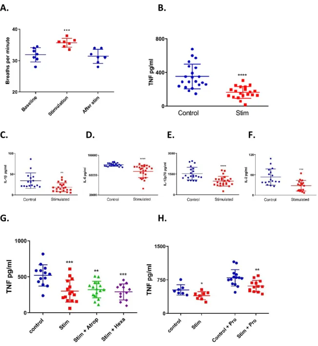
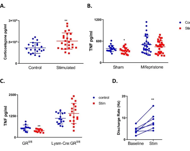
Les corps carotidiens sont des structures bilatérales situées au niveau de la bifurcation de l'artère carotide. Ils sont capables de détecter divers stimuli physiologiques tels que la concentration de gaz dans le sang et la pression sanguine. Suite à la variation de ces paramètres au-delà d’une valeur consigne, le corps carotidien, via un nerf afférent appelé nerf sinuso-carotidien (NSC), déclenche une réponses riposte par des voies nerveuses ou hormonales efférentes. De récentes études ont suggéré l’existence d’une relation entre le système immunitaire et le corps carotidien. Il a en particulier été démontré que le corps carotidien est capable de détecter des effecteurs de l’inflammation tels que les cytokines. En outre, des données in vivo démontrent que la résection bilatérale du NSC diminue la survie suite à un choc endotoxémique. Notre étude a porté sur les capacités immunomodulatrices du corps carotidien. Pour mener à bien cette étude, nous avons utilisé une approche d’électrostimulation du NSC plutôt qu’une une approche pharmacologique moins spécifique. Nous avons en particulier développé une méthode chirurgicale pour isoler le NSC, implanter des électrodes puis stimuler le nerf. Nous avons pu constater que la stimulation électrique du NSC atténuait l'expression des cytokines pro-inflammatoires induites par injection de lipopolysaccharide (LPS), en particuliers le Tumor Necrosis Factor (TNF). Par la suite nous avons pu montrer que l'effet de la stimulation du nerf sinusal de la carotide n’était médié ni par des nerfs sympathiques, ni par des nerfs parasympathiques efférents. En revanche, nous avons découvert que la stimulation du NSC augmentait le taux de corticostérone, une hormone anti-inflammatoire qui active le récepteur des glucocorticoïdes. L’antagoniste du récepteur des glucocorticoïdes, abolit les effets de l’atténuation des cytokines pro-inflammatoires par l’électrostimulation du NSC. De plus, l’utilisation de souris transgéniques ayant des cellules immunitaires myéloïdes dépourvues de récepteurs aux glucocorticoïdes, a montré que l’effet de la stimulation nerveuse du NSC était également perdu indiquant le rôle crucial joué par ces cellules et ce récepteur. Enfin, nous avons pu montrer que la stimulation du NSC conduisait à l’augmentation de l’activité de décharge spontanée des neurones hypothalamiques évoquant un lien entre le NSC et l’axe hypotalamo-hypophysaire. Au final,
Dépôt légal de la thèse
Données complémentaires
3
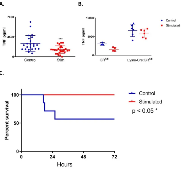
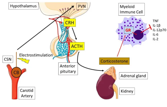
ces résultats indiquent que l’électrostimulation du NSC atténue l’inflammation en agissant sur l’hypothalamus qui, en augmentant la sécrétion de glucocorticoïdes, conduit à une inhibition de la sécrétion de cytokines inflammatoires par les cellules myéloïdes. D’un point de vu thérapeutique, nous avons pu mettre en évidence que les souris qui avaient reçu une électrostimulation du NSC avaient un taux de survive plus important après injection d’une dose élevée de LPS. Ces résultats suggèrent que l’électrostimulation du NSC pourrait être un traitement potentiel considéré par la médecine bioélectronique pour les maladies inflammatoires immunomédiées en agissant sur l’axe hypotalamo-hypophyso-surrénalien.
In English (mandatory)
Thesis title Study of the role of the carotid sinus nerve in inflammation Thesis sub-title Electrostimulation of the carotid sinus nerve in mice attenuates
inflammation by acting on the hypothalamic-pituitary-adrenal axis and myeloid cells.
Keywords carotid body, inflammation, electrostimulation, mouse, carotid sinus nerve, endotoxemia, glucocorticoids, myeloid cells, hypothalamus, bioelectronic medicine
Abstract (from 1700 to 4000 prints including spaces)
The carotid bodies (CB) are located bilaterally at the carotid artery bifurcation. They are polymodal sensors capable of detecting various physiological stimuli – blood gas concentration and blood pressure. The carotid body, via its innervating nerve – the carotid sinus nerve (CSN), signals to the brain to modulate these physiological stimuli through efferent activity. Recent evidence has suggested that there is a relationship between the immune system and the carotid body. It has been shown that the carotid body detects inflammation and functionally responds. Additionally, there is in vivo data, demonstrating that bilateral removal of the CSN decreases survival to endotoxemic shock. We hypothesized that activation of the carotid body would attenuate inflammation. To carry out this study, we electrostimulated the CSN instead of using a non-specific pharmacological approach. A surgical method was developed to isolate the CSN, implant electrodes and then stimulate the nerve. We found that electrical stimulation of CSN attenuated the expression of pro-inflammatory cytokines induced by lipopolysaccharide (LPS) injection, including Tumor Necrosis Factor (TNF). The mechanism by which electrical activation of the CSN attenuated inflammation was unknown. It was investigated if the effect of CSN stimulation was mediated via efferent parasympathetic or sympathetic nerves - it was found that it was not mediated via these nerves. In contrast, it was discovered, that CSN stimulation increased corticosterone – an anti-inflammatory hormone, which activates the glucocorticoid receptor. An antagonist for the glucocorticoid receptor abolished the attenuation of pro-inflammatory cytokines by CSN electrostimulation. This indicated that corticosterone is the mediator of the effect of CSN stimulation.In addition, we found that the stimulation of the CSN led to the increase of the spontaneous discharge activity of the hypothalamic paraventricular nucleus likening the CSN activity to the hypothalamic-pituitary adrenal axis. Using transgenic mice with no glucocorticoid receptor on their myeloid immune cell, it was found that effect of CSN
4
stimulation was additionally prevented. Finally, we were able to show that the stimulation of the CSN led to the increase of the spontaneous discharge activity of the hypothalamic neurons suggesting a link between the CSN and the hypothalamus-pituitary axis. Overall, these results indicate that electrostimulation of CSN attenuates inflammation by acting on the hypothalamus which, by increasing the secretion of glucocorticoids, leads to an inhibition of the secretion of inflammatory cytokines by myeloid cells. From a clinical perspective, using a high dose of lipopolysaccharide, it was found that mice who had received electrostimulation of the CSN were more likely to survive than control mice. This result is particularly interesting as it demonstrates that electrostimulation of the CSN may be a potential therapeutic in bioelectronic medicine for immune-mediated inflammatory diseases.
5
Acknowledgements
More than anything, here, I just want to say thank you to all the mice and congratulations to anytime you got lucky and bit me.
Then in lab, thank you’s to all, in particular Philippe and Glaichenhaus, but also a thank you in particular to Thomas Lorivel who was always kind and willing to help.
Friends and family you all did your part.
Oh, actually, I will thank the tv show Doctor Who for getting me interested in doing a PhD And yeah I will thank SignaLife as well. Cheers.
6
Table of Contents
Description
Page Number
French Abstract 2-3 English Abstract 3-4 Acknowledgments 5 Table of Contents 6-9 Abbreviation list 10 Guide to Introduction 11
Introduction
12-74
1.0.0 – Immune System 12-25 1.A.0 – Overview 121.B.0 – Innate Immune Response 13
1.B.i – Cells 13-15
1.B.ii – Cytokines 15-16
1.C.0 – Adaptive Immune Response 21-22
1.D.0 – Dysregulated immune system 23-24
1.E.0 – Summary 25
2.0.0 – Regulation of the Immune System: Hormones and Nerves 26-49
2.A.0 – Hormones and nerves introduction 26-27
2.B.0 – HPA – Overview 30
2.B.i – Pathway 30-32
2.B.ii – Instigating stimulus for hypothalamus 32-33
2.B.iii – Glucocorticoids 33-35
2.B.iv – Glucocorticoids in Medicine 35-36
2.C.0 – The Inflammatory Reflex 38
2.C.i – Vagus Nerve 38-39
2.C.ii – Afferent Sensors 39-41
2.C.iii – Efferent Activity 41-45
2.C.iv – Evolving theory 45-47
7
3.0.0 – Carotid Body 50-65
3.A.0 – Overview 50
3.B.0 – Anatomy 51-52
3.B.i – Molecular and cellular 52-53
3.C.0 – Physiological functions 55
3.C.i – O2 sensing 55-56
3.C.ii – Metabolism 56-57
3.C.iii – Blood pressure 57-58
3.C.iv – Immune system 58-63
3.D.0 – Summary 65
4.0.0 – Bioelectronic Medicine 66-74
4.A.0 – Overview 66-67
4.B.0 – Bioelectronic medicine and the vagus 68
4.B.i – Pre-clinical: Inflammation 68-69
4.B.ii – Clinical: Inflammation 69-70
4.C.0 – Bioelectronic medicine and the Carotid Body 71
4.C.i – Pre-clinical: Metabolism 71-72
4.C.ii – Clinical: Blood pressure 72-73
4.D.0 – Future of Bioelectronic Medicine. 74
Hypothesis & Aims
75
Introduction Illustrations
Illustration 1 17 Illustration 2 18 Illustration 3 19 Illustration 4 20 Illustration 5 28 Illustration 6 29 Illustration 7 37 Illustration 8 48 Illustration 9 54 Illustration 10 648
Paper
76-108
Abstract 77 Introduction 78-79 Results 80-84 Figures 85-87 Fig.1 88-89 Fig.2 90-91 Fig.3 92-93 Discussion 94-97 References 98-100 Supplementary Figures 101-108 SFig.1 101-102 SFig.2 103 SFig.3 104 SFig.4 105 SFig.5 106 SFig.6 107 SFig.7 108Paper finished
108
Additional Work
109-111
Methods 109 Results 110 SFig.8 110 SFig.9 111Discussion
112-129
Conclusion
130
9
Discussion Illustrations
Illustration 11 120 Illustration 12 121 Illustration 13 124 Illustration 14 127References
131-142
10
Abbreviation list
Acetylcholine ACh
Adrenocorticotrophic hormone ACTH
Adrenergic receptor AR
Adenosine tri-phosphate ATP
Autonomic nervous system ANS
Baroreceptor activation therapy BAT
Bilateral carotid neurotomy BCN
Blood brain barrier BBB
Carotid body CB
Carotid sinus nerve CSN
Collagen-induced arthritis CIA
Choline acetyltransferase expressing T-cells CHAT
Central nervous system CNS
Cortisol-releasing hormone CRH
Dendritic cells DC
Glucocorticoid-glucocorticoid receptor complex GCR
Glucocorticoid response element GRE
Glucocorticoid Receptor GR
Hypothalamic-pituitary adrenal HPA
Inflammatory bowel disease IBD
Immune mediated inflammatory disease IMID
Interferon IFN
Interleukin IL
Interleukin-1 receptor IL-1R
Lipopolysaccharide LPS
Muscarinic ACh receptor mAChR
Natural Killer cells NK
Nicotinic ACh receptor nAChR
Nucleus tractus solarias NTS
Pathogen-associated Molecular patterns PAMPS
Protein Kinase A PKA
Pro-opiomelanocortin POMC
Peripheral nervous system PNS
Parasympathetic nervous system PSNS
Pattern recognition receptors PRR
Paraventricular nucleus PVN
Rostral ventrolateral medulla RVLM
Suprachiasmatic nucleus SCN
Suppressor of cytokines signalling SOCS
Sympathetic nervous system SNS
Transforming growth factor beta TGFβ
T-regulatory cell Treg
Toll-like receptor TLR
Tumour Necrosis Factor alpha TNF
TNF receptor TNFR
Thyroid stimulating hormone TSH
11
Guide to the introduction
My introduction consists of four primary sections: 1. The Immune System;
2. Regulation of the Immune System; 3. The Carotid Body;
4. And Bioelectronic Medicine.
Within these four primary sections, there are numerous sub-sections. When first viewed, these sections appear quite disparate and while they are linked within the text, I felt some clarification was required.
As seen from the abstract, my investigation focuses primarily on the role of the carotid body and its regulation of the immune system. This, however, is investigated through the mechanics of bioelectronic medicine, which in this context means the use of external electrodes placed onto the carotid sinus nerve (the carotid body nerve) and stimulating with electricity.
Therefore, it was necessary to explore bioelectronic medicine in the introduction to examine its impact on current research and within the clinical. Additionally, it was crucial to give an overview on the immune system – though innate immunity and pro-inflammatory cytokines are the focus – and the carotid body – focusing on its relationship to inflammation.
The section on the regulation of the immune response is divided into two large sub-sections – the hypothalamic-pituitary-adrenal axis and the inflammatory reflex. The reason for this is because they are both non-immune based endogenous mechanisms for regulating inflammation. The inflammatory reflex was the initial basis for how we believed the carotid body would regulate inflammation. We know now that it is actually dependent upon the hypothalamic-pituitary-adrenal axis, so it was necessary to talk about these two mechanisms for attenuating inflammation.
Overall, the integration of all sections becomes clear in the “hypothesis and aims” sections which follows the introduction.
12
Introduction
1.A.0 – Overview
The immune system is one of the most diverse, complicated and fascinating systems within the body. The Immune system’s functional components contain some of the smallest proteins to a body-wide vasculature system. Nearly every disease and disorder the body can be afflicted with has an immune system component to it and with that immune component the hope for a cure. New functional information comes out every day about the immune system and it is still surprising and delighting the people who research it.
The immune system is historically divided into two responses of the same system – the innate immune response and the adaptive immune response. In reality, these systems are very much interwoven and the activity of both is typically required for a healthy immune response. Within the innate and adaptive immune response there is four major roles of the immune system: Detecting a threat; responding to a threat; remembering that threat and regulation of activity. These roles of the immune system will be addressed in the coming sections.
13
1.B.0 – Innate Immune Response
The components of the innate immune response are typically considered “the first responders” of the immune system because their primary role is the detection and initial response to a threat. To use the classic example: When we cut a finger, this gives external threats, such as bacteria, access to our bodies. There are resident components, cells, of the immune response that will detect this threat and signal to more components of the immune response to aid in the elimination of this threat and resolution of the immune response. These actions take place via the combined activity of cells and cytokines - which will be addressed in sections 1.B.i/ii respectively.
1.B.i – Innate Immune Response: Cells
There are multiple immune cells, universally referred to as white blood cells (WBC) or leukocytes, within the innate immune response with varied roles. These cells can be found in tissues, as resident cells, within the vasculature or residing in organs of the immune system – lymph nodes and the spleen. To understand the cells of the immune system, understanding their lineage is crucial. All immune cells, innate or adaptive, arise initially from pluripotent haematopoietic stem cells found in the bone marrow. The stem cells first divide into common lymphoid and common myeloid progenitors. These progenitors further sub-divide into functional immune cells. Typically, common lymphoid progenitors are associated with cells of the adaptive immune response, except natural killer (NK) cells which are cells of the innate immune response. Cells of the myeloid lineage are all part of the innate immune response. Aside from NK cells, in the innate immune response there is also mast cells, eosinophils, basophils, neutrophils, dendritic cells (DC) and monocytes [82] [Illustration.1].
Each individual cell of the innate immune response can also have varying states of maturity which augment their physiological response, however in brief each cell will be described. Neutrophils, basophils and eosinophils are characterised as granulocytes, due to the presence of granules within their cell bodies. Basophils and eosinophils are believed to be crucial in defence against large parasites - it is also believed mast cells play a similar role. Neutrophils are incredibly abundant, particularly when the innate immune response has been activated and are crucial to the clearance of an infection through various means. NK cells are
14
particularly effective at clearing viral infections. DCs are capable of activating the adaptive immune response, which will be discussed further in section 1.D.0. [156]. Monocytes, and their derivative – macrophages, will be discussed in detail.
Macrophages migrate during development to tissues and become resident immune cells for that tissue. Additionally, monocytes when travelling to a source of inflamed tissue may differentiate into macrophages [158]. Macrophages are often described as sentinel immune cells, because they are supremely capable at detecting the presence of a threat, either from the environment such as bacteria or damaging tissue such as cancer cells. Macrophages are equipped with multiple sensory molecules know as pattern-recognition receptors (PRR). These PRR detect certain regions well conserved on pathogens – pathogen-associated molecular patterns (PAMP)s, instigating the innate immune response. Perhaps the best categorised of these PRR are the Toll-like receptors (TLR). There are multiple TLR’s, some found on the macrophage cellular membrane and others within the cell [Illustration.2]. TLR4 can detect the PAMP lipopolysaccharide (LPS) a crucial component of gram-negative bacteria [2]. Synthetic components of LPS are often used experimentally to instigate the immune response. Once TLR4 recognises LPS, a complex network of signalling molecules on the cell surface and then internal signalling molecules start activating in a cascade, ultimately activating a pro-inflammatory transcription factor. In brief, LPS is bound by LPS-binding protein, which shuttles LPS to CD14 and facilitates the pairing of the two. CD14 performs a similar role for the TLR4 and MD2 receptor complex that recognises LPS and induces internal signalling. There are two internal signalling pathways – MyD88 dependent or independent pathway [Illustration.3]. The MyD88 dependent pathway uses TLR4/MD2 bound MyD88 to subsequently activate IRAK and TRAF signalling proteins to instigate transcription via NF-κB and AP-1. Whereas, the MyD88 independent pathway utilises TRIF and TRAF signalling proteins to activate pro-inflammatory transcription factor NF-κB and IRF3 [95]. NF-κB, as an example, will then translocate into the nucleus of the cell to generate the production of proteins that can recruit and activate other cells of the immune response [158]. These proteins are considered products of the immune response and are called cytokines and chemokines. LPS-induced production of the pro-inflammatory cytokines – TNF, IL-6 and IL-1β will be addressed in the following section (1.B.ii).
An additional response by macrophages upon activation is phagocytosis. The process of phagocytosis is whereby the macrophage, upon sensing a pathogen will encapsulate it within
15
itself and begin to degrade it. Interestingly, this process of pathogen degradation can augment the cytokine profile of macrophages, as macrophages display remarkable plasticity and can be activated in a pro-inflammatory state as described, but also into an anti-inflammatory state that promotes wound healing and resolution of the immune response [158]. Immune proteins, cytokines, will be discussed in this context of inflammatory states in the next section.
1.B.ii – Innate Immune Response: Cytokines
Cytokines are typically very small proteins involved with the immune response and can signal in an autocrine (self-activation), paracrine (local cell activation) or hormonal fashion (travelling through the vasculature). There are many varieties of cytokines routinely divided into families called interferons (IFN), interleukins (IL) and the tumour-necrosis factor (TNF) family. Within these family groups, cytokines have a wide variety of functions that can be simplified to as pro-inflammatory, anti-inflammatory and chemotactic. When cytokines are considered to have a chemotactic role – the recruitment of additional immune cells – they are labelled as chemokines. One such chemokine is CXCL1, which is produced upon LPS-induced NF-κB activation and recruits’ neutrophils to the infected tissue [155]. Anti-inflammatory cytokines, are essentially any cytokine that prevents further recruitment and/or activation of immune cells, occasionally aiding in wound healing. IL-10 is the primary anti-inflammatory cytokine as it is a potent inhibitor of macrophage function. Interestingly, this inhibition of macrophage function can be considered another activation state of macrophages as they still produce further anti-inflammatory cytokines [158]. Pro-inflammatory cytokines are broadly described as cytokines that promote further immune activity. IL-1β is a particularly potent pro-inflammatory cytokine: it is produced in a proto form by NF-κB transcription and requires cleavage by a separate PRR to be operational – NLRP3 [89]. IL-1β activates components of the adaptive immune response, macrophages and induces fever in the host to aid clearance of an infection – the sickness response [155]. Overall, however, cytokines are pleiotropic, meaning their function varies depending on the cell they activate and the presence of other cytokines in the vicinity [155]. IL-6 is an example of cytokine pleiotropy as it cannot be considered wholly a pro-inflammatory cytokine, as it has anti-inflammatory roles too [143]. IL-6 will induce the production of acute-phase proteins from
16
the liver that are involved in the immune response, but it also augments cells of the adaptive immune response towards an anti-inflammatory phenotype [143].
Another particularly important pro-inflammatory cytokine is TNFα (TNF) and it is routinely used as the gold-standard indicator of inflammation in immunological studies. TNF is produced during LPS-induced TLR4 activation of NF-κB, but it also produced by cells other than macrophages [21]. TNF was initially known as a protein capable of destroying tumours, however it is known now that this is not the sole role of TNF. Overall, TNF is a promotor of inflammation, however one key role is its ability to cause endothelial cells – found in blood vessels – to form looser junctions between themselves [155]. This enables the recruited cells, by chemokines, to leave the blood vessels and enter tissue in a process known as extravasation. It is known that a lethal dose of LPS will kill mice, however if you block TNF, the mice will survive [21]. This indicates that TNF is one of the primary mediators of lethality in an infection and egregious TNF can be dangerous, this will be addressed in section 1.D.0. Signalling for these pro-inflammatory cytokines is crucial. Immune cells express their receptors – IL-1β binds to IL-1 receptor (IL-1R), IL-6 binds to IL-6 receptor (IL-6R) complexed with GP130 to be functional and TNF binds to TNF receptor (TNFR) 1 and 2. IL-1β-induced activation of IL-1R uses the TRAF signalling pathway to activate downstream NF-κB or other pro-inflammatory transcription factor AP-1. Whereas, IL-6-induced activation of IL-6R will mobilise the JAK-STAT signalling pathway to engender gene expression. Similarly, to IL-1β TNF via TNFR2 will mobilise the TRAF pathway to ultimately activate pro-inflammatory transcriptions factors – NF-κB and AP-1. Additionally, however, TNF can activate TNFR1 to induce apoptosis in the activated cell [155]. These cytokines, however, are not the only pro-inflammatory cytokines critical to the innate immune response - a list summarising these cytokines was adapted from Turner et al [155], [Illustration.4].
17
Illustration 1: Immune cell lineage
All immune cells are derived from the one haematopoietic stem cell, which can further subdivide into lymphoid or myeloid progenitors. The lymphoid progenitor will give rise to the lymphocytes – B-cells, T-cells and natural killer cells. The myeloid progenitor will give rise to multiple different proto-myeloid cells culminating with – DCs, monocytes, macrophages, mast cells, basophils, eosinophils and neutrophils. Image made using Servier Medical Art.
18
Illustration 2: TLRs and their PAMPs
A representative table of the main TLR’s and a sample of the pathogens and PAMPs that activate the receptors. It should be noted that all TLR’s respond to more than one stimulus and the above table is representing primary stimuli for each TLR.
19
Illustration 3: TLR signalling
When TLR4 is activated by LPS it will induce a signalling cascade that is dependent or not on MyD88. Various signalling molecules are recruited: MyD88 signalling will predominantly induce the activation of pro-inflammatory transcription factor NF-κB to enhance transcription of pro-inflammatory cytokines; whereas, MyD88 independent signalling activates interferon regulatory factors to induce the expression of interferons – a specific type of pro-inflammatory cytokine. Image adapted from Lu et al, 2008.
20
Illustration 4: Cytokines
A table representing a sample of the cytokines crucial to the immune response. Representing both pro-inflammatory and anti-inflammatory cytokines. Chemokines are also present (IL-8). This table represents primarily information from humans, however most is transferable to rodents, except notably IL-8. This table is adapter from Turner et al, 2014.
21
1.C.0 – Adaptive Immune Response
The adaptive immune response can be considered the “memory” response of the immune system and is primarily concerned with remembering a threat and regulation of response. As outlined previously (section 1.B.i) the lineage of all cells in the adaptive immune response are lymphocytes. The common lymphoid progenitor becomes a B-cell, T-cell or natural killer cell in the bone marrow. B-cells mature in bone marrow, whereas for T-cells it is necessary to travel to the thymus to mature. Once mature, these lymphocytes (not including natural killer cells) reside in the organs of the immune system such as lymph nodes, the spleen and the lymphatic system. The lymphatic system primarily functions as a drainage system for extracellular fluid, however it is also as a vasculature system for WBCs with lymph nodes as the hubs for leukocytes. Mature B and T-cells are often described as naïve, because once activated they differentiate into their functional forms [20, 37].
DCs are at the interface of innate and adaptive immunity by playing a key role in the activation of T-cells. DCs, like macrophages, can phagocytose pathogens and degrade them internally. Once degraded, DCs present a small part of the pathogen on its cell surface – an antigen. Through chemotactic signals, DCs are signalled away from the infected area into the lymphatic system, where they present the antigen to any naïve T-cell they encounter, however only one T-cell will recognise this pathogen and mount the appropriate response [20, 37]. There are two primary subsets of T-cells – CD4+ and CD8+ T-cells. Both Subsets of T-cells are activated in the same way by DCs, however, once activated they perform different roles. CD8+ T-cells, once activated, are referred to at cytotoxic T-cells as they seek the antigen that was presented and kill the pathogen infected cell once encountered. CD4+ cells are described as the T-helper-cells as they orchestrate and direct a successful response by the immune system to a pathogen. Upon activation, CD4+ T-cells differentiate further into specific types of functional T-cells – Th1, Th2 and Tregulatory T-cells (Treg), which are defined by their activities. Th1 cells orchestrate the classically considered pro-inflammatory action of the immune response via production of TNF and IFN-γ, which is a potent activator of macrophages. Th2 cells also activate the immune system but are more supportive to the function of B-cells via IL-4 [56]. Treg cells produce immunosuppressive proteins such as IL-10 and are a crucial component of the overall immune response as overt immune activity can lead to damage and immune disorders which will be addressed in the following section 1.D.0.
22
B-cells, similarly to T-cells, respond to one antigen and once activated undergo clonal expansion, differentiate and start producing antibodies. B-cell antigen activation, however, is a little more complex than T-cell antigen activation and generally requires the aid of specific CD4 T-cells, rather than DCs. The antibodies produced by plasma cells selectively bind to their antigen, perhaps a small section of a bacterial cell wall and once bound, it enables multiple components of the immune response to better recognise pathogens and ultimately remove them. A fraction of these activated B-cells will become memory B-cells and remember this antigen to produce a more rapid response, should the immune system re-encounter the pathogen [20, 37].
23
1.D.0 – Dysregulated Immune Response
The immune system is designed to respond to an infection, react strongly and then return to homeostasis. This design is evident by the short life-span of neutrophils [106], the plasticity of macrophages, switching to a state of near quiescence [158], and the role of Tregs [81]. Tregs are distinguished from other T-cell subsets by their transcription factor FOXP3 and their keen ability to supress – pro-inflammatory cytokines, cytolysis, metabolic disruption and DC function [37]. Tregs produce copious quantities of anti-inflammatory cytokine IL-10 and transforming growth factor β (TGFβ). TGFβ can induce cell differentiation and proliferation: together these cytokines are crucial to the role of Tregs and their loss can exacerbate already established pathologies [37]. Interestingly, CD4+ T-cells can be induced to become a subset of Tregs and perform a similar role to natural Tregs [144]. Ultimately, the actions of Tregs and their production of anti-inflammatory cytokines induce quiescence in additional immune cells and encourage the resolution of inflammation.
It is crucial that the immune system can regulate its reactions, auto-immune disorders being a clear example of when the immune system fails. Auto-immune disorders are characterised by T and/or B-cells recognising a self-antigen and consequently mounting an immune response against that self-antigen. Myasthenia gravis is an example of an auto-immune disorder where antibodies are made against a component of neuro-muscular activity, which consequently, over time, prevents patients from moving their muscles and ultimately causes death. Alternatively, the immune system can fail by its inability to control and stop an immune reaction leading to chronic inflammation. Immune-mediated inflammatory diseases (IMID)s are defined by dysregulation of the immune response and frequently have continuously high levels of pro-inflammatory cytokines. Two examples of IMIDs are osteoarthritis and inflammatory bowel disease (IBD). Osteoarthritis is a disorder distinct of rheumatoid arthritis – which is an autoimmune disorder, however phenotypically they are similar with degradation of cartilage between joints. Mild symptoms of osteoarthritis are pain and swelling in joints, however this can become near insufferable and patients can have loss of movement in their joints. TNF is the crucial mediator of this disorder, due to chronically high levels. Blocking TNF can ameliorate osteoarthritis and improve symptoms; currently anti-TNF blocking drugs are used to treat osteoarthritis [21]. IBD, or more specifically, Chron’s disease is an IMID affecting the intestines that is associated with abdominal pain and
24
diarrhoea. The exact pathogenesis of IBD is unknown, however, it has been shown that chronic inflammation can induce a Chron’s disease-like disorder in mice. Furthermore, giving patients infliximab, an anti-TNF antibody treatment, can alleviate the symptoms of Chron’s disease [21]. It is evident from these disorders that homeostasis is required – too much regulation of activity can result in the development of auto-immune disorders and too little regulation can result in the development of IMIDs. A final example of an immune system disorder due to dysregulation is sepsis, which is a catastrophic failure by the immune system at regulation. Sepsis is defined by a life-threatening infection coupled with organ dysfunction. During the early stage of sepsis, there is a massive pro-inflammatory response, while in the later stage of sepsis there is considerable immunosuppression. Early or late sepsis are both potentially deadly. The immune system tries to correct the overt inflammatory reaction but overcompensates and leaves the patient at risk of a rebound infection [68]. Sepsis is a complicated disorder and researchers are still searching for a reliable means of treatment. Overall, regulation of the immune response prevents chronic inflammation and the development of IMID’s, which is crucial to maintain a healthy individual.
25
1.E.0 – Summary
In the previous sections the primary roles of the immune system – detect, respond to, remember a pathogen and regulation – have been described. It is now evident that resident tissue macrophages will detect pathogens, release chemokines to draw in additional cells, such as DCs, of the innate immune system. DCs will then in turn, phagocytose a pathogen and present the antigen on its cell surface to activate T-cells and ultimately B-cells to aid in the clearance of the infection. Additionally, it is apparent now that the resolution of this inflammatory response is crucial. If the immune system remains unchecked, there can be chronic inflammation, which may lead to IMID’s. These previous sections have described regulation of the immune system as if the immune system was entirely self-governing – this is not true. There are two main non-immune based endogenous mechanisms to regulate inflammation – the hypothalamic-pituitary adrenal (HPA)-axis and the inflammatory reflex, respectively acting via hormones and the nervous system. In the following section, these two mechanisms will be discussed and described in full.
26
2.A.0 – Hormones and nerves introduction
To pass information from one distant area of the body to another there needs to be communication. The two predominant means of distant communication in the body are via the endocrine system and the nervous system [Illustration.5].
The Endocrine system comprises a disparate conglomerate of hormone producing organs and glandular tissue. These hormones travel through the vasculature to distant endocrine organs, glandular tissue or act almost ubiquitously in the body. Hormones are predominantly either proteins or steroids – a derivative of cholesterol. Hormones can be further divided into hydrophobic and lipophilic groups which have their receptors respectively on the surface of a cell or internally. Typically, it can take minutes for hormones to reach their target area, and the desired effect can last up to days. The endocrine system as a whole is responsible for a myriad of functions within the body: Regulating reproduction; growth and development; sleep and the stress response. It can be said that the hypothalamus, an area of the central nervous system (CNS), is the primary controller of the endocrine system and this will be discussed further in sections 2.B.i/ii. In brief, the hypothalamus releases hormones typically in a circadian manner following or day-night cycle, however, separate hormones from the periphery can influence the release of their progenitor hormones. Typically, the neurons of the hypothalamus secrete synthesising hormones onto the pituitary gland – a peripheral zone of the hypothalamus responsible for hormone release into the periphery. An example of this is thyrotropin-releasing hormone released from hypothalamic neurons to activate the pituitary gland to secrete thyroid stimulating hormone (TSH). TSH will then activate the thyroid gland to produce thyroid hormones – thyroxine and triiodothyronine. These two thyroid hormones are crucial to the regulation of metabolism throughout the body, additionally they are essential during periods of development [84]. This is just one example of the crucial roles the endocrine system performs in the body.
The nervous system comprises the CNS and a network of peripheral nerves throughout the body known as the peripheral nervous system (PNS). These peripheral nerves are divided into the somatic nervous system and autonomic nervous system (ANS). The somatic nervous system is the domain of voluntary movement and these nerves are used for controlling the musco-skeletal system. The ANS is subtler and ultimately more complex - these are the nerves that control non-voluntary actions, such as continuous lowering of the heart rate via the vagus
27
nerve, which will be discussed in detail in section 2.C.i. The ANS can be further sub-divided into the sympathetic nervous system (SNS) and the parasympathetic nervous system (PSNS) [Illustration.6]. The SNS and PSNS are so named because respectively they rely on Norepinephrine or acetylcholine (ACh) as their primary. Frequently nerves of the SNS using norepinephrine are called catecholaminergic and nerves of the PSNS using ACh are called cholinergic. PSNS signalling via ACh generally activates either the nicotinic ACh receptor (nAChR) or the muscarinic ACh receptor (mAChR), whereas SNS signalling is generally via norepinephrine acting on the adrenergic receptors (AR) – α or βAR. Traditionally, the PSNS and the SNS can be said to control “rest and digest” and “fight and flight” responses respectively [Illustration.6]. In recent years, however, the roles of the PSNS and SNS have been determined to be more diverse and that these old divisions may not be wholly accurate. In this investigation, nerves will be regarded individually based on their function and efferent nerves will be addressed in greater detail in section 2.C.iii. The nervous system is more rapid than the endocrine system and generally its effects will be induced within seconds and be partially transient.
It is worth noting, however, the nerves described here so far are efferent nerves – nerves that carry information to the periphery. Alternatively, afferent nerves carry information to the CNS and are crucial for processing our sense of the world. It is possible for afferent nerves to be distinct sources from efferent nerves or run in the same nerve bundle as efferent nerves, but information going in opposite directions. Afferent nerves in the context of the vagus nerve will be addressed in greater detail in section 2.C.ii.
28
Illustration 5: Nervous and endocrine systems
A table demonstrating the primary differences between the nervous and endocrine systems as a means of communication within the body.
29
Illustration 6: PSNS and SNS
A. The parasympathetic nervous system typically arises from cranial nerves and travels to
numerous locations in the body to perform “rest and digest” responses. B. The sympathetic nervous system primarily arises from the spinal cord and synapses with one or more ganglions before arriving at its target location to induce “fight or flight” responses. Images taken from Servier Medical Art.
30
2.B.0 – The Hypothalamic-Pituitary Adrenal Axis Overview
The Hypothalamic-Pituitary Adrenal (HPA) Axis is the name of the functional relationship between the hypothalamus, the key regulatory organ of the endocrine system, and the adrenal glands – hormone-producing tissue found bilaterally above the kidneys. One outcome of this system is the controlled production and release of glucocorticoids: a class of hormones that when activating their receptor, glucocorticoid receptor (GR), induce a myriad of effects in the body [74]. The HPA axis plays a major role in multiple systems: the cardiovascular system; metabolism; the nervous system and during neonatal development for lung maturation. Of particular interest to this investigation is the effect that the HPA axis has on the immune system and will be addressed in section 2.B.iii. Bioactive Endogenous Glucocorticoids – corticosterone in mice and cortisol in humans – acutely attenuate the immune response and versions of glucocorticoids have been used to treat inflammatory conditions for 70 years [74]. Considering the potent effect of glucocorticoids, it is no surprise that externally manufactured and exogenous glucocorticoids – prednisolone, dexamethasone and more – are widely used in modern medicine today [118]. This will be discussed at length in section 2.B.iv. The HPA-axis is a key regulator of immune function and will be discussed in full in the following sub-sections.
2.B.i – The HPA axis: Pathway
As stated in the previous section, the HPA axis consists of the hypothalamus signalling to the adrenal glands to produce and/or release glucocorticoid. In reality, the process is more complicated with multiple other factors playing a crucial role in the HPA axis. To summarise: the hypothalamus releases cortisol releasing hormone (CRH), which acts on the anterior pituitary to release adrenocorticotrophic hormone (ACTH) and then goes through the vasculature to the adrenal glands and releases glucocorticoid [Illustration.7]. This pathway will be discussed in detail here.
The hypothalamus is a crucial component of the neuroendocrine system located at the base of the CNS and is only partially protected by the blood-brain barrier (BBB) [17], which is used to protect the CNS from peripheral stimuli. The overall architecture of the hypothalamus is the central out-growth of the CNS with a connecting stalk to the bulb of the pituitary gland –
31
anterior and posterior. The anterior and posterior pituitary glands are important neuroendocrine glands with a variety of roles and responsibilities for hormone production throughout the body. Within the hypothalamus, there is a topographically discrete region called the paraventricular nucleus (PVN) [154]. The PVN consists of neurosecretory regions – the magnocellular and parvocellular PVN. Both the magnocellular and parvocellular PVN have primary roles, however there is some overlap between their functions [154]. The magnocellular PVN primarily produces vasopressin, a hormone that can induce constriction in arterioles of the vasculature and encourages water reabsorption in the kidneys. Whereas, the parvocellular PVN is responsible for producing and releasing CRH. CRH is released at the end of the parvocellular PVN neurons into the hypophyseal portal vein system at the base of the median eminence. This portal vein system allows for release of CRH into capillaries and then direct transfer, via portal vessels, into the capillaries of the anterior pituitary [154]. Within the anterior pituitary, CRH will bind to its receptor on the surface of corticotropes, which will induce the synthesis and release of Pro-opiomelanocortin (POMC) – the precursor of ACTH – and release of ACTH [126].
The adrenal glands are located bilaterally above the kidneys and can be divided into adrenal medulla and the adrenal cortex. The adrenal medulla is a modified tissue of the SNS and can produce the typical neurotransmitters of the SNS – epinephrine and norepinephrine, which are released into the blood and not, strictly, used a neurotransmitter. Whereas, the adrenal cortex is glandular tissue and is divided into three functionally distinct zones: zona glomerulosa to produce mineralocorticoids; zona reticularis to produce sex hormones and zona fasciculata to synthesise glucocorticoids. ACTH will bind to its receptor, mineralocorticoid receptor type 2, in the cells of the zona fasciculata, which induces downstream signalling, leading to the activation of cAMP-dependent protein kinase A (PKA). PKA, once activated, can regulate free cholesterol to aid in the production of steroid hormones and ultimately aid in the release of glucocorticoids [52].
Glucocorticoid will then be released into the vasculature to search for the GR to exert its myriad of effects, which will be discussed in detail in section 2.B.iii. One particularly interesting aspect of the HPA axis is its self-regulation: It functions as a closed feedback loop. There is GR in the PVN and anterior pituitary, which, when activated by glucocorticoids, will block the synthesis and secretion of CRH and ACTH respectively [52]. This control of the CNS directly by glucocorticoids is partly because of their nature, but also because the
32
hypothalamus Is partially unprotected by the BBB. This allows multiple types of peripheral stimuli to influence the activity of the hypothalamus and will be addressed in the following section.
2.B.ii – The HPA axis: Instigating stimulus for the hypothalamus
The hypothalamus is in a somewhat unique position in the CNS, in that its activity can be regulated by the CNS, via nerves, or the periphery, via hormones and cytokines. The HPA axis, hypothalamic activity and ultimate glucocorticoid release, is regulated in a circadian manner. In humans, with a regular day-night routine, there is low glucocorticoid concentration in the blood by late evening and it rebounds after midnight to increase until peaking early morning [118]. The opposite is true in mice however, with their lowest levels during our morning. There is a region of the hypothalamus called the suprachiasmatic nucleus (SCN), which is responsible for the diurnal release of glucocorticoids. The SCN can track time autonomously but in order to function optimally it receives direct retinal innervation [62]. Additionally, the brainstem can send inputs to the hypothalamus to regulate activity [154]. These are the primary means by which the CNS regulates the hypothalamus, peripheral stimuli influencing the hypothalamus will now be discussed.
In the literature it has been well described that a variety of peripheral stimuli can affect the activity of the hypothalamus [154]. The focus will be the specific regulation of CRH and ACTH in response to cytokines. Moreover, as multiple cytokine receptors and cytokines can have an influencing effect on CRH/ACTH secretion, the focus will be on IL-6, TNF and IL-1β. Within the hypothalamus there is expression of the mRNA of IL-6 and IL-6R [51] and there are binding sites for IL-6 in the anterior pituitary [116]. Furthermore, IL-6 expression is enhanced in both the hypothalamus and the pituitary following stimulation by LPS [90]. It has been demonstrated that IL-6 will increase the release of ACTH in vivo in rats [97], this increase remaining evident up to 24 hours later with the concentration of glucocorticoid also enhanced compared to control stimuli [61]. Interestingly, however, it is not indicative that IL-6 will increase the mRNA of CRH or POMC [61]. This result, perhaps indicates that IL-6 acting on its receptor, does not induce the production of CRH or ACTH, but only affects the release of already stored ACTH. The receptor for TNF has been discovered in two cell lines representing the pituitary [80] and TNF expression is found in the hypothalamus and pituitary gland in vivo
33
after LPS injection [90]. In rats it has been demonstrated that TNF will increase the concentration of both ACTH and glucocorticoid in the blood. Furthermore, TNF will increase the secretion of CRH from primary hypothalamus cells tested in vitro and the increase of ACTH and glucocorticoid is blocked by CRH anti-serum [8]. This result indicates that TNF is acting primarily on the parvocellular PVN cells to release CRH, rather than directly on the pituitary. IL-1R was discovered in the murine pituitary gland [119] and LPS will increase the presence of IL-1β in the hypothalamus and pituitary [90]. Moreover, IL-1β can directly increase the mRNA of CRH and ACTH, as well as the secretion of ACTH and glucocorticoid even up to 24 hours after initial IL-1β injection into rats [61].
The relationship between 1β and the hypothalamus is particularly interesting, because IL-1β is one of the primary cytokines responsible for fever induction and the hypothalamus regulates temperature in the body. Researchers have investigated this relationship from multiple angles and this effect was crucial to the discovery of the inflammatory reflex, which will be discussed in section 2.C.ii. It is worth stating that there is a wealth of literature on cytokines affecting HPA activity and what is presented is only representative of the whole, while conflicting data can be found, it is well established that cytokines play a large role in the regulation of HPA activity [154].
2.B.iii – Glucocorticoids
Glucocorticoid is a steroid hormone that binds to the nuclear GR. The GR is almost ubiquitously expressed in the body; however, the receptor has multiple isoforms – GRα, GRβ, GRγ, GR-A and GR-P. Furthermore, within these isoforms of GR there are further subdivisions. Each isoform plays a functionally different role, but in this investigation, unless otherwise stated, GR will be considered as a whole and weaker or negative regulation of glucocorticoids by different GR isoforms will not be focused upon [74]. When describing the functional signalling of glucocorticoids GRα is used as the prime example as it is the main mediator of glucocorticoids effects [74]. How glucocorticoid via GR regulates inflammation will be the focus of this investigation with a brief examination of general GR activity.
The importance of GR signalling is particularly evident in the two glucocorticoid based disorders – Cushing’s syndrome and Addison’s disease. Cushing’s syndrome is marked by increased cortisol levels in the blood, potentially because of continuous increased production
34
of CRH or ACTH. The phenotypical symptoms of Cushing’s syndrome are obesity, high blood pressure, impaired immunological function and moon face [74]. Whereas, Addison’s disease is characterised by chronically low levels of cortisol, generally because the adrenal glands are functioning sub-optimally. Addison’s disease is characterised by weight loss, low blood pressure, nausea, diarrhoea and occasionally the development of auto-immune disorders [74]. It is evident that glucocorticoid signalling requires balance in the body and its dysregulation can cause damage beyond the immune system.
Glucocorticoids and the immune system have a complicated relationship that cannot be stated as just a suppressor of immune activity. The impact of GR activation varies from cell to cell and even cell maturity [32]. The focus in this investigation will be to examine the anti-inflammatory role glucocorticoids impart on macrophages, however in brief additional effects will be described. T-cells require GR to undergo successful clonal expansion and survival [23] and, additionally, there is a controversial role, as yet not wholly elucidated, for GR in T-cell maturation in the thymus [32].
Glucocorticoids activating the GR, found internally in the cytoplasm of a cell, can attenuate inflammation in a genomic and non-genomic fashion. The genomic method for attenuation of inflammation is via transcription of anti-inflammatory proteins. Whereas, the non-genomic methods are mRNA destabilisation, post-transcriptional modifications to proteins or interference with other transcriptional factors binding to DNA [112, 7]. The glucocorticoid-GR complex (GCR) can bind to AP-1, a transcription factor involved in the pro-inflammatory immune response and prevent it from initiating transcription [24]. Additionally, the GCR can block the activity of NF-κB by binding to it [131]. This blocking of typical transcription factors for pro-inflammatory cytokines is interesting because of its speed which highlights the potent anti-inflammatory effect of GCR, which in-turn is further compounded by its genomic activity. Glucocorticoid once bound to the GR can translocate to the nucleus and depending on the glucocorticoid response element (GRE) available different genomic activity will take place. GRE’s can have a positive or negative role. Typically, positive GRE’s will perform gene transcription, whereas negative GRE’s will block it [112]. Positive GRE signalling can be further sub-divided into composite, tethering and simple. Tethering and composite GRE responses can either enhance or block the activity of additional transcription factors [74]. Whereas positive GRE response can increase the expression of powerful anti-inflammatory cytokine IL-10, IκBα – an inhibitor of NF-κB and suppressor of cytokine signalling (SOCS) proteins which
35
can block the signalling molecules of multiple pro-inflammatory cytokines - as well as additional anti-inflammatory proteins [7].
The anti-inflammatory effect of the GCR cannot be overstated, one key experiment to demonstrate its physiological effect on myeloid cells was seen using Lysm-Cre:GRfl/fl mice –
mice with no GR in their myeloid cells. A lethal dose of LPS was administered to Lysm-Cre:GRfl/fl mice and their GRfl/fl negative littermates which demonstrated that the mice lacking
GR had dramatically reduced survival in comparison to their littermate controls. Furthermore, the Lysm-Cre:GRfl/fl mice had significantly higher levels of pro-inflammatory cytokines such as
TNF [13]. The varied and potent anti-inflammatory role of endogenous glucocorticoids has now been outlined: exogenous glucocorticoids produce similar results, though in diverse capacities. In the following section exogenous glucocorticoid use in medicine will be discussed.
2.B.iv – Glucocorticoids in Medicine
Glucocorticoids have been in use since the late 1940’s in medicine: Today the use of glucocorticoids is one of the most widely prescribed drugs. Depending on the desired treatment, different forms of glucocorticoids can be used – hydrocortisone, prednisolone and dexamethasone. Hydrocortisone is the closest synthetic to human cortisol in activation potency and is considered low potency with 20 mg and with a half-life of 8-12 hours. Prednisolone is considered medium strength as its potency is 4 times greater than that of hydrocortisone at a 5 mg dose and a half-life of 12-36 hours. Whereas, dexamethasone is considered high potency as its anti-inflammatory activity is 30 times stronger than that of hydrocortisone at a dose of 0.75 mg and has a long half-life of 36-72 hours [118]. These are just examples of the many forms of synthetic glucocorticoids which can be for short- or long-term use. Additionally, glucocorticoids can be administered orally, topically, inhaled or injected intra-articularly. While, synthetic glucocorticoids are best known as medication against the immune response, there are some strictly non-immune related disorders treated by glucocorticoids. Sufferers of Addison’s disease generally require continuous administration of synthetic glucocorticoids such as hydrocortisone. Ambulatory surgery, or day surgery, is frequently complicated by post-operative and post-discharge nausea and vomiting, for which orally administered dexamethasone can give a sense of well-being, stimulate appetite and
36
reduce the occurrence of nausea or vomiting [147]. Routinely, glucocorticoids are used for their anti-inflammatory properties after surgeries and particularly for solid-organ transplants [96]. Furthermore, glucocorticoids are used for a large range of inflammatory disorders – sudden severe auto-immune disorder, respiratory infections and disorders, inflammatory skin conditions, but also IMID’s such as non-infectious colitis and arthritis [150].
The benefit of glucocorticoids is undoubtable, however there are risks associated with long-term use. There is considerable evidence for adverse effects due to long-long-term high-dose use of glucocorticoids [118], but even low-dose medium-term (>60 days) usage of glucocorticoids can cause adverse effects. In a study based in the United States with 2 446 active participants, 90% of the participants reported an adverse effect, with 55% stating it was bothersome. The predominant adverse effect was weight gain – 70% of all participants reported this outcome, but skin bruising and acne were also commonly reported. More serious conditions such as cataracts and fractures were also reported in a minority of the participants [35]. Long-term, high dose glucocorticoid use is a risk-factor for osteoporosis, T2D, hypertension, infection and iatrogenic Cushing’s syndrome [118]. Iatrogenic Cushing’s syndrome is an artificial or induced form of Cushing’s syndrome due to high-dose glucocorticoid treatment, the symptoms of which are like that of the physiological Cushing’s syndrome, but without the underlying pathology. Alternatively, there is also a risk of the opposite – iatrogenic adrenal insufficiency. Within the hypothalamus, as discussed, there is GR which glucocorticoids can bind to with the same efficiency as their target area. This activation in the hypothalamus performs the role of activating the negative feedback loop and prevents the production of CRH and ACTH, ultimately not producing natural glucocorticoids. This is a concern for patients who are going off long-term high dose usage of synthetic glucocorticoids, as it is possible a reset in the basal levels of cortisol has occurred [118]. Glucocorticoid administration can additionally be a risk factor for the immune system [59], even generating apoptosis in nearly all cells of the immune system in certain conditions [59, 173].
Overall the HPA axis and glucocorticoids are invaluable suppressors of the immune response, both physiologically and pharmacologically.
37
Illustration 7: The HPA axis
The HPA-axis consists of neurons of the PVN, found in the hypothalamus will release CRH into the hypophyseal portal vein system. There, released CRH will immediately arrive at the anterior pituitary to activate ACTH-secreting cells. Subsequently, ACTH is released into the blood and travels to the adrenal glands to induce the release of corticosterone. Corticosterone will then induce its myriad of effects on the body and potentially feedback on the hypothalamus and control its own regulation.
38
2.C.0 – The Inflammatory reflex overview
It is widely known that there is some relationship between the immune system and the nervous system, other than the activity of the hypothalamus, as immune cells have functional receptors for neurotransmitters. Sympathetic activity, via NE, in particular has been known for decades to regulate immune functions – generating a shift from Th1 to Th2 T-cells and discouraging pro-inflammatory activities [43]. The true control the PNS wields over the immune system with the inflammatory reflex was not yet known. It was less than 20 years ago that Kevin Tracey published the initial result that demonstrated the existence of the inflammatory reflex [152] and his team has been involved in practically every major result in this field since [3]. Prior to Tracey’s work on the inflammatory reflex, there was evidence of this connection between nerves and the immune system but it had not been amalgamated. In this investigation, due to the relatively recent nature of these discoveries, the inflammatory reflex will be outlined in a historical fashion in sections 2.C.ii/iii.
In brief the inflammatory reflex, or the cholinergic anti-inflammatory reflex, is characterised by peripheral vagal afferent nerves detecting inflammation and sending this information to the CNS. Consequently, the CNS activates vagal efferent nerves, which synapse at the celiac ganglion to activate adrenergic nerves to the spleen, which in turn releases norepinephrine and ultimately attenuates inflammation [3]. The truly interesting aspect of the inflammatory reflex is that while it is a physiological reflex, it can be induced by electrical activation. In this section of the investigation, the inflammatory reflex will be regarded as a pure physiological reflex and, though electrostimulation, will be mentioned as means of activating the reflex. The evoked potential of electrical activation in the vagus will not be discussed until the bioelectronic medicine section 4.B.i-ii.
2.C.i – The Inflammatory Reflex: Vagus
The vagus nerve is the tenth of twelve cranial nerves, a group of ANS nerves that arise directly from the brainstem. It has an important role in multiple systems aside from the inflammatory reflex. Anatomically, the vagus can be found bilaterally running parallel to the trachea at the cervical level of intervention and additionally, sub-diaphragmatically, it runs parallel to the oesophagus. Furthermore, the vagus nerve splits and separates into various smaller nerves
39
and innervates most major organs – the heart, lungs, liver, stomach, intestines, pancreas [11], non-organ ganglions – like the coeliac ganglion [125] and potentially, via the coeliac ganglion, the spleen [3]. Continuous vagal efferent stimulation inhibits the beat rate of the heart, which physiologically runs around 100 beats per minute. The efferent parasympathetic tone from the vagus keeps the heart rate closer to 60 beats per minute and this is perhaps the best characterised activity of vagal efferent fibres. It should be noted however that up to 90% of fibres within the vagus are afferent [11]. As widely dispersed as the vagus is throughout the body, at most nerve endings there is a sensor for the local environment, which will detect stimuli and induce afferent activity. In the lungs, there are neuroepithelial bodies which can function as mechanosensors for the CNS, which signal through vagal afferent fibres [91]. These mechanosensors can inform the CNS on breath rate and potentially induce an adjustment in the response. Additionally, there is a specific form of mechanoreceptors – baroreceptors, for measuring blood pressure – located in the aortic arch, which signal via afferent vagal fibres. Furthermore, there is also some evidence of chemoreceptors located in the rat aortic arch, though there is no evidence of what they are specifically detecting [26]. These diffuse vagal afferents re-combine with the core vagus nerves sub-diaphragmatically or at the cervical level, before the vagus arrives at the brainstem of the CNS and more specifically the nucleus tractus solarias (NTS) [47].
Interestingly, at the terminal of vagal afferents there are occasionally small tissues know as paraganglia. Paraganglia are non-neuronal cells derived from neural tissue during development. Vagal paraganglia are located densely at the cervical level, near the liver and comparatively sparsely near the pancreas. Paraganglia act as sensors of their environment and induce activity in the vagal afferent fibres [11]. Their specific role in regard to the inflammatory reflex will be outlined in the following section.
2.C.ii – The Inflammatory Reflex – Afferent sensors
In this section, afferent vagal paraganglia will be discussed from a historical perspective. During the mid-1990’s, researchers were particularly interested in how IL-1β could induce the fever response – there were two hypotheses. The first was that IL-1β travelled to the CNS and acted on receptors there – particularly in the hypothalamus [12], in a similar manner to as outlined in section 2.B.ii. The second hypothesis was that IL-1β was being detected in the