HAL Id: tel-02394442
https://hal.univ-lorraine.fr/tel-02394442
Submitted on 4 Dec 2019HAL is a multi-disciplinary open access
archive for the deposit and dissemination of sci-entific research documents, whether they are pub-lished or not. The documents may come from teaching and research institutions in France or abroad, or from public or private research centers.
L’archive ouverte pluridisciplinaire HAL, est destinée au dépôt et à la diffusion de documents scientifiques de niveau recherche, publiés ou non, émanant des établissements d’enseignement et de recherche français ou étrangers, des laboratoires publics ou privés.
Mechanisms of S-nitrosothiols intestinal permeability
and NO store formation within vascular wall to improve
NO oral delivery systems
Yi Zhou
To cite this version:
Yi Zhou. Mechanisms of S-nitrosothiols intestinal permeability and NO store formation within vascular wall to improve NO oral delivery systems. Pharmaceutical sciences. Université de Lorraine, 2019. English. �NNT : 2019LORR0101�. �tel-02394442�
AVERTISSEMENT
Ce document est le fruit d'un long travail approuvé par le jury de
soutenance et mis à disposition de l'ensemble de la
communauté universitaire élargie.
Il est soumis à la propriété intellectuelle de l'auteur. Ceci
implique une obligation de citation et de référencement lors de
l’utilisation de ce document.
D'autre part, toute contrefaçon, plagiat, reproduction illicite
encourt une poursuite pénale.
Contact : [email protected]
LIENS
Code de la Propriété Intellectuelle. articles L 122. 4
Code de la Propriété Intellectuelle. articles L 335.2- L 335.10
http://www.cfcopies.com/V2/leg/leg_droi.php
Ecole Doctorale BioSE (Biologie-Santé-Environnement)
Thèse
Présentée et soutenue publiquement pour l’obtention du titre de
DOCTEUR DE l’UNIVERSITE DE LORRAINE
Mention : « Sciences de la Vie et de la Santé » par
Yi ZHOU
Mechanisms of S-nitrosothiols intestinal permeability and
NO store formation within vascular wall to improve NO
oral delivery systems
Le 17 octobre 2019
Membres du jury:Rapporteurs:
Gilles PONCHEL Pr, UMR CNRS 8612 Université Paris-Sud Céline DEMOUGEOT
Pr, PEPITE EA 4267, Université de Franche-Comté Examinateurs:
Mourad ELHABIRI DR-CNRS, UMR CNRS 7042-LIMA, ECPM, Université de Strasbourg
Cédric BOURA MCU, HDR, UMR CNRS 7039-CRAN, Université de Lorraine
Caroline GAUCHER MCU, HDR, CITHEFOR EA 3452, Université de Lorraine, directeur de thèse
Marianne PARENT MCU, CITHEFOR EA 3452, Université de Lorraine, co-directeur de thèse
---
EA 3452 CITHEFOR "Cibles thérapeutiques, formulation et expertise préclinique du médicament", Campus Brabois Santé 9, avenue de la Forêt de Haye - BP 20199
Acknowledgements
The completion of my PhD thesis would not have been possible without the support of many people whom I wish to thank for their contributions and diligent efforts. I would like to express my sincerely thanks to Pr. Ponchel (Université Paris-Sud), Pr. Demougeot (Université de Franche-Comté), Dr. Elhabiri (DR-CNRS, Université de Strasbourg) and Dr. Boura (MCU, Université de Lorraine) for accepting to be my reviewers of this work. To Pr. Ponchel and Dr. Boura, thank you for their participation in this thesis committee and valuable comments which have enriched this work.
I would like to express my deepest and sincere gratitude to my supervisors: Caroline Gaucher and Marianne Parent, who supported me during the all the research and writing of this thesis. Caroline had a large impact on my work by sharing her insight and experience. I deeply appreciate her smart mind and efficient work capacity. Her bright ideas and optimism on work always inspired me. I learned a lot from Marianne about Pharmaceutics. She let me know the importance of standardization and details in experiments. She always spends her time listening to all the developments in my work and providing help and support every step of the way. I am lucky to have these two supervisors and friends: not only guided me in the thesis, but also helped me make my casual life easier in France.
I gratefully acknowledge Pr. Hu (my supervisor during master study in Wuhan University), Pr. Maincent (Université de lorraine) and Pr. Leroy (Université de lorraine). They provided me this precious opportunity to start my PhD here. Pr. Hu taught me a lot about basic experiment knowledge during the three years of master study in Wuhan. Pr. Maincent gave me a lot of useful suggestions of the formulation part and Pr. Leroy let me know the importance of serous and detailed attitude in the experiments.
I would like to express my thanks to Ariane Boudier (leader of EA3452), Anne Sapin-Minet and Igor Clarot for their supporting and advices during the experiments and life. Their smile and words always comfort and inspire me especially during the discussion.
I would like to express my gratitude to Isabelle Lartaud, Isabelle Fries and Caroline Perrin-Sarrado for their help and contributions about the work during pharmacological evaluations.
My grateful words should go to Philippe Giummelly. for his help on GSNO preparation, apparatus validation as well as the operation teaching on different apparatus.
I also need to express truthful gratitude to Arnaud, Romain, Justine, Jordan and Margaux… for their help and suggestions during the cooperation and daily experiments. Special gratitude is sent to Hui and Haiyan, who accompanied and help me from my master study to PhD work.
I also own my sincere gratitude to Pascale, Nathalie and all my colleges for their help at any time for any cases.
At last, the deepest thanks and heartfelt goes to my parents in China, who have always supported me about every decision, that motivated me to keep going.
Table of contents
Scientific works ... 5
Publications ... 5
Oral presentations (international congress) ... 5
Poster communications ... 6
List of tables, figures and abbreviations ... 7
Tables ... 7 Figures ... 9 Abbreviations ... 16 Aperçu de la thèse………. 19 Chapter 1. Introduction ... 31 1.1 Nitric oxide ... 32
1.2 Nitric oxide and cardiovascular diseases (CVD) ... 33
1.2.1 Nitric oxide in the cardiovascular system ... 33
1.2.2 NO signaling in vascular beds in health and disease. ... 35
1.3 NO storage ... 37
1.4 Nitric oxide donors ... 39
1.4.1 Direct Donors ... 40
1.4.2 Donors Requiring Metabolism ... 41
1.4.3 New NO donors ... 42
Article 1: Synthesis of Novel Mono and Bis Nitric Oxide Donors with High Cytocompatibility and Release Activity ... 44
The stability and biological chemistry of RSNOs ... 49
1.5 GSNO ... 52
1.5.1 Chemical Synthesis of GSNO ... 52
1.5.2 In vitro stability ... 53
1.5.3 In vivo metabolism ... 53
1.6 GSNO related delivery systems and therapeutic potentials ... 57
1.6.1 Intravenous administration ... 57
1.6.2 Topical administration ... 61
1.6.3 Implants ... 65
1.6.4 Oral administration ... 67
1.6.5 Gastrointestinal barrier: ... 70 1.7 Nanoparticles/microparticles for oral delivery of biological molecules . 71
1.7.1 Nanoparticles targeting the gastrointestinal tract ... 71
1.7.2 Microparticles for oral delivery ... 73
Chapter 2: Intestinal permeability of S-nitrosothiols ... 75
2.1 Transport mechanism through intestinal barrier ... 76
2.2 Intestinal permeability measurement with Caco-2 cell monolayer model 78 2.3 Biopharmaceutical Classification System... 79
Article 2: Intestinal absorption of S-nitrosothiols: permeability and transport mechanisms ... 82
Chapter 3: Vascular NO storage and its vasoactivity ... 95
3.1 NO storage ... 96
3.2 Endothelial dysfunction ... 99
Article 3: S-nitrosothiols as potential therapeutics to induce a mobilizable vascular store of nitric oxide to counteract endothelial dysfunction. ... 101
Chapter 4: Nano or micro? Three different particles to deliver GSNO through oral route. ... 125
Eudragit® RL PO ... 126
Double emulsion (W/O/W and S/O/W) ... 127
Water in oil in water (W/O/W) double emulsion ... 127
Solid in oil in water (S/O/W) double emulsion ... 128
Article 4: Three different particle types to protect and deliver S-nitrosoglutathione: nanoparticles, water-in-oil-in-water microparticles and solid-in-oil-in-water microparticles ... 129
General discussion, conclusions and perspectives ... 171
References ... 185
Scientific works
Publications
(1) *Bonetti, J. & *Zhou, Y., Parent, M., Clarot, I., Yu, H., Fries-Raeth, I., Leroy, P., Lartaud, I. & Gaucher, C. (2018). Intestinal absorption of S-nitrosothiols: Permeability and transport mechanisms. Biochemical pharmacology (IF = 4.8), 155, 21-31. * equal contribution
(2) Sahyoun, T., Gaucher, C., Zhou, Y., Ouaini, N., Schneider, R., & Arrault, A. (2018). Synthesis of novel mono and bis nitric oxide donors with high cytocompatibility and release activity. Bioorganic & medicinal chemistry letters (IF = 2.53), 28(20), 3329-3332
(3) *Perrin-Sarrado. C. & *Zhou, Y., Salgues. V., Parent, M., Giummelly.P., Lartaud.I. & Gaucher, C. S-nitrosothiols as potential therapeutics to induce a mobilizable vascularstore of nitric oxide to counteract endothelial dysfunction (under review). Biochemical pharmacology (IF = 4.8), * equal contribution
(4) Zhou, Y., Gaucher, C., Fries-Raeth, I. Hobbekaya, M., Martin, C., Sapin-Minet, A… & Parent, M. Enhancing intestinal permeability of nitric oxide with storable microparticles for S-nitrosoglutathione oral delivery (submitted) Journal:
International Journal of Pharmaceutics (IF = 4.2).
Oral presentations (international congress)
(1) Zhou, Y., Gaucher, C., Fries-Raeth, I. & Parent, M. Nano or micro: 3 different particles to deliver and protect S-nitrosoglutathione for oral route administration: COST meeting, 25th to 27th March 2019 in Luxemburg. (published in Proceedings)
(2) Parent, M., Zhou, Y., Bonetti, J., Perrin-Sarrado, C., Lartaud, I., Sapin-Minet, A. & Gaucher, C. Antioxidant properties of S-nitrosoglutathione and
nanotechnologies: COST meeting, 25th to 27th March 2019 in Luxemburg. (published in Proceedings)
(3) Zhou, Y., Gaucher, C., Fries-Raeth, I., Hobbekaya, M-A., Martin, C. & Parent,
M. Nano- versus micro-particles for S-nitrosoglutathione formulation: PDDS meeting, 24th to 26th June 2019 in Marne-La-Vallée, France.
Poster communications
(1) Zhou, Y., Gaucher, C., Parent, M. :Système d'administration orale de S-nitrosoglutathion pour les maladies cardiovasculaires. DocLor – Doctorial de Lorraine 2018, April 16th to 20th, 2018 in La Bolle Saint-Dié (France)
(2) Zhou, Y., Gaucher, C., Fries-Raeth, I., Hobbekaya, M-A., Martin, C. & Parent, M: Nano- versus Micro-particles for S-nitrosoglutathione formulation. Formulation Days 2019, January 10th to 11th, 2019 in Lyon (France).
List of tables, figures and abbreviations
Tables Introduction
Table 1. GSNO related intravenous delivery systems...59
Table 2. GSNO related topical delivery systems...63
Table 3. GSNO loaded implants. ...66
Table 4. GSNO related oral delivery systems. ...69
Article 2 Table 1. Standard curves validation parameters for S-nitrosothiols (RSNO), nitrite ions (NO2-) and nitrate ions (NO3-) in HBSS with Ca2+/Mg2+. Mean ± sem; n = 3...85
Table 2. Values of apparent permeability coefficient (Papp) for NOx species (RSNO + NO2-+ NO3-) and the RSNO molecular form after 4 h of permeation from the apical to the basolateral compartment. nd: not determined, LOQ: Limit of quantification. Mean ± sem of four independent experiments done in duplicate...86
Table 3. Mass balance for each treatment (initial amount: 50 nmol) after 4 h of permeation from the apical to the basolateral compartment. Mean ± sem of four independent experiments done in duplicate...87
Table 4. Values of apparent permeability coefficient (Papp) for NOx species (RSNO + NO2-+ NO3-) and the RSNO form after 4 h of permeation from basolateral to apical compartment. nd: not determined, LOQ: Limit of quantification. Mean ± sem of four independent experiments done in duplicate...88
permeability from the basolateral to the apical compartment. Mean ± sem of four independent experiments done in duplicate...89
Table 6. Values of apparent permeability coefficient (Papp) for NOx species (RSNO + NO2-+ NO3-) and the RSNO form after 4 h of permeability study from the apical compartment (pH 6.4) to the basolateral compartment (pH 7.4). Mean ± sem of three independent experiments done in duplicate...90
Table 7. Mass balance for all tested treatments (initial amount 50 nmol) after 4 h of permeability from the apical compartment at pH 6.4 to the basolateral compartment at pH 7.4. Mean ± sem of three independent experiments done in duplicate………..…91
Article 3
Table 1. Pharmacodynamic parameters calculated from phenylephrine concentration curves established on endothelium-intact and endothelium removed aortae subjected or not to 2 μM of S-nitrosoglutathione (GSNO), S-nitroso-N-acetylcysteine (NACNO),
S-nitroso-Nacetylpenicillamine (SNAP) or sodium nitroprusside (SNP) treatment...121
Article 4
Table 1. Mass balance of NOx species for GSNO, GSNO-NP and GSNO-MPs conditions after 1 h of permeability study (GSNO initial amount 50 nmol). Results are presented as mean ± sem, n=3 in duplicate...131
Table 2. Values of apparent permeability coefficient for RSNO, nitrite ions, nitrate ions and NOx (sum of all species) after 1 h of free GSNO, GSNO-NP or GSNO-MPs incubation. Results are presented as mean ± sem, n=3 in duplicate...132
Table 3. Conditions used in supercritical trials to decrease the size of GSNO grain and subsequent results. Gaseous antisolvent (GAS) and supercritical antisolvent (SAS) processes rely on the use of the supercritical CO2 as an anti-solvent. The crystallization of GSNO is induced by the mass transfer of the solvent (DMSO) in the supercritical phase...133
Figures Introduction
Figure 1. An overview of nitric oxide (NO) in various systems...32
Figure 2. The nitric oxide synthases reactions………..………32
Figure 3. NO in cardiovascular system………...35
Figure 4. NO signaling in cardiovascular system under physiological (left) and pathological (right) conditions……….……….36
Figure 5. S-nitrosothiols (RSNO) formation and degradation in the body...37
Figure 6. Potential mechanisms of S-nitrosothiols and S-nitrosoproteins formation and degradation...39
Figure 7. Structures of conventional organic nitrate and nitrite esters...42
Figure 8. Structure of amidoxime function...42
Figure 9. Structure of 4-chlorobenzamidoxime...43
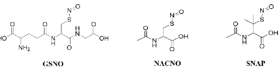
Figure 10. Structure of GSNO, NACNO and SNAP...49
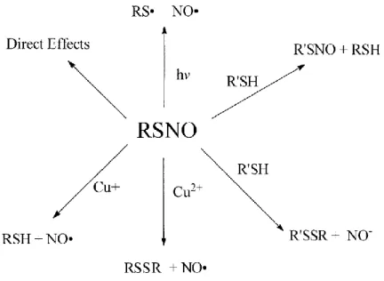
Figure 11. Biological activity of RSNO...50
Figure 12. In vivo metabolism of GSNO through different enzymes: 1. GSNO reductase (GSNOR)/carbonyl reductase 1 (CR1); 2. thioredoxin system (Trx); 3. protein disulfide isomerase (PDI); 4, γ-glutamyltranspeptidase (GGT)……….…...54
Figure 14. mechanisms of molecules crossing through the intestinal barrier131. (a) receptor-mediated transport; (b) carrier-mediated transport; (c) paracellular transport; (d) transcellular transport; and (e) M cell mediated transport (i.e., phagocytosis by M cells)...76
Figure 15. drugs classification according to the Biopharmaceutical Classification System (BCS)...80
Figure 16. NO storage in the form of S-nitrosothiols (RSNO), dinitrosyl iron complexes (DNIC) with protein ligands, and low molecular weight DNIC...98
Figure 17. The various factors that affect the endothelium and the consequences of endothelial dysfunction...99
Figure 18. Scheme of the preparation procedure for drug loaded microparticles by the double emulsion solvent evaporation method...128
Article 1:
Fig. 1. In vivo oxidation of L-Arginine by NO Synthases...46 Fig. 2. Synthesis of amidoximes...46
Fig. 3. Structure of 4-chlorobenzamidoxime...46 Fig. 4. Concentration of released nitrite ions from compounds 2 and the reference after a 10 min incubation with microsomes. Results are presented as mean ± SEM of three to five independent experiments and compared with a One-way ANOVA; * p < 0.05 versus reference...47
Fig. 5. Viability of the smooth muscle cells after 24 h incubation at concentrations ranging from 0.001 to 100 μM. Results are presented as mean ± SEM of three to four tests and compared with a two-way ANOVA for compound 2a; * p < 0.001 100 μM versus all the concentrations...47
Fig. 6. RSNO intracellular concentration after a 1 h incubation of smooth muscle cells with 100 μM of each compound except 2d that was used at 50 μM. Results are presented as mean ± SEM of three independent experiments and compared with a One-way ANOVA; * p < 0.05 versus reference; # p < 0.05 versus GSNO (Dunnett’s multiple comparisons post-test)...47 Article 2:
Fig. 1. Schematic representation of the bidirectional permeability of S-nitrosothiols across the Caco-2 monolayer. (a) From the apical (intestinal lumen) to the basolateral (bloodstream) compartment (on the left, A to B) to study physiological intestinal permeability, (b) from the basolateral to the apical compartment (on the right, B to A) to study S-nitrosothiol efflux...85 Fig. 2. Cytocompatibility of S-nitrosothiols with Caco-2 cells. Cell activity was assessed with the MTT test, 24 h after incubation with different S-nitrosothiols or NaNO2. Values are expressed as mean ± SD of three independent experiments done in duplicate..86 Fig. 3. Apical to basolateral compartment - Quantification in the basolateral compartment of permeated (A) RSNO, (B) NO2- and (C) NO3- after 1 h and 4 h of exposure to 50 nmol of each treatment. Results are shown as mean ± SD of four independent experiments done in duplicate and are compared using two-way ANOVA (ptreatment (GSNO, NACNO, SNAP; excluding NaNO2), ptime (1 h, 4 h) and pinteraction). *vs. GSNO; # vs. SNAP at the same time; p < 0.05 (Bonferroni’s multiple comparisons test...86 Fig. 4. Apical to basolateral compartment - Quantification in the apical compartment of remaining (A) RSNO, (B) NO2- and (C) NO3- after 1 h and 4 h of exposure to 50 nmol of each treatment. Results are shown as mean ± SD of four independent experiments done in duplicate and are compared using one-way ANOVA (excluding NaNO2) Bonferroni post-test. *vs. GSNO; # vs. SNAP at the same time; p < 0.05...87 Fig. 5. Apical to basolateral permeability - Intracellular quantifications of (A) RSNO, (B) NO2-, (subtracted from the control cells) after 1 h and 4 h of exposure to 50 nmol of each treatment. Results are shown as mean ± SD of four independent experiments done in duplicate and are compared using two-way ANOVA (ptreatment (GSNO,
NACNO, SNAP; excluding NaNO2), ptime (1 h, 4 h) and pinteraction) *vs. GSNO; # vs. SNAP at the same time; p < 0.05 (Bonferroni’s multiple comparisons test)...87 Fig. 6. Basolateral to Apical permeability - Quantification in the apical compartment of permeated (A) RSNO, (B) NO2- and (C) NO3- after 1 h and 4 h from 50 nmol of each treatment. Results are shown as mean ± SD of four independent experiments done in duplicate and are compared using two-way ANOVA (ptreatment (GSNO, NACNO, SNAP; excluding NaNO2), ptime (1 h, 4 h) and pinteraction). *vs. GSNO; # vs. SNAP at the same time; p < 0.05 (Bonferroni’s multiple comparisons test)...88
Fig. 7. Basolateral to apical permeability - Quantification in the basolateral compartment of remaining (A) RSNO, (B) NO2- and (C) NO3- after 1 h and 4 h of exposure to 150 nmol of each treatment. Results are shown as mean ± SD of four independent experiments done in duplicate and are compared using one-way ANOVA (excluding NaNO2). *vs. GSNO; # vs. SNAP at the same time; p < 0.05 (Bonferroni post-test...89 Fig. 8. Apical pH 6.4 to Basolateral permeability - Quantification in the basolateral compartment of remaining (A) RSNO, (B) NO2- and (C) NO3- after 1 h and 4 h of exposure to 50 nmol of each treatment. Results are shown as mean ± SD of three independent experiments done in duplicate and are compared using two-way ANOVA (ptreatment (GSNO, NACNO, SNAP; excluding NaNO2), ptime (1 h, 4 h) and pinteraction). *vs. GSNO; # vs. SNAP at the same time; p < 0.05 (Bonferroni’s multiple comparisons test)...90 Fig. 9. Apical to basolateral compartment - Quantification in the apical compartment of remaining (A) RSNO, (B) NO2- and (C) NO3- after 1 h and 4 h of exposure to 50 nmol of each treatment. Results are shown as mean ± SD of three independent experiments done in duplicate and are compared using one-way ANOVA (excluding NaNO2). *vs. GSNO; # vs. SNAP at the same time; p < 0.05 (Bonferroni post-test)...91 Fig. 10. Summary of NOx species permeability for each S-nitrosothiol treatment. The colour code of each arrow from the left to the right is the amount of RSNO, NO2- and (C) NO3-. The 4 h-permeability for each treatment is represented from (A) apical to
basolateral compartment, (B) basolateral to apical compartment and (C) apical (pH 6.4) to basolateral compartment (pH 7.4)...92 Article 3:
Fig. 1. Quantity of nitric oxide-derived (NOx) species in endothelium-intact (A) or endothelium-removed (B) rat aortae after incubation or not (control) with 2 μM of S-nitrosoglutathione (GSNO), acetylcysteine (NACNO), S-nitroso-N-acetylpenicillamine (SNAP) or sodium nitroprusside (SNP) for 30 min. Results are presented as mean ± SD of n = 3-15 per group, from 3-15 different rats in each group and compared with a Kruskal-Wallis test; * p < 0.05 versus control...122 Fig. 2. Vasorelaxant effects of N-acetylcysteine (10-5 M) in endothelium-intact (A) and endothelium-removed (B) rat aortae after incubation or not (control) with 2 μM of S-nitrosoglutathione (GSNO), acetylcysteine (NACNO), S-nitroso-N-acetylpenicillamine (SNAP) or sodium nitroprusside (SNP) for 30 min followed by one hour of washing with Krebs’ solution. Results are expressed as the percentage of 10-6 M phenylephrine preconstriction, presented as mean ± SD of n = 8-10 per group, from 4-8 different rats in each group and compared with one-way ANOVA; *p<0.05 versus control, #p<0.05 versus NACNO (Bonferroni’s multiple comparisons test)...122 Fig. 3. Concentration-dependent responses curves to phenylephrine of endothelium intact (A) and endothelium-removed (B) rat aortae after incubation or not (control) with 2 μM of S-nitrosoglutathione (GSNO), N-acetylcysteine (NACNO), S-nitroso-N-acetylpenicillamine (SNAP) or sodium nitroprusside (SNP) for 30 min. Results are presented as mean ± SD of n = 6-17 per group, from 3-7 different rats in each group and analyzed using the Hill equation...123 Fig. 4. Schematic representation of S-nitrosoglutathione (GSNO), S-nitroso-N-acetylcysteine (NACNO), S-nitroso-N-acetylpenicillamine (SNAP) and sodium nitroprusside (SNP) metabolism to produce a NO store of S-nitrosated proteins (Pr-SNO) in cells. Protein disulfide isomerase (PDI), gamma-glutamyltransferase (GGT), L-type amino acid transporter (L-AT), S-nitrosocysteine (Cys-NO)...123 Fig. 5. Schematic representation of N-acetylcysteine (NAC)-induced vasorelaxation either in endothelium-intact (A) and in endothelium-removed aortae (B). NAC enters
inside endothelial cells (A) or smooth muscle cells (B) using the L-type amino acid transporter (LAT). Then, NAC is deacetylated in cysteine residues that, in the presence of a NO store (PrSNO), forms NO by transnitrosation process. The unstable Cys-NO immediately releases Cys-NO that either diffuse to smooth muscle cells to activate the soluble guanylate cyclase (sGC) (A) or activate directly the sGC in the case of endothelium-removed aortae (B)...124 Article 4:
Figure 1. Characterization of GSNO-loaded particles before and after lyophilization. Size and polydispersity index (PDI) of the GSNO-NP (a) as well as size and span of the GSNO-MP (b), encapsulation efficiencies (c) are presented as mean ± sd (n=3). Representative Scanning Electron Microscopy images are also presented (d)...165 Figure 2. Stability (size (a) and remaining GSNO content (b)) of GSNONP, GSNO -MPW and GSNO-MPS suspensions stored at 4 °C. Results are presented as mean ± sd (n = 3) and compared to the values obtained just after preparation (day 0, 100%). The grey areas delimit the sizes between 90% and 110% of the initial values. The percentages of remaining GSNO were analyzed with One-way ANOVA p < 0.0001 (Dunnett's multiple comparisons test): * p < 0.05 versus day 0...165
Figure 3. Evolution of size and GSNO remaining for the GSNO-NP (a), GSNO-MPW (b) and GSNO-MPS (c) stored at 4 °C under inert atmosphere after lyophilization. Values measured immediately after lyophilization are considered as 100%. The grey areas delimit the sizes between 90% and 110% of the initial values. Results are presented as mean ± sd (n = 3). The data about GSNO remaining were analyzed with One-way ANOVA p < 0.01 (Dunnett's multiple comparisons test: GSNO-NP: p = 0.0045; GSNO-MPW: p = 0.0016; GSNO-MPS: p = 0.0003): * p < 0.05 versus week 0 (100%)...166 Figure 4. Drug release in PBS. Results are presented as mean ± sd (n = 3, free GSNO as control), two-way ANOVA (Turkey's multiple comparisons test): * p < 0.05 versus free GSNO and GSNONP...167
Figure 5. Cytocompatibility of Caco-2 cells after 24 h of incubation with GSNO (a), GSNO-NP (b), GSNO-MPW (c), GSNO-MPS (d). Control condition = culture medium
without fetal bovine serum. Results are presented regarding GSNO loading, in equivalence of free GSNO from 25 μM to 2500 μM. Results are shown as means ± sem, n = 3 in duplicate. ND = not determined...168
Figure 6. Quantity of GSNO, nitrite ions nitrate ions remaining in the apical medium (a, b and c, respectively), as well as in the basolateral medium (e, f and g, respectively), and GSNO (d) remaining inside the particles after 1 h of permeability study (GSNO initial amount = 50 nmol in all cases). Results are shown as means ± sem, n=3 in duplicate, one-way ANOVA (Tukey's multiple comparisons test): *p < 0.05 versus GSNO and # p < 0.05 versus GSNO-NP...169 Figure 7. Summary of NOx species permeability for each condition (free GSNO, GSNO-NP, GSNO-MPW and GSNO-MPS). The width of each section of the arrows is correlated with the amounts (from left to right) of NO3-, NO2- and GSNO...169
Abbreviations
Abbreviations Full names
BCS Biopharmaceutical Classification System
BSA Bovine serum albumin
cGMP Cyclic guanosine monophosphate CYP450 Cytochrome P-450 system
CVD Cardiovascular diseases CBR1 or CR1 Carbonyl reductase CysNO S-nitrosocysteine
DAN 2,3-Diaminonaphthalene DNIC Dinitrosyl iron complexes
EDRF Endothelium-derived relaxing factor EDTA Ethylene diaminotetraacetic acid eNOS Endothelial nitric oxide synthase GGT γ-glutamyltransferase GSH Reduced glutathione GSNO GSNO-NP GSNO-aNCP GSNO-cNCP GSNO-acNCP GSNOR S-nitrosoglutathione GSNO-loaded nanoparticles
GSNO-loaded alginate nanocompsite particles GSNO-loaded chitosan nanocompsite particles GSNO-loaded chitosan nanocompsite particles GSNO reductase GSSG GTP Glutathione disulphide Guanosine-5’-triphosphate Hb Hemoglobin
Hb-α Hemoglobin-α
HBSS Hank's Balanced Salt Solution HPMC Hydroxypropyl methylcellulose IBD Inflammatory bowel diseases iNOS Inducible NO synthase
IV Intravenous
L-Arg L-arginine
log P Logarithmic value of partition coefficient
MTT 3(4,5-Dimethylthiazol-2-yl)-2,5-diphenyltetrazolium bromide
NAC N-acetyl-L-cysteine
NACNO S-nitroso-N-acetyl-L-cysteine
NADPH Dihydronicotinamide-adenine dinucleotide phosphate NHA N-hydroxy-L-arginine
nNOS NOS
Neuronal NO synthase Nitric oxide synthase
NOx species Nitrogen oxide related species Papp
PCL
Apparent permeability coefficient Poly(ε-caprolactone)
PDI PEG
Protein disulfide isomerase Poly(ethylene glycol)
PKG Protein kinase G
pKa PLGA
Negative logarithmic value of acid ionisation constant Poly(lactide-co-glycolide)
PVAT PVA
Perivascular adipose tissue Poly(vinyl alcohol)
PVP Poly(vinyl pyrrolidone) RSNOs S-nitrosothiols
SDS Sodium dodecyl sulfate sGC
SMCs
Soluble guanylate cyclase Smooth muscle cells SNAP SNP S-nitroso-N-acetyl-D-penicillamine Sodium nitroprusside S/O/W Trx TrxR Solid-in-oil-in-water Thioredoxin Thioredoxin reductase
VSMCs Vascular smooth muscle cells TEER Transepithelial electrical resistance W/O/W Water-in-oil-in-water
Aperçu de la thèse
Le monoxyde d’azote (NO) est un gaz radicalaire impliqué dans la signalisation des systèmes cardiovasculaires, immunitaires et nerveux. Au sein du système cardiovasculaire, la première cible de NO est la guanylate cyclase soluble (GCs) dont l’activation permet la synthèse de GMPc, un second messager médiant la relaxation vasculaire. Ceci constitue la voie de signalisation NO/GCs/GMPc. Cependant, le mécanisme d’action de NO emprunte également des voies de signalisation indépendantes de la voie NO/GCs/GMPc, comme la S-nitrosation. La S-nitrosation correspond à la formation d’un lien covalent entre la fonction thiol d’un résidu cystéine et NO. Elle est considérée comme une modification post-traductionnelle des protéines, aussi importante que la phosphorylation pour la modulation des activité/expression protéiques. La S-nitrosation forme des S-nitrosothiols comme les S-nitrosoprotéines,
S-nitrosopeptides ou S-nitrosoacides aminés. En plus de moduler les voies de
signalisation, ces S-nitrosothiols représentent également une forme de stockage et de transport de NO dont la demi-vie à l’état radicalaire n’est que de quelques secondes. Ainsi, à l’état radicalaire, le rayon d’action de NO ne peut dépasser quelques micromètres. Ces S-nitrosothiols sont depuis quelques années considérés comme des candidats médicaments. IIs constituent en effet des prodrogues de NO, dont la biodisponibilité est considérablement diminuée au cours des pathologies cardiovasculaires. Les donneurs de NO comme les dérivés nitrés sont utilisés depuis de nombreuses années dans le traitement de l’angor dans le cadre de sa prévention et en cas de crise. Cependant, les dérivés nitrés sont sujets au phénomène de tolérance (perte d’effet thérapeutique lors d’administration chronique), présentent une faible biodisponibilité et une faible sélectivité. Ils sont également responsables de l’amplification du stress oxydant déjà installé au cours des pathologies cardiovasculaires.
Ainsi, d’autres molécules donneuses de NO sont actuellement à l’étude. Parmi celles-ci nous nous sommes d’abord intéressés aux amidoximes. La fonction amidoxime est physiologiquement produite lors de la synthèse de NO catalysée par les NO synthases. En effet, au cours de cette catalyse en deux étapes, l’arginine est d’abord N-hydroxylée en N-hydroxyarginine présentant une fonction amidoxime. Cette fonction amidoxime est capable de libérer NO sous l’action oxydative des cytochromes
P450. Les résultats de l’étude de nouvelles molécules synthétiques, mono ou bis-amidoxime, sont présentés dans l’article 1.
Article 1: Synthesis of Novel Mono and Bis Nitric Oxide Donors with High Cytocompatibility and Release Activity
Tanya Sahyoun, Caroline Gaucher, Yi Zhou, Naïm Ouaini, Raphaël Schneider, Axelle Arrault.
Quatre composés comportant une ou deux fonctions amidoximes ont été synthétisés : (1) Le composé 2a-b contenant une fonction amidoxime aromatique, (2) le composé 2c présentant une fonction amidoxime aliphatique, et (3) le composé 2d présentant deux fonctions amidoxime, une aromatique et une autre aliphatique. La capacité de ces composés à libérer du monoxyde d’azote (NO) a été évaluée in vitro en tirant partie du métabolisme oxydant de cytochromes P450 de microsomes de foie de rats. Les résultats obtenus démontrent que toutes les fonctions amidoxime des composés synthétisés sont capables de libérer NO, avec une plus grande capacité pour le composé 2a qui possède une fonction amidoxime aromatique. De plus, toutes les amidoximes testées sont cytocompatibles vis-à-vis de cellules musculaires lisses humaines. En utilisant la formation de S-nitrosothiols intracellulaires comme marqueur de la biodisponibilité de NO, les composés 2a et 2c ont montré leur capacité à délivrer une plus grande quantité de NO que la molécule de référence (4-chlorobenzamidoxime). Ainsi, les amidoximes sont capables de traverser la membrane cellulaire pour être ensuite oxydées par les CYP450 des cellules musculaires lisses. En revanche, la bis-amidoxime aromatique/aliphatique 2d est de grand intérêt car sa capacité à délivrer NO et par la suite à augmenter le stockage de NO dans les cellules
via la formation de S-nitrosothiols intracellulaires est similaire aux amidoximes 2a et
2b. En effet, en considérant que la bis-amidoxime 2d a été étudiée à une concentration deux fois plus faible que celle des composés 2a et 2b, nous pouvons conclure que le composé 2d est la molécule la plus performante de tous les composés donneurs de NO testés ici.
D’autres donneurs de NO dérivant des S-nitrosothiols, formes physiologiques de transport et de stockage de NO, sont également à l’étude. Parmi ces S-nitrosothiols, nous pouvons citer le S-nitrosoglutathion (GSNO), la S-nitroso-N-acétyl-L-cystéine (NACNO) et le S-nitroso-N-acétyl-pénicillamine (SNAP). Les S-nitrosothiols sont considérés comme des candidats médicaments d’avenir, puisqu’ils ne présentent pas les effets indésirables de dérivés nitrés précédemment énumérés. Les RSNO sont dénués de toxicité aigüe, et de par l’existence physiologique de certains d’entre eux, ils ne présentent probablement aucun risque dans le cadre d’application répétée. Ceci est d’autant plus important que les pathologies cardiovasculaires sont, pour la plupart, des affections chroniques nécessitant un traitement sur le long terme. Dans ce contexte, la voie d’administration orale de ces molécules est la plus indiquée du point de vue des patients (facilité, faible contrainte conduisant à une meilleure observance). De plus, la voie orale présente des avantages physiologiques comme une grande surface d’absorption (300 à 400 m2). Néanmoins, la barrière intestinale constitue un obstacle important à franchir pour ces molécules de nature peptidique ou protéique, avec une liaison nitrosothiol fragile. Ainsi, une deuxième étude a été menée sur les mécanismes d’absorption intestinale des S-nitrosothiols (Article 2).
Article 2: Intestinal absorption of S-nitrosothiols: permeability and transport mechanisms
Absorption intestinale des S-nitrosothiols : Mécanismes de transport et de perméabilité
Justine Bonetti#, Yi Zhou#, Marianne Parent, Igor Clarot, Haiyan Yu, Isabelle Fries-Raeth, Pierre Leroy, Isabelle Lartaud, Caroline Gaucher*
# Les deux auteurs ont contribué à parts égales à ce travail.
Au cours du vieillissement ou du développement des pathologies cardiovasculaires, une diminution de la biodisponibilité du monoxyde d’azote (NO) est observée. Afin de restaurer une concentration physiologique de NO, les S-nitrosothiols (RSNO) font partie des donneurs de NO envisagés comme principes actifs potentiels. Les RSNO sont la forme physiologique de stockage et de transport de NO dans l’organisme, lui assurant ainsi une demi-vie supérieure à sa forme libre radicalaire (< 0,5 s). La plupart des pathologies cardiovasculaires nécessitent un traitement chronique pour lequel la voie orale est plébiscitée. Cependant, l’absorption intestinale des RSNO, première limite à leur biodisponibilité vasculaire, n’a jamais été étudiée. Ainsi, à l’aide d’un modèle in vitro de barrière intestinale (Caco-2), cette étude vise à élucider les mécanismes de perméabilité intestinale (passifs ou actifs, voie paracellulaire ou transcellulaire) et à prédire le site d’absorption intestinale préférentiel des RSNO. Dans notre étude, trois RSNO, le S-nitrosoglutathion (GSNO), la S-nitroso-N-acétylcystéine (NACNO), la S-nitroso-N-acétyl-D-pénicillamine (SNAP), qui diffèrent de par le squelette transportant NO, ont été évalués. Ces squelettes transporteurs de NO confèrent des propriétés physico-chimiques (lipophilie) et des activités biologiques (antioxydantes et/ou anti-inflammatoires) différentes aux RSNO. Ainsi, en montrant à l’aide de ce modèle la perméabilité apparente moyenne des RSNO, associée à leur forte solubilité, cette étude a permis de placer ces molécules entre la classe I et la classe III du Système de Classification Biopharmaceutique. L’évaluation bidirectionnelle de la perméabilité a également solutionné le mécanisme de perméabilité de RSNO selon un phénomène passif. De plus, GSNO et NACNO suivent préférentiellement la voie transcellulaire alors que SNAP préfère la voie paracellulaire. Finalement, le squelette transportant NO a une influence sur le site d’absorption des RSNO, puisque la perméabilité de NACNO est favorisée à pH 6,4, pH proche de la partie proximale du jéjunum, alors que celles de GSNO et SNAP sont meilleures à pH
7,4, pH proche de sa partie distale. Au travers de cet article, nous avons déterminé les mécanismes de perméabilité intestinale des RSNO et prouvé que leur administration par voie orale était possible. Après avoir franchi la barrière intestinale, les S-nitrosothiols rejoignent le compartiment vasculaire où ils joueront leurs rôles de transporteurs puis de donneurs de NO pour restaurer le pool vasculaire de NO. Ce pool se retrouve ainsi sous deux formes, une forme circulante dans le flux sanguin et une forme tissulaire stockée dans la paroi vasculaire. Ainsi, la prochaine étape de cette étude a consisté à comparer la capacité des différents S-nitrosothiols à promouvoir la formation d’un stock au sein de la paroi aortique et de vérifier si ce stock est mobilisable pour la vasodilation, faisant preuve de sa biodisponibilité. Enfin, les propriétés protectrices des fonctions endothéliales des S-nitrosothiols ont été évaluées sur des aortes dénuées d’endothélium (Article 3).
Article 3: S-nitrosothiols as potential therapeutics to induce a mobilizable vascular store of nitric oxide to counteract endothelial dysfunction.
Les S-nitrosothiols : de potentiels médicaments induisant la formation d’un stock mobilisable de monoxyde d’azote au sein des vaisseaux afin de contrer la dysfonction endothéliale.
Caroline Perrin-Sarrado#, Yi Zhou#, Valérie Salgues, Marianne Parent, Philippe Giummelly, Isabelle Lartaud, Caroline Gaucher*
# Les deux auteurs ont contribué à parts égales à ce travail.
Article soumis au journal Biochemical Pharmacology le 28 juin 2019, en révision
La dysfonction endothéliale prédispose au développement de certaines pathologies cardiovasculaires. Elle est définie comme un déséquilibre entre la production de substances vasodilatatrices comme le monoxyde d’azote (NO) et de substances vasoconstrictrices. Afin d’assurer ses fonctions physiologiques, NO, un radical gazeux à demi-vie très courte, a besoin d’être stocké et transporté jusqu’à son site d’action. Les S-nitrosothiols (RSNO) comme le S-nitrosoglutathion (GSNO) représentent la principale forme physiologique de stockage de NO au sein du système vasculaire. Le stock de NO formé par les RSNO reste alors biodisponible pour déclencher la vasorelaxation. Ainsi, les RSNO représentent une classe émergente de donneurs de NO pour restaurer la biodisponibilité de NO au cours des troubles cardiovasculaires. Le but de cette étude est de comparer la capacité des S-nitrosothiols à produire un stock vasculaire de NO au sein d’artères dont l’endothélium est intact ou d’artères dont l’endothélium a été retiré. Un RSNO physiologique, le GSNO, ainsi que deux RSNO synthétiques, la S-nitroso-N-acétylcystéine (NACNO) et la
S-nitroso-N-acétylpénicillamine (SNAP) ont été testés, permettant ainsi d’évaluer le potentiel thérapeutique des RSNO pour compenser la dysfonction endothéliale. Le nitroprussiate de sodium (SNP), un médicament bénéficiant déjà d’une autorisation de mise sur le marché, a été utilisé comme donneur de NO contrôle non RSNO. Les deux types d’artères (avec ou sans endothélium) utilisées dans cette étude ont été isolées de rats Wistar mâles normotendus et exposées aux donneurs de NO. Ensuite, les espèces dérivées de NO (NOx) représentant le stock intravasculaire de NO, ont été quantifiées en utilisant la sonde diaminonaphthalène couplée aux ions mercuriques. La biodisponibilité du stock de NO et sa capacité à produire la vasorelaxation ont été
testés à l’aide de la N-acétylcystéine. Ensuite la capacité de ce stock à s’opposer à la vasoconstriction produite par la phényléphrine (PHE) a été appréciée.
Tous les RSNO étudiés ont été capables de générer un stock de NO trois à cinq fois supérieur à la quantité basale de NOx au sein des artères contrôle. NACNO était le plus performant de tous les RSNO pour produire un stock vasculaire biodisponible pour la vasorelaxation et pour induire une hyporéactivité à la PHE des artères sans endothélium. L’efficacité de GSNO et SNAP était équivalente, et supérieure à celle de SNP. Au sein des artères avec endothélium, le stock de NO s’est également formé, cependant il s’est avéré moins disponible pour la vasorelaxation et n’a pas influencé la vasoconstriction induite par la PHE.
En conclusion, les RSNO, et NACNO plus particulièrement, sont capable de restaurer la biodisponibilité de NO en créant un stock fonctionnel de NO au sein de la paroi des vaisseaux sanguins et de façon plus marquée quand l’endothélium est absent. Ce phénomène a été associé à une hyporéactivité de l’artère à un agent vasoconstricteur comme la PHE. Des traitements à base de RSNO pourraient donc présenter un bénéfice pour restaurer les fonctions NO-dépendantes dans des états pathologiques associés à un endothélium lésé.
Ainsi, toutes ces études mettent en évidence que NACNO est le S-nitrosothiol qui franchit le mieux la barrière intestinale et qui produit le stock de NO vasculaire le plus grand et le plus biodisponible.
Dans la dernière partie de ce travail, nous nous sommes intéressés au développement de formulations particulaires adaptées à la voie orale et permettant d’augmenter la biodisponibilité des S-nitrosothiols. Malgré la supériorité de NACNO dans les études précédentes, c’est le GSNO qui a été utilisé pour cette étude. En effet, il présente certains avantages. D’une part, il est physiologique (contrairement à NACNO) et a été utilisé dans la majorité des essais cliniques. D’autre part, seul GSNO peut être obtenu sous la forme d’un produit à la fois pur et solide (ce qui permet plus de libertés dans la formulation).
En ce qui concerne le procédé de formulation galénique, il doit être adapté à ces molécules particulièrement fragiles et hydrophiles, tout en respectant le cahier des charges pour la voie orale : polymère adapté, charge en principe actif suffisante, libération appropriée, stabilité des caractéristiques des particules obtenues pour faciliter leur utilisation, amélioration de la perméabilité intestinale sans cytotoxicité…
Dans ce contexte, trois stratégies de formulation de GSNO basées sur des procédés de double émulsion/évaporation de solvant ont été comparées (Article 4).
Article 4: Enhancing intestinal permeability of nitric oxide with storable microparticles for S-nitrosoglutathione oral delivery.
Des microparticules préparables à l’avance pour la délivrance de S-nitroglutathion par voie orale et capables d’augmenter la perméabilité intestinale du monoxyde d’azote
Yi Zhou, Caroline Gaucher, Isabelle Fries, Mehmet Hobekkaya, Charlène Martin, Clément Leonard, Frantz Deschamps, Anne Sapin-Minet, Marianne Parent*
Article soumis au journal International Journal of Pharmaceutics le 21 août 2019 Trois sortes de particules encapsulant du S-nitrosoglutathion (GSNO), un donneur physiologique de monoxyde d’azote, ont été préparées à partir des mêmes matériaux mais avec des procédés modifiés de double émulsion/évaporation de solvant : eau dans huile dans eau (E/H/E) ou solide dans huile dans eau (S/H/E). Les nano- et micro-particules obtenues par la méthode E/H/E ont été comparées aux micromicro-particules obtenues par la méthode S/H/E en ce qui concerne leur taille, leur capacité à encapsuler le GSNO et à le libérer in vitro. La cytocompatibilité des particules vis-à-vis des cellules intestinales ainsi que leur capacité à améliorer la perméabilité intestinale du GSNO in vitro (modèle de barrière intestinale constituée par les cellules Caco-2 différenciées) ont été évaluées.
L’efficacité d’encapsulation du GSNO est la même (environ 30 %) pour les trois sortes de particules mais les microparticules (S/H/E > E/H/E) présentent un profil de libération plus lent du GSNO in vitro et améliorent significativement la perméabilité intestinale de GSNO. Cette perméabilité apparente élevée lorsqu’il est encapsulé dans les microparticules, associée à une solubilité élevée, fait passer GSNO en classe III de la classification biopharmaceutique.
La réduction de la taille des grains de GSNO (de 40 µm à moins de 10 µm) initiée en collaboration avec la société StaniPharm devrait, d’après la littérature, conduire à une meilleure encapsulation et à une libération prolongée de GSNO avec les microparticules S/H/E. Cependant, les premiers essais en fluide supercritique présentés dans cet article n’ont pas été concluants, à cause de la grande fragilité et de l’hydrophilie du GSNO.
Par ailleurs, les paramètres critiques des particules (taille et contenu en GSNO) sont maintenus jusqu’à 1 mois après lyophilisation et stockage à 4°C pour les microparticules et jusqu’à 3 mois pour les nanoparticules. Les suspensions fraîches
de particules n’étant stables qu’une journée, cette stabilisation représente une avancée majeure, car elle permettra d’optimiser les campagnes de préparation des particules et de les envoyer éventuellement vers d’autres laboratoires pour la réalisation de tests précliniques. Les particules chargées en GSNO développées pourraient ainsi représenter une nouvelle opportunité thérapeutique pour un traitement chronique, par voie orale, des pathologies associées avec un déficit en monoxyde d’azote
Pour conclure, les objectifs de ce travail étaient de mettre en évidence les mécanismes de la perméabilité intestinale de différents S-nitrosothiols et leur capacité à former un stock de NO mobilisable dans la paroi vasculaire, ainsi que de développer des formulations adaptées pour le traitement oral des maladies cardiovasculaires. Nous avons démontré : 1) que les trois RSNO étudiés peuvent être administrés par voie orale (avec une perméabilité intestinale moyenne) ; 2) qu’ils peuvent tous trois former un stock vasculaire de NO mobilisable pour une vasorelaxation ou pour induire une hyporéactivité à un agent vasoconstricteur. Tout ceci dans le cadre d’un endothélium lésé, ce qui est prometteur dans le contexte des maladies cardiovasculaires, souvent liées à une dysfonction endothéliale ; et 3) qu’il est possible d’augmenter significativement la perméabilité intestinale du GSNO grâce à des formulations, en particulier les microparticules obtenues par la méthode d’émulsion solide-huile-eau. Ce travail ouvre de nombreuses perspectives à la fois pharmacologiques et galéniques. Par exemple, une localisation plus fine du stock vasculaire de NO ainsi qu’une identification des protéines nitrosées (par une technique de biotin-switch associée à la spectrométrie de masse, déjà développée dans l’équipe) pourrait être entreprise afin de mettre au jour de nouvelles cibles de NO. Idéalement, les méthodes analytiques devraient également permettre de réaliser une pharmacocinétique complète des RSNO administrés (libres ou formulés). En ce qui concerne la formulation, de nombreuses optimisations pourraient être recherchées (meilleure encapsulation, libération plus longue, encapsulation d’autres donneurs de NO – RSNO, amidoximes, ou autres-, co-encapsulation de différents principes actifs, modification des polymères par exemple avec des groupements donneurs de NO, …). Cependant, les formulations développées dans ce travail pourraient d’ores et déjà être administrées chez l’animal sain, pour vérifier la formation du stock vasculaire de NO, ou dans des modèles pathologiques (athérosclérose ou accident vasculaire cérébral, par exemple).
1.1 Nitric oxide
Nitric oxide (NO) is a gaseous radical, whose biosynthesis and cellular signaling mechanisms were discovered in the 1980’s1–4. As one of the most important signaling molecule, NO is involved in different organs and systems (such as cardiovascular system5,6, immune system2,3 and nervous system7) where it is responsible for modulating various effects8,9 (Figure 1). Among others, NO can be regarded as a potent vasodilator, a regulator of platelet adhesion, a neurotransmitter, an antimicrobial agent, a tumor mediator and an angiogenesis promoter10,11. Thus, researchers from different disciplines have proposed NO as a potential therapeutic.
Figure 1. An overview of nitric oxide (NO) physiological activities in various systems10.
As an important second messenger, NO is synthesized by three different isoforms of nitric oxide synthases (NOS): neuronal NOS (nNOS), inducible NOS (iNOS), and endothelial NOS (eNOS)12. nNOS was first discovered in neuronal tissue and eNOS in vascular endothelial cells. iNOS is the inducible isoform found in a wide range of tissues and cells. NO is constitutively generated by nNOS or eNOS at relatively small
concentration (10 nM) for signaling. However, NO synthesized by the inducible isoform (iNOS), works as a response to inflammation, leading to high (more than 1 µM) and cytotoxic local concentrations of NO12,13. These three NOSs isoforms catalyze the synthesis of NO through a two-step reaction starting from L-arginine (L-Arg) transformed in an amidoxime intermediate, the N-hydroxy-L-arginine (NHA) to finally form L-citrulline and NO14 (Figure 2).
Figure 2. The synthesis of nitric oxide by nitric oxide synthases12.
1.2 Nitric oxide and cardiovascular diseases (CVD) 1.2.1 Nitric oxide in the cardiovascular system
In cardiovascular system15–17, eNOS is mainly expressed in endothelial cells but also in platelets, cardiac myocytes and smooth muscle cells (SMCs); nNOS is basically expressed in the sarcoplasmic reticulum from cardiac myocytes, in autonomic cardiac neurons as well as ganglia or within vascular smooth muscle cells (VSMCs) (Figure 3). However, only under oxidative or pro-inflammatory conditions, iNOS will be expressed in a large range of cells such as endothelial cells, cardiac myocytes, nerve cells, VSMCs, leukocytes and fibroblasts. NO regulates cardiovascular system via two distinct pathways: one is the activation of the soluble guanylate cyclase (sGC) followed with the downstream stimulation of Protein Kinase G (PKG), and one direct pathway through proteins S-nitrosation (the binding of a NO moiety on a thiol group from a cysteine residue to form an RSNO).
In vessels, NO synthesized by eNOS triggers the relaxation of VSMCs as well as inhibits their proliferation17,18. After its diffusion to the vessel lumen, it also mediates
angiogenesis and inhibits platelet aggregation and thrombosis.
In erythrocytes, NO reacts with the Fe2+ heme of oxyhemoglobin and produce nitrate ions. Then, the nitrate reductase existing in mammalian tissues reduces nitrate ions (NO3−) to nitrite ions (NO2−). In fact, deoxygenated hemoglobin has a nitrite reductase activity, which reduces NO2− to NO in hypoxic and acidic environments. In addition, reversible S-nitrosation of the Cys 93 of hemoglobin might transport NO for its subsequent release in hypoxic tissues. The reduction of NO2− and/or S-nitrosation of hemoglobin promotes NO release followed by-immediate relaxation of hypoxic tissues19. The diffusion and bioavailability of NO in VSMCs are regulated by hemoglobin-α (Hb-α) at the myoendothelial junction and by cytoglobin in muscle cells. In addition, NO synthesized through nNOS in the VSMCs also help to regulate the vascular tone20–22.
In cardiac myocytes, the main roles of NO are the regulation of cardiac contractility through the modulation of the excitation–contraction coupling, relaxation, and mitochondrial respiration. In addition, the action of nNOS expressed in cardiac nerves and postsynaptic eNOS can reinforce the parasympathetic (vagal) transmission through modulating the sympathetic–parasympathetic balance, which finally reduces the heart rate18.
Figure 3. NO in the cardiovascular system15. NO, nitric oxide; eNOS, endothelial nitric
oxide synthase; VSMC, vascular smooth muscle cell; NO2−, nitrite ions; Hb-α, haemoglobin-α;
CYGB, cytoglobin; nNOS, neuronal nitric oxide synthase;β1, adrenergic receptor β1; ACh,
acetylcholine; m2, muscarinic acetylcholine receptor m2; NA, noradrenaline; SNO, S-nitrosothiol; SR, sarcoplasmic reticulum; T-tubule, transverse-tubule.
1.2.2 NO signaling in vascular beds in health and disease.
Under physiological conditions (Figure 4a), NO synthesized by eNOS inhibits platelet aggregation and thrombosis. Basically, NO activates soluble guanylate cyclase in VSMCs by coordination to the ferrous prosthetic of the heme moiety (which is nitrosylation) which converts the guanosine-5’-triphosphate (GTP) to cyclic guanosine monophosphate (cGMP). Then, the PKG will be activated in the SMCs. Both cGMP and PKG lowered the intracellular Ca2+ levels through stimulating its reuptake by sarcoplasmic/ endoplasmic reticulum calcium-ATPase and its extrusion through the
plasma membrane calcium-transporting ATPase15. In addition, the efflux of K+ will also be triggered by PKG through the large-conductance Ca2+-sensitive potassium channel, which will hyperpolarize the cell and reduce Ca2+ entry through the L-type calcium channel. At last, PKG promotes the dephosphorylation of the myosin light chain through the associated phosphatase. Together these modifications mediate vasorelaxation. Perivascular adipose tissue (PVAT) contributes to this effect under healthy conditions by releasing NO and adiponectin, which activates NO production in SMCs15,18,21.
Figure 4. NO signaling in cardiovascular system under physiological (left) and pathological (right) conditions15. NO, nitric oxide; eNOS, endothelial nitric oxide synthase; sGC, soluble
guanylate cyclase; PKG, protein kinase G; SMCs, smooth muscle cells; SERCA, sarcoplasmic/ endoplasmic reticulum calcium-ATPase; PMCA, plasma membrane calcium-transporting
ATPase; BKCa, Ca2+-sensitive potassium channel; LTCC, L-type calcium channel; MLCP,
myosin light chain through the associated phosphatase; PVAT, Perivascular adipose tissue; APN, adiponectin; NADPH ox, NADPH oxidase; O2−, superoxide anion; TNF, Tumour necrosis
factor; LK, leukocyte; MLCK, myosin light chain kinase; SR, sarcoplasmic reticulum. Under pathological condition (Figure 4b), NO bioavailability will be reduced by different cardiovascular risk factors through the uncoupling of eNOS and the activation of Reduced nicotinamide adenine dinucleotide phosphate (NADPH) oxidase, resulting in superoxide anion production. Reduced NO bioavailability promotes platelet aggregation, thrombosis, and vasoconstriction. Tumor necrosis factor produced from PVAT further activates NADPH oxidase. Subsequent endothelial dysfunction,
increased permeability, and leukocyte diapedesis contribute to the initiation of atherosclerosis19. As one of the key factors for the initiation and progress of cardiovascular diseases including atherosclerosis, heart failure, hypertension, arterial thrombotic disorders and stroke27,28, NO deficiency may result from both endothelium dysfunction (decreased production) or NO oxidative consumption (increased metabolism)15.
1.3 NO storage
The first target of NO is sGC, the soluble isoform of the guanylate cyclase5. However, it is now known that many actions of NO are in fact independent of cGMP. It has been reported that the cGMP-independent NO signaling pathways are attributed to S-nitrosation of structurally and functionally important cysteine residues9. In fact, the
S-nitrosation process will produce different S-nitrosothiols (RSNOs), which are the
main components of ‘NO storage pool’ in the body. NO synthesized from NOSs or coming from exogenous sources will be transferred into different NO storage forms like nitrite ions, RSNOs, N-nitroso proteins and other compounds like iron-nitrosyl-complexes (Figure 5). These compounds can act as NO donors or may even possess intrinsic NO-like activity16,25.
As physiological compounds, RSNOs are generated through the S-nitrosation of thiols of low molecular weight (e.g. glutathione, cysteine) or of high molecular weight (cysteine residues of proteins like albumin, hemoglobin, …) in the body. The S-nitrosation reaction between NO and a thiol function is a one-electron oxidation reaction. As shown in Figure 4, the NOS activation results not only in NO production but also in NOS auto-S-nitrosation, followed with the transnitrosation of NO. RSNOs such as S-nitrosoglutathione (GSNO) or S-nitrosoproteins can be generated from NO through different ways: transition metal catalyzed pathway26, thiyl radical recombination, NO oxidation or transnitrosation from other RSNOs27,28. Indeed, once formed, GSNO or S-nitrosated proteins can transfer NO to other proteins through transnitrosation reactions. RSNOs can degrade through different ways (Figure 6): for example, GSNO degrades through direct denitrosation by enzymes like GSNO reductase (GSNOR) or Carbonyl reductase (CBR1 or CR1)27, which lead to the formation of non-bioavailable NO. Other enzymes like γ-glutamyltranspeptidase (GGT) also help the degradation of GSNO (will be discussed in the chapter about GSNO) In addition, the Thioredoxin (Trx) can denitrosate the RSNOs and regenerate through the activity of thioredoxin reductase (TrxR)26. The protein disulfide isomerase (PDI) also catalyzes transnitrosation and denitrosation processes through thio-disulfide exchange reactions.
Figure 6. Potential mechanisms of S-nitrosothiols and S-nitrosoproteins formation and degradation26. NOS, nitric oxide synthases; GSNO, S-nitrosoglutathione; GSNOR, GSNO
reductase; CBR1, carbonyl reductase 1; Trx, thioredoxin; TrxR, thioredoxin reductase. 1.4 Nitric oxide donors
An exogenous supply of NO is needed to restore deficient NO levels. Indeed, by applying gaseous NO directly via inhalation, the pulmonary hypertension was relieved without systemic vasodilation and hypotension29. However, this application is still limited by several shortcomings: such as, its rapid inactivation when stopping NO administration due to the short half-time of NO; complex operation conditions with high cost (specific tanks); lack of possibility to treat pathologies at distant sites (NO cannot reach more than lungs when applied through inhalation). Therefore, a lot of compounds showing the ability to release NO have been investigated for cardiovascular diseases treatment and some of them (eg, nitroprusside and nitroglycerin) are or even have
been used in clinic for decades30–32. According to different mechanism of NO formation, some of them are direct NO donors (including NO gas) whereas others require metabolism33. This will be discussed in the following sections. In addition, two families (related to our work) among the several new NO donors will be presented: amidoximes and RSNOs.
1.4.1 Direct Donors
Compounds, which release NO directly are pharmacological agents with either a nitroso (S-N=O) or nitrosyl (metal·NO) functional group. These direct donors can spontaneously release NOx species (nitric oxide related species: different redox forms of nitrogen monoxides produced by the donor molecule). The most common members of direct donors are NO gas, sodium nitroprusside, and sodium trioxodinitrate (Angeli’s salt). NO gas is the most direct form but the complex conditions for inhalation and the short half time of NO (within second) limits its application34. In sodium nitroprusside (SNP), NO is coordinated to iron as a nitrosyl group (also connected to 5 nitrile ions) in a square bipyramidal complex35. Under physiological conditions, NO can be released spontaneously from the parent chemical structure. For decades, SNP was widely used for the acute treatment of heart failure and hypertension. However, the application of SNP was limited due to parenteral administration, tolerance and side effects (potential thiocyanate toxicity for long-time administration)36. Thus, SNP has been withdrawn from European market in 2017. In Angeli’s salt, NO-, a reduced form of NO, is released spontaneously under physiological conditions. Distinct from other redox forms of NO, NO- has an unique effect on vascular smooth muscle: the relaxation mediated by NO- from Angeli’s salt were more prolonged than those induced by NO37,38. Besides, compounds like Diethylamine/NO and diethylenetriamine/NO (which belong to diazeniumdiolate or NONOate [N(O)NO] class) can also release NO spontaneously39. In these two compounds, NO is covalently linked to diethylamine and diethylenetriamine. It has been proved that diazeniumdiolates can protect the neuro system from the neurotoxicity induced by hydrogen peroxide through a rat model40. Other heterocyclic compounds include the oxatriazolium class (sydnonimines) and the furoxan class41. These two kinds of NO donors can release NO directly using some cofactors (oxidants for sydnonimines and thiols for furoxans) 42.
1.4.2 Donors Requiring Metabolism
NO donors requiring metabolism to release NO are also widely used. For example, the two classic nitrovasodilator families, nitrite esters and organic nitrates (like amyl nitrite, isosorbide dinitrate and isosorbide 5-mononitrate nitroglycerin, shown in Figure 7), have been used in cardiovascular diseases for a long time. However, there are several limitations for these NO donors, including tolerance (i.e. the loss of therapeutic effect under chronic administration), limited bioavailability, low selectivity as well as potentially adverse hemodynamic effects43,44. Though these limitations, these drugs are still one the main therapeutics for cardiovascular diseases treatment. As these organic nitrate esters are prodrugs, they require enzymes to generate NO in the body. The main enzyme systems involved in the metabolism of organic nitrate/ nitrite esters is located within microsomal membranes. A lot of evidences suggest that the cytochrome P-450 (CYP450), the presence of NADPH oxidase as well as the activity of glutathione-S-transferase, are necessary in the metabolic processes of both de-nitration and reduction of organic nitrate esters to authentic NO45,46. It has also been proved that the tolerance of these NO donors is related to the increasing angiotensin II-dependent vascular superoxide anion production from NAD(P)H oxidase and eNOS47,48. Superoxide anion then reacts with NO generated from the NO donors, producing peroxynitrite ions (OONO-)49. In addition, the tolerance to organic nitrates is also associated with the cross-tolerance to endothelium-derived NO, both by the oxidative inactivation of this endogenous NO to peroxynitrite ions and by the “uncoupling” of eNOS activity50,51.
Figure 7. Structures of conventional organic nitrates and nitrite esters42.
1.4.3 New NO donors Amidoximes
Amidoximes are oximes of amides with general structure as in Figure 8. These molecules present many important biological effects and have the capacity of releasing NO when oxidized by enzymes such as CYP45052.
Figure 8. Structure of amidoxime function. The oxidation of amidoxime compounds has two pathways53: Nitric oxide Synthase pathway
As described in the Figure 2, nitric oxide is produced by a single pathway during the oxidation of guanidine function of the L-arginine. The oxidation is catalyzed by eNOS, in two steps. The first step is the N-hydroxylation of one of the two guanidino nitrogens of the L-arginine. This step consumes one mol of NADPH and of O2. The second step is the oxidation of the intermediate product, NHA, which bears an
amidoxime function. This step consumes 0.5 mol of NADPH and 1 mol of O2 for the oxidative cleavage of theC=N-OH bond to produce NO and citrulline. However, it seems the eNOS can only oxidize NHA, no other amidoximes.
Cytochromes P450 pathway
The first step of the oxidation by NOS resembles a classical P-450 dependent monooxygenation, which is not the case of the second step since it consumes only 0.5 mol of NADPH. This reaction has been the subject of study of many researchers, which have proven that the oxidation of not only the NHA, but also other arginine-mimetics like amidoximes, can be done by the CYP450.
It has been proven by previous work, that many molecules presenting the C=N-OH function like benzamidoximes, ketoximes, guanidoximes, and arylamidoximes are capable of releasing NO in the presence of CYP450, NADPH and dioxygen.
One of the studied molecules that released important quantities of NO is 4-chlorobenzamidoxime54 (Figure 9).
Figure 9. Structure of 4-chlorobenzamidoxime.
In this thesis, some new amidoximes molecules have been evaluated in the article 1, as follows: Cl N H2 N O H