HAL Id: tel-03179255
https://tel.archives-ouvertes.fr/tel-03179255
Submitted on 24 Mar 2021HAL is a multi-disciplinary open access archive for the deposit and dissemination of sci-entific research documents, whether they are pub-lished or not. The documents may come from teaching and research institutions in France or abroad, or from public or private research centers.
L’archive ouverte pluridisciplinaire HAL, est destinée au dépôt et à la diffusion de documents scientifiques de niveau recherche, publiés ou non, émanant des établissements d’enseignement et de recherche français ou étrangers, des laboratoires publics ou privés.
Deciphering the role of the death receptor Fas/CD95 in
T cell co-stimulation
Giorgia Miloro
To cite this version:
Giorgia Miloro. Deciphering the role of the death receptor Fas/CD95 in T cell co-stimulation. Cellular Biology. Université Côte d’Azur, 2020. English. �NNT : 2020COAZ6036�. �tel-03179255�
Déterminer le rôle du récepteur de mort
Fas/CD95 dans la co-stimulation des
cellules T
Giorgia MILORO
Institut de Biologie de Valrose
Présentée en vue de l’obtention du grade de docteur en
Interactions cellulaires et moléculaires d’Université Côte d’Azur
Dirigée par : Anne-Odile Hueber Co-encadrée par: Aurélie Rossin Soutenue le : 11/12/2020
Devant le jury, composé de :
Dr Hai-Tao He, Directeur de recherches CNRS, CIML, Marseille
Dr Frédéric Rieux-Laucat, Directeur de Recherche INSERM, Institute Imagine, Paris Dr Aurélie Rossin, Chargé de Recherche CNRS, IBV, Nice
Dr Anne-Odile Hueber, Directeur de Recherche INSERM, IBV, Nice
Déterminer le rôle du récepteur de mort
Fas/CD95 dans la co-stimulation des cellules T
Deciphering the role of the death receptor
Fas/CD95 in T cell co-stimulation
Jury:
Rapporteurs
Dr Hai-Tao He, Directeur de recherches CNRS, CIML, Marseille Dr Frédéric Rieux-Laucat, Directeur de Recherche INSERM, Institute Imagine, Paris Co-Encadrante
Dr Aurélie Rossin, Chargé de Recherche CNRS, IBV, Nice Directeur de thèse
I
Résumé
Fas (CD95 / TNFRSF6), un récepteur transmembranaire de type I de la superfamille des récepteurs au TNF (TNFR), est un activateur de mort cellulaire bien connu. Cependant, il a également été impliqué dans des fonctions de non-mort cellulaires, telles que la survie, la différenciation et la migration. Alors que la cascade moléculaire qui initie l'apoptose lors de l'engagement de Fas avec son ligand FasL est particulièrement bien décrite, les informations concernant les mécanismes moléculaires sous-tendants les voies non
apoptotiques médiées par Fas sont rares.
Comme indiqué par les manifestations d’auto-immunité et de lymphoprolifération chez les patients ALPS porteurs de mutations dans le récepteur ou dans son ligand, le système Fas / FasL joue un rôle majeur dans l'homéostasie des lymphocytes T et dans le contrôle de l'auto-immunité et du cancer. D'un côté, la mort médiée par Fas a été décrite comme critique pour (i) la suppression des lymphocytes autoréactifs, et donc dans le maintien de la tolérance périphérique; (ii) le contrôle du nombre de lymphocytes activés par des antigènes faibles lors d'infections par des pathogènes.
De l'autre côté, certaines fonctions de non mort de Fas ont été décrites dans les cellules T, parmi lesquelles le rôle de Fas comme récepteur co-régulateur de l’activation du TCR. Malgré l'importance potentielle de ce rôle dans les stratégies immunothérapeutiques, seules quelques études controversées liées à cette implication ont été réalisées. En effet, alors que plusieurs études ont décrit Fas comme un récepteur co-stimulateur du TCR, d'autres ont défini une inhibition de l'activation des lymphocytes T lors d’une
stimulation concomitante de Fas et du TCR. Dans ce contexte, l'objectif de mon projet de thèse consistait à disséquer moléculairement la co-signalisation Fas-TCR.
En utilisant à la fois des cellules T primaires et des lignées cellulaires portant un TCR transgénique spécifique, nous avons pu définir Fas comme un récepteur co-stimulateur. En exploitant les approches biochimiques ainsi que la cytométrie en flux et la microscopie, nous avons déchiffré la co-stimulation Fas-TCR à la fois au niveau fonctionnel et moléculaire. Premièrement, nous avons montré que la co-stimulation Fas-TCR se produit à la fois dans les cellules T naïves et les cellules T mémoire ainsi que dans les sous-populations CD4 + et CD8 +. Moléculairement, nous avons décrit que Fas renforce la signalisation TCR dès les étapes précoces, puisque la phosphorylation des premières protéines impliquées dans l'activation du TCR est augmentée. En outre, les formes membranaires et solubles de FasL sont capables d'initier le signal co-stimulateur de Fas. Enfin, nous avons pu exclure l'implication de FADD et Caspase-8, premiers acteurs de la signalisation Fas, dans la co-activation, et , de manière importante, l'implication du domaine de mort de Fas, suggérant le rôle d'un autre domaine de Fas .
Décrire les mécanismes moléculaires et le contexte dans lequel la co-stimulation Fas-TCR se produit pourrait être d'une importance cruciale dans la compréhension de la physiopathologie de Fas dans les cellules T, mais également pour l’établissement de futures stratégies immunothérapeutiques.
Mots clés : Récepteur de mort Fas, activation des lymphocytes T, point de contrôle immunitaire du TCR, immunothérapie, co-stimulation du TCR, système immunitaire.
II
Abstract
Fas (CD95/TNFRSF6), a type-I transmembrane receptor of the tumor necrosis factor receptor (TNFR) superfamily, is a well-known cell death activator. However, it has been also implicated in non-cell death processes including cell survival, differentiation, migration. Whereas the molecular cascade that initiates apoptosis upon Fas engagement with its ligand FasL is particularly well described, the informations concerning the molecular mechanisms underlying the Fas mediated non-apoptotic pathways are sparse.
As indicated by the induction of autoimmunity and lymphoproliferation in ALPS patients harboring mutations in either the receptor or its ligand, the Fas/FasL system plays a major role in T cell immune homeostasis and thus, in the control of autoimmunity and cancer. On one side, the Fas mediated death has been described critical for (i) the deletion of autoreactive lymphocytes, and thus in the maintenance of peripheral tolerance; (ii) the control of the number of lymphocytes activated by weak antigens during pathogen infections.
On the other side, and beyond cell death induction, some Fas non-death pathways have been described in T cells, among which the role of Fas as co-regulatory receptor for the TCR during its activation. Despite the potential importance of this role in immunotherapeutic strategies, only few and controversial studies related to this involvement were done. Indeed, whereas several studies have described Fas as a TCR co-stimulatory receptor, others defined an inhibition of T cell activation by Fas-TCR concomitant stimulation. In this context, the aim of my PhD project consisted into molecularly dissect the Fas-TCR co-signaling.
By using both primary T cells and cell lines bearing a specific transgenic TCR, we could define Fas as a co-stimulatory receptor. By exploiting biochemical approaches as well as flow cytometry and microscopy we could decipher the Fas-TCR crosstalk both at functional and molecular level. First, we show that Fas-TCR co-stimulation occurs in both naïve and in memory T cells as well as in both CD4+ and CD8+ T cell subpopulations. Molecularly, we could describe that Fas enhances the TCR signaling at membrane proximal level, since the phosphorylation of the first proteins involved in TCR activation is increased. Furthermore, both membrane-bound and soluble FasL are capable to initiate Fas co-stimulatory signal. Lastly, we could exclude the involvement of FADD and Caspase-8, first actors of Fas signaling, in the co-activation, and even more importantly, the involvement of the death domain of Fas cytoplasmic tail, unveiling the implication of another Fas receptor domain.
To describe the molecular mechanisms and the context where Fas-TCR co-stimulation occurs might be of an outstanding importance in the comprehension of Fas physiopathology in T cells and for future studies that might involve its potential for immunotherapeutic strategies.
Keywords: Death receptor Fas, T cell activation, TCR immune checkpoint, immunotherapy, TCR co-stimulation, immune system.
III
Table of content
Résumé ………I Abstract ………..II Table of content……….III List of Figures and Tables……….VII List of Abbreviations………IX INTRODUCTION
T cell mediated immune response……….1
A. Fas/FasL system: structure, functions and signaling………..3
1. TNF/TNFR superfamilies………..3 1.1 Generalities……….3 1.2 TNFs………..4 1.3 TNFRs………5 1.3.1 Death Receptors……….6 1.3.2 Non-Death Receptors……….6 1.3.3 Decoy Receptors..……….6 1.4 Tissue repartition……….7 2. Fas/FasL proteins………..8
2.1 Fas protein structure……….8
2.2 FasL protein structure……….11
3. Fas/FasL in physiopathology………13
3.1 Role of Fas and FasL in immune system……….13
3.1.1 Regulation of Fas/FasL expression in immune cells……….13
3.1.2 CTL and NK use FasL to kill……….14
3.1.3 Lpr and gld mice models in the discovery of Fas/FasL as mediators of T cell homeostasis….15 3.1.4 Autoimmune lymphoproliferative disease: the human deficiency of Fas/FasL system………..16
3.1.5 RICD in vitro and in vivo: controversies………17
3.1.6 Fas in preventing and promoting autoimmunity………..19
3.1.7 Fas/FasL in T cell activation………..21
3.1.8 Chemoattraction property of FasL………..21
3.1.9 FasL in immunoprivileged sites………..22
IV
3.2.1 Fas/FasL in cancer suppression………..23
3.2.2 Pro-tumoral roles of Fas………..25
4. Fas/FasL signaling has multiple outcomes………..29
4.1 Fas/FasL interaction………..29
4.1.1 Fas/FasL interaction site………..29
4.1.2 FasL forms that bind to Fas receptor………..30
4.2 The Fas/FasL death signaling………31
4.2.1 The apoptotic pathway ……….31
4.2.2 Molecular dissection of Fas-induced apoptosis………33
4.3 Fas pro-survival signal: an overview of outcomes and pathways involved………..36
4.3.1 Production of proinflammatory cytokines………37
4.3.2 T cell differentiation……….38
4.3.3 Cell migration………39
4.4 Regulation of Fas/FasL signaling: events that modulate Fas signaling………41
4.4.1 Fas signaling in polarized cells………..41
4.4.2 Lipid raft localization………42
4.4.3 Fas internalization……….43
4.4.4 Fas phosphorylation……….45
4.5 Regulation of Fas/FasL signaling: proteins that modulate Fas signaling………46
4.5.1 Role of DISC-interacting proteins ………..46
4.5.2 Generic apoptosis inhibitor: anti apoptotic Bcl-2 family………..49
4.5.3 Other examples of Fas associated proteins in regulation of cell death and cell survival ……50
B. T cell receptor: structure, functions and signaling……….51
1. Generalities……….51
1.1 TCR discovery and early studies………..51
1.2 Genetic localization and organization of TCR chains……….51
2. Structure of TCR and partner proteins……….52
2.1 TCR-CD3 complex………..52
2.2 Major histocompatibility complex and antigen presenting cells………..54
2.3 CD4 and CD8 co-receptors………..55
3. TCR activation in physiology……….56
3.1 T cell development………56
3.2 Mature T cell activation in the periphery………..57
4. TCR signaling………..59
V
4.1.1 Ligand discrimination based on the sensing of TCR affinity………59
4.1.2 Models for signal initiation……….61
4.2 Molecular dissection of T cell activation………..64
4.2.1 Lck activation and regulation……….65
4.2.2 ZAP70 activation and regulation……….66
4.2.3 LAT signalosome……….67
4.2.4 PLCγ activation……….68
4.2.5 Vav1 activation, rearrangement of cytoskeleton and cell polarization………69
4.2.6 Inside-out Integrin signal………..70
4.2.7 TCR-Microclusters and IS formation………..70
4.2.8 FADD and Caspase-8 in T cell activation……….72
4.3 Modulation of TCR signaling………..74
4.3.1 Role of tuning molecules………74
4.3.2 Role of receptor internalization………77
4.4 Differential signals between naïve and memory T cells……….79
C. Regulation of the T cell response by immune checkpoint receptors……….80
1. Role of the immune checkpoints in TCR regulation……….80
2. Spatio-temporal expression and regulation………..83
2.1 Immune checkpoints expression in different T cell subsets……….83
2.2 Competition among co-receptors and ligands………87
2.3 Co-localization with TCR upon activation………..87
3. Molecular mechanisms of co-receptor signaling………..89
3.1 Qualitative and quantitative models ………..89
3.2 IgSF co-receptors………90
3.2.1 CD28……….90
3.2.2 ICOS………..92
3.2.3 CTLA-4……….92
3.2.4 PD-1………..93
3.3 TNFRSF co-receptors: 4-1BB and OX-40……….94
4. Immune checkpoints in cancer therapy………..96
5. Fas-TCR crosstalk………..100
5.1 Inhibitory or co-stimulatory Fas signaling?...100
VI
THESIS AIM and RESULTS
1. Thesis aim……….104
2. Results……….107
3. Additional results……….143
3.1 Qualitative or quantitative signal? ………..143
3.1.1 Investigation on Fas localization at membrane level during co-stimulation………..143
3.1.2 CD28 comparison ………145
3.2 Fas partners during co-stimulation………..147
3.2.1 SHP-1 hypothesis……….147
3.2.2 Defining Fas binding protein in Fas-TCR co-stimulation using the the Genetic Code Expansion technology (GCE)………..149
DISCUSSION and PERSPECTIVES 1. Fas as an immune checkpoint……….152
1.1 Co-stimulatory or co-inhibitory receptor?...152
1.2 Fas role on different T cell subsets……….154
2. Impact of Fas agonist on Fas-TCR co-stimulation ………..……….155
3. Molecular insight of Fas-TCR co-stimulation………157
3.1 Fas co-stimulates TCR signaling in a quantitative way………..157
3.2 Fas-TCR co-stimulation is FADD and Caspase-8 independent mechanism……….159
3.3 Role of Fas domains and post-translational modifications in Fas-mediated TCR co-stimulation.160 4. Future perspective: Fas as immunotherapeutic target………..163
BIBLIOGRAPHY………165
ANNEXES……….200 Acknowledgements
VII
List of Figures and Tables
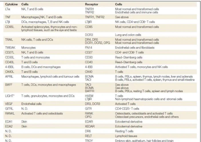
Fig.1 Timeline of the discovery of various members of the TNF superfamily and their receptors. Aggarwall et al 2012.
Table 1: Cellular expression of ligands and receptors of the tumor-necrosis factor superfamily. Aggarwall et al 2003.
Fig.2 Schema of some DR and non-DR of TNF/TNFR superfamilies and their tissue localization. Croft and Siegel 2017.
Fig.3 Human Fas Structure.
Fig.4 Comparison of human/mouse aa sequence. Fig.5 Human FasL structure.
Fig.6 Fas expression in naïve and activated T cells. Inaba et al 1999.
Fig.7 Mechanism of CTL mediated cytotoxicity. Adapted from Golstein and Griffiths 2018. Fig.8 RICD model. Adapted from Yi et al 2018.
Fig.9 Pro and anti-autoimmunity Fas/FasL activities. Adapted from Rossin et al 2019. Fig.10 Cancer immunoediting phases. Pennell 2015.
Fig.11 Fas/FasL roles in tumors. Adapted from Rossin et al 2019.
Fig. 12 Fas/FasL residues for binding. Schneider et al 1997 and Starling et al 1997. Fig.13 intrinsic and extrisic apoptotic pathways. Strasser et al 2009.
Fig.14 Receptor cluster and two models for Fas DD-FADD binding. Hymowitz and Dixit 2010. Fig.15 Fas conformation in 4:4 model. Scott et al 2008.
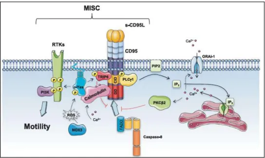
Fig.16 Fas mediated cytokines production. Brint et al 2013.
Fig. 17 Molecular deciphering of MISC complex. Guégan and Legembre 2018. Fig.18 Fas in AJ. Gagnoux-Palacios et al 2018.
Fig.19 Model for Fas mediated apoptosis. Schutze et al 2008.
Fig.20 Schematization of signaling complex I and II in TNFR and TRAIL/Fas. Adapted from Siegmund et al 2017.
Fig.21 DISC-interacting proteins mediated regulation of Fas response. Adapted from O’Reilly et al 2016. Fig.22 Schematic representation of TCR chain locus before and after rearrangement. Bosselut 2019. Fig.23 TCR/CD3 complex. Gelkop et al 2012.
Fig.24 Reconstruction of human TCR/CD3 complex. Dong et al 2019. Fig.25 T cell development in the thymus. Germain 2002.
Fig.26 Cytokines involved in T cell differentiation and produced by T cell subsets. Pennock et al 2013. Fig.27 Co-receptor scanning model. Courtney et al 2018.
VIII
Fig.28 Receptor deformation and CD3 tail conformational change models. Courtney et al 2018. Fig.29 Schematization of aggregation models. van der Merwe and Dushek 2011.
Fig.30 Major TCR signaling pathways. Gaud et al 2018. Fig.31 Model of Lck activity regulation. Gaud et al 2018.
Fig.32 Schema of Immune synapse formation. Hashimoto-Tane and Saito 2016. Fig.33 SHP-1/PTPN phosphatases targets. Gaud et al 2018.
Fig.34 Example of LAT protein endocytosis and movement through TCR/CD3 islands during TCR activation. Zucchetti et al 2019
Fig.35 Schema of main TCR immune checkpoint. Adapted from Chen and Flies 2013.
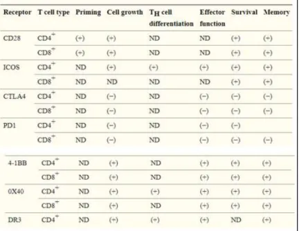
Table 2. Igs and TNFRs described as TCR immune checkpoints. Adapted from Chen and Flies 2013. Table 3. Examples of co-stimulatory and co-inhibitory receptor function in stages of T cell differentiation. Adapted from Chen and Flies 2013.
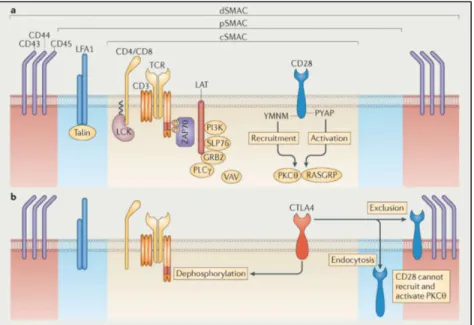
Fig.36 Localization of CD28 and CTLA-4 in the cSMAC upon TCR activation. Chen and Flies 2013.
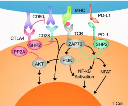
Fig.37 Scheme of CD28 molecular pathways induced upon concomitant TCR ligation. Bluestone et al 2011. Fig.38 Schematization of CTLA-4 and PD-1 mechanism of action. Adapted from Borcherding et al 2018. Fig.39 Examples of ICB strategies of CTLA-4 and PD-1. Waldman et al 2020.
Fig.40 ACT technique and example of second and third generation of CARs. Adapted from Waldman et al 2020.
IX
List of Abbreviations
ACT: Adoptive Cell transfer
ADAM: A disintegrin and metalloprotease
ADAP: Adhesion and degranulation promoting adapter protein Ag: Antigen
AJ: Adherens junction
ALPS: Autoimmune lymphoproliferative disease AP-1: Activator Protein 1
Apaf-1: Apoptotic protease-activating factor 1 APC: Antigen presenting cell
Arp2/3: Actin related protein 2/3 Bak: Bcl-2 homologous antagonist/killer Bax: Bcl-2 associated X protein
Bcl-2/10/XL: B cell lymphoma 2/10/XL BCR: B cell receptor
Bid/tBid: BH3 interacting-domain death agonist/ truncated BIM: Bcl-2-like protein 11
BTK: Bruton tyrosine Kinase
BTLA-4: B- and T-lymphocyte attenuator CAF: Cancer associated fibroblast
CARMA-1: Caspase recruitment domain-containing membrane-associated guanylate kinase protein-1 CAR-T: Chimeric antigen receptor T
Cbl b/c: Casitas B-cell lymphoma b/c
Cdc42: Cell division control protein 42 homolog
cFLIPL/S/R: cellular FLICE-like inhibitory protein Long/Short/Raji c-IAP1/2: cellular Inhibitor of apoptosis protein-1/2
CKI: Casein kinase I substrate CMV: Cytomegalovirus
CRAC: Calcium release-activated channels CRD: Cysteine rich domain
CSK: C-term src kinase CTL: Cytotoxic T lymphocyte
X
CTLA-4: Cytotoxic T-Lymphocyte Antigen 4 DAG: Diacylglycerol
DAXX: Death domain associated protein 6 DC: Dendritic cell
DcR: Decoy receptor DD: Death Domain
DED: Death Effector Domain
DISC: Death-inducing signaling complex Dlg: Disc large protein
DNT: Double negative T cell DP: Double positive T cell DR: Death receptor
DRM: Detergent resistant membrane
EAE: Experimental autoimmune encephalomyelitis ECD: Extracellular domain
EMT: Epidermal to mesenchymal transition ER: Endoplasmic reticulum
ERK1/2: Extracellular regulated kinases 1/2 ERM: Ezrin, radixin and moesin
FADD: Fas-associated protein with death domain Fap-1: Fas associated phosphatase-1
GADS: Grb2-related adaptor downstream of Shc GBM: Glycosphingolipid binding motif
GCE: Genetic code expansion
gld: Generalized lymphoproliferative disease Grb2: Growth factor receptor-bound protein 2 GSK3β: Glycogen synthase kinase 3 beta GVHD: Graft versus host disease GVT: Graft versus tumor
HLA: Human leukocyte antigen HVEM: Herpesvirus entry mediator ICAM: Intercellular Adhesion Molecule 1 ICB: Immune checkpoint blockade
XI
ICD: Intracellular domain
ICOS: Inducible T-cell COStimulator IFNα/β/γ: Interferon α/β/γ
IgG: Immunoglobulin G
IgSF: Immunoglobulin superfamily IkB: Inhibitor of nuclear factor kappa B IKK: IκB kinase
IL-1R1: Interleukin-1 receptor1 IL-2/4/6/8/10: Interleukin-2/4/6/8/10 IP3: Inositol triphosphate
IP3R: Inositol triphosphate receptor IS: Immune synapse
ITAM: Immunoreceptor Tyrosine-based Activation Motif ITIM: Immunoreceptor Tyrosine-based inhibition Motif Itk: IL-2-inducible T-cell kinase
ITSM: Immunoreceptor Tyrosine-based Switch Motif JNK: c-Jun N-terminal kinase
LAT: Linker for activation of T cells
Lck: Lymphocyte-specific protein tyrosine kinase LFA-1: Lymphocyte function-associated antigen 1 Lpr: Lymphoproliferation
LRR: Lysine rich region LTα: Lymphotoxin
LUBAC: Linear ubiquitination assembly complex
MALT-1: Mucosa-associated lymphoid tissue lymphoma translocation protein 1 MAPK: Mitogen-activated protein kinase
MC: Microcluster
mbFasL: membrane bound FasL
MHC I / II /pMHC: Major histocompatibility complex I / II / peptide MISC: Migration-inducing signaling complex
MMP: Metalloprotease
MTOC: Microtubule-organizing center
XII
NFAT: Nuclear factor of activated T cells
NF-κB: Nuclear factor kappa-light-chain-enhancer of activated B cells NK: Natural killer
NMR: Nuclear magnetic resonance PARP: Poly (ADP-ribose) polymerase PBMC: Peripheral blood mononuclear cell PCD: Programmed cell death
PD-1: Programmed cell death protein 1 PI3K: Phosphoinositide 3-kinases
PIP2: Phosphatidylinositol (4,5)-bisphosphate PIP3: Phosphatidylinositol (3,4,5)-trisphosphate PKC β/θ: Protein Kinase C β/θ
PLAD: Pre-ligand binding assembly domain PLCγ: Phospholipase C gamma
PP2A: Protein phosphatase 2 A PRD: Proline rich domain PRR: Proline rich region
PTEN: Phosphatase and tensin homolog PTM: Post translational modification
PTPN: Protein tyrosine phosphatases, non-receptor type Rac1: Ras-related C3 botulinum toxin substrate 1 Rap1: Ras-related protein 1
RICD: Restimulation induced cell death
RIPK1-3: Receptor-interacting serine/threonine-protein kinase 1/3 ROS: Reactive oxygen species
sFasL: soluble FasL SH2/3: Src homology 2/3
SHP-1/2: Src homology 2 domain-containing protein tyrosine phosphatase 1/2 SKF: Src kinase family
SLE: Systemic lupus erythematosus
SLP-76: SH2 domain-containing leukocyte phosphoprotein of 76kDa SMAC c/p/d: Supramolecular activation cluster central/peripheral/distal SMase: Sphingomyelin phosphodiesterase
XIII
SP: Single positive
STAT-1/3/4: Signal transducer and activator of transcription 1/3/4 TAB2/3: TGF-Beta Activated Kinase 1 (MAP3K7) Binding protein 2/3 TAK1: Mitogen-activated protein kinase kinase kinase 7
TCM: Central memory T lymphocytes TCR: T cell receptor
TEFF: Effector T lymphocyte
TEM: Effector memory T lymphocyte Tfh: Follicular helper T lymphocyte TGFβ: Transcription growth factor β Th: Helper T lymphocyte
THD: TNF homology domain TIL: Tumor infiltrating lymphocyte TIM: TRAF interacting motif
TKB: Tyrosine kinase binding domain TM: Memory T lymphocyte
TM/TMD: Transmembrane/ Transmembrane domain TN: Naive T lymphocyte
TNF(SF): Tumor necrosis factor (Superfamily)
TNFR(SF): Tumor necrosis factor receptor (Superfamily)
TRADD: Tumor necrosis factor receptor type 1-associated Death domain TRAF: TNF receptor associated factors
Treg: Regulatory T lymphocyte
VCAM: Vascular cell adhesion protein 1
1
T cell mediated immune response
Vertebrates have evolved multiple strategies to counteract invasion and aggression coming from a large panel of insults, from attack by microorganisms such as bacteria and viruses, to chemical and physical injuries. Immune system consists of a large network of chemical (humoral) and cellular mediators all finely regulated. Each cell type plays a specific role to respond efficiently to external invasions or even to internal modifications not recognized as physiologic. Both cellular and molecular events are differently involved in the two main types of immunity: the innate and the adaptive immunity.
The first barrier against non-self-invasion is fulfilled by innate immunity, which recognizes intrinsic or pathogenic patterns, without any specificity for the type of agent. Conversely, the adaptive immunity requires specificity against a determined pathogen, in order to amplify the strength of the response and efficiently defeat the foreigner invaders. Adaptive immunity is based on cell-cell cooperation mechanisms where different cell populations participate to maintain homeostasis and assure constant surveillance of the organism. Innate and adaptive immunity, furthermore, collaborate with each other, where first recognition of an unknown agent by innate system can lead to activation of the adaptive one as will be simplistically described below.
One vital characteristic of lot of cells that are part of the innate immunity is the ability to ingest (phagocytosis) their target or part of them, processing it inside their cytoplasm and exposing on their surface pathogen-specific peptides (antigens). All these cells act indeed as antigen presenting cells (APCs), able to induce and activate the adaptive immunity. The APCs expose the antigen to be presented to the adaptive cells through the Major Histocompatibility Complex (MHC). MHC can be divided in two main subclasses ( and) and shows one of the highest genetic variability of our genome, in order to load on their extracellular part specific antigens that will be recognized by membrane receptors of the adaptive immune cells, the T and B lymphocytes and natural killer (NK) cells. In this manuscript we will focus on the role of T lymphocytes, and how their interaction with APCs is important to modulate the immune response of an organism.
T lymphocytes that have completed their maturation process, briefly described in the second chapter of this thesis, are called naïve or resting T cells (TN). Upon encounters and recognition of antigens presented
by APCs by their T cell receptor (TCR), T lymphocytes get activated and they can clonally expand to efficiently respond to the infection becoming effector cells (TEF). Mature T lymphocytes can be mainly divided in two
classes: the one expressing the CD4 receptor and the one expressing the CD8 receptor. When CD8+ cells get activated they differentiate in cytotoxic T lymphocytes (CTL) that together with the cells of the innate system and NK cells can act in destroying the non-self invaders, by inducing their death. On the other side, CD4+ cells, once activated, will differentiate in one of the subclasses of “helper” population. Multiple classes of effector cells exist and their differentiation into one or the other subtype depends on intrinsic and extrinsic
2
factors such as type and strength of initial TCR activation, cytokines present in the stroma and the presence of different TCR co-receptors (immune checkpoint receptors) which act in modulating T cell fate. The main effector cell categories known so far are T helper 1, 2, 9, 17 (respectively called Th1, Th2, Th9 and Th17), the T helper follicular cells (Tfh) and the regulatory T cells (Treg) of which this last presents suppressive functions against other T helpers in order to avoid an excessive reaction of the immune system to any type of injury. Moreover, T lymphocytes can mutually downregulate themselves by induction of programmed cell death (PCD) at the end of the immune response, a mechanism known as T cell contraction. Furthermore, after the shutdown of the immune response, T lymphocytes can further differentiate into cells that keep the recognition pattern of the external agent that they were in contact with, to be able to respond rapidly to a second attack, the so called memory cells (further divided into effector and central memory cells TEM and
TCM), that circulate in the bloodstream as prepared and equipped guardians. These cells are, however, just a
small fraction of the pool.
One of the main roles of helper T cells is, therefore, to « help » other T cells and B lymphocytes, the other main lymphocyte subclass, to get properly activated and expand. Once they get activated, by their B cell receptor (BCR) in concert with other co-receptors, B lymphocytes can differentiate into plasma cells, a population that is able to create, expose and secrete antibodies, main actors of the humoral response. One important feature of the B lymphocytes is that, once activated, they can act as APCs, amplifying the propagation of the signal activating in a feedback loop other T lymphocyte.
Both deficiency or exacerbation of the immune response are cause of insurgence of severe diseases, ranging from immunodeficiency-related pathologies, when patients cannot properly activate the immune system upon infections, to autoimmune diseases when the immune response cannot be shut down.
Fas and FasL proteins are components of the Tumor Necrosis Factor Receptor/Tumor Necrosis Factor Superfamilies (TNFRSF/TNFSF) respectively. The Fas/FasL couple plays a fundamental role in the homeostasis of the immune cells. Fas/FasL interaction is actually one of the main mediators of the peripheral tolerance in the elimination of autoreactive T cells, and their deficiency is the main cause of the appearance of autoimmune disorders such as autoimmune lymphoproliferative diseases (ALPS) and systemic lupus erithematosus (SLE), pathologies characterized by the presence of excessive number of lymphocytes and production of autoantibodies. This subject will be deeply faced in the section of the first chapter dedicated to the role of Fas in physiopathology. Fas/FasL interaction is, furthermore, mediator of multiple pro-survival pathways, which are strictly context and cell type dependent. As for other members of TNFR superfamily, a role for Fas in the modulation of the T lymphocyte activation has been established, but nor its importance and neither its features as well as the molecular mechanism have never been elucidated so far. It is thus, of an extremely interest to go deeper into this field and try to define how these players influence T cell fate.
3
A. Fas/FasL system: structure, functions and signaling
1. TNF/TNFR superfamilies 1.1 Generalities
More than thirty years ago some scientists started to suspect that specific proteins secreted by immune cells could have a critical role in some human diseases such as tumor development, inflammations, autoimmunity and pathogen infection (Berke et al 1972, Erard et al 1984, Decker et al 1987, Goeddel et al 1986). In a tumoral context it was for instance observed that factors expressed by activated macrophages and lymphocytes were able to induce tumor rejection by necrotic phenotype (Granger et al 1969, Carswell et al 1975). Their identification led to the cloning, in 1984, of two proteins respectively named the tumor necrosis factor (TNF) and the lymphotoxin (LTα) (Pennica et al, Grey et al 1984). These two factors, that shared 50% of homology at the protein level, were the first two members of a superfamily that count nowadays 19 members (see list in Table 1) and that was named tumor necrosis factors superfamily (TNFSF)(Aggarwal 2003).
Fig.1 Timeline of the discovery of various members of the TNF superfamily and their receptors.
Aggarwall et al 2012.
One year later, the identification of the receptors that could bind to these two polypeptides was done by Aggarwal’s team and gave rise to the identification of the tumor necrosis factor receptor (TNFR) superfamily (TNFRSF)(Aggarwal et al 1985) that comprises currently 29 members (Aggarwal 2003) (Table 1). The figure 1 represents the timeline of the discovery of several members of TNFSF and TNFRSF. The receptor/ligand couples possess unique structural attributes that induce diverse signaling outcomes ranging from death to cell proliferation, survival, and differentiation (Alderson et al 1993, Aggarwal et al 1995, Klebanoff et al 2016). It is therefore not surprising that they play a fundamental role in a plethora of physiologic and pathologic processes from development and organogenesis to tumorigenesis and chronic diseases (Desbarats and Newell 2000, Kleber et al 2008, Straus et al 1999, Fisher et al 1995, Rieux-Laucat et al 1995). The notion that both ligands and receptors can bind multiple partners renders this network quite complex and with a high level of regulation.
4
Table 1: Cellular expression of ligands and receptors of the tumor-necrosis factor superfamily. Aggarwall et al 2003.
1.2 TNFs
Despite several differences in their conformation, in their roles and in their evolutionary conservation among the species, all members of the TNFSF share structural common features.
The TNFSF members are type II transmembrane proteins. They exist under different forms that were all described as active despite that they differ in their physiologic role: a membrane-bound form and soluble forms (Schneider et al 1998, Holler et al 2002, O’Reilly et al 2009). These latter can be secreted either as full-length proteins or as proteolytically shed extracellular fragments generated upon cleavage by metalloproteases (Gearing et al 1994).
Even if TNFSF proteins exist also as monomers their proactive forms require homo or hetero trimerization (Pennica et al 1984).
The most conserved region among the TNFSF members (25-30% of homology) resides in the extracellular part that is involved in the trimer assembly and that was therefore called TNF homology domain (THD) (Cha et al 1999, Jones et al 1989, Schneider et al 1998). Each monomer adopts a “jelly roll” conformation organized in beta sheets that form the THD. This structure allows a naturally occurring trimer formation (Bodmer et al
5
2000, Eck and Sprang 1989). In contrast, the domain which shows the lowest similarity in amino acid sequence is the C-terminus extracellular part (Reviewed in Locksley et al 2001).
Six members of TNFSF contain in their cytoplasmic region a casein kinase I substrate (CKI) domain that has been described to be involved in reverse signaling giving to these ligands a role of receptors that could transmit pro-survival signals involved in multiple cellular context, such as T lymphocyte activation and induction of gene expression (Smith et al 1994, Suzuki et al 1998, Watts et al 2005, Eissner et al 2004).
1.3 TNFRs
On the other side, the TNFRs are type I transmembrane proteins. The extracellular portion is characterized by the presence of 6 or more cysteines which form bisulfide bonds and define the cysteine-rich-domains (CRDs), a structural common feature among all the members of this superfamily. The number of CRDs variates among the different TNFRs (from 1 to 6) and plays a fundamental role for receptor-ligand interaction, as will be described later (Ashkenazi 2002).
The TNFRSF receptors cluster on the cell surface independently of the presence of their ligand. Their trimerization is mediated by their pre-ligand assembly domains (PLADs) formed by the N-terminal, the CRD1 and part of the CRD2 (Chan et al 2000, Clancy et al 2005, Siegel et al 2000). Conversely, the binding to the ligand was suggested to be mediated by a conformational change that occurs when receptors trimerize (Locksley et al 2001, Scott et al 2009).
These receptors can be furthermore divided in 3 categories according to the nature of the domain present in their cytoplasmic part: the death receptors, the non-death receptors and the decoy receptors. A schema comprising the main TNFRs/TNFs interaction and their cellular localization can be found in figure 2. The death receptors (DR) harbor a region called death domain (DD) (see the definition below). The ones that do not contain this domain are instead defined by the presence of a TRAF-interacting motif (TIM) (non-DR). In addition, the members that do not have any cytoplasmic domain cannot signals directly although they can compete with the signaling members and they have therefore been called decoy receptors (see below) (Ashkenazi 2002, Kwon et al 1998).
6
1.3.1 Death Receptors
Members of this subclass of TNFR are TNFR1, Fas, TRAIL-R1, TRAIL-R2, DR6, NGFR and EDAR (Wajant 2003). The DD is a region of around 80 amino acids, composed of 6 alpha helix bundle structure (Park et al 2007, Kohl and Grutter 2004). This domain constitutes a scaffold for DD-containing adaptor proteins upon engagement of the ligand. This homotypic DD-DD interaction (described below at page 33) initiates different molecular pathways that, depending on partners, cellular context, membrane localization or receptor’s post-translational modifications lead to cell death or survival signaling (Chakrabandhu et al 2016).
1.3.2 Non-Death Receptors
Non-DRs are characterized by the presence of a motif which binds other classes of adaptor proteins belonging to the TNF receptor associated factors (TRAF) family. TRAFs, through adaptor and ubiquitin ligase activity (Wallach et al 1999), can initiate the activation of Nuclear factor kB (NF-κB), Jun N-terminal kinase (JNK), Extracellular signal-regulated kinase (ERK) and Phosphoinositide-3 kinase (PI3K) (Dempsey et al 2003). Even though, this concept has been revaluated because again, depending of multiple factors, these TNFRs are able also to induce cell death, as, on the other side, the DRs can mediate non-death signals (Aktas et al 2006, Guicciardi et al 2009).
1.3.3 Decoy Receptors
As mentioned above, these receptors do not contain any functional intracellular motif, but through the binding to the TNFSF ligands they can compete with the other TNFRs, acting as negative regulators of the signaling. Four members of this subcategory have been identified so far: DcR1 (TRID/TRAIL-R3), DcR2 (TRUNDD/TRAIL-R4), DcR3 and Osteoprogenin (OPG) (Ashkenazi et al 2002).
7
Fig.2 Schema of some DR and non-DR of TNF/TNFR superfamilies and their tissue localization. Croft and Siegel 2017.
1.4 Tissue repartition
Concerning their tissue expression, TNFR members have quite ubiquitous localization, with higher level in thymus, liver, heart and kidney. Conversely, the members of the TNFSF are mostly restricted to immune cells such as activated lymphocytes, monocytes, dendritic cells and natural killer (NK) cells (Aggarwall et al 2003) (Fig.2). Additionally, they are expressed in tissues defined as immunoprivileged sites, such as eyes and testis, where their expression avoids unwanted immune response (Griffith et al 1996). Variation of the expression level of some TNF/TNFR members can be also found in a tumoral context, a feature that will be discussed later in this manuscript (page 26).
8
2. Fas/FasL proteins
Fas receptor was discovered by Yonehara and coworkers in 1989. They found that another receptor was able to bind an antibody originally directed against TNFR1 and to mediate cell apoptosis (Yonehara et al 1989).
By molecular and biochemical approaches, the team of Nagata and the one of Krammer respectively, were able to clone the Fas protein (Inazawa et al 1992, Lichter et al 1992). Its specific ligand, called Fas ligand (FasL), was cloned the year after by the team of Suda where they demonstrated its belonging to TNFSF (Suda et al 1993). This discovery initiated a new field, the one of cell death by “apoptosis” (see page 31). Since that moment hundreds of papers came out, by defining partners and way of action of this interaction.
In the following years, accumulation of molecular informations modified the paradigm of the role of Fas/FasL interaction, initially restricted to cell death induction, and comprising instead nowadays a large number of cell mechanisms in physiology and diseases, including non-apoptotic roles such as cell proliferation, differentiation and cell survival (Alderson et al 1993, Klebanoff et al 2016).
2.1 Fas protein structure
Human Fas receptor, also known as CD95/TNFRSF6/APO-1, is one DR of the TNFRSF. It is a type I transmembrane protein of 335 amino acids with a molecular weight that ranges from 42 to 56 kD, depending on its differential level of glycosylation. At the genomic level, the FAS gene is located at the 10th chromosome (10q23) and consists of 9 exons (Inazawa et al 1992, Yan et al 2005) that give rise to at least 6 different protein isoforms created by alternative splicing. These isoforms, being soluble truncated forms, might act as Fas full-length negative regulators or play unidentified roles (Papoff et al 1996).
Fig.3 Human Fas structure.
High homology level with Fas protein was found for the soluble decoy receptor DcR3, which lacks the transmembrane domain and that can compete for the ligand binding with Fas protein (Pitti et al 1998).
PDZ bs
1 47 83
N-term
CRD1
CRD2
CRD3
PLAD
Ligand binding
C-term
DD
174 166 190TM domain
127 230 314 335 Y232 Y291CD
95
C199 K K K K K9
Fas protein, schematized in Fig.3, is composed by: N-terminus extracellular part (1-173), containing:
A) the PLAD, by which the pre-associated homotrimer can form from homotypic interaction.
B) three CRDs critical for the interaction with the ligand (see page 29 for more detailed explanations). C) a glycosphingolipid binding motif (GBM) that plays an important role in Fas membrane localization
and clathrin-dependent internalization (Chakrabandhu et al 2008).
One transmembrane domain (174-190) that can form also stable homotrimer in the lipid bilayer. At the position 183 a proline is present to allow sufficient flexibility for the accommodation of the hydrophobic core (Fu et al 2016).
C-terminus cytoplasmic domain (191-335), which includes:
A) in its most membrane proximal part a lysine rich region (LRR) important for lipid raft localization, together with the cysteine present at position 199.
B) From position 202 starts a 68 amino acid DD, fundamental for the binding of the adaptor protein FADD and the initiation of the apoptotic signaling. Fas DD-FADD interaction models will be described in the section dedicated to the apoptotic signal. Interestingly the DD contains two different tyrosines at the position 232 and 291 subjected to phosphorylation by the Src kinase family members (Chakrabandhu et al 2016).
C) Finally, a PDZ binding site (SLV) at the extreme C-terminus end, important for protein stability and interaction with other partners, mainly PDZ-containing proteins (Gagnoux-Palacios et al 2018). Beside the glycosylation, Fas protein is subjected to different kind of post translational modifications (PTMs), such as phosphorylation, palmitoylation, nitrosylation and glutathionylation which modulate its stability, cellular localization and functions (reviewed by Seyrek-Lavrik 2019). The role of Fas post translational modifications will be developed in the section concerning Fas signal regulation (pages 42 and 45).
As all members of TNFRSF, Fas exists as monomer but it has to trimerize in order to accomplish its function. We have already mentioned the presence of a PLAD in the extreme N-terminus of Fas (Papoff et al 1999) that is essential for its ability to self-associate independently of its ligand binding (Siegel et al 2000, Chan et al 2000). Recently, Fu et al defined also the importance of the TM domain for transduction of optimal intracellular signal, by nuclear magnetic resonance (NMR) technique, that resides in the presence of prolines which allow a proper steric conformation of the liganded trimers (Fu et al 2016).
At cellular level Fas is mostly localized at the plasma membrane, but it can internalize and so it can be found also in the membrane of endosomes or of other intracellular compartments (Reviewed by Tchikov et al 2011).
10
Fig.4 Comparison of human/mouse aa sequence. In bold the common aa between the species.
The molecular mechanisms underlying Fas internalization will be further described later in this manuscript (pag 43).
In mouse, the FAS gene is located at the distal part of the chromosome 19 and it contains 13 exons. Mouse genome does not contain a sequence for DcR3, which is conserved instead just in primates. Mouse Fas is a protein of 327 amino acids. Despite the conservation of the function of apoptosis inducer of this protein among vertebrates, their amino acid sequences differ (Fig.4). Murine homologue of Fas protein is indeed quite divergent from the human one in term of primary sequence. These differences are mostly localized in the cytoplasmic region rather than in the extracellular one. Indeed, murine Fas is able to bind with human FasL. Therefore, data obtained with murine Fas have to take into account these differences before transposing the results on humans, considering that the signaling regulation can variate in a consistent way.
Despite the primary sequence divergence, most of the functional and structural informations given for the human Fas are also present in mouse Fas, from the post translational modifications (to be adapted at the species-specific amino acid sequence), the necessity of trimerization to be able to signalize, the ubiquitous expression in all the tissues of the organism, to the presence of the three regions divided in: N-term extracellular domain (1-169), one transmembrane domain (170-186) and a C-term cytoplasmic tail (187-327). Interestingly the PDZ-binding site at C-terminus expressed in human Fas is not found in mouse.
11
2.2 FasL protein structure
Fas ligand (FasL/ APO-1L/ CD95L/CD178/TNFSF6) is a cytokine of the TNFSF and the only known ligand for Fas. Conversely, FasL can bind also to DcR3. It is a type II transmembrane protein of 281 amino acids with a molecular weight that ranges from 38 to 42kD depending on its glycosylation status.
Fig.5 Human FasL structure.
FasL exists in 2 main forms: as a membrane-bound protein, and as a soluble protein. Furthermore, the membrane-bound form can be expressed at the plasma membrane or in membrane of exosomes that can be released by the cells in the stroma, a process that occurs in NK and CTL cells during their cytotoxic action (Wasem et al 2001, Bossi et al 1999, Blott et al 2001). The soluble form, on the other side, can be a full-length secreted protein or a cleaved one lacking its intracellular domain (Schulte et al 2007, Kirkin et al 2007).
As the other members of TNFSF, FasL is subjected to ectodomain shedding. This process is mediated by both matrix metalloproteases, such as MMP7, and disintegrins of the ADAM family. ADAM 10 was identified as the main FasL sheddase in T cells (Schulte et al 2007, Kirkin et al 2007) that releases in the stroma a 26-29kD form (Schulte et al 2007). The functional meaning of this cleavage was initially attributed to a down-modulation of the apoptotic signal since the soluble form of FasL resulted to be unable to induce apoptosis. Lines of evidence supporting functional role of the cleaved form of FasL are now several and attributed mostly to induction of non-apoptotic signals. Examples of these evidence will be found in this manuscript especially in the section dedicated to the role of Fas and FasL in pro-survival signaling (from page 36). The cleavage can be constitutive (Huovila et al 2005) or induced by several stimuli ranging from variation of Ca2+ level (Endres et al 2003, Nagano et al 2004), activation of growth factors (Fisher et al 2003), and even binding of ligands to receptors (Janes et al 2005). ADAM10 cleavage was found to be coupled to intracellular shedding, mediated by the signal peptide peptidase-like 2a (SPPL2A), which generates a free intracellular FasL form that can translocate into the nucleus and modulate the expression of target genes (Kirkin et al 2007). Furthermore, this mechanism generates a reverse signaling that was found to be involved in the down-modulation of the immune response (Luckerath et al 2011).
At the genomic level, FASLG resides in chromosome 1 (1q24.3) and consists in 4 exons, and only another isoform of the protein, created by alternative splicing, has been identified.
TM domain
C-termTHD
SA
PRD
N-term 1 37 70 82 103 130 183 275 291Soluble form
CD
95
L
12
FasL, as all the members of TNFSF, acts as a trimer in order to interact with its binding partner. FasL monomer, schematized in Fig.5, consists of:
N-terminus intracellular domain (1-80), the longest among the TNFSF members (Takahashi et al 1994) containing:
A) a proline rich domain (PRD), a protein-protein interaction motif for SH3 or WW bearing proteins, important for secretory lysosome storage and trafficking to the plasma membrane (Blott et al 2001). B) a CKI-S described to be necessary for FasL reverse signaling and regulation of gene expression
(Luckerath et al 2011).
One transmembrane domain (81-102) with SPPL2A cleavage site at position 81-82 C-terminus extracellular domain (103-281) containing:
A) the THD, conserved among the members of TNFSF, necessary for binding to CRDs of Fas B) a self-assembly (SA) motif, necessary for ligand trimerization together with the THD.
The site for ADAM10 cleavage for the production of the soluble form is located at the position 129-130. Other PTMs identified for FasL are the phosphorylation on tyrosine (Zuccato et al 2007), palmitoylation and monoubiquitination, important for FasL localization at membrane level and for internalization (Cahuzac et al 2006 and Reviewed in Voss et al 2008).
At cellular level FasL is a membrane-bound protein, but its localization and level of expression are finely regulated. Because its constitutive expression would cause inappropriate cell death in Fas positive cells, it can be stored in secretory lysosome by immune cells, such as cytotoxic T lymphocytes and Natural killer cells, until they receive the signal that allows its membrane exposure or secretion (Wasem et al. 2001, Bossi and Griffith 1999, Blott et al 2001). FasL expression regulation in immune system will be described below at page 13 in this manuscript.
Differently from its receptor counterpart, FasL is extremely conserved compared to its mouse homologue, sharing more than the 77% of primary sequence (Nagata et al 1997), and consequently most of the structural and functional characteristics. Palmitoylation represents an exception, which is absent in murine FasL. The mouse protein consists of 279 amino acids. Human and mouse FasL have been considered as interchangeable polypeptides. Its gene locus resides also in mouse at the chromosome 1 and is divided in 4 exons.
13
3. Fas/FasL in physiopathology
3.1 Role of Fas and FasL in immune system
3.1.1 Regulation of Fas/FasL expression in immune cells
Even if Fas expression among the tissues is mostly ubiquitous, it is well established that it plays a central role in the immune system, mainly by regulating lymphocytes homeostasis, via its expression on T cells, B cells and APCs in general.
It is expressed in all the immune cell types, even though its expression level is finely regulated among the different subpopulations to guarantee an efficient response to the FasL engagement at the precise timepoint.
Fig.6 Fas expression in naïve and activated T cells. Inaba et al 1999.
It has been described that human naïve T and B lymphocytes that have never been activated show extremely low level of Fas and are insensitive to FasL induced apoptosis (Fig.6). Interestingly, it is still controversial if these basal levels are anyway able to initiate a non-death signal, as the T cell co-stimulation, but this subject will be further discussed later. When T cells get activated upon the recognition of the antigens, they start to upregulate Fas (Trauth et al 1989), thus becoming sensitive to apoptosis, in order to shut down in a proper way the immune response and avoid an inappropriate response that can be detrimental for body homeostasis. Even though, the involvement of Fas in the elimination of the T lymphocytes is still controversial and will be faced in the section 3.1.5. Even the dendritic cells (DC) follow the same trend. Once they have been recognized by T cells, they start to upregulate Fas to become sensitive to apoptosis. This is a mechanism to avoid T cell over-activation by modulating the activated APCs circulation.
Expression of FasL is even more tightly regulated. We have already mentioned that FasL expression is restricted to few cell populations, such as the activated lymphocytes and neutrophils or other antigen
14
presenting cells. It is upregulated in T cells upon activation but its form and cellular localization are critical to regulate its activity. In cells bearing secretory lysosomes (NK and CTL) FasL is retained in these organelles until they receive a signal to release them at the plasma membrane, making them able to kill Fas bearing cells, in a mechanism known as « kiss of the death » (Bossi and Griffith 1999, Lowin et al 1996). The explanation of this process will be further discussed below in this chapter.
3.1.2 CTL and NK use FasL to kill
NK and CTL are immune cells that have an extremely important complementary role in the innate and adaptive immune system respectively. They have the capacity to recognize and eliminate virus infected cells and tumoral cells.
CTL and NK use the same systems to kill their target: the perforin (pore forming protein) /granzyme (serine proteases) and TNF/TNFR system where FasL mediated cell death is broadly exploited. These cells, as mentioned above, contain in their cytoplasm secretory lysosomes where FasL protein and perforins/granzymes are stored (Wasem et al 2001, Bossi and Griffith 1999, Blott et al 2001). Whether FasL and perforin/granzyme share the same granules and are released upon the same stimuli remains a matter of debate (review of Lettau 2015).
Fig.7 Mechanism of CTL mediated cytotoxicity. Adapted from Golstein and Griffiths 2018.
In response to non-self Ag recognition (for NK cells) or T cell restimulation (for CTL) (He et al 2007) the secretory lysosomes move from the cytoplasm to the cell membrane and release in the extracellular
15
environment the lytic agents by a process known as degranulation or expose at the plasma membrane the FasL (Fig.7). Interestingly, the FasL that is transported at the cell surface upon restimulation is mostly the preformed protein contained in the secretory lysosome more than the de novo synthetized, since the inhibition of new transcription by cyclosporin A after restimulation minimally affect the exposure of FasL at the membrane (Wasem et al 2001). Once exposed on the plasma membrane FasL can mediate its apoptotic function by localizing in the lipid raft (Nachbur et al 2006, Cahuzac et al 2006) or can be cleaved by MMPs (Mariani et al 1995) which enhances FasL mediated inflammation.
Another compartment of FasL storage in these type of cells are the multivescicular bodies (MVB) that can be directly targeted on the immune synapse upon T cell restimulation and allow the release of FasL-bearing exosomes, together with other proteins involved in T cell activation, directly at the contact zone with the target cell (Blanchard et al 2002).
3.1.3 Lpr and gld mice models in the discovery of Fas/FasL as mediators of T cell homeostasis The discovery by Andrews et al in 1978 of a naturally occurring mouse of the MLR strain developing lymphadenopathy and splenomegaly opened the gates in the description of Fas role in the immune system (Andrews et al 1978). This mouse was called lpr for lymphoproliferation and a second mutant with a similar phenotype gld for generalized lymphoproliferative disease was then discovered (Roths JB 1984). Indeed, mutation in the FAS or FASLG gene were identified as the origin of the systemic lupus erythematosus (SLE), phenotype observed in lpr and gld mice respectively (Ramsdell et al 1994b, Watanabe-Fukunaga et al 1992a). Hallmarks of these mutations are the accumulation of normal CD4+ and CD8+ lymphocytes and of the unusual double negative T lymphocyte population (CD4-/CD8-/B220+), that are causing the lymphadenopathy and splenomegaly. They also show autoimmune features, such as the presence of autoreactive T cells, amplification of B cell activation and proliferation, as well as the presence of a high content of autoantibodies of the families IgG and IgM. All these dysregulations lead to severe glomerulonephritis and arthritis symptoms that finally provoke death of the mice few months after their birth. It was later proved that the severity of the phenotype was strictly dependent on the mouse genetic background (Nagata et al 1995).
Adachi et al in 1993 identified that the cause of the extremely low level of Fas mRNA and protein (leaky mutation) was due to the insertion of a transposable element in the intron 2 of the FAS locus that was creating a truncated and loss of function protein (Adachi et al 1993). Another Fas mutation was identified in another strain called lprcg. It consists of a single point mutation (T to A) inside FAS exon 9 (just after
recognized as coding for the death domain) thus creating a full-length protein without its apoptotic capacity. The gld strain, on the other side, was caused by a point mutation, T to C, of the FASLG gene in the external region of the translated protein (C-term) making it unable to bind the Fas receptor.
16
It was therefore clear that the Fas/FasL system was playing a fundamental role in maintaining the homeostasis of immune cells and in controlling autoimmunity. Even though, lpr and gld mice did not show apparent defects in the thymocyte positive and negative selection (Singer and Abbas 1994, Sidman et al 1992). Instead, the role of Fas in preventing autoimmunity insurgence was addressed to the peripheral tolerance, in the clonal deletion of autoreactive T cells mediated by APCs (Vignaux et al 1994).
3.1.4 Autoimmune lymphoproliferative disease: the human deficiency of Fas/FasL system
The crucial role of the Fas/FasL couple in a human context was due to the discovery that patients lacking a functional Fas receptor developed a chronic non-malignant autoimmune lymphoproliferative syndrome (ALPS) (Rieux-Laucat et al 1995). It is also known as Canale and Smith syndrome referring to the names of the two scientists that initially described it in 1967 (Canale and Smith 1967). The most frequent symptoms that appear (usually before the age of 5) in this rare genetic disease are a benign lymphoproliferation through the accumulation of double negative TCRαβ lymphocyte (DNT) in the lymphoid organs that lead to spleen and lymph nodes enlargement. These DNT lymphocytes have some defined features, such as the presence of the MHC, high level of CD28 protein, the presence of the markers CD57, associated with senescent cells and, on the other side, of the naïve cell marker CD45RA (Rensing-Ehl et al 2014 and reviewed by Rieux-Laucat et al 2018). These features led to the hypothesis that this population might have arisen from abnormal differentiation and accumulation of CD4+ and CD8+ single positive cells in absence of Fas (Rensing-Ehl et al 2014, Volkl et al 2016). In two third of the patients, autoimmunity features are found with a high level in the serum of FasL, IL-10 and B123 vitamin and hyper immunoglobulinemia G and A. These manifestations could be the cause of c hepatitis, glomerulonephritis and dermatitis (Fisher et al 1995). In addition, ALPS patients are more prone in the onset of certain type of cancer such as Hodginks and non Hodginks lymphomas (Straus et al 2001).
The identification of mutations in other genes than in FAS in ALPS patients allow the classification of this disease in different subgroups:
The ALPS-0 and Ia subgroups (ALPS-FAS) describe the ALPS presenting respectively recessive and dominant inherited mutations of the FAS gene. The ALPS Ia form found in 70% of the patients is the most common one (Straus et al 1999). The Fas translated protein, unable to trimerize, fails to induce the apoptotic cascade. In ALPS type 0 a form of recessive mutation of Fas is caused by a large deletion on the last exon (Rieux-Laucat et al 1995). This latest type of mutation is, furthermore, prone to become homozygous by loss of heterozygosity (LOH) and thus can cause the development of severe symptoms. The insurgence of symptoms in heterozygous siblings with healthy heterozygous parents enhanced the point that Fas mutations could be both dominant or recessive and that the incomplete penetrance of the mutation was interfering with the onset of the disease
17
(Bettinardi et al 1997). The discovery that ALPS insurgence could have arisen from somatic mutation in the hematopoietic progenitors, detectable in DNT lymphocytes, increased the level of complexity of this pathology (ALPS-sFAS Im). Indeed, even if in a small proportion compared to the wild type T cells, these apoptotic resistant mutants could have caused the manifestation of the symptoms for their selective advantages. Up to now, somatic ALPS represent the 15% of the cases (Dowdell et al 2010).
The ALPS-Ib group (ALPS-FASLG) contains the mutations in the gene encoding Fas-ligand. Nowadays there are 8 cases of this rare form of the disease and only one is a dominant mutation, which however does not reflect totally the criteria of classical ALPS (low DNT accumulation and no splenomegaly) (Del-Rey et al 2006, Nabhani et al 2014).
The ALPS-II group involves patients that bear mutations within Fas apoptotic cascade such as in Caspase-8 (-IIb) and 10 (-IIa) (Wang et al 1999, Salmena et al 2005). The latest, however, is often associated with Fas mutation in order to be symptomatic (Cerutti et al 2007, Martinez-Feito et al 2016). Moreover, Caspase-8 deficiency was not reflecting totally the ALPS features, since was leading to combined immune deficiency with defect in lymphocyte proliferation and recurrent bacterial and viral infections. Homozygous missense mutation of FADD, described to lead to an autosomal recessive inherited disorder, was identified to show biological features of ALPS with different clinical features (no lymphoproliferation and autoimmunity) such as predisposition to bacterial and viral infections (Bolze et al 2010).
The ALPS-III group (ALPS-U) collect all the cases that show clinical phenotype close to ALPS but for which the genetic defects were not yet defined. It was suggested anyway that might be associated in some cases with reduced CTLA-4 expression at the cell surface of activated effector T cells and Tregs (Kuehn et al 2014, Schubert et al 2014).
Even if in this manuscript I focused just on the Fas-related ALPS types, as we saw for the ALPS-U group, not all the forms of ALPS are strictly related with Fas pathway. Insurgence of ALPS-like disease was, indeed, described for mutations in other molecules such as KRAS or NRAS which give rise to the so called Ras-Autoimmune Leukoproliferative Disease (RALD) (ALPS IV)(Oliveira et al 2010, Takagi et al 2011, Calvo et al 2015).
3.1.5 RICD in vitro and in vivo: controversies
Activated peripheral T cells are eliminated in order to silence the immune response at the end of an infection, a mechanism known as T cell contraction (Krueger et al 2003). Indeed, upon the first TCR stimulation T cells start to upregulate Fas and FasL expression, in a way that, after another contact with the same Ag presented by the MHC on the APCs they might be eliminated by apoptosis, in a process known as restimulation induced cell death (RICD) (Fig.8) (Zheng et al 2017). The observed apoptosis was first attributed
18
to the FasL/Fas system. In line, central memory T cells, which are known to be fundamental in activating a second immune response upon reinfection with the same antigen, are less sensitive to Fas induced cell death in vitro.
Nowadays, the role of Fas in RICD has been revaluated. If Fas is able to induce apoptosis in restimulated cells in vitro, this is not necessary true for in vivo models. More recent studies showed that Fas-induced apoptosis is the main mechanism for cell death only after repetitive contacts with low affinity Ags for their TCR receptor (Fortner et al 2011), which occur for cells that have escaped to selection in the central tolerance setting (autoreactive T cells) or in response to a chronic infection (Varanasi et al 2014).
Fig.8 RICD model. Adapted from Yi et al 2018.
The self Ags which show a high affinity for TCR are mostly eliminated by negative section in the thymus during cell development, in a process that involves the mitochondrial apoptotic pathway and the deprivation of cytokines in the microenvironment and is independent from Fas. In the same way, acute infections show a high Ag affinity for T cell receptor and upon this interaction, T cell death has been found to be more dependent from the proapoptotic protein BIM, known member in the induction of mitochondrial dependent cell death (Hildeman et al 2002, Pellegrini et al 2003, Strasser et al 2009), as well from the withdraw of survival cytokines, such as IL-2 and IL-7. Pellegrini and coworkers defined, indeed, that upon acute infection with Herpes simplex virus (HSV) Fas and TNFR1 were dispensable for the response and elimination (Pellegrini et al 2003). Furthermore, the viral response in a double knock-out of both Caspase-8 and RIPK3, fundamental proteins for induction of Fas mediated cell death, was not impaired and still worked at high strength (Feng et al 2018).
Conversely, in chronic response, there is a persistence of the pathogen in the host, and a « state of cohabitation » is achieved to keep a tolerated titer and avoid damageable response (Sprent and Tough 2001).
19
Hughes and his team in 2008 found that upon challenge with CMV (which causes a chronic infection) both Fas/FasL system and the apoptotic BIM protein are necessary for clonal T cell deletion (Hughes et al 2008). However, the need of Fas in response to repetitive stimulations is also questioned. In the paper of Stranges of 2007 by using a mouse model where Fas was removed in total T cells or just in activated T cells (Th1 CD4+ or CD8+), they did not find any selective advantage in the survival of these cells compared to control mice (Fas sufficient mice) in response to primary or repetitive antigenic stimulations, doubting on the role for Fas in the elimination of T cells in both T cell contraction (primary response to stimulation) or upon restimulation (Stranges et al 2007). What was instead confirmed is the effect of Fas deficiency in the onset of autoimmune features, confirming a role for Fas in the deletion of autoreactive cells (Stranges et al 2007).
3.1.6 Fas in preventing and promoting autoimmunity
Fig.9 Pro and anti-autoimmunity Fas/FasL activities. Adapted from Rossin et al 2019.
At the state of the art it is well established that Fas/FasL proteins play a fundamental role in preventing the systemic autoimmunity insurgence and progression. However, Fas activation can also promote autoimmunity development in other specific cases rendering the situation quite complex (Fig.9).
As both lpr and gld mice models show evident autoimmune features, the role of Fas/FasL system was at first highly investigated in its property to prevent autoimmunity. Fas/FasL system has been shown to be determinant in the control of peripheral tolerance as its deficiency is causing spontaneous autoimmune