HAL Id: tel-02533666
https://tel.archives-ouvertes.fr/tel-02533666
Submitted on 6 Apr 2020
HAL is a multi-disciplinary open access archive for the deposit and dissemination of sci-entific research documents, whether they are pub-lished or not. The documents may come from teaching and research institutions in France or abroad, or from public or private research centers.
L’archive ouverte pluridisciplinaire HAL, est destinée au dépôt et à la diffusion de documents scientifiques de niveau recherche, publiés ou non, émanant des établissements d’enseignement et de recherche français ou étrangers, des laboratoires publics ou privés.
interfaces in the musculo-skeletal system based on tissue
engineered osteotendinous junctions
Alejandro García García
To cite this version:
Alejandro García García. Multiscale analysis of multi-layered tissues constructs : interfaces in the musculo-skeletal system based on tissue engineered osteotendinous junctions. Bioengineering. Univer-sité de Technologie de Compiègne, 2019. English. �NNT : 2019COMP2488�. �tel-02533666�
Par Alejandro GARCÍA GARCÍA
Thèse présentée
pour l’obtention du grade
de Docteur de l’UTC
Multiscale analysis of multi-layered tissues
constructs : interfaces in the musculo-skeletal system
based on tissue engineered
osteotendinous
junctions
Soutenue le 11 juin 2019
Spécialité : Bio-ingénierie, Biomécanique, Biomatériaux : Unité
de Recherche Biomécanique et Bio-ingénierie (UMR-7338)
TISSUES CONSTRUCTS: INTERFACES IN THE
MUSCULO-SKELETAL SYSTEM BASED ON TISSUE
ENGINEERED OSTEOTENDINOUS JUNCTIONS
PRESENTED BY : GARCIA GARCIA ALEJANDRO
THESIS SUPERVISORS :BEDOUI FAHMI LEGALLAIS CECILE
JURY MEMBERS :
Dr. AMEDEE VILAMITJANA JOELLE, DR INSERM (REFEREE) Dr. GANGHOFFER JEAN-FRANCOIS, PROFESSOR (REFEREE) Dr. PAUTHE EMMANUEL, PROFESSOR
Dr. VAYSSADE MURIEL, PROFESSOR Dr. BEDOUI FAHMI, MCF HDR Dr. LEGALLAIS CECILE, DR CNRS
THESE PRESENTEE POUR L’OBTENTION DU GRADE DE DOCTEUR DE L’UNIVERSITE DE TECHNOLOGIE DE COMPIEGNE SPECIALITE
BIO-INGENIERIE, BIOMECANIQUE, BIOMATERIAUX 11 Juin 2019
2
Résume
L’objectif de cette thèse était le développement d'un substitut bio-hybride pour la reconstruction du continuum tendon-os sur le principe de la ingénierie tissulaire. Après une analyse bibliographique exhaustive des structures natives et de leur environnement, nous avons d'abord proposé la réalisation de chaque système séparément en utilisant des scaffolds en polycaprolactone réalises par electrospinning.
Dans un premier temps, nous avons combiné l’electrospinning et l’electrospraying pour produire un scaffold composé de polycaprolactone et d’hydroapatite avec une structure en forme de nid d'abeille. Notre hypothèse était de doter le substitut d'une structure biomimétique favorisant l'adhésion, la colonisation et la différenciation cellulaire. L'analyse mécanique et biologique in vitro réalisée avec une lignée cellulaire progénitrice et des tests organotypiques a confirmé notre approche originale. Ensuite, le matériel ensemencé avec des cellules souches de moelle osseuse a été implanté avec succès par nos collaborateurs d'Amiens dans le but de traiter un défaut maxillo-facial chez un modèle de rongeur.
Parallèlement, pour la reconstruction du tendon, nous avons réalisé différents scaffolds d'electrospinning, dont la taille et l'organisation (aléatoire/alignée) des fibres varient. Dans une perspective bio-inspirée, nous avons combiné les scaffold avec l'étirement dynamique pour reproduire l'entraînement physique. Sous ces stimulations mécaniques, établies d'abord avec la même lignée cellulaire progénitrice, nous avons démontré dans une deuxième étude que les CSM s'alignaient sur l'axe d'étirement et produisaient une matrice extracellulaire, ce qui a permis de conserver les propriétés mécaniques de la matrice biohybride pendant les deux semaines de la culture.
Nous avons démontré que la différenciation cellulaire vers la lignée tendineuse et osseuse a été réalisée avec succès en l'absence de tout facteur de différenciation, étant spécifiquement lié aux propriétés des matériaux et à la mécanotransduction. Par conséquent, l'étape suivante, qui consiste à assembler les deux échafaudages avec une zone de transition, devrait conduire à la reconstruction de ce continuum osseux-tendon. Mots clés Ingénierie tissulaire, os, tendon, interface, electrospinning, biomécanique, cellules souches, polymères
3 The objective of this thesis was the development of a biohybrid substitute for the reconstruction of the bone-tendon continuum based on tissue engineering strategies. After an exhaustive bibliographic analysis of the native structures and their environment, we first proposed the realization of each system separately using electrospun polycaprolactone scaffolds.
At first, we combined electrospinning with electrospraying techniques to produce a PCL-hydroapatite scaffold with honeycomb cavities. Our hypothesis was to provide the substitute with a biomimetic structure favoring cell adhesion, spreading and differentiation. The in vitro mechanical and biological analysis performed with a progenitor cell line and with organotypic assays confirmed our original approach. Then, the material seeded with bone marrow stem cells was successfully implanted by our collaborators in Amiens with the objective of treating a maxillofacial defect in a rodent model.
In parallel, for the tendon reconstruction, we investigated several electrospinning processes, varying fibers’ size and organization (random/aligned). In a bioinspired perspective, we combined the choice of the scaffold with dynamic stretching to reproduce physical training. Under those mechanical stimulations, established first with the same progenitor cell line, we demonstrated in a second study that MSCs aligned with the stretching axis and produced extracellular matrix, which in turn allowed to keep the mechanical properties of the biohybrid scaffold all over the 2 weeks of culture.
We demonstrated that cell differentiation towards tendon and bone lineage was successfully achieved in the absence of any differentiation factor, being specifically related to materials properties and mechanotransduction. Therefore, the next step consisting in the assembly of both scaffolds with a transition area should lead to this bone-tendon continuum’s reconstruction.
Key words
Tissue engineering, bone, tendon, interface, electrospinning, biomechanics, stem cells, polymer
4
Laboratories
UMR CNRS 7338: Biomécanique et Bioingénierie (BMBI) Alliance Sorbonne Université
Université de Technologie de Compiègne Centre de Recherches
Rue du Docteur Schweitzer 60200 Compiègne
FRE CNRS 2012 : Roberval Alliance Sorbonne Université
Université de Technologie de Compiègne Centre de Recherches
Rue du Docteur Schweitzer 60200 Compiègne
5
Publications:
Garcia Garcia A., Beldjilali-Labro M., Farhat F., Bedoui F., Grosset J.F., Dufresne M., Legallais C. Biomaterials in Tendon and Skeletal Muscle Tissue Engineering: Current Trends and
Challenges. Materials 2018, 11, 1116; doi: 10.3390/ma11071116.
Garcia Garcia A, Hebraud A., Duval J.L., Wittmer C.R., Gaut L., Duprez D., Egles C., Bedoui F., Schlatter G., Legallais C. Poly(Ɛ-caprolactone)/Hydroxyapatite 3D Honeycomb Scaffolds for
a Cellular Microenvironment Adapted to Maxillofacial Bone Reconstruction. ACS Biomater.
Sci. Eng. 2018,4,3317-3326 ; doi : 10.1021/acsbiomaterials.8b00521.
Garcia Garcia A., Perot J.B., Beldjilali-Labro M., Dermigny Q., Naudot M., Le Ricousse S., Legallais C., Bedoui F. Monitoring mechanical stimulation towards optimal tendon tissue
engineering: Mechanical and biological multiscale study. Draft completed.
International and national congress:
Garcia Garcia A., Schlatter G., Hebraud A., Egles C., Bedoui F., Legallais C. Bone reconstruction with Polycaprolactone/hydroxyapatite scaffolds. ESB Lyon 2016. Poster.
Garcia Garcia A., Schlatter G., Hebraud A., Egles C., Bedoui F., Legallais C. Bone tissue engineering with Polycaprolactone/hydroxyapatite biomimetic scaffolds. 43th ESAO Congress 2016, Warsaw, Poland. Oral presentation.
Garcia Garcia A., Beldjilali-Labro M., Farhat F., Perot J.B., Dermigny Q., Dufresne M., Grosset J.F., Bedoui F., Legallais C. Multi-scale approach to reconstruct a bioartificial system of system: the example of the bone-tendon-muscle continuum. SOSE 2018 Paris, France. Poster.
6 Garcia Garcia A., Perot J.B., Naudot M., Le Ricousse S., Legallais C., Bedoui F. Mechanical stimulation monitoring towards optimal tendon tissue reconstruction. XLV ESAO Congress
2018, Madrid, Spain. Oral presentation.
Garcia Garcia A., Perot J.B., Naudot M., Le Ricousse S., Legallais C., Bedoui F. Mechanical stimulation of tendon-engineered construct. Termis World Congress 2018, Kyoto, Japan. Poster.
Other scientific communications:
Garcia Garcia A., Naudot M. Comportement de cellules souches mésenchymateuses sur des matériaux électrospinnés pour une application en chirurgie maxillo-faciale. Quatrième
journée scientifique de l’Institut Faire Faces 2017, Amiens, France.
Fête de la science 2017, Université de Technologie de Compiègne. Participation au stand :
De Capitaine Crochet à Iron Man.
Awards
yESAO Exchange Program, European Society for Artificial Organs. Coaxially electrospun and
directional solidified scaffolds for bone and vascular tissue engineering. 43th ESAO Congress 2016, Warsaw, Poland.
7
List of Figures ... 9
List of Tables ... 11
Abbreviations ... 12
Introduction: from bone tissue engineering to the reconstruction of the musculo-skeletal system of system ... 15
Objective of the thesis ... 15
Chapter I: State of the art ... 19
1. Bone Tissue Engineering: Current Trends and Challenges ... 19
1.1. Bone Composition and Structure ... 19
1.2. Bone Remodeling and Injuries... 20
1.2.1. Remodeling ... 20
1.2.2. Injuries ... 21
1.2.3. Bone Grafts and Subtitutes for Bone Reconstruction ... 21
1.3. Bone Tissue Engineering ... 22
1.4. Biomimetic Electrospun Scaffolds for Bone Tissue Engineering ... 26
2. Review: Biomaterials in Tendon and Skeletal Muscle Tissue Engineering: Current Trends and Challenges ... 43
3. Bone-Tendon Interface: Bioinspired Approach ... 93
3.1. The Enthesis: Composition and Structure ... 93
3.2. First Approaches for the Reconstruction of the Bone-Tendon Interface ... 94
Chapter II: Material and Methods ... 97
1. Scaffold Production by Electrospinning ... 97
1.1. Electrospinning Device ... 97
1.2. Materials used for electrospinning ... 98
1.3. Electrospinning Method ... 98
1.4. Electrospun Scaffold Characterization ... 100
2. Cell Culture ... 101
2.1. The Origin of the Cells ... 101
2.2. Cell Culture over Electrospun Scaffolds ... 102
8
2.4. Monitoring Cell Activity ... 102
Chapter III: (Article) Poly (ɛ-caprolactone)/Hydroxyapatite 3D Honeycomb Scaffolds for a Cellular Microenvironment Adapted to Maxillofacial Bone Reconstruction ... 110
Chapter IV: Towards the Development of a Tendon Tissue-Engineered Construct ... 122
1. Effect of Fiber Size ... 123
1.1. Synthesis and Characterization of the Electrospun Scaffolds ... 123
1.2. Early Cells Response ... 125
1.3. Discussion ... 127
2. Towards a Biomimetic Model ... 128
2.1. Electrospun Fibers Alignment... 128
2.2. Dynamic Culture as a Key Factor Guiding Tendon Differentiation ... 134
3. Conclusion ... 144
Chapter V: (Submitted Article) Monitoring Mechanical Stimulation for Optimal Tendon Tissue Engineering: a Mechanical and Biological Multiscale Study. ... 146
Chapter VI: Conclusion and Perspectives ... 177
1. Conclusion ... 177
2. Perspectives ... 179
9
FIGURE 1.BONE STRUCTURE, ADAPTED FROM SEVIER MEDICAL ART. ... 20
FIGURE 2.THE THREE PILLARS OF BONE TISSUE ENGINEERING: CELLS ARE IDEALLY CULTIVATED ON A BIOMIMETIC SCAFFOLD IN ORDER TO GUIDE THEIR PERFORMANCE AS CLOSE AS IN THE NATIVE BONE.THE MECHANICAL AND BIOCHEMICAL ENVIRONMENT ARE OF KEY RELEVANCE IN ELICITING TARGETED RESPONSES. ... 23
FIGURE 3.A.SCHEME OF THE DIFFERENT PARAMETERS INVOLVED IN THE ELECTROSPINNING PROCESS.B.GENERAL REPRESENTATION OF THE COMPONENTS OF AN ELECTROSPINNING SYSTEM.C.THREE TYPES OF NEEDLES EMPLOYED IN THE ELECTROSPINNING TECHNIQUE.FROM RIGHT TO LEFT: SINGLE NEEDLE, CO-AXIAL NEEDLE AND A MULTIPLE NEEDLE SYSTEM.D.DIFFERENT TYPES OF COLLECTORS FOR THE REALIZATION OF DIFFERENT ORGANIZATION OF ELECTROSPUN FIBERS.FROM RIGHT TO LEFT AND FROM TOP TO BOTTOM: FLAT COLLECTOR, PARALLEL COLLECTOR, AND ROTATORY DRUM COLLECTOR AT HIGH SPEED AND ROTATORY COLLECTOR AT LOW SPEED. ... 27
FIGURE 4.REPRESENTATION OF THE TWO TYPES OF ENTHESIS.A.FIBROUS ENTHESIS.B.FIBROCARTILAGINOUS ENTHESIS. ... 94
FIGURE 5.ELECTROSPINNING DEVICE OF LABORATOIRE ROBERVAL. ... 97
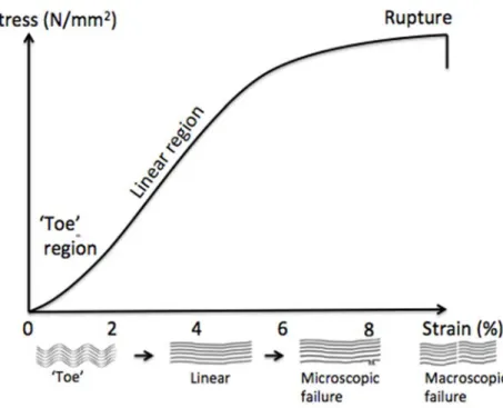
FIGURE 6:SCHEMATIC REPRESENTATION OF STRESS VS STRAIN. ... 108
FIGURE 7:GEOMETRIC REPRESENTATION OF E’ AND E’’ AND THEIR RELATIONSHIP WITH E*. ... 108
FIGURE 8:SEM IMAGES OF THREE SCAFFOLDS OBTAINED BY ELECTROSPINNING DIFFERENT CONCENTRATIONS OF PCLA,D.PCL10%. B,E.PCL12.5%.C,F.PCL15%.TOP IMAGES: SCALE BAR OF 500µM.BOTTOM IMAGES: SCALE BAR OF 10µM ... 123
FIGURE 9:AVERAGE FIBER DIAMETER RELATIVE TO THE PCL CONCENTRATION.FIBERS’ SIZE DISTRIBUTION FOR THE DIFFERENT ELECTROSPUN SCAFFOLDS (10% IN BLUE,12.5% IN RED AND 15% IN GREEN). ... 124
FIGURE 10.AVERAGE ELASTIC MODULUS RELATIVE TO PCL CONCENTRATION ANALYZED IN DRY (BLEU) OR WET CONDITIONS (RED).THE P VALUES ARE INDICATED FOR THE TESTS SHOWING A SIGNIFICANT DIFFERENCE.(***FOR P VALUE <0.001). ... 125
FIGURE 11.MTT ANALYSIS FOR COMPARING PROLIFERATION OF C3H10T1/2 ON DIFFERENT PCL SCAFFOLDS:10-WT %(BLEU), 12.5-WT %(RED) AND 15-WT %(GREEN) AT DAY 1,2 AND 3.THE P VALUES ARE INDICATED BETWEEN EACH DAY TESTS, SHOWING A SIGNIFICANT DIFFERENCE (*** FOR P-VALUE <0.001). ... 126
FIGURE 12.GENE EXPRESSION OF TENDON- AND BONE-RELATED MARKERS IN C3H10T1/2 CELLS CULTURED ON THE DIFFERENT SCAFFOLDS.THE DATA OBTAINED FOR GENE EXPRESSION OF THE CELLS CULTURED OVER THE SCAFFOLDS WERE COMPARED TO THOSE OF THE CONTROL CELLS (TISSUE CULTURE PLATE) WITH THE MANN-WHITNEY NONPARAMETRIC STATISTICAL TEST.(* FOR P VALUE <0.05,** FOR P VALUE <0.01). ... 127
FIGURE 13.SEM IMAGES OF TWO SCAFFOLD OBTAINED BY ELECTROSPINNING PCL10-WT % UNDER DIFFERENT ROTATATION SPEEDS. .A.RANDOM PCL10-WT % AT MEDIUM SPEED (1000RPM).B.ALIGNED PCL10-WT % AT HIGH SPEED (2000 RPM).SCALE BARS OF 10µM. ... 129
FIGURE 14.FIBER SIZE DISTRIBUTION FOR RANDOM 10-WT %(UP) AND ALIGNED 10-WT %(DOWN) SCAFFOLDS. ... 130
FIGURE 15.AVERAGE ELASTIC MODULUS RELATIVE TO FIBER ALIGNMENT.A.REPRESENTATIVE STRESS VS STRAIN CURVES FOR EACH MORPHOLOGY.B.AVERAGE ELASTIC MODULUS.THE P VALUES ARE INDICATED FOR THE TESTS SHOWING A SIGNIFICANT DIFFERENCE.(***FOR P VALUE <0.001). ... 131
10
FIGURE 16.EFFECT OF FIBERS ALIGNMENT ON CELL MORPHOLOGY.FLUORESCENCE STAINING OF ACTIN CYTOSKELETON (RED) AND NUCLEI (BLUE) OF C3H10T1/2.THE CELLS WERE CULTURED FOR 2 DAYS (A,C) OR 5 DAYS (B,D) FREE OF GROW FACTORS OVER RANDOM (A,B) OR ALIGNED PCL10-WT % SCAFFOLDS (C,D).SCALE BARS 50 µM. ... 132
FIGURE 17.GENE EXPRESSION OF TENDON- AND BONE-RELATED MARKERS IN C3H10T1/2 CELLS CULTURED ON ALIGNED OR RANDOM SCAFFOLDS FOR 1 OR 3 WEEKS (N=6). 10-WT % RANDOM SCAFFOLD (RANDOM 1 WEEK) WAS CHOSEN AS A CONTROL.THE DATA OBTAINED FOR GENE EXPRESSION WERE COMPARED WITH THE MANN-WHITNEY NONPARAMETRIC STATISTICAL TEST.(*
FOR P VALUE <0.05,*** FOR P VALUE <0.001). ... 133
FIGURE 18.(A)BOSE BIODYNAMIC CELL CULTURE ROOM.A.BIOREACTOR CHAMBER.B.RODS.C.CLAMPING SCEWS OF GLAZED WALLS.D.GLAZED WALLS.E.CLAMPS.(B)MOUNTED BIOREACTOR INSIDE AN INCUBATOR. ... 136
FIGURE 19.DYNAMIC CULTIVATION PROTOCOL.(A).REPRESENTATION OF THE APPLIED STRAIN.AMPLITUDE AND 0,STATICARE SET TO 0.5% STRAIN.THE OFFSET 0 IS 1% AND THE PERIOD T1S.(B).REPRESENTATION OF THE RESULTING STRESS. ... 137
FIGURE 20.FLUORESCENCE STAINING OF ACTIN CYTOSKELETON (RED) AND NUCLEI (BLUE) OF C3H10T1/2 AFTER 5 DAYS OF STATIC
(A-B) OR DYNAMIC CULTURE (C-D).SCALE BARS OF 50µM... 138
FIGURE 21.GENE EXPRESSION OF TENDON- AND BONE-RELATED MARKERS IN C3H10T1/2 CELLS CULTURED ON RANDOM SCAFFOLDS UNDER STATIC OR DYNAMIC CULTURE CONDITIONS FOR 5 DAYS INSIDE THE BOSE BIODYNAMIC 5100 BIOREACTOR
(N=6). STATIC CULTURE WAS CHOSEN AS A CONTROL. ... 138
FIGURE 22.STRAIN VS TIME (A) AND STRESS VS TIME (B) FOR A 5 DAYS MECHANICALLY STIMULATED CONTROL SCAFFOLD WITHOUT CELLS... 139
FIGURE 23. STRAIN VS TIME (A) AND STRESS VS TIME (B) FOR A 5 DAYS MECHANICALLY STIMULATED CELL-CONSTRUCT. ... 140
FIGURE 24.REPRESENTATION OF THE STRAIN AND STRESS VS. TIME (A) AND STRESS VS. STRAIN (B) CURVES OF THE FIRST SINUS FROM THE FIRST CYCLE AT DAY 1 AND THE LAST SINUS FROM THE LAST CYCLE AT DAY 5.SCAFFOLD 10-WT % CONTROL WITHOUT CELLS
... 141
FIGURE 25.A.REPRESENTATION OF THE STRAIN AND STRESS VS. TIME (A) AND STRESS VS. STRAIN (B) CURVES OF THE FIRST SINUS FROM THE FIRST CYCLE AT DAY 1 AND THE LAST SINUS FROM THE LAST CYCLE AT DAY 5.SCAFFOLD 10-WT % WITH CELLS. ... 142
FIGURE 26.EFFECT OF 5 DAYS OF DYNAMIC CULTURE (WITH OR WITHOUT CELLS) ON THE VISCOSITY OF THE MATERIAL.(A).STRESS VS.
TIME AND DISPLACEMENT VS. TIME CURVES.(B).PHASE LAG BETWEEN THE APPLIED DISPLACEMENT AND THE OBTAINED FORCE. BLUE REPRESENTS THE CONTROL EXPERIENCE WITHOUT CELLS.RED REPRESENTS THE CELL-CONSTRUCT EXPERIENCE. ... 143
11
TABLE 1.MATERIAL CHARACTERISTICS OF ELECTROSPINNING BASED STRATEGIES FOR BONE TISSUE ENGINEERING.(RNFS =RANDOM
NANO FIBERS;ANFS =ALIGNED NANO FIBERS. ... 31
TABLE 2.IN VITRO PERFORMANCES OF BIOHYBRID SCAFFOLDS IN BONE TISSUE ENGINEERING ... 34
TABLE 3.IN VIVO PERFORMANCES OF BIOHYBRID CONSTRUCTS IN BONE TISSUE ENGINEERING. ... 35
TABLE 4.PARAMETERS FOR THE REALIZATION OF THE DIFFERENT PCL SCAFFOLDS. ... 99
TABLE 5.ELECTROSPINNING AND ELECTROSPRAYING PARAMETERS STEP BY STEP.SOLUTION A:PCL AT 15-WT %W/V IN DCM/DMF 60/40%;SOLUTION B:HA AT 10%W/V IN ETHANOL. ... 99
TABLE 6.FLUORESCENT STAINS USED FOR CELL VIABILITY ANALYSIS. ... 103
TABLE 7.FLUORESCENT STAINS USED FOR CELL MORPHOLOGY ANALYSIS. ... 103
TABLE 8.FLUORESCENT ANTIBODIES USED FOR CELL MORPHOLOGY ANALYSIS. ... 105
12
Abbreviations
3D: Three-dimensional space ADSC: Adipose derived stem cells ALP: Alkaline phosphatase
ANF: Aligned nanofiber ANOVA: Analysis of variance Aqp1: Aquaporin 1
BCIP/NBT: 5-Bromo-4-chloro-3-indolyl phosphate / 4-Nitro blue tetrazolium chloride BGLAP: Bone gamma-carboxyglutamate protein
BMP: Bone morphogenetic protein BMSC: Bone marrow stromal cell BSP: Bone sialoprotein
BTE: Bone tissue engineering Cbfa1: Core-binding factor alpha 1
cDNA: Complementary deoxyribonucleic acid COL1: Collagen type 1
CS: Chitosan
DCM: Dichloromethane Dlx5: Distal-less Homebox 5 DBM: Demineralized bone matrix DMF: Dimethylformamide DNA: Deoxyribonucleic acid E’: Storage modulus
E’’: Loss modulus E*: Complex modulus ECM: Extracelular matrix
EDS: Energy-dispersive X-ray spectroscopy ESC: Embryonic stem cell
EthD-1: Ehidium homodimer-1 FBS: Fetal bovine serum
13 G (needle): Gauge
GPa: Gigapascal HA: Hydroxyapatite HCl: Hydrochloric acid
hFOB: human fetal osteoblastic IBPS: Institut de Biologie Paris-Seine IBSP: Integrin binding sialiprotein
ICPEES: Institut de chimie et procédés pour l’énergie, l’environnement et la santé IGF: Insulin growth factor
IMP: Institute für MehrphasenProzessen iPS: induced pluripotent stem
kV: kilovolt MPa: Megapascal
MSC: Mesenchymal stem cell
MTT: 3-(4,5-dimethylthiazol-2-yl)-2,5-diphenyltetrazolium bromide MW: Molecular weight ND: Non-determined NF: Nanofiber OCN: Osteocalcin OPN: Osteopontin PAF: Paraformaldehyde PBS: Phosphate-buffered saline PCL: Polycaprolactone
PEO: Polyethylene oxide PHB: Polyhydroxybutyrate PLA: Poly lactic acid
PLGA: Poly lactic-co-glycolic acid PLLA: Poly L-lactic acid
RNA: Ribonucleic acid RNF: Random nanofiber RP: Rapid prototyping
14 Rpm: Revolutions per minute
RT-qPCR: Reverse quantitative transcription polymerase chain reaction SAPC : Service d’analyse physico-chimique
SC: Stem cell Scx: Scleraxis
SEM: Scanning electron microscopy SF: Silk fibroin
SOSs: System of systems
SP7: Transcription factor SP7, or Osterix
SPP1: Secreted phosphoprotein 1, or Osteopontin TCP: Tricalcium phosphate
TDSC: Tendon derived stem cells, or TSPC TGF: Transforming growth factor
TM: Tensile modulus
TNMD: Tenomodulin, or Tnmd TS: Tensile strength
TSPC: Tendon stem/progenitor cell, or TDSC UPJV: Université de Picardie Jules Verne UTC: Université de Technologie de Compiègne VEGF: Vascular endothelial growth factor WJSC: Wharton Jelly stromal cell
15
the musculo-skeletal system of system
In a previous study (PhD thesis of T. Baudequin), our laboratory developed a bio-hybrid tissue made of granules of hydroxyapatite on which cells had been seeded and grown within a bioreactor, to produce a hand-able tissue sheet for the further application in maxillofacial reparatory bone surgery. With the synthesis of their own extracellular matrix, cells encompass the particles forming a sheet-like tissue after one month of culture.1 Very attractive regarding biocompatibility and osteoinduction, the handle-sheet bone like tissue were far from the specifications established by the laboratory regarding its mechanical properties.
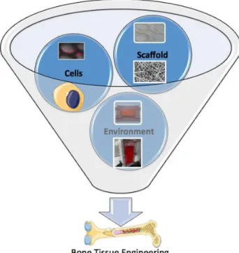
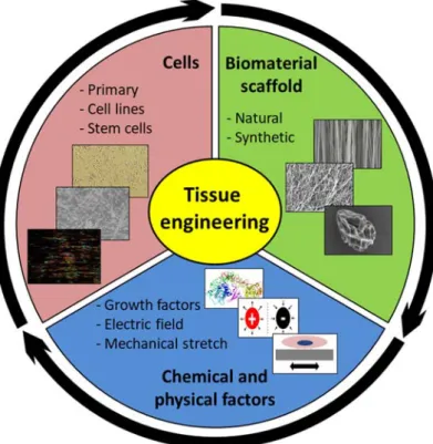
To improve these mechanical properties keeping the sheet aspect, electrospun matrices appeared as a clear alternative to granules for maxillofacial regeneration. The combination of elastic properties and fibrous nature similar to the extracellular matrix raised interesting process and solution for tissue engineering. The research group focused then on two polymers, PCL and PLLA, both polyesters with high biocompatibility currently employed in the biomedical field. Interestingly, the investigated polymers not only appears to be promising for bone tissue regeneration, but also seemed to be good candidates for other tissues such as tendon or muscle, other major systems in the musculo-skeletal system of system. These results allowed us to set the framework for the development of a tissue-engineered based model of musculo-skeletal system, with a future emphasis on the reconstruction of the interfaces between the different subsystems, i.e. the osteo-tendinous and the myotendinous junctions. The whole project, involving several researchers at BMBI and Roberval laboratories was selected as a “challenge and funded by the Labex MSST (Maitrise des Systèmes de Systèmes Technologiques)”.
Objective of the thesis
In this context, the aim of this PhD thesis was to propose an overall methodology to design and validate a bioartifical system representing the continuum tendon-bone, itself composed of biohybrid systems at different scales. It represents an example of complex bioinspired system of systems (SoSs) in which the different systems are in continuous remodeling and
16 interactions. The structural complexity (as a reconstructed multi-layered tissue) of the proposed bioengineered system presents a major challenge to understand and predict its mechanical and biological behavior.
To this purpose, the following objectives were defined:
Characterize the tissues of interest, their components and the key features that should guide their reconstruction. These living tissues are themselves SoSs, formed by the cells, their extracellular matrix and their interaction and are continuously evolving. For this purpose, we perform the review of literature and interact with the experts in physiology and mechanics of musculo-skeletal system at BMBI laboratory Establish the requirements and design the appropriate bio-inspired scaffold based on
the native structure. In the framework of this PhD thesis, we focus in the development of two sub-systems: reconstructed bone and tendon.
Analyze the interactions involved within the different subsystems. They are contingent to the biological properties of the developed tissues, but also to the chemical and mechanical properties of the material scaffolds. These interactions will be monitored through the culture process at the relevant scale to first tune the different cell response toward the desired type of tissue and tailor the mechanical properties of the global SoS.
Having in mind this idea of continuum and based on our previous studies, all scaffolds were prepared by electrospinning techniques. Chapter I was dedicated to the literature review regarding first bone and bone tissue engineering, focusing on electrospun scaffolds. Then, a similar survey was achieved for tendon and tendon tissue engineering, under the form of a review paper in which muscle and the myotendinous junction are also described. Finally, the enthesis issues were also briefly analyzed. Chapter II summarized the Materials and Methods employed for this PhD.
For bone bio-hybrid constructs (chapter III), we elaborated 3D scaffolds with a honeycomb-like architecture, based on a technique associating electrospun poly-ε-caprolactone (PCL) nanofibers and electrosprayed hydroxyapatite (HA) nanoparticles. These constructs were designed and built thanks to our collaboration with ICPEES (Dr. G. Schlatter, Dr. A. Hébraud). The biomimetic topography served as a niche for the growth and differentiation of cells. For
17 collaboration with Dr. Delphine Duprez (IBPS), was chosen since these cells are able to differentiate to bone or tendon lineage. Then, in vivo studies were performed by our collaborators at EA 4666 (UPJV) in the framework of a project led by C. Legallais and funded by Région Hauts de France (previously Picardie).
For tendon biohybrid constructs, aligned and random PCL electrospun scaffolds were seeded with cells, in the absence of any differentiation factors. Cyclic stretching was applied to the scaffolds in a bioinspired vision to simulate training. In a first series of investigation (Chapter IV), we also used C3H10T1/2 so as to define culture and stretching conditions in favor of tendon differentiation. Then, we transfer the protocol to rat mesenchymal stem cells (chapter V), to go closer to preclinical in vivo studies. In addition, as these cells present high production of the extracellular matrix, we aimed at studying over time the impact of this neo-synthesis on the mechanical properties of the biohybrid constructs.
18
19
1. Bone Tissue Engineering: Current Trends and Challenges
1.1. Bone Composition and Structure
Bone is a hierarchical and complex hard, dense and highly mineralized connective tissue that support and protect vital organs, allow the body movement, provides the body reservoirs of minerals and growth factors and a site for hematopoiesis.2 Bone matrix is composed of 70% minerals, 25% of organic compounds and 5% of water.3 The mineral part is composed mainly of crystals of calcium phosphates (Hydroxyapatite crystals). Type 1 collagen represents around 90% of the organic part of the extracellular matrix. Other collagens such as collagen III, V, XI and XIII, glycoproteins such as Bone Sialo Protein (BSP), vitronectin, osteonectin, thrombospondin, fibronectin and osteonpontin have been also found as constituent for the organic phase of bone matrix.4
The skeleton of human adults is composed of around 213 bones that can be classified according to their location, shape, consistency or size.5 Macroscopically, bones can be categorized into two groups: the cortical and the trabucular bones.6 Cortical bone, which represents the 80% of bones, is dense and compact with a low porosity (10%), has a slow turnover rate and a high mechanical resistance; and constitutes the outer part of all skeletal structures. Cortical bone provide mechanical strength and protection to the body, but it can also participate in metabolic response, particularly when there is severe or prolonged mineral deficit.7 It is organized in cylindrical structures called osteons that have a diameter of around 200µm in a human adult. Each osteon consists of concentric layers of compact matrix called lamellae that surrounds a central canal called the harvesian canal, which contains blood vessels and nerves fibers. The remaining 20% is the trabecular bone; less dense, more elastic and porous (30-90%), it has a high turnover rate than cortical bone and constitutes the inner part of long bones, vertebras, pelvis and other large flat bones. Trabecular bones contribute to mechanical support, act as a scaffold for the hematopoietic cells and provide supplies of mineral in acute deficiency states.7,8 Without osteons, lamellae are organized in a “rod and plate” structure called trabeculae.9 Relative to its structure and composition there are characteristic mechanical properties for each group of bone. Thus, the cortical bone presented a Young´s modulus of 15-20 GPa and a compressive strength of
20 100-200MPa and the trabecular bone has a Young´s modulus of 0.1-2GPa and a compressive strength of 2-20MPa.10
Figure 1. Bone structure, adapted from Sevier Medical Art.
Four different cell types constitute bone tissue: Osteoblasts, bone lining cells, osteocytes and osteoclasts.11 Osteoblasts, derived from mesenchymal stem cells (MSC), are responsible for new bone synthesis. In their mature form, osteoblasts could undergo two possible pathways: apoptosis or become osteocytes or bone lining cells. Osteocytes, the most abundant cells in bones (90%) are mature osteoblasts located within lacunae surrounded by mineralized bone matrix. Osteocytes are connected creating a network through the lacunae, facilitating the intercellular transport of small signaling molecules, oxygen and nutrients. As osteocytes, bone lining cells derived from osteoblasts. They cover the surface of bones, preventing osteoclasts to enter in contact with bone matrix when matrix resorption should not occur. Osteoclasts are terminally differentiated myeloid cells responsible uniquely to bone resorption.
1.2. Bone Remodeling and Injuries 1.2.1. Remodeling
Bone is a dynamic tissue involved in a continuous cycle of remodeling responsible to the maintain of adult skeleton and mineral homeostasis.12 This tightly interconnected cycle is constituted of three consecutive phases which involves different phases: (I) Old bone resorption by osteoclasts, (II) a transition phase from resorption to new bone formation, and
21 an inherent ability to regenerate without scaring and this newly formed bone is almost indistinguishable from adjacent uninjured tissue.14
1.2.2. Injuries
However in some cases due to trauma, injury, disease or aging damage, bone could present a significant loss of its repair ability.15 These conditions may lead to non-union, scar formation and long-term persistent bone defects.16 In these cases in which the self-regeneration of bone tissue is compromised, bone graft appears as the most widespread solution.
1.2.3. Bone Grafts and Subtitutes for Bone Reconstruction
The main function of bone grafts is to ensure growth and healing, whilst providing mechanical support as long as the processes take place.17 Bone grafts are classified in different categories: autografts, allografts, xenografts and bone grafts substitutes. Each type differs with regard to their properties of osteogenesis, osteoinduction and osteoconduction.
18 Osteogenesis refers to a material which contain living cells that will produce new bone.
The osteoconduction is the ability to allow bone growth from vascular and osteogenic host cells. Finally, osteoinduction refers to the ability to induce cell differentiation leading to mature bone cells. 19
Autographs are considered as the gold standard: they are harvested from the donor and present the three characteristics expected from the bone grafts, they are osteogenic, osteoconductive and osteoinductive. Autograft includes aspirate bone marrow, cortical or cancellous bone and vascularized grafts. 20 In spite of the respect of the specifications for the bone grafts, the limits in terms of quantity and the risk of morbidity make it considerable other ways to overcome bone regeneration and reconstruction.21
Bone allografts refer to bone tissue coming from another individual of the same species. Without the preceding limits in terms of quantity and risk of morbidity, allografts are present in various forms including cortical or cancellous bone and derivatives forms as demineralized bone matrix (DMB). 22 While they lack of osteogenicity, allogeneic cortical and cancellous bone presented osteoconductive properties. DMB are processed in such a way that they
22 provide osteoinduction.23 As main drawbacks, allografts do not reach a regeneration as complete as autografts, they are immunogenic and they present a risk for the transmission of diseases. 24 Another alternative, xenografts, refers to bony tissue harvested from other species. Moreover, its high immunogenic response turns this possibility into something marginal. Current efforts are made in terms of complete decellularization, deproteination and defatting protocols. 19
In order to overcome these limitations different alternatives have been deployed as the use of synthetic bone grafts substitutes. Among these biomaterials, we can distinguish calcium phosphate ceramics, bioactive glass and calcium sulfate.25 Calcium phosphates are the most widespread ceramics available on the market. They are a family of calcium salt compounds consisting of calcium ions and organophosphates. The calcium phosphate family includes monocalcium phosphate, dicalcium phosphate, tricalcium phosphate (TCP), hydroxyapatite (HA) and tetracalcium phosphate.22 In general, most calcium phosphates are forming mixed compounds in form of blocks, cements or powders.26,27 Interestingly, HA (Ca10[PO4]6[OH]2) is
a mineral of natural origin which comprises about 50% of bone weight. Despite their differences in terms of composition, calcium phosphates are osteoconducive, presented a good osteointegration and could promote osteoinduction.28 Bioactive glasses or bioglass, are synthetic silica-based materials developed in 1970s with osteoconductive and unique bone binding properties.29 When implanted in the bone a silica-rich layer occurs and on top of this layer, a layer of HA is formed.22,30 This layer of HA absorbs proteins and attracts osteo-progenitor cells.31 Calcium sulfate or plaster of Paris (CaSO4) is a osteoconductive and biodegradable ceramic with the faster resorption rate of ceramics, a faster rate than actual bone deposition which compromise bone healing.22 However their low cost and easy procurement make it an interesting candidate for bone reconstruction if combined with other materials.31
1.3. Bone Tissue Engineering
While the aforementioned substitutes for bone allows to a greater or lesser extent for bone reconstruction, none is exempted of further ameliorations to achieve the ideal regeneration: low morbidity, osteoconductive, osteoinductive and osteogenic properties, size restriction, quick accessibility and reasonable cost.32 In order to achieve all the ideal requirements for
23 the last 30 years. BTE requires the combination of the appropriate scaffold, cells and/or signaling factors. The goal is to provide the organism with the right biohybrid scaffold that provides both mechanical support and enough information to allow hosted cells to organize a new bone tissue.
Figure 2. The three pillars of bone tissue engineering: cells are ideally cultivated on a biomimetic scaffold in order to guide their performance as close as in the native bone. The mechanical and biochemical environment are of key relevance in eliciting targeted responses.
1.3.1. Materials and Scaffolds Manufacture
The “right” scaffold should provide a similar three-dimensional structure as bone tissue with osteoconductive, osteoinductive and osteogenic properties. From bioactive inorganic materials as calcium phosphates33–37 or bioactive glass38–41 to polymers both naturals as collagen, fibrin, silk, chitosan, alginate or hyaluronic acid, or synthetics as polycaprolactone (PCL)42,43 polylactic-acid (PLLA)44,45, poly-lactic-co-glycolic acid (PLGA)46,47 and polyurethanes (PUs)48–50 or composites (polymers and inorganic compounds)51–58 and natural derived tissues (decellularised or demineralized bone), a wide range of materials has been used for BTE applications.
24 To produce the ideal scaffold, different techniques have been developed during the last years as solvent casting, gas foaming, freeze drying, electrospinning, melt-blown process and rapid prototyping process.59 Despite the differences among the different processes, it has been highlighted that the optimal scaffold for the bone tissue regeneration must have a sufficient porosity to allow cell colonization, nutrient supply, vascularization and tissue ingrowth, while taking into account the mechanical requirements of the bone tissue. The porosity ranges varies from 100-500 µm and the pore distribution is related to the manufactured technique60.
1.3.2. Cells
Several cell sources have been investigated for bone tissue engineering. The ideal cell source must meet a series of requirements such as being easily isolated and expanded, as well as presenting an interesting activity in the required field, in this case bone regeneration. Depending on their state of differentiation, we can distinguish between stem cells (SC) or terminally differentiated cells. Only stem cells present a differentiation potential (pluripotent, multipotent or unipotent) and they could be classified into two groups depending of it origin: embryonic or adult.
Embryonic stem cells (ESCs), which are pluripotent, are isolated from the blastocyst. Despite being able to differentiate towards the three germ layers, their use is not exempt of limits such as the risk of immune rejection, as well as ethics’ concerns61. Recently, induced pluripotent stem (iPS) cells appeared as an alternative to ESCs. iPS are derived from adult somatic cells via the introduction of a series of transcription factors. Recently, some works successfully focused on the differentiation of iPS-derived cells towards bone lineage62,63. However, due to the potential for mutagenesis and the low efficiency of transfection, they are not yet authorized for clinical application in humans64.
Adult mesenchymal stem cells (MSCs) are promising cell sources presenting the ability to differentiate into osteoprogenitors and mature osteoblasts. MSCs could be isolated from several autologous sources as bone marrow, adipose tissue, synovial membrane, dental pulp, skin, cartilage and other sources as umbilical cord. In addition their potential for self-renewal and clonogenicity, make MSCs a relevant source for clinical application. Bone marrow derived stem cells (BMSCs) are the predominant source for adult MSC for bone tissue engineering and are already used in preclinical studies65. However they present
25 individuals. Compared to BMSCs, adipose derived stem cells (ADSCs) can be obtained in larger number and in a less invasive procedure. However, further studies should be carried to confirm their bone-forming capacity and safety concerns66.
Immortalized cells lines are also commonly used for bone tissue engineering. From animal (MC3T3-E167–71, C3H10T1/272) or from human (hFOB73–75), cells lines offer advantages as they could be grown for prolonged period in vitro and provide homogenous cell population with well-known properties76. However, as they have been genetically modified, their phenotype, their native functions, as well as their response capacity to stimuli may be disturbed77. These cells are often used in early investigations as proof of concept, or in fundamental studies.
1.3.3. Chemical and Mechanical Environment
Bone healing occurs immediately after a fracture, orchestrated by a cascade of events guided by cytokines, involving different types of cells as inflammatory cells, vascular cells, mesenchymal progenitor cells and osteocytes. According to their activity, we can distinguish among (I) pro-inflammatory, (II) angiogenic and (II) osteogenic factors78. The main families regrouping the different growth factors include transforming growth factor superfamily (TGFβs and bone morphogenetic proteins (BMPs)),79,80 fibroblast growth factor (FGF)81, vascular endothelial growth factor (VEGF)82 and insulin-like growth factor (IGF)83.
TGFβ superfamily comprises over forty members such as the three isoforms of TGF (TGFβ1, TGF 2 and TGF 3) and BMPs. TGFβ stimulates matrix protein synthesis, enhances the proliferation of mesenchymal cells and osteoblasts in fractures and plays an important role in bone remodeling with it facilitating or suppressing role over the activity on osteoclasts84,85. Among TGFBβ superfamily, BMPs are involved in different signaling pathways in bone formation, from promoting recruitment and migration of mesenchymal cells to osteogenic differentiation. While more than 20 different BMPs have been identified, isoforms BMP-2, BMP-4 and BMP-7 are the best characterized ones. BMP-2 and BMP-7 have been incorporated in FDA-approved systems for bone regeneration and are commercially available for surgical use on collagen sponge carrier86. FGF displays an important role in bone regeneration and homeostasis. While it does not directly induce osteoblast differentiation, FGF plays a role on osteoblast differentiation. It has been suggested that isoforms FGF-2 and
26 FGF-9 play an important role on osteoblasts proliferation and angiogenesis87,88. Another pro-angiogenic factor is VEGF that stimulates neovascularization by stimulating the proliferation and migration of endothelial cells82. IGF regulates different processes as bone development, growth and healing by stimulating proliferation and differentiation of osteoblast precursors. Interestingly, it has also been reported a role on osteoclast survival and remodeling processes89.
Besides chemical stimulation, it is well known that mechanical stimulation has a preponderant role in bone homeostasis and remodeling. Indeed, during standing and physical activities, mechanical forces are exerted on the bones. This mechanical environment result in a maintenance or gain of bone mass90. Lack of physical activity leads to weakening of the bone and consequently, bone fractures could occur91. Duncan et al. (1995) summarized the different ranges of physical strains that affect bone homeostasis. Those strains were reported in terms of microstrain (μstrain) where 1000 μstrain corresponds to 0.1% in terms of elongation percentage. Bone resorption occurs below 200 μstrain, the physiological range up to 2500 μstrain, an overuse appears around 5000 μstrain and over this threshold, pathological states appear92. Therefore, during the last years, different works have focused on the effect of mechanical stress on bone tissue and the application of a mechanical environment for bone tissue engineering as a key factor towards the proper-engineered construct. This mechanical environment has been generated using different bioreactors that our team summarized in a previous review.93
1.4. Biomimetic Electrospun Scaffolds for Bone Tissue Engineering
Electrospun based scaffolds present micron to sub-micron fibres which are similar to the extracellular matrix 94. In addition to the fibrous nature of the material, the versatility of electrospinning to produce scaffolds with tailored morphology and porosity from a wide range of different polymers could explain its success during the last years in the field of tissue engineering95. Different scaffolds have indeed been developed for applications in skin96, cartilage97, tendon/ligament98, nerves99 and bone100 reconstruction.
In the electrospinning process, a polymer solution is introduced in a syringe. Under a constant flow rate, the solution is extruded through a thin needle. At the exit of the needle, the solution is held by its surface tension. When subjected to an electric field, generally over
27 overcome the surface tension threshold, the high voltage allows the formation of a stable Taylor cone. The polymer is thus carried out towards the collector presenting opposite charges, in the form of a thin and unique continuous fiber. The solvent evaporates as long as the fiber becomes thinner. As a result, a network of fibers is formed on the collector 101. The fibers and their spatial configuration are influenced by many parameters such as concentration in polymer, type of solvent, humidity, type of collector used, etc.102
Figure 3. A. Scheme of the different parameters involved in the electrospinning process. B. General representation of the components of an electrospinning system. C. Three types of needles employed in the electrospinning technique. From right to left: single needle, co-axial needle and a multiple needle system. D. Different types of collectors for the realization of different organization of electrospun fibers. From right to left and from top to bottom: flat collector, parallel collector, and rotatory drum collector at high speed and rotatory collector at low speed.
While the common electrospinning device consists in a single solution flowing through a capillary and a flat collector, variations of the system are possible. Since the electric field mainly contributes to the attraction towards the collector, the device can be placed vertically or horizontally. The collector can take the form of a rotating cylinder, which will allow greater uniformity of the fiber mat, but especially their alignment at high rotational speed. In addition to these techniques where part of the assembly is in motion, aligned
28 fibers may be formed using two spaced apart electrical sources operating alternately. Other geometrical modifications of the ground collector could be performed by playing with the surface topography, where fibers are deposited following the design of the surface. Electrospinning also makes it possible to spin polymer mixtures (co-spinning), coaxial fibers or to use multiple jets simultaneously.
We hereafter propose a review of the different materials used for scaffold production, the mechanical properties of the biohybrid constructs, as well as both in vitro and in vivo outcomes. We sorted the references in tables, according to increasing scaffold’s complexity.
1.4.1. Materials and Manufacture
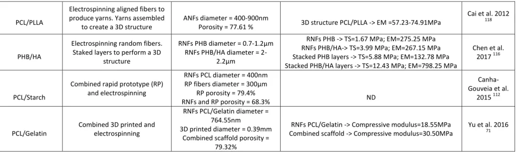
A wide range of polymers has been used to produce electrospun fibrous scaffolds for bone tissue engineering (Table 1). Generally, these polymers are classified according in two groups: natural or synthetic. Thanks to their origin, natural polymers (collagen103–105, gelatin71,106, silk107,108,67,73 or chitosan109), present correct biocompatibility allowing cell adhesion and proliferation. However, their poor mechanical properties, rapid degradation and costs associated to their isolation and purification limit their use. In contrast, synthetic polymers appeared interesting during the last years because they can easily tailor scaffolds with reproducible porosity, shape and better mechanical properties59. Among the examples of synthetics polymers used for bone tissue engineering, we can cite PCL70,110–112, PLLA72,74,113, co-polymers as PLGA114 or PLCL115; polystyrenes69 and polyhydroxyalkanoate as PHB116. However, synthetic polymers are less favorable to cell adhesion and proliferation. Therefore, composites of both natural and synthetic polymers have been deployed to meet the entire requirement for bone tissue engineering, i.e. suitable mechanical properties but also bioactivity70,71,74,104–106,112.
In addition to its fibrous structure, bone specificity depends on its mineralized ECM. Electrospinning setup, by incorporating mineral particles at the polymer solution, is able to produce fibers with nano- to micro-particles, such as TCP111 or HA73,75,109,116, already widely used as biomaterials for bone regeneration or repair. Others successfully incorporated demineralized bone matrix (DMB) into a PLLA solution to produce composite scaffolds113. Added into the polymer solution108 or encapsulated by the fibers109, electrospinning has successfully used as a vehicle for growth factors delivery.
scaffold
PCL/Gelatin Electrospinning random and
aligned fibers RNFs diameter = 344-347nm
ANFs diameter = 355-356nm
RNFs 344nm -> TM=38.6 MPa; TS=7.9 MPa RNFs 334nm -> TM=33.4 MPa; TS=5.5 MPa RNFs 347nm -> TM=26.0 MPa; TS=2.0 MPa ANFs 355nm -> TM=45.3 MPa; TS=20.9 MPa ANFs 335nm -> TM=36.3 MPa; TS=15.6 MPa ANFs 363nm -> TM=30.4 MPa; TS=13.4 MPa
Guo et al. 2015 70
PCL/Gelatin Electrospinning random fibers RNFs diameter = 10-1000nm
Gelatin NFs -> YM=105 MPa; TS=2.50 MPa PCL NFs -> YM=4.98 MPa; TS=2.70 MPa PCL/Gelatin NFs -> YM=30.8; TS=1.29 MPa
Zhang et al. 2005 106
PCL/Collagen
Electrospinning random fibers. 3D scaffolds obtained by
wrapping fibers mats
RNFs diameter = 513nm
RNFs -> YM=4.59 MPa
Wrapped scaffold in the axial direction => 0.61 MPa Wrapped scaffold in the radial direction =>1.0 MPa
Ekaputra et al. 2009 104
PCL/Collagen
Electrospinning random co-axial fibers. Inner PCL and outer fiber
Collagen RNFs coaxial = 442nm ND
Haslauer et al. 2011 105
PLCL/Fibrinogen
Electrospinning random and aligned fibers. 3D honeycomb
scaffolds RNFs diameter = 195-462nm ANFs diameter = 195-491nm HC diameter = 213-445nm ND Nedjari et al. 2017 117
Collagen Electrospinning random fibers RNFs diameter = 50-1000nm ND
Shih et al. 2006 103
PS Electrospinning random and
aligned fibers RNFs diameter= 1-3.5μm ANFs diameter = 1.5-4.5μm ND Terranova et al. 2016 69 PCL, PLLA and PCL/PLLA
Electrospinning random, aligned and coaxial fibers
RNFs PCL diameter = 665-1159nm RNFs PLLA diameter = 681nm ANFs PCL diameter = 1032nm CNFs PCL/PLLA diameter = 1928-2461nm RNFs PCL -> YM = 21-30MPa RNFs PLLA -> YM = 24MPa ANFs PCL -> YM = 15MPa CNFs PCL/PLLA -> YM = 32-60MPa Baudequin et al. 2017 72
30
Chitosan/HA Electrospinning random fibers
RNFs chitosan diameter = 138nm RNFs chitosan/HA diameter = 214nm ND Zhang et al. 2008 75 PLLA PLLA/HA
PLLA/Collagen/HA Electrospinning random fibers
RNFs PLLA diameter = 860nm RNFs PLLA/HA diameter = 845nm RNFs PLLA/Collagen/HA diameter = 310nm RNFs PLLA -> TS = 4.69MPa RNFs PLLA/HA -> TS = 3.10MPa RNFs PLLA/Collagen/HA -> TS = 2.05MPa Prabhakaran et al. 2009 74 PLLA/PCL
PLA/PCL/HA Electrospinning random fibers
RNFs PLA/PCL diameter = 776nm RNFs PLA/PCL/HA diameter = 332-583nm ND Fang et al. 2010 68 PLA/Demineralized
Bone Powders Electrospinning random fibers RNFs diameter = 300-700nm ND
Ko et al. 2008
113
Fibroin/Chitosan/H a/BMP2
Electrospinning co-axial fibers
Thick fibers diameter = 534nm; outer layer 167nm Thin fibers diameter = 546nm;
outer layer 101nm
ND Shalumon et
al. 2015 109
Silk/PEO/HA/BMP2 Electrospinning random fibers
RNFs Silk/PEO diameter = 590nm RNFs Silk/PEO diameter = 575nm RNFs Silk/PEO/BMP2 diameter = 570nm RNFs Silk/PEO/HA diameter = 510nm RNFs Silk/PEO/HA/BMP2 diameter = 520nm ND Vepari et al. 2006 108 Silk
Electrospinning over a modified water bath collector
RNFs diameter = 200-500nm Pores sizes = 0-500μm Porosity = 90-93% ND Park et al. 2010 67 Silk/HA
Electrospinning over a modified water bath collector
RNFs diameter = 1.49μm Pores sizes = 42.3-301.1μm
Compressive modulus of Silk mats= 3.4-102KPa Compressive modulus of Silk/HA mats = 8.1-29.3KPa
Yang et al. 2015 73
31
PCL/PLLA produce yarns. Yarns assembled
to create a 3D structure
ANFs diameter = 400-900nm
Porosity = 77.61 % 3D structure PCL/PLLA -> EM =57.23-74.91MPa
118
PHB/HA
Electrospinning random fibers. Staked layers to perform a 3D
structure
RNFs PHB diameter = 0.7-1.2μm RNFs PHB/HA diameter =
2-2.2μm
RNFs PHB -> TS=1.67 MPa; EM=275.25 MPa RNFs PHB/HA-> TS=3.99 MPa; EM=267.15 MPa Stacked PHB layers -> TS=5.88 MPa; EM=132.78 MPa Stacked PHB/HA layers -> TS=12.43 MPa; EM=798.25 MPa
Chen et al. 2017 116
PCL/Starch
Combined rapid prototype (RP) and electrospinning RNFs PCL diameter = 400nm RP fibers diameter = 300μm RP porosity = 79.4% RNFs and RP porosity = 68.3% ND Canha-Gouveia et al. 2015 112 PCL/Gelatin
Combined 3D printed and electrospinning
RNFs PCL/Gelatin diameter = 764.55nm
3D printed diameter = 0.39mm Combined scaffold porosity =
79.32%
RNFs PCL/Gelatin -> Compressive modulus=18.55MPa Combined scaffold -> Compressive modulus=30.50MPa
Yu et al. 2016
71
Table 1. Material characteristics of electrospinning based strategies for bone tissue engineering. (RNFs = Random Nano Fibers; ANFs = Aligned Nano Fibers.
32
Cells Culture Media Mechanical Properties of
Biohybrid Constructs Major Outcomes Ref
MC3T3-E1 α-MEM, 10% FBS
ND
Aligned fibers resulted in better cell attachment, proliferation, alignment ad ALP activity when
compared to random fibers.
Guo et al. 2015
70
BMSC from NZ
Rabbit DMEM; 15% FBS ND
Better cell attachment and deeper infiltration was found on PCL/gelatin scaffolds, compared to PCL
alone.
Zhang et al. 2005 106
BMSC from Pig
DMEM; 10% FBS; 10nM dexamethasone; 50µM ascorbic acid;
10mM β-glycerophosphate.
ND
Tubular PCL/Col/BMSCs cell constructs presented positive levels of collagen deposition and osteogenic
differentiation with calcium deposition and osteocalcin production.
Ekaputra et al. 2009 104
ADSCs from Human
α-MEM; 10% FBS; 0.1µM dexamethasone; 50µM ascorbic acid;
10mM β-glycerophosphate. ND
Collagen covering PCL fibers enhanced early cell
spreading and increased calcium deposition Haslauer et al. 2011 105
ADSCs from Human
DMEM/F12; 10% FBS; 100nM dexamethasone; 10mM β-glycerophosphate; 50µM ascorbic
acid.
ND
Cell proliferation didn't differs among random, aligned or honeycomb scaffolds, cells cultured over
honeycomb-like scaffolds up-regulated osteogenic differentiation, mineralization and ALP activity.
Nedjari et al. 2017 117 BMSCs from Human IMDM; 10% FBS; 0.1mM dexamethasone; 10mM β-glycerophosphate; 0.2mM ascorbic acid. ND
Collagen nanofibers supported osteogenic differentiation with positive levels of transcripts as osteocalcin, osteonectin and osteopontin, and ALP
activity. Shih et al. 2006 103 MC3T3-E1 α-MEM; 10% FBS; 10nM; dexamethasone; 50µg/ml ascorbic acid ND
Cell attachment was better on large fibers Cell proliferation was better on aligned fibers and large random fibers compared to smallest random fibers. Large aligned fibers showed better ALP activity
compared to small random fibers.
Terranova et al. 2016 69
33
C3H10T1/2 DMEM; 10% FBS ND presented up-regulated bone transcripts while fibers
from micrometric range up-regulated tendon transcripts
Baudequin et al. 2017 72
hFOB DMEM/F12; 10% FBS ND Incorporation of HAP on chitosan fibrous scaffolds
improves cell proliferation and mineral deposition.
Zhang et al. 2008 75
hFOB DMEM/F12; 10% FBS ND
Cells cultured on PLLA/Coll/HAP nanofibrous scaffolds showed higher cell proliferation, increased
ALP activity and mineralization.
Prabhakaran et al. 2009 74
MC3T3-E1 α-MEM; 10% FBS ND PCL/PLA/HAP nanofibrous scaffolds provide a better
cell spreading and proliferation.
Fang et al. 2010 68 MSCs from Human DMEM; 10% FBS; 10nM dexamethasone; 10mM β-glycerophosphate; 50µg/ml ascorbic acid. ND
PLA/DPB scaffolds support the growth of MSCs without compromising their osteogenic differentiation. Mineralization was higher compared
to PLA scaffolds. Ko et al. 2008 113 MSCs from Human DMEM; 10% FBS; 0.1µM dexamethasone; 10mM
β-glycerophosphate; 50µM ascorbic acid ND
While either SF/CS/HAP and SF/CS/HAP/BMP-2 resulted in good biocompatibility, cell proliferation, ALP-activity and mineralization; the combination of SF/CS/HAP/BMP-2 resulted in up-regulated bone
differentiation. Shalumon et al. 2015 109 BMSCs from Human DMEM; 10% FBS; 10nM dexamethasone; 50µg/ml ascorbic acid; 7mM β-glycerophosphate. ND
The combination of HAP and BMP-2 in silk scaffolds induce up-regultated bone transcripts and highest
amount of calcium deposition.
Vepari et al. 2006 108
MC3T3-E1 α-MEM; 10% FBS ND
Medium pores size scaffolds (100-200 µm), improves cell proliferation, cell viability and ALP
activity.
Park et al. 2010
67
hFOB
DMEM/F12; 10% FBS
ND Silk scaffolds provides a good environment for cell
survival and colonization.
Yang et al. 2015 73
34 Table 2. In vitro performances of biohybrid scaffolds in bone tissue engineering
hMSCs derived from ESCs DMEM; 10% FBS; 10nM dexamethasone; 50µg/ml ascorbic acid; 10mM β-glycerophosphate ND
3D scaffolds provide better environment for cell
proliferation and ECM mineralization. Cai et al. 2012 118
BMSCs from Rabbit
DMEM; 10% FBS; 100nM dexamethasone, 10mM β-glycerophosphate and 0.2mM ascorbic
acid.
ND
Composite scaffolds of NHB/HAP showed better cell proliferation, up-regulated bone transcripts and
improved ALP activity than PHB scaffolds.
Chen et al. 2017 116 WJSCs from Human α-MEM; 10% FBS; 10nM dexamethasone, 10mM β-glycerophosphate and 50µg/ml ascorbic acid. ND
Combining PCL nanofibers meshes with TCP improves cells attachement, proliferation and differentiation towards bone lineage. This effect is
accentuated in the presence of an osteogenic medium. In addition, dynamic culture improves cell
differentiation and ALP activity even without osteogenic supplementation medium.
Canha-Gouveia et al. 2015 112
MC3T3-E1 α-MEM; 10% FBS ND
The combination of 3D printing and electrospinning
35 Animal Model and Tissue
Site Implantation
Mechanical Stimulation before
Implantation
Mechanical Properties of the Biohybrid
Construct Following Implantation Biological Outcomes Ref
Murine (Rat) full-thickness
bony calvaria defect None ND
After 12 weeks, the PLA/DBP nanofibrous scaffolds implanted presented almost complete bone healing with more bone density than
PLA scaffolds alone.
Ko et al. 2008
113
Ectopic transplation in a
mice mode None ND
SF/CS/HAP/BMP-2 induces ectopic bone formation even in absence of cells. Pre-cultured scaffolds resulted in
higher ECM deposition and OCN expression.
Shalumon et al. 2015 [13]
Murine (Rat) full-thickness
bony calvaria defect None ND
After 7 weeks, medium pores sizes scaffolds presented more bone
formation and good scaffold resorption after.
Park et al. 2010 67
Murine (Rat) full-thickness
bony calvaria defect None ND
New bone formation was more prominent than control after 8 weeks. The addition of BMP-2 augmented the
amount of new bone formation.
Yang et al. 2015 73
Rabbit bone tibia deffect None ND
After 6 weeks, tibia bone defect filled with 3D scaffold presented cortical-bone like tissue and vascularization.
Cai et al. 2012
118
Ectopic transplation in a
mice mode None ND
After 2 months into an ectopic bone formation mice model, PHB/HAP composite scaffolds presented more
vascularization and bone-like tissue formation.
Chen et al. 2017 116
Combining natural and synthetic polymers to produce hybrid scaffolds
Table 1 highlights the different potential association of natural and synthetic polymer to produce electrospun fibers. In general, it should be noticed that comparisons are very difficult to make since the diameter of the fibers (from 10 nm to 4500 nm) and their configuration (random / aligned) varied from one study to the other.
Zhang et al. (2004) were the first to produce a combined gelatin and PCL electrospun scaffold with fibers diameters from a range of 10 to 1000nm, to combine the advantages of natural and synthetic polymers. This blend scaffold presented a lower tensile strength of 1.29 MPa than those of PCL and gelatin alone, 2.70 MPa and 2.50 MPa respectively, although its Young Modulus was higher than PCL alone (30.8MPa vs. 4.98MPa)106. Results are uneasy to interpret since PCL concentration was different in the blend. Ekaputra et al. (2009) proposed composite scaffolds of PCL and collagen with a fiber diameter of 513 ± 83 nm. When electrospun together, the Young Modulus was lower compared to PCL alone (4.59 ± 1.46MPa vs. 12.35 ± 3.31MPa)104. Prabhakaran et al. (2009) combined PCL with collagen and HA nanoparticles. The addition of collagen resulted in smaller fibers’ diameter (310 ± 125nm) compared to the scaffold without collagen (845 ± 140nm) and to decreased tensile strength (2.05 ± 0.10MPa vs. 3.10 ± 0.15MPa).74 While these authors added either collagen or gelatin together with the synthetic polymer, Haslauer et al. (2011) proposed co-axial electrospinning where the inner part was PCL and the outer layer collagen. Co-axial scaffolds resulted in increased fiber diameter (442 ± 45nm vs. 280 ± 51nm)105. Unfortunately, the mechanical properties of such scaffolds were not assessed. In our group, Baudequin et al. (2017) prepared coaxial electrospun fibers combining PCL and PLLA polymers. When PCL was the outer layer and PLLA was the inner one, fibers with a diameter of 1928nm and a Young modulus of 38MPa were obtained. When PLLA was the outer layer, fibers presented a larger diameter (2461nm) but Young Modulus became lower, with 30 MPa.72
Due to the wide range of mechanical properties and methods of assessment, it seems thus impossible here to conclude on the benefit of a mixture regarding pure polymer, as far as the mechanical properties are concerned.
37 Biomimetic approaches have been developed in order to mimic the native mineralized matrix of bones. Li et al. (2006) produced silk scaffolds loaded with hydroxyapatite nanoparticles. The addition of nanoparticles resulted in fibers with reduced diameter (510 ± 60nm) compared to nude silk scaffolds (590 ± 60nm).108 Zhang et al. (2008) obtained similar results on chitosan scaffolds for which the addition of hydroxyapatite reduced the fiber diameter from 214 ± 25nm to 138 ± 15nm.75 Fang et al. (2010) produced combined scaffolds of PCL and PLA with added nanoparticles of HA. From a ratio of 0.1 to 1, increased amounts of nanoparticles resulted in smaller electrospun fibers. For the maximal concentration of HA fibres presented a diameter of 332 ± 11.3nm, significant smallest compared to 776 ± 15.6nm for PCL and PLA scaffolds without nanoparticles.68
In other work, Ko et al. (2008) analyzed the impact of demineralized bone powders (DPBs) to mimic the composition of the bone matrix. Addition of DPBs in a solution of PLLA resulted in reduced fibers when compared to PLLA scaffold alone.113
The mechanical response of scaffolds might be affected by the addition of nanoparticles. Phrabhakaran et al. (2009) showed that when HA nanoparticles were added to PLLA scaffolds, the tensile strength was reduced from 4.69 ± 0.19MPa to 3.10 ± 0.15MPa.74 However, recently Chen et al. (2017) found opposite effects when PHB scaffolds were loaded with HA nanoparticles. PHB fibres diameter tends to augment with the addition of HA (2-2.2μm vs. 0.7-1.2 μm). When nanoparticles were added tensile strength passes from 1.67 ± 0.65MPa for PHB to 3.99 ± 0.57MPa for PHB/HA while the elastic modulus remained constant.116
Again, as far as materials and mechanical properties are concerned, it is very difficult to draw any conclusion since the changes in the electrospinning process, adding nanoparticles, affected other characteristics of the fibers such as their diameter.
Combining polymers with Growth Factors
Growth factors can be included during the electrospinning process in order to endow the fibers with a biological activity. Li et al. (2006) directly added BMP-2 into the polymeric solution of silk. The addition of BMP-2 did not affect the size of the different electrospun fibers.108 Instead of combining the growth factors with the polymer solution entrapping the
38 factors within the fibers, Shalumon et al. (2015) encapsulated BMP-2 within the fiber by coaxial electrospinning where the outer layer was made of a combination of silk fibroin, chitosan and hydroxyapatite nanoparticles, and the inner layer containing BMP-2 in a phosphate-buffered saline solution. Two sizes of the outer layer were investigated, a thick one (167 ± 41nm) and a thin one (101 ± 9nm), keeping the whole fiber diameter constant (about 540 nm). In both types of fibers, 80 % of BMP-2 was released during the first 2 weeks but more BMP-2 was released at each time point for the thinner layer, which seems logical since the diffusive length was decreased.109
Effect of fibers alignment
Different studies have been carried on the effect of fiber diameter and structure, which have an influence on scaffolds mechanical properties and cell activity. Baudequin et al. (2017) investigated the effect of fiber diameter and alignment on PCL, PLLA and blended scaffolds of both polymers. It appeared that the larger the fibers’ diameter, the higher the Young Modulus. When comparing aligned vs random PCL scaffolds with the same concentration in PCL, fibers with similar diameters (1032nm vs 1159nm) presented higher Young Modulus when aligned (63MPa vs 36MPa).72 Similar results were obtained by Guo et al. (2015) comparing aligned vs random PCL/gelatin scaffolds : aligned fibers presented higher tensile strength (13.45 ± 3.49MPa vs. 2.05 ± 0.31MPa), and higher tensile modulus (30.45 ± 9.15MPa vs. 26.03 ± 5.73MPa) compared to random scaffolds.70
From these few studies, it seems that fiber alignment provided improved mechanical properties to the scaffold, probably because the fibers already aligned decreased the overall material elasticity, while random fibers could be submitted to a larger extension during equivalent stretching strain.
Effect of complex 3D structures
Although the micro- to nano-fibers present advantages such as similarity with the extracellular matrix, electrospinning is not devoid of limitations. Its major drawbacks remain in the reduced pores size of the whole layer, which reduces cellular migration and infiltration through the scaffold119 and in the mechanical properties far under those of the native bone.