Conception and validation of a collagen-based
Pleiotrophin controlled release system for vascular
applications
Thèse en cotutelle
Doctorat en génie des matériaux et de la métallurgie
Francesco Copes
Université Laval
Québec, Canada
Philosophiæ doctor (Ph. D.)
et
Univ Stu Piemonte O A Avogadro
Vercelli,Italie
Conception and validation of a
collagen-based Pleiotrophin controlled release system
for vascular applications
Thèse en cotutelle
Doctorat en génie de matériaux et de la métallurgie
Francesco Copes
Sous la direction de :
Prof. Diego Mantovani, PhD., Université Laval, directeur de recherche
Prof. Francesca Boccafoschi, PhD., Università del Piemonte Orientale,
Résumé
Lors de maladies cardiovasculaires avancées, telle que l’athérosclérose, les patients doivent subir une chirurgie, plus précisément un pontage artériel, afin de rétablir le flux sanguin. Cette opération consiste à remplacer l’artère malade, obstruée par des dépôts, par un substitut. Cependant, des complications post-implantation telles que la thrombose et l’hyperplasie intimale, subsistent et entrainent l’échec de la greffe vasculaire Pour palier à ce problème, l’approche proposée serait d’avoir une endothélialisation rapide du substitut vasculaire. Pour ce faire, la méthode proposée dans cette thèse est d’enrichir les substituts vasculaires avec une molécule pro-endothélialisation et de valider par la suite leurs propriétés biologiques. La pléiotrophine (PTN), une cytokine de croissance / différenciation, a été spécifiquement choisie dans ce travail, car elle est décrite comme un puissant facteur pro-angiogénique. Cependant, ses effets réels sur l'endothélialisation ne sont pas encore complètement connus. Aussi, afin d’avoir un effet efficace et à long terme, il apparait crucial de rechercher le meilleur moyen d’obtenir un substitut chargé en PTN, tout en conservant et maximisant son activité biologique. Les systèmes d'administration de médicaments à base de polymères naturels représentent une option intéressante pour une telle application. De plus, les gels de collagène de type 1 sont couramment utilisés comme échafaudages pour l'ingénierie des tissus vasculaires et pour le développement de systèmes à libération contrôlée grâce à leurs propriétés biologiques favorables. Pour mieux contrôler la libération de PTN, des interactions spécifiques non covalentes peuvent être utilisées pour stabiliser et immobiliser des médicaments dans l’échafaudage de collagène, grâce à l’utilisation d’agents de liaison. L'héparine apparait comme molécule de liaison idéale, déjà largement utilisée dans la formulation de systèmes d'administration de médicaments, en raison de sa capacité à séquestrer, à stabiliser et à protéger les facteurs de croissance et les cytokines.
En se basant sur les travaux précédents du Laboratoire des Biomatériaux et de la Bioingénierie de l'Université Laval, l'objectif de ce travail était donc de développer un système de libération contrôlée de PTN à base de gels de collagène de type I modifiés par l'héparine.
Dans un premier temps, les effets de la PTN sur la viabilité et la capacité de migration des cellules endothéliales ont été étudiés, et seront comparés aux effets de ceux du facteur 1, dérivé du stroma (SDF-1), facteur d’endothélialisation couramment utilisé lors de greffes
vasculaires. Ensuite, un gel de collagène de type I a été utilisé comme échafaudage pour le développement d’un système à libération contrôlée pour la PTN. Pour augmenter son interaction avec le gel et prolonger sa libération dans le temps, de l'héparine en solution a été ajoutée à la formulation de gel standard. Des évaluations mécaniques et structurelles ont été ensuite réalisées afin d’évaluer les effets de l'addition d'héparine sur les propriétés du gel de collagène. La PTN libérée à partir des gels de collagène modifiés par l'héparine a été d’abord quantifiée puis son effet sur la viabilité des cellules endothéliales et des cellules musculaires lisses a été évalué. Enfin, des tests d'hémocompatibilité ont été effectués pour analyser les effets combinés de l'héparine et PTN sur les propriétés thrombogènes des gels de collagène.
Abstract
Arterial bypass graft is the primary therapy for patients with advanced vascular occlusion diseases such as atherosclerosis. Post-implantation vascular graft failure is mainly caused by in-graft thrombosis and intimal hyperplasia. A fast endothelialization has the benefit of reducing these adverse events. Grafts enrichment with pro-endothelialization molecule has been proposed as an effective solution. Pleiotrophin (PTN) is a growth/differentiation cytokine that has been described as a potent angiogenic factor. However, its pro-endothelialization effects have not been fully explored, and efficient ways to deliver PTN for graft enrichments have to be studied. Natural polymer-based drug delivery systems represent an interesting option for such an application. Type 1 collagen gels are commonly used as scaffolds for vascular tissue engineering and for the development of controlled release systems thanks to their favorable biological properties. To better control the release of PTN, specific non-covalent interactions can be used to stabilize and immobilize drugs within the collagen scaffold, through the use of binding agents. Heparin has been widely used in the formulation of drug delivery systems due to its ability to sequester, stabilize and protect growth factors and cytokines.
Based on previous work of the Laboratory for Biomaterials and Bioengineering at Laval University, the objective of this work was to develop a controlled release system for PTN based on a heparin-modified Type I collagen gels.
At first, the effects of PTN on the viability and migration ability of endothelial cells have been studied by comparing them with the effects exerted by stromal derived factor 1 (SDF-1), a known pro-endothelialization factor already used for vascular graft enrichment. Following, a type I collagen gel was used as scaffold for the development of a controlled release system for PTN. To increase its binding to the gel and to prolong its release over time, heparin have been freely added to the standard gel formulation. Mechanical and structural assessments were performed to evaluate the effects of the addition of heparin. Quantification of the released PTN from the heparin-modified collagen gels was studied along with the effects of the released PTN on the viability of endothelial and smooth muscle cells. Finally, hemocompatibility tests have been performed to analyze the effects of the addition of both
Table des matières
Résumé ... ii
Abstract ... iv
Table des matières ... v
Liste des figures ... viii
Liste des tableaux ... xi
Liste des abréviations, sigles, acronymes ... xii
Remerciements ... xv
Avant-propos ... xvi
Introduction ... 1
Context ... 1
The Cardiovascular System ... 2
The Heart ... 2
Vascular System ... 3
The Blood ... 5
Main Pathologies affecting the Cardiovascular System ... 6
Current Clinical Treatments ... 7
Vascular Tissue Engineering ... 9
Vascular Grafts Enrichment ... 10
Pleiotrophin ... 12
Collagen-based Releasing Systems... 13
Strategy and Structure of the Project ... 17
Chapter 1: Collagen-based biomaterials for vascular tissue engineering ... 22
1.1 Introduction ... 22
1.2 Collagen ... 23
1.2.1 Structure and biosynthesis ... 23
1.2.2 Collagen as a biomaterial ... 25
1.3 Collagen as a material for vascular tissue engineering ... 26
1.3.1 Collagen scaffolds for vascularization and artificial blood vessel development ... 26
1.3.3 Collagen-based drug delivery systems ... 29
1.4 Conclusions ... 31
Chapter 2: Pleiotrophin: analysis of the endothelialization potential ... 32
2.1 Résumé ... 33
2.2 Abstract ... 34
2.3 Introduction ... 35
2.4 Materials and methods ... 36
2.4.1 Cell Culture ... 36
2.4.2 Cell Viability Assay ... 37
2.4.3 Wound Healing Assay ... 37
2.4.4 Transwell Migration Assay ... 38
2.4.5 Western Blot ... 38
2.4.6 Statistical Analysis ... 39
2.5 Results and Discussion ... 39
2.6 Conclusions ... 47
Chapter 3: Collagen gels for controlled release of Pleiotrophin: Potential for vascular applications ... 48
3.1 Résumé ... 49
3.2 Abstract ... 51
3.3 Introduction ... 52
3.4 Materials and Methods ... 53
3.4.1 Cell Isolation and Culture ... 53
3.4.2 Collagen Gels preparation ... 55
3.4.3 Unconfined stress/relaxation compression mechanical tests ... 56
3.4.4 Immunofluorescence ... 56
3.4.5 Conditioned Medium Collection ... 57
3.4.6 ELISA quantification ... 57
3.4.7 Indirect Viability assay ... 57
3.4.8 Direct Viability Assay ... 58
3.4.9 Migration Assay ... 58
3.4.11 Statistical Analysis ... 59
3.5 Results ... 60
3.5.1 Mechanical and Structural characterization ... 60
3.5.2 Released PTN quantification ... 60
3.5.3 Indirect viability test ... 62
3.5.4 Direct Viability Assay ... 63
3.5.5 Migration Assay ... 65 3.5.6 Hemocompatibility Assay ... 65 3.6 Discussion ... 66 3.7 Conclusions ... 70 3.8 Acknowledgments ... 70 General Discussion ... 72
Assessment of PTN effects on the viability and migration ability of ECs ... 74
Selection of cell lines as ECs model ... 74
Choice of PTN concentrations ... 75
Comparison of the pro-endothelialization effects of PTN compared to SDF-1... 75
Development of a DDS for the controlled release of PTN based on type I collagen ... 76
Addition of Heparin to the collagen gel ... 76
Choice of cell lines as ECs model ... 77
Choice of PTN concentrations ... 77
Performances of the heparin-modified collagen-based PTN controlled release system ... 78
Conclusion ... 79
References ... 82
Annexes ... 100
A.1 Pleiotrophin dose/response curve on EA.hy926 cells ... 100
A.2 Heparin-modified collagen gels pH analysis ... 100
Liste des figures
Figure 0.1: Cardiovascular system. [A] ... 2
Figure 0.2: Blood vessels structure: a) Artery; b) Vein; c) Capillary. ... 4
Figure 0.3: Atherosclerosis: Progression of the atherosclerotic lesion affecting an artery. ... 6
Figure 0.4: Angioplasty: Main steps of balloon and stent angioplasty procedure. ... 7
Figure 0.5: Bypass surgery: Use of autologous and synthetic grafts for bypass surgery. ... 8
Figure 0.6: Schematic representation of collagen structure. ... 14
Figure 0.7: Heparin major disaccharide repeating units [F]. ... 16
Figure 1.1: Schematic collagen structure. A) Collagen fiber formed by assembled collagen fibrils. B) Collagen Fibrils. C) Assembled tropocollagen. D) Collagen triple helix. E) Hydrogen bond in between collagen α chains. ... 25
Figure 2.1: Viability Assay. Ea.hy926 cells were treated up to seven days with: basal growth medium (CTRL); growth medium enriched with 50 ng/ml PTN (PTN 50ng/ml); growth medium enriched with 50 ng/ml SDF-1 (SDF-1 50ng/ml). Cell viability was measured after 24 hours, 3 and 7 days with MTT Assay. The graphic shows the mean absorbance recorded for each condition. * p<0.01 vs. 24 hours CTRL; ** p<0.001 vs. 3 days CTRL and p<0.01 vs. 3 days SDF-1 50ng/ml; ***p<0.001 vs. 7 days CTRL; # p<0.01 vs. SDF-1 50ng/ml. ... 40
Figure 2.2: Western Blot Analysis for RPTP β/ζ and CXCR4. The images show the results of the Western Blot analysis for the expression of the two receptor in samples obtained by EA.hy926 cells treated with: MEM (CTRL), MEM containing 50ng/ml PTN (PTN 50ng/ml) and C-D-MEM containing 50ng/ml SDF-1 (SDF-1 50ng/ml). Lysates were collected after 24 hours (24h) and 3 days (3d). Data were normalized over tubulin, used as loading control. A) Expression of SDF-1 receptor CXCR4. B) Expression of PTN receptor RPTP β/ζ. ... 42
Figure 2.3: Wound Healing Assay. A) Migration of EA.hy926 induced by treatment with: SF-D-MEM (CTRL), SF-D-SF-D-MEM enriched with SDF-1 (SDF-1 50ng/ml) or SF-D-SF-D-MEM enriched with PTN (PTN 50ng/ml). The pictures were acquired right after the scratch (T0) and 6, 12 and 24 hours after the scratch. (magnification: 20X). B) Percentage of wound closure. Graphic represents the area as mean ± SD of the percentage reduction of original wound at T0, 6, 12 and 24 hours with the different treatments. * p<0.001 vs 12 hours CTRL; **p<0.001 vs. 24 hours CTRL; # p<0.01 vs. 24 hours SDF-1 50ng/ml. ... 43
Figure 2.4: Transwell Migration Assay. A) Brightfield images showing the migrated cells, stained with Crystal Violet, after 24 hours of incubation with different treatments: SF-D-MEM (CTRL); SF-D-MEM enriched with 50ng/ml SDF-1; SF-D-MEM enriched with 50ng/ml PTN. B) Quantitative analysis of migrated cells expressed as mean ± SD of number of migrated cells per field. *p<0.05 vs. 6 hours CTRL; **p<0.001 vs. 24 hours CTRL. ... 44
Figure 2.5: Western Blot Analysis for PCNA on EA.hy926. A) Cells were treated with: C-D-MEM (CTRL), C-D-MEM containing 50ng/ml PTN (PTN) and C-D-MEM containing 50ng/ml SDF-1 (SDF-1). Lysates were collected after 24 hours (24h) and 3 days (3d). Data were normalized over tubulin, used as loading control. B) Densitometric analysis showing the absolute quantification for PCNA expression with un-blocked receptors for PTN and SDF-1. C) Densitometric analysis showing the absolute quantification for PCNA expression with the blocked receptors for PTN and SDF-1. ... 45
Figure 2.6: Western Blot Analysis for Rac-1 on EA.hy926. A) Cell were treated with: C-D-MEM
(CTRL), C-D-MEM containing 50ng/ml PTN (PTN) and C-D-MEM containing 50ng/ml SDF-1 (SDF-1). Lysates were collected after 24 hours (24h) and 3 days (3d). Data were normalized over tubulin, used as loading control. B) Densitometric analysis showing the absolute quantification for Rac-1 expression with un-blocked receptors for PTN and SDF-1. C) Densitometric analysis showing the absolute quantification for Rac-1 expression with the blocked receptors for PTN and SDF-1. ... 46
Figure 3.1: Mechanical and Structural Characterization. The images show the immunofluorescent
staining of the type 1 collagen fibers (green color) in the two gel formulations tested: CTRL and Hep 10µg/ml gels. Images were taken after 24 hours at a 20X magnification. In yellow are reported the values of the Equilibrium Elastic Modulus for the two gels formulation. ... 60
Figure 3.2: PTN ELISA Quantification. The graphic shows the results for the quantification of
PTN release by the Hep 10 µg/ml (H10), PTN 150 ng/ml (P150) and Hep 10 µg/ml PTN 150 ng/ml (H/P) collagen gels after 1, 3, 7 and 10 days of incubation. A) The graphic shows the mean
cumulative release ± SD measured at each time point. *p<0.001 vs. 1 Day P150; **p<0.01 vs. 3 Days P150; # p<0.01 vs. 10 Days P150. B) The graphic shows the % of released PTN ± SD
measured at each time point. ... 61
Figure 3.3: Indirect Viability Assay. HUVECs and HUASMCs were treated with conditioned
medium collected after 1, 3 and 7 days of incubation with the following collagen gel conditions: control collagen gel (CTRL Gel); collagen gel with 10 µg/ml of heparin (H10 gel); collagen gel with 150 ng/ml of PTN (P150 gel); collagen gel containing 10µg/ml of heparin and 150 ng/ml of PTN (H/P gel). Cell viability was measured after 24 hours by means of a resazurin salt solution assay. A) The graphic shows the relative viability ± SD recorded from HUVECs treated with the different experimental conditions. * p<0.01 vs. Day 1 CTRL Cell, CTRL Gel and H10 gel; ** p<0.001 vs. Day 3 CTRL Cell and CTRL Gel; ¥ p<0.01 vs. Day 3 CTRL Gel; # p<0.05 vs. Day 7 CTRL Cell and CTRL Gel; § p<0.001 vs. Day 7 CTRL Cell and CTRL Gel. B) The graphic shows the mean fluorescence ± SD recorded from HUASMCs treated with the different conditions. ... 63
Figure 3.4: Direct Viability Assay. HUVECs and HUASMCs were directly seeded on the
following collagen gel conditions: control collagen gel (CTRL Gel); collagen gel with 10µg/ml of heparin (H10 gel); collagen gel with 150ng/ml of PTN (P150 gel); collagen gel containing 10µg/ml of heparin and 150ng/ml of PTN (H/P gel). Cell viability was measured after 1, 2 and 7 days by means of a resazurin salt solution assay. A) The graphic shows the mean fluorescence ± SD recorded from HUVECs treated with the different experimental conditions. * p<0.01 vs. Day 1 CTRL Cell and H10 gel; ** p<0.001 vs. Day 3 CTRL Cell, CTRL Gel and H10 gel; # p<0.01 vs. Day 3 CTRL Cell, CTRL Gel and H10 gel; § p<0.01 vs. Day 7 CTRL Cell, CTRL Gel and H10 gel.
B) The graphic shows the mean fluorescence ± SD recorded from HUASMCs treated with the
different experimental conditions. ... 64
Figure 3.5: Transwell Migration Assay. A) Quantitative analysis of migrated HUVECs expressed
as mean ± SD of number of migrated cells per field. Results have been normalized against the CTRL Gel condition. *p<0.01 vs. CTRL gel and H10 gel. B) Quantitative analysis of migrated HUASMCs expressed as mean ± SD of number of migrated cells per field. Results have been normalized against the CTRL Gel condition. **p<0.01 vs. CTRL gel; # p<0.001 vs. CTRL gel and P150 gel. ... 65
Figure 3.6: Hemocompatibility Test. Whole human blood was put in contact with the following
collagen gel conditions: control collagen gel (CTRL Gel); collagen gel with 10µg/ml of heparin (H10 gel); collagen gel with 150ng/ml of PTN (P150 gel); collagen gel containing 10µg/ml of heparin and 150ng/ml of PTN (H/P gel). Blood was incubated with the gels for 10, 25 and 50 minutes. At each time point blood was solubilized and absorbance was recorded at 540nm. The graphic shows the relative free hemoglobin ± SD. * p<0.001 vs. 10 minutes P150 gel; ** p<0.05 vs. 10 minutes CTRL gel and p<0.001 vs. 10 minutes P150 gel; ***p<0.001 vs. 25 minutes CTRL gel and P150 gel; # p<0.001 vs. 50 minutes CTRL gel and P150 gel. ... 66
Figure A.1: Pleiotrophin dose/response curve on EA.hy926 cells. Ea.hy926 cells were treated up to
seven days with: D-MEM (CTRL); D-MEM enriched with 10 ng/ml PTN (PTN 10ng/ml); C-D-MEM enriched with 50 ng/ml PTN (PTN 50ng/ml), C-C-D-MEM enriched with 100 ng/ml PTN (PTN 100ng/ml). Cell viability was measured after 24 hours, 3 and 7 days with MTT Assay. The graphic shows the mean absorbance recorded for each condition. * p<0.001 vs. 24 hours CTRL and p<0.05 vs 24 hours PTN 10nh/ml; ** p<0.001 vs. 3 days CTRL; # p<0.01 vs. 3 days PTN 10ng/ml; ***p<0.001 vs. 7 days CTRL; § p<0.05 vs. PTN 10ng/ml. ... 100
Figure A.2: pH measurements. The graphic shows the results of the pH measurements performed
on the different collagen gel preparations: control collagen gel (CTRL gel); CTRL gel containing 0.1mg/ml heparin (H 0.1); CTRL gel containing 0.25mg/ml heparin (H 0.25); CTRL gel containing 0.5mg/ml heparin (H 0.5). ... 101
Figure A.1: Pleiotrophin dose/response curve on HUVECs. Cells were treated up to seven days
with HUVEC-M199 (CTRL); HUVEC-M199 enriched with 50 ng/ml PTN (PTN 50ng/ml); HUVEC-M199 enriched with 100 ng/ml PTN (PTN 100ng/ml), HUVEC-M199 enriched with 150 ng/ml PTN (PTN 150ng/ml), HUVEC-M199 enriched with 250 ng/ml PTN (PTN 250ng/ml), HUVEC-M199 enriched with 500 ng/ml PTN (PTN 500ng/ml). Cell viability was measured after 24 hours, 3 and 7 days with resazurin salt viability assay. The graphic shows the mean fluorescence recorded for each condition. * p<0.01 vs. 3 Days CTRL; ** p<0.05 vs. 3 Days CTRL; ***p<0.01 vs. 7 Days CTRL. ... 102
Liste des tableaux
Table 0.1: Bio-active molecules used for pro-endothelialization of vascular grafts. ... 11
Table 0.2: Collagen-based matrices/scaffolds for drug, cell and gene delivery used in different tissue engineering applications. ... 15
Table 0.3: Heparin-modified scaffolds for growth factor delivery in different tissue engineering applications ... 17
Table 1.1: Main Collagen types and their distribution in the human body. ... 24
Table 1.2: Collagen-based drug delivery systems. ... 30
Liste des abréviations, sigles, acronymes
ACE: Angiotensin-converting enzyme ALK: Anaplastic lymphoma kinase bFGF: Basic fibroblast growth factor
BM-MSC: Bone marrow mesenchymal stem cells BMP2: Bone morphogenetic protein 2
CHU: Centre hospitalier universitaire CTRL: Control
CVDs: Cardiovascular diseases CXCL12: C-X-C motif chemokine 12 CXCR4: C-X-C chemokine receptor type 4 DDS: Drug delivery system
D-MEM: Dulbecco’s modified Eagle’s medium EC: Endothelial cell
ECM: Extra cellular matrix
ELISA: Enzyme-linked immunosorbent assay EPC: Endothelial progenitor cell
ePTFE: expanded polytetrafluoroethylene
FACIT: Fibril Associated Collagens with Interrupted Triple helices FB: Fibroblast
FBS: Fetal bovine serum FGF: Fibroblast growth factor
HB-EGF: heparin binding epidermal growth factor-like growth factor HB-GAM: heparin binding growth associated molecule
HGF: Hepatocyte growth factor
HUASMC: Human umbilical artery smooth muscle cell HUVEC: Human umbilical vein endothelial cell
IGF-1: Insulin like growth factor-1
LBB: Laboratory for Biomaterials and Bioengineering
NCPRM: NSERC CREATE program for regenerative medicine NMR: Nuclear magnetic resonance
NSC: Neural stem cells
NSERC: Natural Sciences and Engineering Research Council of Canada PBS: Phosphate buffer saline
PCNA: Proliferating cell nuclear antigen PDGF: Platelet derived growth factor PET: Polyethylene terephthalate PGA: Polyglycolic acid
PTN: Pleiotrophin
Rac-1: Ras-related C3 botulinum toxin substrate 1 RER: rough endoplasmic reticulum
RPTP β/ζ: protein tyrosine phosphatase beta/zeta RT: Room temperature
SDF-1: Stromal derived factor 1 SMC: Smooth muscle cell
SM-α-actin: Smooth muscle alpha actin TGF-β: Transforming growth factor beta TSR: Thrombospondin type I sequence UPO: Università del Piemonte Orientale VEGF: Vascular endothelial growth factor VSMC: Vascular smooth muscle cell VTE: Vascular tissue engineering VWF: Von Willebrand factor WHO: World health organization
Remerciements
Tout d’abord, je voudrais remercier les membres du jury, les professeurs Catherine Le Visage, Lia Rimondini et Jesse Greener pour le temps qu’ils ont consacré à la lecture de ma thèse et pour les commentaires et corrections pertinents. Je voudrais également remercier le professeur Eduard Ghali d'avoir présidé le jury.
Je tiens à remercier les professeurs Francesca Boccafoschi et Diego Mantovani, mes superviseurs de thèse, de m'avoir donné la possibilité de faire ce doctorat, pour m'avoir accueilli dans leurs laboratoires et de m'avoir soutenu et guidé tout au long de mon parcours. Je tiens à remercier Pascale Chevalier, Lucie Levesque, Caroline Loy et Daniele Pezzoli pour leur aide et la rigueur scientifique qu’ils ont apportées à mes recherches et, enfin, pour leur amitié.
Je voudrais également remercier toutes les personnes que j’ai croisées au long de ces quatre années dans le laboratoire d’anatomie humaine de l’Università del Piemonte Orientale et dans le laboratoire de Biomatériaux et de Bioingénierie de l’hôpital Saint-François d’Assise et au département de génie des mines, des matériaux et de la métallurgie de l’Université Laval. Merci d’avoir partagé ce parcours avec moi.
Un merci spécial à ma famille: maman, papa et Silvia. Merci pour votre amour, de ne jamais me laisser manquer votre soutien et de toujours croire en moi.
Avant-propos
Vascular bypass/replacement surgery, despite the improvements observed over the last years, is still burden by thrombosis and neointima hyperplasia at the implantation site, leading to the ultimate failure of the implants. The fast re-establishment of a functional endothelial cells layer (ECs), along with the inhibition of the proliferation of smooth muscle cells (SMCs), is of crucial importance to reduce these adverse outcomes. The implants modifications with molecules and growth factors capable of speeding up the re-endothelialization process and limiting the SMCs proliferation have been proposed over the last years. However, clinical trials of angiogenic factor delivery have been mostly disappointing, underscoring the need to investigate a wider array of angiogenic factors. In this work, a drug release system based on a type I collagen hydrogel has been proposed for the controlled release of pleiotrophin (PTN), a cytokine known for its pro-angiogenetic effects that has recently been shown to be a good candidate for vascular grafts enrichment compared to already used pro-endothelialization factors. Heparin, in virtue of its ability to sequester, protect and release growth factors, has been used to better control the release of PTN from the collagen gel. The biological performances of the PTN-based drug delivery systems on both the ECs and the SMCs have been investigated. The first part of this project was conducted at the Laboratory of Human Anatomy of the University of Piemonte Orientale led by Professor Boccafoschi and installed in the premises of Palazzo Bellini in Novara (Italy) while the second part was conducted at the Laboratory of Biomaterials and Bioengineering (LBB) of Laval University led by Professor Diego Mantovani and installed in the premises from the research center of Saint François d'Assise Hospital in Quebec City (Canada). This work was supported by the Natural Sciences and Engineering Research Council of Canada (NSERC), the NSERC CREATE program for regenerative medicine (NCPRM) and the CHU de Québec Research Center. The introduction of this thesis presents the general context in which this work fits, the vascular tissue engineering approaches to vascular grafts enrichments and the issues associated with them. Chapter 1 presents a literature review on the use of collagen for vascular medicine applications. The Chapters 2 and 3 present respectively the validation of Pleiotrophin as a pro-endothelialization molecules and the design and development of heparin-modified collagen-based delivery system for the controlled release of Pleiotrophin.
These chapters are the subject of two scientific papers, one of which has already been accepted for publication.
As the first author of these two publications, I conducted this work from their design to the validation and writing in collaboration with co-authors.
Chapter 2: Pleiotrophin: Analysis of the endothelialization potential
Authors: Francesco Copes, Martina Ramella, Luca Fusaro, Diego Mantovani, Mario Cannas,
Francesca Boccafoschi
Journal: This article has been accepted for publication on Advances in Medical Sciences -
Volume 64/1 due in March 2019.
For this article, I, Francesca Boccafoschi and Diego Mantovani conceived the design of the study. Under their supervisions and assistance, I performed all the experiments as well as the writing. Martina Ramella and Luca Fusaro helped me in the development of the protocols and the analysis of the results. Francesca Boccafoschi and Diego Mantovani contributed to the manuscript preparation and to its correction. The other authors have made their corrections to the manuscript.
Chapter 3: Collagen gels for controlled release of Pleiotrophin: Potential for vascular
applications
Authors: Francesco Copes, Pascale Chevallier, Caroline Loy, Daniele Pezzoli, Francesca
Boccafoschi, Diego Mantovani
Journal: This article has been submitted to Frontiers in Bioengineering and Biotechnology
in the Biomaterials specialty.
For this article, I, Pascale Chevallier, Francesca Boccafoschi and Diego Mantovani conceived the design of the study. I performed all the experiments as well as the writing. Caroline Loy and Daniele Pezzoli helped me in the development of the protocols, the analysis of the results and the preparation of the manuscript. Pascale Chevallier, Francesca Boccafoschi and Diego Mantovani revised the manuscript. The other authors have made their corrections to the manuscript.
Chapter 4: general discussion of the work done throughout this thesis, highlighting the main
Introduction
Context
Cardiovascular diseases are the leading cause of death in Western countries, accounting for 17.9 million deaths each year [1]. Among the different cardiovascular diseases, atherosclerosis, a condition in which plaques build up inside the arteries leading to the partial or complete obstruction of blood flow, is the major cause of deaths. The increase of risk factors associated with the pathology (obesity, diabetes, hypertension and smoking) coupled with the increase in average life expectancy has led to the urgent search for a durable and effective solution. Despite the advances made in the clinical treatment of this pathology over the last decades, endoluminal healing techniques (balloon angioplasty and stents) are not enough. Faced with the failure of such procedures, surgeons resorted to the replacement of the injured vessel (vascular bypass surgery). Autologous vessels are preferred as graft materials; However, this approach requires multiple surgical procedures and up to 40% of patients needing bypass surgery may not have healthy arteries suitable as autografts. Synthetic prostheses are the most established vascular substitutes because of their high availability, but the poor clinical efficacy of existing synthetic grafts for small diameter (<6 mm) artery surgery limits their use [2]. Since the 1980s, researchers have joined their knowledge in the fields of medicine, biology and materials science to develop artificial living tissues to be used as alternatives to autografts. This led to the development of vascular tissue engineering. The aim of vascular tissue engineering is to develop living vascular substitutes showing biological, structural and mechanical properties as close as possible to those of the native vessels. This project is part of the activities of the Laboratory of Biomaterials and Bioengineering (LBB) at Laval University in Quebec City. One of the objectives of the LBB is to develop vascular substitutes developed from natural polymers able to present the same mechanical and biological behavior than that of the physiological healthy tissue. This work is based on previous work in the laboratory, which has enabled the development of a physiological-like tri-culture in vitro vascular wall model based on type I collagen gel featuring a multi-layered hierarchical organization composed of a fibroblast-containing adventitia-like layer, a media-like layer seeded with smooth muscle cells and an intima-like endothelial cell monolayer. The overall aim of the present research project is to enhance the endothelialization of the scaffolding during the regeneration of the vascular tissue by the enrichment with pleiotrophin, a pro-angiogenic cytokine, in order to
efficiently recruit endothelial cells and to promote their adhesion and growth in the matrix.
In this introductory chapter, after briefly introducing the cardiovascular system and its component, its most common diseases, their currently available treatment and the strategies under development will be presented and discuss.
The Cardiovascular System
The cardiovascular system, known also as circulatory system, is an organ system composed of the heart and blood vessels responsible for the circulation of blood in the human body. This ensure the transportation of nutrients, oxygen and signaling molecules to the body cells and allows the clearance of carbon dioxide and other waste products of cells metabolism, ultimately concurring in the maintenance of the homeostasis. The main components of the vascular system will be described hereafter.
The Heart
The heart is a muscular organ that resides in the mediastinal space within the thoracic cavity. Its main function is to ensure, through automatic and rhythmic contractions, blood circulation in two main closed circuits: systemic and pulmonary circulation. A third heart-specific circulation system, the coronary system, is also present to supply blood to the heart. The human heart consists of four distinct compartments: two atria, that collect blood and pump it into a pair of ventricles,
which pump blood into the vessels. Cardiac valves, placed at the interface between atria/ventricles and ventricles/blood vessels, prevent the backflow of blood.
The oxygenated blood is ejected from the heart by the left ventricle to the aorta and is then distributed to the organs by the systemic arteries. Following the gas exchange, the deoxygenated blood returns to the heart through the vena cava. It accumulates in the right atrium, then in the right ventricle where
it is ejected in the pulmonary artery to be diffused towards the lungs. Once enriched with
through the pulmonary veins (Figure 0.1). The slightest damage to this system can cause an alteration in the functioning of the heart and lead to the death of the organism.
Vascular System
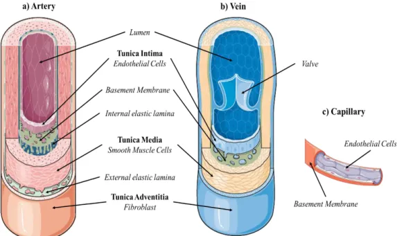
The vascular system is a network of blood vessels that allow the circulation of blood. Vessels are divided into three main categories: Arteries, Veins and Capillaries. The different types of vessels are distinguished by thickness, cellular and protein composition of their walls. This heterogeneous, yet highly organized structure allows the blood vessels to effectively perform their vital functions [3]. Arteries are blood vessels that carry blood away from the heart. Blood carried by arteries is usually highly oxygenated, having just left the lungs on its way to the body’s tissues. Arteries face high levels of blood pressure as they carry blood being pushed from the heart under great force. To withstand this pressure, the walls of the arteries are thicker, more elastic, and more muscular than those of other vessels. Capillaries are the smallest and thinnest of the blood vessels. They connect to arterioles on one end and venules on the other. Capillaries carry blood very close to the cells of the tissues of the body in order to exchange gases, nutrients, and waste products. The walls of capillaries consist of only a thin layer of endothelium that acts as a filter to keep blood cells inside of the vessels while allowing liquids, dissolved gases, and other chemicals to diffuse along their concentration gradients into or out of tissues. Veins are large return vessels of the body. Because the arteries absorb most of the force of the heart’s contractions, veins are subjected to very low blood pressures. This lack of pressure allows the walls of veins to be much thinner, less elastic, and less muscular than the walls of arteries. Veins rely on gravity, inertia, and the force of skeletal muscle contractions to help push blood back to the heart. Some veins contain many one-way valves that prevent blood from flowing back. Arteries and veins are mainly composed of 3 tunics: Intima, Media and Adventitia (Figure 0.2).
Figure 0.2: Blood vessels structure: a) Artery; b) Vein; c) Capillary.
a) The Intima
The tunica intima is the innermost lining of blood vessels. It is composed by the endothelium, which is almost always simple squamous epithelium. Endothelial cells (ECs) are flattened and polarized cells whose apical surface faces the lumen of the vessel. ECs have many important roles that ensure the functionality of the blood vessel [4]. These flat cells are tightly adherent and form a smooth surface that minimizes friction between the blood and the inner surface of the vessels. They are covered on their apical surface with a thin negatively charged layer called glycocalyx [5]. This surface is mainly composed of anticoagulant proteins secreted by endothelial cells. ECs also ensure mechano-transduction, the conversion by cells of a mechanical signal given by the blood flow into cellular physiological signals [6] acting on the ECs themselves (pro-inflammatory and proliferative signals) and on cells from other layers of the vessel wall (contraction of the media layer). In the arteries, a layer of elastic tissue called the internal elastic membrane marks the outer boundary of the tunica intima.
b) The Media
The tunica media is the middle layer of blood vessels. It consists of concentric sheets of contractile smooth muscle cells (SMCs) supported by an
III and proteoglycans [7]. The contraction of SMCs is controlled by the autonomic nervous system, hormones and local chemicals. Contraction of the SMCs is called vasoconstriction and results in a decrease in the size of the lumen and a decrease in blood flow. Relaxation of the SMCs is called vasodilation which has opposite effects. Small variations in the diameter of the vessels have marked effects on blood flow or pressure, thus the media plays an important role in the regulation of circulation in tissues and organs. Arteries sometimes have an outer layer of elastic tissue called the external elastic membrane. When present this marks the boundary between the tunica media and tunica adventitia.
c) The Adventitia
The adventitia is a connective tissue that forms the outermost layer of the vessel. It is composed mainly of collagen type III, elastin and fibroblasts (FBs). The composition of the ECM of this layer allow the blood vessels to stretch while preventing excessive dilation and often blend into the surrounding tissues. In thick-walled vessels, the adventitia may include nerve fibers and blood vessels to supply oxygen and nutrients to the outer layers. These vessels are called vasa vasorum [8].
The Blood
The primary function of blood is to deliver oxygen and nutrients to and remove wastes from body cells. The specific functions of blood also include immunological defense, distribution of heat, and maintenance of homeostasis [9]. Blood constitutes approximately 8% of adult body weight. Blood is a connective tissue and like all connective tissues, it is made up of cellular elements and an extracellular matrix. The cellular elements—referred to as the formed elements—include: 1) Red blood cells (also known as erythrocyte), which are anucleated biconcave disks containing hemoglobin whose primary function is to transport oxygen from the lungs to the body’s tissues; 2) White blood cells (or leucocyte), which are a major component of the immune system implied in the defenses against infectious diseases and 3) platelets (or thrombocytes), cell fragments critical for the process of hemostasis, the stoppage of blood outflow from a damaged vessel (hemorrhage). The extracellular matrix, called plasma, makes blood unique among connective tissues because it is fluid. Plasma, which is mostly composed of water,
perpetually suspends the formed elements and enables them to circulate throughout the body within the cardiovascular system.
Main Pathologies affecting the Cardiovascular System
Cardiovascular diseases (CVDs) are one of the leading causes of death in the world. According to the World Health Organization (WHO), CVDs were responsible for 17,9 million deaths in 2017, almost 31% of the total global mortality [1]. CVDs include numerous pathologies, many of which are related to a process called atherosclerosis. Atherosclerosis [10] affects arteries like the aorta, coronary, brain, pelvis, legs, arms or kidneys arteries. It is a condition that develops when a plaque builds up in the walls of the arteries, causing a partial or total occlusion (stenosis) of the artery lumen and thus reducing the blood flow (ischemia). If the oxygen supply to the heart muscle is reduced (hypoxia), a heart attack can occur. Lipids, cholesterol, foamy cells, cellular debris and calcium accumulate over time in the artery wall. Up today, the mechanisms underlying the insurgence of atherosclerosis are not yet fully understood. These substances stimulate SMCs to proliferate and produce other proteins and chemo-attractive biomolecules, resulting in the accumulation of more cells in between the intima and media layers of the artery where the atherosclerotic lesions form. The arterial wall becomes markedly thickened by these accumulating cells and surrounding materials, leading to ischemia (Figure 0.3). Often, the rupture of the fibrous cap of the plaque will lead to the formation of a blood clot that will block the artery (thrombus), stopping the blood flow.
Current Clinical Treatments
Risk factors and the occurrence of a stenosis, or its aggravation, can be prevented. Heart-healthy lifestyle changes can help in preventing or limiting atherosclerosis. These include: Healthy eating, weight loss, managing stress, physical activity and quitting smoking. In the presence of a genetic predisposition or when the first clinical signs have already appeared, pharmaceutical drugs such as statins (to lower cholesterol levels) [11], angiotensin-converting enzyme (ACE) inhibitors and β-blockers (antihypertensive drugs) [12] are recommended.
In presence of severe atherosclerosis, a medical procedure or surgery to remove the atherosclerotic plaque is highly suggested. Balloon angioplasty is a procedure used to re-open blocked or narrowed coronary arteries. This procedure can improve blood flow to the heart and relieve chest pain. Through an incision in the femoral artery, a catheter on which is placed a balloon is inserted. Once inflated, the balloon compresses the plaque, re-opening the artery lumen [13]. Often a stent, a small mesh tube made of metal (stainless steel, magnesium alloy or titanium), is placed in the artery to keep it open after the surgical procedure [14]( Figure 0.4).
Figure 0.4: Angioplasty: Main steps of balloon and stent angioplasty procedure.
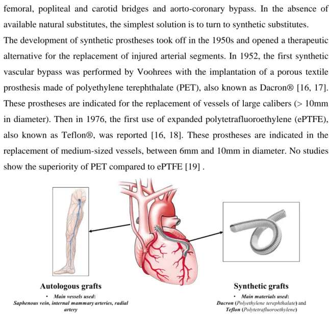
When the vessel's occlusion is severe and other treatments are not suitable, replacement or bypass surgery are the only current solution. During surgery, the diseased part of the artery is removed and replaced or bypassed by a graft or a prosthesis to restore blood circulation (Figure 0.5).
Autologous grafts are the ideal substitute solution with a success rate of 90% at one year [15]. Most often, the saphenous vein of the patient is used in this procedure. It is a vein running through the leg and its diameter and length make it a suitable substitute for
femoral, popliteal and carotid bridges and aorto-coronary bypass. In the absence of available natural substitutes, the simplest solution is to turn to synthetic substitutes. The development of synthetic prostheses took off in the 1950s and opened a therapeutic alternative for the replacement of injured arterial segments. In 1952, the first synthetic vascular bypass was performed by Voohrees with the implantation of a porous textile prosthesis made of polyethylene terephthalate (PET), also known as Dacron® [16, 17]. These prostheses are indicated for the replacement of vessels of large calibers (> 10mm in diameter). Then in 1976, the first use of expanded polytetrafluoroethylene (ePTFE), also known as Teflon®, was reported [16, 18]. These prostheses are indicated in the replacement of medium-sized vessels, between 6mm and 10mm in diameter. No studies show the superiority of PET compared to ePTFE [19] .
Figure 0.5: Bypass surgery: Use of autologous and synthetic grafts for bypass surgery.
Although many improvements have been made over the years, the clinical performance of these prostheses, especially for small diameter vessels bypass/substitution (Ø<6 mm) [20], is still hampered by high rate of graft lumen occlusion due to intra-graft thrombosis and intimal hyperplasia, especially at the anastomosis site, that are regularly observed in the months or years following surgery [2, 21]. The formation of a functional endothelial layer, known as endothelialization, in the lumen of the vascular substitutes would significantly improve small-diameter graft survival by: i) Preventing thrombus formation on the graft surface; ii) Enhancing internal healing and limiting intimal hyperplasia [22]. Developing prostheses for patients without suitable autologous vessels that can be used for bypass surgery following vascular pathologies has become necessary [23].
Vascular Tissue Engineering
Vascular tissue engineering (VTE) it is an inherently multidisciplinary area of research combining the know-how of several research fields with the aim of developing technologies for vascular medicine applications featuring biological and mechanical properties as close as possible to those of the native vessels, in order to ameliorate the integration and healing of the implanted substitutes. The ultimate goal of VTE is the development of artificial vascular substitutes. The ideal substitute must fulfil strict specifications such as: Absence of thrombogenicity and immunogenicity; Suitable mechanical properties; High availability, resistance to suture and infections and low costs [24-27]. The tissue engineering derived vascular substitutes can be classified according to various criteria. One of these criteria is the origin of the polymer used as a scaffold for the vascular substitutes. Polymers can be of synthetic or of natural origin. Synthetic polymers include biodegradable synthetic polymers, such as polyglycolic acid (PGA) , or permanent composite synthetic polymers, such as PET, ePTFE and polyurethane [23]. Natural polymers are usually proteins that constitute the original architecture of native ECM. The generation of protein polymers that mimic native structural proteins and adopt the characteristics of the arterial wall offers a unique approach to develop a vascular graft. There are three major types of natural vascular substitutes derived from tissue engineering: Decellularized matrices, derived from living tissues that undergo chemical and mechanical treatments to remove cells from the tissue obtaining biological scaffolds that are expected to maintain the complex 3-D structure and biomechanical properties of the ECM [28, 29], allowing the re-cellularization with autologous cells to produce a new immunologically compatible tissue adequate for implantation [30]. Self-assembly derived substitutes, [31, 32] in which the scaffolds are obtained by wrapping cellular sheets made of cells and synthesized collagen. Cells (SMCs and FBs) are cultured to form cell sheets that are subsequently rolled around a mandrel. These tubular structures are further cultured for a maturation period before undergoing endothelialization. Scaffolding system based on natural polymers, where cells (most often SMCs and FBs), are seeded in a protein solution which is then gelified [33]. This approach aims to create protein-bound cell matrices similar to vascular tissue. Type I collagen is the most used protein because it is present in many tissues and can be isolated, solubilized and reconstituted according to numerous protocols [34]. Fibrin is also used for these applications [35, 36] as also elastin [37]. The use of gel solutions based on proteins makes it easy to inoculate
cells. This cell suspension can then be molded to obtain the desired shape, for example tubular structures. The cells are uniformly dispersed and immobilized in the matrix during gelation. A reorganization of the matrix by the cells is then observed. The clinical application of protein-based gels, however, is still limited owing to their low mechanical properties [38-44]. Numerous methods have therefore been developed to reinforce these matrices [45] avoiding the use of non-biological solutions, such as the use of chemicals or synthetic polymers.
Despite the progresses made in the last years in the development of artificial blood vessels, further work is still required to ensure success towards clinical translation. In fact, despite showing promising biological performances, these products still miss important characteristics, mainly mechanical properties, that will ensure a successful implantation of the tissue engineered vessels[46, 47].
However, the strategies developed in the pursue of an artificial blood vessel can still find application in vascular medicine, representing interesting in vivo model for study needing the complex hierarchy typical of the blood vessel architecture [48], or even provide material useful for the amelioration of existing vascular graft [49] or for the development of drug delivery systems (DDS) [50].
Vascular Grafts Enrichment
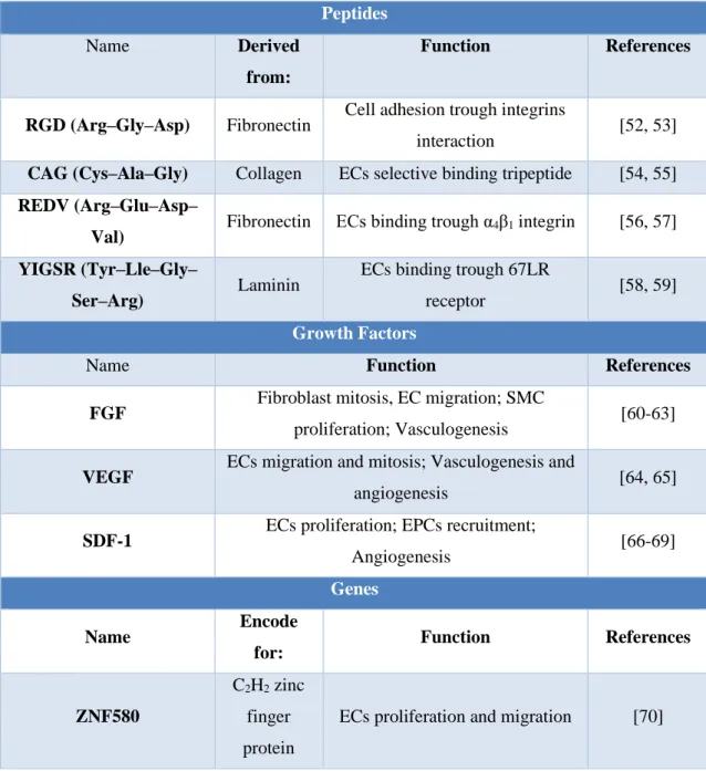
As previously stated, the lack of endothelialization is one of the major causes of small caliber vascular grafts failure. The absence of a complete and functional ECs layer on the luminal surface of the grafts leads to early thrombus formation and late restenosis, limiting the use of current vascular substitutes in clinic. Spontaneous endothelialization of vascular grafts or long segments of de-endothelialized arteries does not occur, leading to early thrombus formation, and the progressive SMC proliferation results in intimal hyperplasia, causing the occlusion (restenosis) of the grafted substitute. There is a critical need to control these phenomena by eliciting ECs ingrowth to optimize the physiologic functions of the vascular substitute. Physiological ECs ingrowth is regulated by complex interactions among growth factors and cytokines released by the cells and extracellular matrix within the local vascular microenvironment. For these reasons, the modification of the grafts luminal surface to induce a faster re-endothelialization has been proposed. In recent years vascular substitutes have been additionally engineered by enrichment with growth factors, cytokines, regulatory proteins, bioactive peptide and genes able to
ultimately guiding the optimal integration and functionality of the implant [51]. Several of these approaches have been shown to be able to promote endothelial cells recruitment for vascular grafts enrichment applications. In Table 0.1 some of the most used molecules are listed.
Table 0.1: Bio-active molecules used for pro-endothelialization of vascular grafts.
Peptides
Name Derived
from:
Function References
RGD (Arg–Gly–Asp) Fibronectin Cell adhesion trough integrins
interaction [52, 53]
CAG (Cys–Ala–Gly) Collagen ECs selective binding tripeptide [54, 55]
REDV (Arg–Glu–Asp–
Val) Fibronectin ECs binding trough α4β1 integrin [56, 57] YIGSR (Tyr–Lle–Gly–
Ser–Arg) Laminin
ECs binding trough 67LR
receptor [58, 59]
Growth Factors
Name Function References
FGF Fibroblast mitosis, EC migration; SMC
proliferation; Vasculogenesis [60-63]
VEGF ECs migration and mitosis; Vasculogenesis and
angiogenesis [64, 65]
SDF-1 ECs proliferation; EPCs recruitment;
Angiogenesis [66-69]
Genes
Name Encode
for: Function References ZNF580
C2H2 zinc finger protein
ECs proliferation and migration [70]
Abbreviations: FGF: Fibroblast growth factor; VEGF: Vascular endothelial growth factor; SDF-1: Stromal
derived factor; ECs: Endothelial cells; SMC: Smooth muscle cells; EPCs: Endothelial progenitor cells.
While the discovery of these effects initially had promising prospects for their use in therapy, clinical trials of angiogenic factor delivery have been mostly disappointing, underscoring the need for a wider array of angiogenic factors [71].
Pleiotrophin
Pleiotrophin (PTN), also known as “heparin binding growth associated molecule” (HB-GAM), is a growth/differentiation cytokine first discovered in 1989 that exert different biological effects on various cell types and is expressed mainly, but not exclusively, during embryogenesis [72]. The corresponding gene, Ptn, have been sequenced [73] and it encodes an 18 kDa protein of 168 amino acids. The conservation of PTN amino acid sequences among different species (human, bovine, rat, mouse, and chick) is the highest of any of the known cytokines [74-76]. PTN is known for its high affinity to heparin [72] and with extracellular matrix, from which it can be released into solution by heparin [74]. The binding sites of PTN to heparin were found to be located within the β-sheet domains of the protein, instead of the previously believed sites at the lysine rich N- and C-termini [77]. PTN has been shown to interact with and/or affect cell functions through several cell surface receptors such as syndecans [78], the anaplastic lymphoma kinase (ALK) [79], integrins [80], nucleolins [81] and neuropilin-1 [82]. However, the primary receptors for PTN is protein tyrosine phosphatase beta/zeta (RPTP β/ζ), whose activation can regulate multiple functions [83-86]. Over the years, PTN has been proved to be able to regulate multiple cell functions. The ability to promote cell growth was first described on FBs in 1989 [87]. After this first discovery, PTN has been found to be a mitogen for different cell types, including ECs, epithelial cells and different FBs cell lines [88-90]. PTN is also involved in cell motility [82, 91] and, by means of its structural similarity to the thrombospondin type I (TSR) sequence, PTN can mediate cell-to-extracellular matrix and cell-to-cell interactions [77]. It has been demonstrated to affect neural cells, stimulating neurite outgrowth from different cultured neuronal cell types [72, 92] and promoting process outgrowth when added to glial progenitor cells in primary cultures [93]. It also effects bone tissue: PTN has been found to take part in bone repair [94] and is also an osteocyte-derived factor that participate in mediating the osteogenic response to mechanical loading in bone [95]. Effects of PTN on skeletal muscle regeneration, mainly in the formation of neuro-musculature junctions, have been reported [96], as for the enhancement of cell survival and large-scale propagation of cultured human embryonic stem cells [97]. PTN is known for its effects on the immune system, like promoting lymphocyte survival and driving immune cell chemotaxis [83], and has also been shown to exert beneficial effects in the hematopoietic niche, regulating the retention
and self-renewal of hematopoietic stem cells in the bone marrow [98] and their regeneration [99].
PTN is also highly expressed in multiple human cancers, including breast, pancreas and lungs [84][14], in fact it is implicated in tumor angiogenesis and as proliferative driver and as factor in the resistance to apoptosis. PTN induce extracellular matrix remodeling, increase angiogenesis and stimulate the proliferation of stromal cells in the tumor microenvironment [100, 101].
• Pleiotrophin and the Cardiovascular System
Over the years, the effects exerted by PTN on the cardiovascular system, in particular on blood vessel and endothelial cells, have been widely studied. These effects of PTN on angiogenesis were firstly investigated in 1998, by virtue of its expression by ECs during ischemic brain injury healing process [102]. PTN was found to be able to stabilize the formation of capillary-like structures by cultured endothelial cells [103]. Subsequently, it was demonstrated that the induced overexpression of PTN in a rat model of myocardium ischemia was able to promote neovascularization of the infarcted area [104] and it can induce angiogenesis in adult and senescent rat aortic rings ex vivo [105]. In recent years, the involvement of PTN in physiological angiogenesis has been studied [106]. An important effect of PTN is the ability to chemotactically attract EPCs at angiogenic sites [107]. PTN seems to play a key role also in inflammation-induced neovascularization. The in vitro ability of PTN to induce a downregulation of monocytes/macrophages cell markers and an upregulation of endothelial cell characteristics, resulting in their transdifferentiation into functional endothelial cells has been reported [108] and further confirmed by other studies [109, 110][42, 43]. Moreover, PTN has been shown to be involved in the biosynthesis of catecholamine and in the regulation of the Renin/Angiotensin system in the mouse aorta [111, 112], thus, implying his involvement in the regulation of aortic blood pressure.
Collagen-based Releasing Systems
To efficiently deliver drugs or bioactive molecules, the choice of a suitable scaffold is of crucial importance. Among the different options available nowadays, natural polymers are one of the most interesting and promising candidates. One of such polymers is collagen. Collagen comprises 25% (by dry weight) of total protein content in vivo [113]; In some types of collagen, the entire molecule is a triple helix (Figure 0.6), while in other
types only a portion have this structure [114]. Of the various types of collagen, type I is by far the most prevalent form [115]. Mature type I collagen is composed of about 1000 amino acids and present a triple helix structure.
Figure 0.6: Schematic representation of collagen structure.
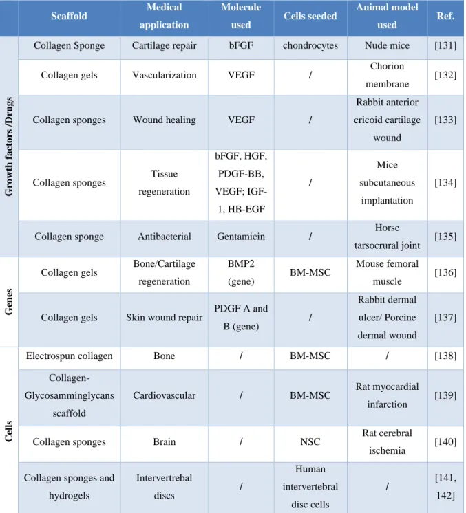
It is the main component of extracellular matrices, conferring their strength and shape to connective tissues such as bone, teeth, cartilage, tendons, ligaments and fibrous matrices of the skin and blood vessels. Type I collagen is widely chosen as a biomaterial for medical applications due to its ease of extraction, weak antigenicity, robust biocompatibility, and its ability to be physically and chemically modified for a variety of applications [116-118]. Among the applications the following can be mentioned: Substrate for cell cultures [119], suture material [120], tissue engineering scaffold for bladder reconstruction [121], bone tissue engineering scaffold [122], tendons reconstruction [123, 124], sponges are used to treat burns and other injuries (58) and scaffold for the regeneration of vascular tissue [125]. Due to its favorable biological properties, collagen-based matrices have been thoroughly investigated as a releasing system for therapeutic drug delivery applications. These investigations date back to the ‘70s [126] and over the years collagen-based drug delivery systems, including injectable microspheres, implantable collagen gels, interpenetrating networks of collagen and collagen membranes, have been studied for the delivery of growth factors, proteins, drugs, genes and cells [50] for application in several tissues such as bone [127], eye [128], ischemic heart [129], ischemic brain [130] and others. In Table 0.2 some of the use of collagen-based releasing systems are reported.
Table 0.2: Collagen-based matrices/scaffolds for drug, cell and gene delivery used in different tissue
engineering applications.
Scaffold Medical application
Molecule
used Cells seeded
Animal model used Ref. G ro wt h f a ct o rs /Dr ug s
Collagen Sponge Cartilage repair bFGF chondrocytes Nude mice [131]
Collagen gels Vascularization VEGF / Chorion
membrane [132]
Collagen sponges Wound healing VEGF /
Rabbit anterior cricoid cartilage
wound
[133]
Collagen sponges Tissue regeneration bFGF, HGF, PDGF-BB, VEGF; IGF-1, HB-EGF / Mice subcutaneous implantation [134]
Collagen sponge Antibacterial Gentamicin / Horse
tarsocrural joint [135]
G
en
es
Collagen gels Bone/Cartilage regeneration
BMP2
(gene) BM-MSC
Mouse femoral
muscle [136]
Collagen gels Skin wound repair PDGF A and
B (gene) / Rabbit dermal ulcer/ Porcine dermal wound [137] Cells
Electrospun collagen Bone / BM-MSC / [138]
Collagen-Glycosamminglycans
scaffold
Cardiovascular / BM-MSC Rat myocardial
infarction [139]
Collagen sponges Brain / NSC Rat cerebral
ischemia [140] Collagen sponges and
hydrogels Intervertrebal discs / Human intervertebral disc cells / [141, 142]
Abbreviations: bFGF: Basic fibroblast growth factor; VEGF: Vascular endothelial growth factor; HGF:
Hepatocyte growth factor; PDGF-BB: Platelet derived growth factor-BB; IGF-1: Insulin like growth factor-1; BMP2: Bone morphogenetic protein 2; HB-EGF: Heparin binding epidermal growth factor-like growth factor; BM-MSC: Bone marrow mesenchymal stem cells; NSC: Neural stem cells.
Among the different structure used, collagen gels are widely used for soft tissue applications. As already mentioned in the introduction, collagen gels are flowable, allowing to mold them to obtain different shape, like tubular structures, or to easily inject them. The most readily available forms of such collagen gels are suspensions of collagen fibers. However, fibrillar collagen gels have an effective pore size too large to allow a controlled release of the loaded protein-based drugs, such as growth factors and cytokines, by hindered diffusion. To control release, it is necessary to rely on binding of the active agent to collagen. Over the years, many types of drug delivery systems based on collagen have been developed for the control release of small molecule and protein-based drugs for biomedical applications [143]. The use of specific non-covalent interactions to stabilize drugs and immobilize them within a biocompatible matrix, thus protecting their biological activity and slowing their diffusion from the matrix, has been widely investigated in the last years. To achieve this result, several approaches have been used to modify the collagen scaffold.
• Heparin
One of such approaches rely on the modification of the collagen scaffold with heparin [144]. Heparin is a linear polysaccharide synthesized only in mast cells, where it is cleaved from the core protein serglycin [145]. Heparin polymer chains are made up of repeating disaccharides, the most common are 2-O-sulfated iduronic acid and glucosamine with varying degrees of sulfation and N-acetylation (Figure 0.7).
Figure 0.7: Heparin major disaccharide repeating units [F].
Heparin is best known for its anticoagulant properties, but has also been shown to promote cell adhesion, inhibit smooth muscle cell proliferation and to moderate inflammation [146]. Moreover, heparin is also known to sequester, stabilize and protect growth factors and
cytokines [144] and has been widely used in conjunction with different scaffolds to enhance their retention ability (Table 0.3).
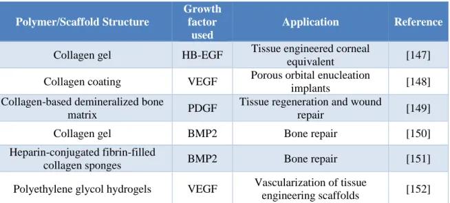
Table 0.3: Heparin-modified scaffolds for growth factor delivery in different tissue engineering
applications Polymer/Scaffold Structure Growth factor used Application Reference
Collagen gel HB-EGF Tissue engineered corneal
equivalent [147]
Collagen coating VEGF Porous orbital enucleation
implants [148]
Collagen-based demineralized bone
matrix PDGF
Tissue regeneration and wound
repair [149]
Collagen gel BMP2 Bone repair [150]
Heparin-conjugated fibrin-filled
collagen sponges BMP2 Bone repair [151]
Polyethylene glycol hydrogels VEGF Vascularization of tissue
engineering scaffolds [152]
Abbreviations: HB-EGF: Heparin binding epidermal growth factor-like growth factor; VEGF: Vascular
endothelial growth factor; PDGF: Platelet derived growth factor; BMP2: Bone morphogenic protein-2. Heparin interactions with proteins are largely electrostatic, however, there are clearly contributions from hydrophobic effects and hydrogen bonding, as well as promoting secondary structure in the proteins binding to heparin, which imparts some selectivity and specificity [153]. Finally, of importance for its use in cardiovascular applications, heparin itself has been shown to be able to stimulate angiogenesis [154, 155].
Strategy and Structure of the Project
Vascular bypass/replacement surgery is the primary clinical therapy for patients with advanced vascular occlusion diseases such as atherosclerosis. For this application, the need for functional small-caliber grafts is highly demanded. Autologous replacement vessels are the gold standard for this application [156, 157]. However, because of the usual bad condition of the vascular system in the patient these vessels are not always available to be used [21, 158, 159]. In this scenario, the only alternative is the use of an artificial vascular substitute. Despite the improvements observed over the last years, the use of synthetic grafts is still burden by high rates of implants failure. After implantation, the low patency of the vascular substitutes is mainly related to early in-graft thrombosis, caused by platelet deposition and
blood coagulation, and to late intimal hyperplasia near theanastomotic regions [160, 161]. Failure of vascular grafts is mainly determined by the lack of endothelial cells, as these cells effectively inhibit thrombosis and intimal hyperplasia [162, 163].
Therefore, the rapid establishment of an endothelial lining on the luminal surface of a vascular prosthesis would be beneficial to prevent early thrombosis and failure and for the long-term patency of the grafts. Several approaches have been used to achieve the formation of an ECs monolayer by seeding with harvested ECs or endothelial progenitor cells (EPCs) followed by in vitro maturation steps, resulted in an improvement of the patency [164, 165]. Despite these positive results, in vitro endothelialization involves multiple specialized procedures and cell cultures require long incubation period, making it cost ineffective, inconvenient and limited to adequately equipped facilities.
In recent years, to overcome the problem, implants modifications with bioactive molecules (growth factors, cytokines and other regulatory proteins) capable to efficiently recruit resident ECs, to promote their adhesion and growth speeding up the re-endothelialization process and, ultimately, guide the optimal integration and functionality of the grafted vessel [166-169] have been proposed over the last years. However, clinical trials of pro-endothelialization enrichment of vascular grafts have been mostly unsatisfactory, thus the need to investigate a wider array of angiogenic factors and new approaches for this application [71].
Pleiotrophin (PTN) is an 18-kDa growth/differentiation cytokine able to regulate multiple functions including cell adhesion, cell migration, cell proliferation and cytoskeletal stability [170] and has been described to be a potent enhancer of angiogenesis and neovasculogenesis [104, 105].
However, the concentration of biomolecules must be fine-tuned in order to accomplish the desired effects on the migration and proliferation of ECs, thus the need to release these molecules in a controlled way. The development of drug delivery systems (DDS) capable of controlling the release of therapeutic agents have been widely explored in recent years. The use of collagen as a scaffold for DDS applications is justified by several favorable properties that characterized this natural polymer [116-118]. To further modulate the release of the loaded proteins, specific non-covalent interactions can be used to stabilize drugs and
![Figure 0.1: Cardiovascular system. [A]](https://thumb-eu.123doks.com/thumbv2/123doknet/2904304.75099/20.892.471.744.711.1023/figure-cardiovascular-system-a.webp)


![Figure 0.7: Heparin major disaccharide repeating units [F].](https://thumb-eu.123doks.com/thumbv2/123doknet/2904304.75099/34.918.300.641.723.906/figure-heparin-major-disaccharide-repeating-units-f.webp)
![Table 1.1: Main Collagen types and their distribution in the human body. Modified from [177]](https://thumb-eu.123doks.com/thumbv2/123doknet/2904304.75099/42.918.129.794.132.638/table-main-collagen-types-distribution-human-body-modified.webp)
