HAL Id: tel-01066888
https://tel.archives-ouvertes.fr/tel-01066888
Submitted on 22 Sep 2014
HAL is a multi-disciplinary open access
archive for the deposit and dissemination of sci-entific research documents, whether they are pub-lished or not. The documents may come from teaching and research institutions in France or abroad, or from public or private research centers.
L’archive ouverte pluridisciplinaire HAL, est destinée au dépôt et à la diffusion de documents scientifiques de niveau recherche, publiés ou non, émanant des établissements d’enseignement et de recherche français ou étrangers, des laboratoires publics ou privés.
Functional characterization of GPI-anchored proteins of
the SKU5/SKS gene family
Ke Zhou
To cite this version:
Ke Zhou. Functional characterization of GPI-anchored proteins of the SKU5/SKS gene family. Plants genetics. Université Paris Sud - Paris XI, 2013. English. �NNT : 2013PA112096�. �tel-01066888�
UNIVERSITE PARIS-SUD
ÉCOLE DOCTORALE : SCIENCES DU VÉGÉTAL
Laboratoire de Catherine PERROT-RECHENMANN, ISV, CNRSDISCIPLINE : BIOLOGIE
THÈSE DE DOCTORAT
Soutenance prévue le 21/06/2013 par
Ke ZHOU
Functional characterization of GPI-anchored proteins of the
SKU5/SKS gene family
Composition du jury :
Directeur de thèse : Catherine PERROT-RECHENMANN DR CNRS Rapporteurs : Sébastien MONGRAND Professeur
Pierre CAROL Professeur Examinateurs : Dao ZHOU Professeur Jacqui SHYKOFF DR CNRS
Abstract
:ABP1 (Auxin Binding Protein1), who can bind auxin, is essential for the development of plants. It was proved to have the ability to bind auxin and transduce auxin signal into the cells. It is supposed to be localized and functions at the outer surface of plasma membrane through unknown component.
In my theiss, we tried to invesitgate the interaction between ABP1 and the candidate of the unknown component, CBP1 (From maize), which is GPI-acnhored and already identified as the binding ability to synthesized C-terminus peptide of ABP1 in 2006.
The orthologous of CBP1 in arabidopsis belongs to a gene family with 19 members, in which only three of them were prediceted to be GPI anchored. We did the functional characterisation of these three GPI-anchored members. Data suggested that GPI-anchored SKS proteins were involved in cell orientation, gametophyte and embryo development.
Résumé:
L’ABP1 (auxine Binding protein 1), qui peut se lier à l'auxine, est essentielle pour le
développement des plantes. Il a été prouvé d'avoir la capacité de se lier signal d'auxine auxine et transduce en les cellules. Il est supposé être localisée et des fonctions à la surface extérieure du plasma membrane à travers composante inconnue.
Dans mes Theiss, nous avons essayé de invesitgate l'interaction entre ABP1 et le candidat de le composant inconnu, CBP1 (à partir de maïs), qui est GPI-acnhored et déjà identifiée comme étant la capacité de liaison de synthèse peptidique C-terminale du ABP1 en 2006. L'orthologue de CBP1 chez Arabidopsis appartient à une famille de gènes avec 19
membres, dont seulement trois d'entre eux ont été prediceted être ancré GPI. Nous avons fait le fonctionnel caractérisation de ces trois membres. Les données suggèrent que GPI
Protéines SKS ont été impliqués dans l'orientation de la cellule, gamétophyte et le développement de l'embryon.
Acknowledgments
First I would like to thank members of the jury who have accepted to spend some of their time to review my thesis.
I would like to thank Catherine, my supervisor, who teach me and support me a lot along my PhD.
And thank all the members of our group, especially Catherine PRIMARD, who help me a lot both in life and experiment; Philippe MULLER, who did a lot of work for me in gene
expression analyses by Real time PCR; and Sebastien PAQUE, Alexandre TROMAS and Anne-Laure QUETTIER, who gave me a lot of scientific advice. Also thank the other members who were in our group or are still in our group.
And thank the CSC (China Scholarship Council), who granted me for three years. And finally, I want to thank everybody in our institute for helping me such a lot.
TABLE CONTENTS
List of abbreviations... 4
Introduction ... 5
-I-1 Auxin responses and signalling ... - 6 -
I-1-1 Auxin in plants. ... - 6 -
I-1-2 Auxin Biosynthesis and Storage ... - 7 -
I-1-3 Auxin transport... - 8 -
I-1-4 Auxin signalling pathway for transcriptional regulation... - 9 -
I-2 The role of ABP1 in auxin signalling... - 11 -
I-2-1 ABP1 identification... - 11 -
I-2-2 ABP1 functions ... - 11 -
I-2-3 ABP1 localization and potential ABP1 docking protein... - 11 -
I-3 GPI-Anchored Proteins... - 13 -
I-3-1 GPI and GPI-anchored proteins ... - 13 -
I-3-2 GPI biosynthesis... - 14 -
I-3-3 Transport and localization of GAPs ... - 17 -
I-3-4 GAPs in Mammals and Yeasts... - 19 -
I-3-5 GAPs in Plants ... - 19 -
I-4 Thesis project... - 23 -
Results... 24
Chapter I: In silico analyses ... 25
-I-1 CBP1 in Maize... - 25 -
I-2 Homologous genes of CBP1 in Arabidopsis. ... - 28 -
I-2-1 SKU5 gene family in Arabidopsis... - 28 -
I-2-2 In Silico Expression Data ... - 31 -
I-2-3 SKU5/SKS proteins... - 33 -
I-2-4 SKU5/SKS proteins through evolution ... - 35 -
Conclusion and Discussion ... - 37 -
Chapter II: Investigating the interaction between GPI-anchored SKU5/SKS
proteins and ABP1... 42
-II-1. CBP1 and GPI-anchored SKU5/SKS proteins were candidates for being ABP1 binding or docking proteins... - 42 -
II-2. Yeast two Hybrid System... - 42 -
II-3 Interaction detection by yeast two hybrid system ... - 44 -
II-3.1 Gene cloning and modification for yeast two hybrid... - 44 -
II-3-2 Yeast transformation and searching for interaction ... - 49 -
Conclusion and Discussion: ... - 54 -
Chaper III: Functional Analysis of SKS genes by reverse genetic
approaches ... 56
-III-1 Expression analysis of SKS1, SKS2, SKU5 in Arabidopsis thaliana:... - 56 -
III-2 Characterization of sks1, sks2 and sku5 null mutants. ... - 58 -
III-2-1 sku5/sks single null mutants ... - 58 -
III-2-2 Generating double and triple mutants for sks1, sks2 and sku5. ... - 61 -
III-2-3 SKS1, SKS2 and SKU5 expression in mutant plants... - 63 -
III-2-4 Analyses of sks1, sks2 and sku5 single and double mutants ... - 64 -
III-2-5 Analyses of sks1,sks2,sku5 triple mutant... - 69 -
Chapter IV: SKS1, SKS2 and SKU5 are required for fertilization and
embryogenesis... 82
-IV-1 Introduction to fertilization, pollen tube guidance and embryogenesis... - 82 -
IV-1-1 Male and female gametophytes... - 82 -
IV-1-2 Pollen tube guidance ... - 84 -
IV-1-3 Double fertilization of flowering plants ... - 86 -
IV-1-4 Arabidopsis embryogenesis... - 87 -
IV-1-5 seed maturation and dessication ... - 88 -
IV-2 Absence of SKS1, SKS2 and SKU5 affect fertilization and embryogenesis. ... - 90 -
IV-2-1 SKS1, SKS2, SKU5 in fertilization ... - 93 -
IV-2-2 SKS1, SKS2 and SKU5 in embryogenesis. ... - 100 -
Conclusion and Discussion ... - 105 -
Chapter V. Functions of GPI-anchored SKS and the roles of GPI anchors
by overexpression analyses... 107
-V-1 Tools for transient assay and SKS overexpression ... - 107 -
V-1-1 SKS9 was cloned from Arabidopsis... - 107 -
V-1-2 SKS genes modifications... - 108 -
V-1-3 Vectors for SKS overexpressors... - 111 -
V-2 GPI-anchors for localization by transient assay... - 112 -
V-3 Overexpressors in Ws background acquisition and analyses... - 114 -
V-3-1 Overexpressors acquisition... - 114 -
V-3-2 Expression levels of SKS Overexpressors in Ws background... - 115 -
V-3-3 Phenotype analyses of overexpressors... - 118 -
V-4 Overexpressors in sks1,sku5 background acquisition... - 119 -
Conclusion and Discussion ... 120
ChapterVI Discussion and Perspectives... 121
-
List of abbreviations
ABP1: Auxin Binding Protein 1 IAA: Indole 3 acetic acid GAP: GPI-anchored protein
GPI: GlycosylPhosphatidylInositol PT: Pollen tube
Introduction
I-1 Auxin responses and signalling
I-1-1 Auxin in plants.The word ‘Auxin’ was generated from a Greek word which means ‘to grow’. Its function was firstly described by Charles and Francis Darwin in ‘The Power of Movement in Plants’ in 1880 (Darwin and Darwin, 1880). Long-term studies revealed that auxin is the most important plant hormone and functions as a key morphogen in regulating plant growth and development. Several native auxins were found in plants. According to the chemical structures of native auxin, several chemicals were synthesized and functioned in plants (Simon and Petrášek, 2011). Some of the native and synthetic auxins were shown in Table I-1.
I-1-2 Auxin Biosynthesis and Storage
Indole-3-acetic acid (IAA) is the main naturally occurring auxin in plants and the best studied. It is biosynthesized through two major routes, from Tryptophan (Trp) using various Trp-dependent pathways and from an indolic Trp precursor via Trp-inTrp-dependent pathways (Woodward and Bartel, 2005; Korasick et al., 2013) (Figure I-1).
After the synthesis, IAA is stored in the auxin pool, which includes free auxin, conjugated auxins to amino-acid residues or glucose, inactive auxin precursor IBA and the inactive methyl ester form of IAA, MeIAA; but only a small fraction of auxin exists as free active signalling molecule (Korasick et al., 2013) (Figure I-2).
Figure I-1. Potential IAA biosynthetic pathways.
Arrows in pathways for which enzymes have been identified are solid and arrows in pathways that have not been identified are dashed and can represent single or multiple steps. (Korasick et al., 2013)
I-1-3 Auxin transport
IAA is weak acid with the pKa 4.85. In plants, the pH at the apoplast is approximately 5.5, where the IAA is 83% dissociated and 17% protonated. In the cytosol, the pH is
approximately 7, where the almost entire IAA is shifted to dissociated form.
Proton-associated IAA molecule can enter the cell by lipophilic diffusion, but dissociated IAA anion cannot go across the plasma membrane (Robert et al., 2010). So the auxin carriers are
necessary for the movement of auxin and are responsible for short distance oriented transport of auxin. In Arabidopsis, AUXIN1/LIKE-AUX1 (AUX1 and LAX) family of auxin
transporters are major auxin influx carriers whereas PIN-FORMED (PIN) and P-GLYCOPROTEIN (PGP) family members are major auxin efflux carriers (Ugartechea-Chirino et al., 2012).
In Arabidopsis, AUX1 belongs to a small multigene family comprising four highly conserved genes, AUX1, LAX (LIKE AUX1)1, LAX2 and LAX3; they show well conserved gene structure. AUX1/LAX has 11 transmembrane segments with N-terminal residing inside the cell and C-terminal outside. All proteins they encoded display auxin uptake functions and
Figure I-2. Potential IAA storage form pathways.
Arrows at steps for which enzymes have been identified are solid and arrows in pathways that have not been identified are dashed and may be single or multiple steps. (Korasick et al., 2013)
bind IAA with PH dependence; the maximal binding takes place between pH5 and 6 (Peret et al., 2012; Swarup and Peret, 2012).
In Arabidopsis, eight PIN genes were identified to encode PIN proteins with a similar structure; they all have amino- and carboxy-terminal hydrophobic membrane-spanning domains separated by a central hydrophilic domain (Krecek et al., 2009). They mainly differ by the hydrophilic loop, in terms of sequence and length. PIN-FORMED 1 (PIN1) gene, named as naked and pin-like inflorescences with its mutation, was found as the first member of the PIN gene family (Okada et al., 1991).
PIN-LIKE genes(PILs), identified on the basis of their similarity with a subclass of PIN genes, encode proteins containing auxin carrier domain; evidence showed that they were involved in auxin transports but possibly in the endoplasmic reticulum (Barbez et al., 2012) Auxin transport could be blocked by various chemicals. Auxin transport inhibitor N-1-naphthylphthalamic acid (NPA) was identified as a compound able to inhibit polar auxin transport and at the cell level was shown to block auxin efflux. NPA treatment arrests lateral root development by blocking primordium initiation ; the initiation could be recovered by additional NAA treatment (Casimiro et al., 2001). At a later stage of development application of NPA to the plant can mimick the phenotype of the pin1 mutant.
I-1-4 Auxin signalling pathway for transcriptional regulation
TIR1 was first identified from the tir1 mutant that exhibited a weak resistance to the auxin inhibitor NPA. Later on, TIR1 identified as an auxin receptor (Dharmasiri et al., 2005;
Kepinski and Leyser, 2005), the mechanism by which it works is much clearer than for ABP1 (Figure I-3).
Auxin response factors (ARF) are transcription factors that regulate the expression of auxin response genes and Aux/IAAs are transcriptional repressors. With the binding of Aux/IAAs and other co-repressors as TOPLESS (TPL) to ARF, the transcription is repressed (Guilfoyle and Hagen, 2007; Causier et al., 2012).
TIR1 and the related auxin F-box (AFBs) genes encode F-box proteins that are part of E3 ubiquitine ligase complexes names SCFTIR1/AFBs. Auxin promotes the interaction between TIR1/AFBs and Aux/IAAs , acting as a molecular glue (Tan et al., 2007). TIR1/AFBs and Aux/IAA form auxin co-receptors. Aux/IAAs are polyubiquitinated by SCFTIR1/AFBs
complexes and then enter the ubiquitin-proteasome degradation pathway (Santner and Estelle, 2011). After the removal of Aux/IAA repressors, ARFs promote the transcription of auxin response genes.
Figure I-3. SCFTIR1 and related SCF complexes bind auxin and target Aux/IAA proteins for degradation. (a) At low cellular levels of auxin, transcription of auxin response genes is repressed by the Aux/IAAs. (b) When auxin cellular levels increase, auxin binds to TIR1and Aux/IAAs, and promotes their ubiquitination and subsequent degradation, thus permitting the ARF proteins to promote transcription. (Santner et al., 2009)
I-2 The role of ABP1 in auxin signalling
I-2-1 ABP1 identificationAUXIN BINDING PROTEIN 1 (ABP1) was identified on its auxin binding activity nearly 30 years ago (Lobler and Klambt, 1985). Although the binding activity was confirmed by biochemical methods or co-crystallization with auxin (Jones and Venis, 1989; Woo et al., 2002), the mechanism of how it work was still largely unclear.
I-2-2 ABP1 functions
A lot of efforts were made to perform functional analyses of ABP1. The importance of ABP1 is generally revealed. Exogenous ABP1 protein and synthesized C-terminus peptide of ABP1 were shown to induce an hyperpolarization response of tobacco protoplasts (Leblanc et al., 1999b; Leblanc et al., 1999a). ABP1 was involved in auxin-induced protoplast swelling, the antibody to ABP1 and C-terminus peptide could prevent the auxin-induced swelling (Steffens et al., 2001), ABP1 is also essential for the control of the cell cycle; with inactivation of ABP1 by conditional immunomodulation, the cell cycle was arrested in BY2 cells in culture (David et al., 2007).
ABP1 is essential for embryogenesis and null mutation of abp1-2 causes embryo lethality (Chen et al., 2007). ABP1 functional inactivation was obtained and studied using conditional immunomodulation of ABP1 function in Arabidopsis generated by the controlled production (ethanol induction) of recombinant antibodies blocking the protein (Braun et al., 2008). By this tool, ABP1 was found to be essential at all stages of post-embryonic development. Knockdown for ABP1 results in severe alterations of leaf initiation and growth, as well as inhibition of primary root growth (Braun et al., 2008; Tromas et al., 2009). These defects result from default in cell division and cell expansion. Recently was protein was also shown to control clathrin dependent endocytosis and to alter recycling of plasma membrane proteins, including PIN efflux carriers (Robert et al., 2010; Chen et al., 2012). Whereas the recycling of PIN is potentially affected by ABP1 inactivation, no significant effect was reported onthe polar localisation of PIN or on the auxin gradient formed at the root apex (Tromas et al., 2009)
I-2-3 ABP1 localization and potential ABP1 docking protein
The localization of ABP1 is an interesting aspect. ABP1 genes in different plants species encode soluble proteins with N-terminal signal peptides allowing synthesis to the ER. In flowering plants, a KDEL ER-retention signal is also found at the C-terminus suggesting that
the protein could be retained within the lumen of the ER (Tromas et al., 2010). This KDEL sequence seems however to be recent as it is not present in non-flowering plants (Panigrahi et al., 2009). Despite the absence of hydrophobic domain or post-translational modification allowing interaction with membrane fractions, ABP1 is always found to be associated with membranes. In addition, all functional evidences point out a localization of ABP1 at the plasma membrane (Tromas et al., 2010). The hypothesis that ABP1 might interact with a membrane protein or a protein complex was proposed (Schiebl et al., 1997)(Figure I-4).
To identify the putative “docking protein”, a photoaffinity crosslinking study was
performed using a synthetic peptide corresponging to the C-terminus of the maize ABP1. GPI-anchored protein C-TERMINAL PEPTIDE BINDING PROTEIN 1 (CBP1) was identified as exhibiting the capacity to bind the synthetic C-terminus peptide of ABP1 (Shimomura, 2006).
Interestingly, GPI-anchor is a structure which can associate protein to the outer face of the plasma membrane. However, no more evidence confirmed the direct interaction between ABP1 and CBP1.
Figure I-4. Rapid auxin responses mediated by ABP1.
Auxin is perceived at the outer face of the plasma membrane (Plm) by ABP1 which interacts with an unknown membrane-associated protein or protein complex (docking protein). Binding of auxin to ABP1 activates a cascade of events including activations of the Plm proton pump ATPase and of potassium inward rectifying channels. The activation of the H+ ATPase induces hyperpolarization of the Plm and acidification of the extracellular space which contributes to cell wall loosening. The increase of intracellular K+ contributes to the uptake of water which is essential for cell expansion. It is not known whether these early events are part of a signaling cascade controlling downstream ABP1-mediated cellular responses (Tromas et al., 2010).
I-3 GPI-Anchored Proteins
I-3-1 GPI and GPI-anchored proteins
Proteins following the secretory pathway can be associated to the plasma membrane by various mechanisms. Proteins exhibiting hydrophobic transmembrane domains can be integrated in the plasma membrane bilayer. Proteins without transmembrane region can be attached to the membrane through post-translational lipid modifications occurring in the lumen of the endoplasmic reticulum. One of the best characterized process is the attachment of glycosylphosphatidylinositol (GPI) anchor that allows tethering the modified protein to the to external surface of the plasma membrane (Ferguson et al., 1988b, 1988a). This glycolipid modification includes a fatty acid tail anchored in the inner membrane leaflet of the
endoplasmic reticulum (lumen side) then within the outer leaflet of the plasma membrane bilayer after secretion of the modified protein (Figure.I-5A). GPI moiety from different species exhibit conserved core structure. Mannose residues are linked to the carboxyl-terminus of the protein through an amide bond with ethanolamine phosphate group (Holder and Cross, 1981; Holder, 1983). Glucosamine and phosphatidylinositol make the junction between sugar residues and the fatty acid that can be diacylglycerol or ceramide (Ferguson et al., 1988a) (Figure.I-5B). GPI modification is present among lower and higher eukaryotes, about 0.5% of total proteins are predicted to be GPI anchored proteins (GAPs) (Eisenhaber et al., 2001).
I-3-2 GPI biosynthesis
The GPI anchor biosynthesis is decided by the special amino acid sequence of the precursors of non-mature GPI-anchored proteins. All the GAPs are encoded with a cleavable
hydrophobic signal peptides at N-terminus and hydrophobic GPI-anchoring signal at carboxy-terminus (Figure I-6) (Caras et al., 1987; Caras and Weddell, 1989; Mayor and Riezman, 2004). The N-terminal hydrophobic signal lead the precursors enter the ER lumen and the carboxy-terminal hydrophobic GPI-anchoring signal forms the transmembrane region when the precursor synthesized passing through the ribosome into the lumen of ER. The signal peptidase recognize and cleave the N-terminal signal peptide and GPI protein transamidase
A
B
Figure I-5. GAP and Core structure of GPI moiety (from Mayor and Riezman, 2004).
A | GPI-anchored proteins are embedded in the extracellular or lumenal leaflet of membranes through their glycolipid moieties and are not directly accessible from the cytosolic face of the membrane. B | The GPI core structure is shown. The conserved core consists of ethanolamine phosphate in an amide linkage to the carboxyl terminus of the protein, three mannose residues (orange), glucosamine (blue) and
phosphatidylinositol (purple). It can be modified and is subject to various remodelling reactions of the lipid moiety. Variations can occur by addition of extra sugars or ethanolamine phosphates to the mannose residues; acylation of the inositol ring; changes in the fatty acids (length, saturation, hydroxylation), or their types of linkage to the glycerol backbone (acyl to alkyl); or remodelling of the entire diacylglycerol to ceramide.
recognizes and cleaves this region at ω-site, and then link the free –COOH of the mature protein to the synthesized GPI moiety (Mayor and Riezman, 2004).
When GPI protein transamidase recognizes the precursors, the biosynthesis of GPI moiety begins. The diacyl-PI of lipid bilayer, which is present at the cytoplasmic side of the ER membrane, would flip into the luminal side of the endoplasmic reticulum membrane, where later biosynthesis and linkage to the protein occur (step 1-11 in Figure. I-7); a series of enzymes were identified for their contribution to the different steps of this complex progress in various species (Fujita and Kinoshita, 2010). Table I-2 summarizes identified enzymes in mammals, Saccharomyces cerevisiae and Arabidopsis.
Figure I-7. Biosynthesis and remodeling of mammalian GAPs. (Fujita and Kinoshita, 2010)
Steps 1–11, TA, ID, GR, FR1, and FR2 correspond to those shown in Table1. TA, transamidation; ID, inositol deacylation; GR, glycan remodeling; FR, fatty acid remodeling; Dol-P-Man,
dolichol-phosphate-mannose; PE, phosphatidylethanolamine.
Figure I-6 Structure of GPI-anchor-protein precursor (Mayor and Riezman, 2004).
GPI-anchored proteins are synthesized as precursors with a cleavable, hydrophobic amino-terminal signal sequence that targets the protein to the lumen of the endoplasmic reticulum (ER) and a cleavable,
carboxy-terminal signal sequence that directs GPI anchoring. The GPI-anchoring signal consists of a hydrophobic region separated from the GPI-attachment site (ω-site) by a hydrophilic spacer region. Amino-acid residues with small side chains are highly preferred for the two amino acids that follow the ω-site.
Step Donor substrate Mammals S. cerevisiae Arabidopsis
PIG-A GPI3 SETH1
PIG-C GPI2 SETH2
PIG-H GPI15 AT4G35530
PIG-P GPI19 AT1G61280AT2G39445
PIG-Q GPI1 –
PIG-Y ERI1 –
1 GPI-GlcNAc transferase (GPI-GnT) UDP-GlcNAc
DPM2 – –
2 GlcNAc-PI de-N-acetylase PIG-L GPI12 –
3 Flippase Not identified ARV1? –
4 Inositol acyltransferase Palmitoyl-CoA PIG-W GWT1 –
5 PI remodeling enzyme ? Not identified – –
PIG-M GPI14
PNT1 PNT2 PNT3 PNT4
6 α1-4mannosyltransferase I (GPI-MT I) Dol-P-Man
PIG-X PBN1 AT5G46850
PIG-V GPI18 AT1G11880
7 α1-6mannosyltransferase II (GPI-MT II) Dol-P-Man
– PGA1 –
8 EtNP transferase I (GPI-ET I) PE PIG-N MCD4 –
9 α1-2mannosyltransferase III (GPI-MT III) Dol-P-Man PIG-B GPI10 –
M4 α1-2mannosyltransferase IV (GPI-MT IV) Dol-P-Man PIG-Z (SMP3) SMP3 –
PIG-O GPI13 –
10 EtNP transferase III (GPI-ET III) PE
PIG-F GPI11 AT1G16040
PIG-G (GPI7) GPI7 –
11 EtNP transferase II (GPI-ET II) PE
PIG-F GPI11 AT1G16040
PIG-K GPI8 –
GAA1 GAA1 AT5G19130
PIG-S GPI17 AT3G07180
PIG-T GPI16 –
TA GPI transamidase
PIG-U GAB1 AT1G12730AT1G63110
ID Inositol deacylase PGAP1 BST1 AT2G44970AT3G27325
AT3G52570
GR 2nd EtNP phosphoesterase PGAP5 CDC1 TED1 –
–
FR1 GPI-phospholipase A2 PGAP3 PER1
–
PGAP2 CWH43-N? –
FR2 Lyso-GPI acyltransferase I Stearyl-CoA
Not identified GUP1 –
CR Ceramide remodelase Ceramide? CWH43 –
FR3 GPI phospholipase A1 – – –
FR4 Lyso-GPI acyltransferase II – – –
I-3-3 Transport and localization of GAPs
After the GAPs were synthesized inside the ER, they will be packaged into ER-derived vesicles, which are distinct from the other secreted proteins, and exit endoplasmic reticulum through the Golgi to the surface of plasma membrane (Muniz et al., 2001). In animal cells, GAPs are present at the plasma membrane surface as monomers and a smaller fraction were reported to be associated to nanoscale cholesterol-sensitive clusters (Sharma et al., 2004).
Figure I-8. Trafficking and dynamics of GPI-APs on the membrane in mammalian cells. (extracted from(Fujita and Kinoshita, 2012)). (1) From the TGN to the plasma membrane.
GPI-APs are sorted and clustered into lipid-mediated carriers at the TGN. Sorting of raft-associated proteins, such as palmitoylated proteins and lipids, including glycosphingolipid and cholesterol, and exclusion of non-raft components,would occur. Oligomer formation of GPI-APs would be required for apical targeting. Luminal lectins, may help this sorting. (2) At the plasma membrane. The plasma
membrane appears to be compartmentalized by a cortical actin cytoskeleton. GPI-APs exist as monomers and as lipid assembled nanoclusters (5–10 nm) that are small and dynamic. In response to external signals, they can be actively induced to form large-scale rafts (40–300 nm). Signaling molecules are then recruited to the domains. The plasma membrane also appears as a mosaic of membrane domains, including caveolae and transient confinement zones. (3) Endocytosis. At steady state, GPI-APs are internalized as clathrin-independent carriers (CLICs) that seem to be regulated by Arf1, Cdc42, actin, and GRAF1. They are then incorporated into GEECs (GPI-APs enriched early endosomal compartment) . GPI-APs are subsequently transported to the recycling endosomes (RE), followed by recycling back to the plasma membrane, which is regulated by sphingolipids and cholesterol. Alternatively, GEECs are trafficked to the late endosomes (LE).
Interestingly, the transport efficiency of GAP is dependent on the presence of GPI anchor; without GPI-anchoring signal, the precursor of GAPs display the normal secretory protein pathway, for protein also having a signal peptide at the N-terminus and being targeted to the outer face of the plasma membrane; the absence of the GPI anchor reduces the secretion of the GAPs from the ER to the outer surface. (McDowell et al., 1998)
Although GPI-anchor is supposed to localize protein at the plasma membrane, actually it is not strictly true. In animal cells, the GPI-anchor of plasma membrane located GAPs can be cleaved by PI-specific phospholipase thus releasing the protein as a soluble protein in the medium of mammal cells in culture (McDowell et al., 1998). In yeast, only few GAPs reside permanently at the plasma membrane, a majority of them gets further processed and
integrated into the cell wall by a covalent attachment to cell wall glucans (Pittet and Conzelmann, 2007). Several annotations of Arabidopsis genes suggest that PI-specific phospholipase exist also in plants. There is however no functional characterization of these genes confirming their putative role in the cleavage of GPI anchors in vivo. Plant GAPs were detected at the plasma membrane and also at the cell wall, however mis-localization of reporter proteins cannot be ruled out (Roudier et al., 2005).
GPI anchor is not only important for the localization but also the functions of GAPs. The GPI anchor cleavage by GPI-PLC could change the antigenicity of Variant Surface Glycoprotein (VSG), which suggests that GPI lipid moiety affect the major conformation of VSG (Butikofer et al., 2001). And when the GPI anchor is replaced by a transmembrane (TM) region, the lymphocyte function-associated antigen 3 (LFA3) isoform TM FLA3 could reduce the Jurkat cell adhesion to planer bilayer (Tozeren et al., 1992).
I-3-4 GAPs in Mammals and Yeasts
In mammals and yeasts, GAPs play various and important roles in many biological processes, such as :
1. cell surface receptors: folate receptor in mammal cells participates in the cellular accumulation of 5-methyltetrahydrofolic acid in a number of epithelial cells in vitro through the process of caveolin-dependent potocytosis (Weitman et al., 1992); CD14, the first
identified Pattern Recognition Receptor (PRR) in mammals, acts as a co-receptor (along with the Toll-like receptor TLR 4 and MD-2 signalling complex) for the detection of bacterial lipopolysaccharide (LPS) and endocytosis (Zanoni et al., 2011).
2. cell adhesion molecules isoforms: fasciclin I directly mediates homophilic drosophila cells adhesion (Elkins et al., 1990), and the Algal-CAMs, which contain the homological repeat of fasciclin I and were found in Volvox plant, are also involved in cell adhesion (Huber and Sumper, 1994). But no GPI anchor was predicted in Algal-CAMs.
3. cell surface hydrolases.
4. GPI linked PrPC and PrPSc to plasma membrane, and surface conversion of PrPC to PrPSc requires PrPC, but not PrPSc to be GPI anchored (Radford and Mallucci, 2010).
And so on.
As GPI-anchored proteins play that many important roles, the defect of GPI anchor biosynthesis gene could result in severe phenotypes; the defect of X-linked GPI anchor
biosynthesis gene PIG-A, could result in paroxysmal nocturnal hemoglobinuria (PNH) disease (an acquired disorder that leads to the premature death and impaired production of blood cells), and it causes a lethal effect if the defect occurs in embryos (Kawagoe et al., 1996).
I-3-5 GAPs in Plants
248 potential GAPs are predicted by proteomic and genomic analysis, and classified as several families in Arabidopsis, including classical AGPs (arabinogalactan proteins), AG peptides, Fasciclin-like AGPs, COBRA family, SKU5 family, or receptor-like GAPs(Borner et al., 2002; Borner et al., 2003). 47 of them are expressed in pollen, in which 15 of them are detected specifically in flowers and pollens; the defect of GPI anchor biosynthesis pathway specifically block male transmission and pollen function (Lalanne et al., 2004). Plant GAP proteins were shown to be implicated in cell wall synthesis and morphogenesis (Gillmor et al., 2005). However not so many of these proteins have been studied in plant to date.
A large proportion of predicted GAPs (about 40%) are potentially also modified by O- linked arabinogalactans (AGPs)(Figure I-9) (Borner et al., 2003; Eisenhaber et al., 2003; Ellis
et al., 2010). AGPs are one of the most complex families in plants because of the high diversity of glycans that is decorating protein backbone (Ellis et al., 2010).
According to their protein backbone, 13 classical AGPs, 10 AG (acid glycoprotein) peptides, 21 Fasciclin-like AGPs, and 3 Lys-rich AGPs are classified in Arabidopsis through
bioinformatic analysis (Schultz et al., 2002). They are proved to be involved into many processes of growth and development, such as cell adhesion, cell wall composition, pollen tube guidance, cell division and programmed cell death (Seifert and Roberts, 2007).
Salt Overly Sensitive5 (SOS5), also called fasciclin-like AGPs4 (FLA4), was suggested to play a role in cell-to-cell adhesion; the absence of this gene causes thinner cell walls, swelling of root cells and increased sensitivity to salt stress (Shimomura, 2006).
POWDERY MILDEW RESISTANT 6 ( PMR6) in Arabidopsis encodes a pectate lyase-like protein, which is required for susceptibility to Powdery Mildew. The pmr6 mutant shows altered leaf morphology and decreased size, and alterations of cell wall composition that were proposed to activate defense response. PMR6 represents an interesting link between the cell wall and pathogen resistance, however the mode of action is unknown (Vogel et al., 2002).
Figure I-9 extracted from (Sardar et al., 2006).
A hypothetical cell surface network model involving interactions between LeAGP-1 and the cytoskeleton (i.e. MTs and F-actin). GPI-anchored LeAGP-1 is localized to lipid rafts, which contain lipids such as glycosphingolipids and sterols (such as stigmasterol, campesterol, and β-sitosterol), and interacts with MTs and F-actin in the cytoplasm either by a transmembrane protein (in the phospholipid bilayer) or by molecules (A and B) associated with the lipid rafts. GPI-anchored LeAGP-1 in lipid rafts may mediate interactions in two possible ways: A, binding of LeAGP-1 to a transmembrane receptor in the lipid microdomain mediates the interactions; and B, ligand-LeAGP-1 receptor complex-induced translocation of a signal molecule outside the lipid raft to activate a cytoplasmic kinase. Also shown are the different constituents of LeAGP-1 that include a Pro/Hyp-rich protein backbone decorated with arabinogalactan polysaccharides, short arabinosides, and an unglycosylated Lys-rich peptide region.
COBRA belongs to a multigene family consisting of 12 members in Arabidopsis, all predicted to be GAP. Analysis of COBRA mutants (cob) revealed that the protein is essential for anisotropic expansion in most organs. Further studies suggested that COBRA Elongating cells of cob mutants, especially in fast elongating zone as in root or hypocotyl, expand
radially instead of longitudinally thus resulting in very severe phenotypes (Schindelman et al., 2001; Roudier et al., 2002). The complete loss of function of COBRA causes severe growth and developmental defects, like cell selling and extreme dwarfism through the whole plant body (Ko et al., 2006). COBRA protein was found to be localized in limited domains of the plasma membrane and only at the lateral side of elongating cells (perpendicular to the axis of elongation). The patterning of COBRA is dependent on cortical microtubule organization (Roudier et al., 2005) and reciprocally COBRA was proposed to be required for orientation of cellulose microfibrils (Roudier et al., 2005). GPI-anchored COBRA-like proteins are also found in other species. The absence of Brittle culm1 in rice, Brittle stalk2 in maize, provoke alterations of cell wall composition and structure of secondary cell wall affecting mechanism strength of the tissues, resistance to bending (Sindhu et al., 2007; Panigrahi et al., 2009). In tomatoe, SlCOBRA-like was also reported to play an important role on fruit cell wall architecture and fruit development (Cao et al., 2012).
LORELEI belongs to a small plant-specific family containing 4 GPI-anchored members, LORELEI, LLG1, 2 and 3. In Arabidopsis, LORELEI encodes a putative GPI-anchor-containing membrane protein involved in signaling mechanisms at the synergid cell membrane by which the female gametophyte recognizes the arrival of a compatible pollen tube and promotes sperm release (Capron et al., 2008). LORELEI was shown to have a role in double fertilization and early seed development (Tsukamoto et al., 2010). Loss of function results in mostly failure of fertilization, which is mainly caused by pollen tubes continuing to grow after reaching the embryo sac and embryo sac continuing to attract additional pollen tubes (Capron et al., 2008; Tsukamoto et al., 2010). Despite a delay in the initiation of embryo development, the embryos recover and complete embryo development; no significant defect is detected in seedling and root development (Tsukamoto and Palanivelu, 2010).
SKU5 family belongs to a family of 19 highly conserved members, but only 3 members, SKU5, SKS1 (SKU5-similar 1) and SKS2 (SKU5-similar 2) are predicted to be modified by a GPI anchor (Borner et al., 2003). Among these, SKU5 was shown to be modified by a GPI.
The protein was released from the microsomal membrane fraction by action of a
phosphatidylinositol-specific phospholipase C that cleaves the lipid of the anchor (Sedbrook et al., 2002).
To date, SKU5 is the only GPI-anchored member of this family that was studied. Loss-of-function mutant has twisted roots and exhibits directional growth defects compared to wild type. The expression of the protein was correlated with expanding cells and the protein was localized at the plasma membrane and in the cell wall (Sedbrook et al., 2002). SKS6, a non-GPI-anchored member of this family, participates in cotyledon vascular patterning; the T-DNA insertion mutants have decreased numbers of full vascular loops in their cotyledon (Jacobs and Roe, 2005).
I-4 Thesis project
In my thesis, three questions were going to be addressed:
1. Whether GPI-anchored SKS proteins are ABP1 binding/docking proteins.
2. If not, the functions of GPI-anchored SKS in Arabidopsis are still interesting for us. 3. The role GPI anchors play in localization and function of GPI-anchored SKS proteins. To solve these questions, experiments were designed.
1. Yeast two hybrid system were utilized for investigating the interaction between ABP1 and GPI-anchored SKS protein in vitro.
2. Null mutant of GPI-anchored SKS genes were ordered and crossed to investigating the function of them.
3. The omega-domain, which is the signal for GPI synthesis, was cleaved from SKS genes and visualized by fluorescent proteins to investigate the function of GPI in localization and function.
Chapter I: In silico analyses
I-1 CBP1 in Maize.
As mentioned in the introduction, GPI-anchored protein C-terminal peptide-binding protein 1 (CBP1) from maize was identified through its capacity to bind synthetic C-terminal peptides of ABP1 in vitro and was thus proposed to be an ABP1 binding or docking protein in Maize (Shimomura, 2006).
CBP1 encodes a precursor protein with a signal peptide at the N-terminus and a hydrophobic C-terminal domain preceded by an ω-site. The leader peptide leads CBP1 to be addressed into the ER during synthesis. The C-terminal hydrophobic domain anchors the protein in the inner face of the ER membrane before its cleavage at the ω-site and replacement by a GPI anchor. The protein is then predicted to enter the secretion pathway and to be localized at the outer surface of the plasma membrane by its GPI anchor (Figure I-1).
The mature protein of CBP1 is predicted to exhibit conserved domains showing high similarities with multicopper oxidases, ascorbate oxidase and laccase (by NCBI Conserved Domain Database). The multi-domain consists of three domains, which are distributed along the sequence and are related to multicopper-oxidase of Cu-oxidase type 3, type 1 and type 2 (from N-ter to C-ter), respectively. All are structurally related to COX2 (Cytochrome c oxidase subunit 2) superfamily (Figure I-2), which is the second subunit of cytochrome c oxidase. It provides the substrate-binding site and contains a copper centre called Cu(A) that transfers electrons from cytochrome c to the catalytic subunit 1 (Capaldi et al., 1983) .
HPD SP Mature protein ω-site
A B
Figure I-1. GPI-Anchored Protein CBP1.
A- CBP1 protein scheme. CBP1 has a signal peptide at the N-terminus (SP) and a hydrophobic domain (HPD) at the C-terminus with a
cleavage site named ω-site. B- CBP1 is synthesized and targeted into the ER, After cleavage of the C-terminus and addition of a GPI anchor, the protein is entering the secretion pathway and is located at the outer surface of the plasma membrane.
: CBP1 ER
secretion pathway
In this series of proteins, Ascorbate oxidase (AO) has been studied deeply. It is proved to have copper binding and oxidase enzymatic activities. Comparison between CBP1 and AO was performed and it revealed that most critical residues of copper binding center motifs are absent or mutated in CBP1. These residues are labeled as * in figure I-2 (Messerschmidt and Huber, 1990). It suggests that copper binding to these centers in CBP1 is very unlikely.
CBP1 has several homologous genes in Maize (Figure I-3). Only several of them are predicted to encode GPI-anchored proteins (Table I-1), but none of them has been described.
FigureI-2. Putative conserved domains of CBP1 (Maize) and ascorbate oxidase (Zucchini), and copper binding center.
A, B- Conserved domains of CBP1 and AO
C- Amino acid alignment and copper binding sites
Figures A and B are from Conserved Domain Database of NCBI. Grey bars: amino acid sequence; Cyan fragments: compositionally biased regions not used for domain database search; conserved domains and superfamilies are indicated in dark red and pink bars;
PLN00044 and ascorbase are the name of multidomains.
The Histidine residues in copper binding centers of CBP1 are mostly absent (pointed by *) (Messerschmidt and Huber, 1990)
A-C-terminal peptide-binding protein 1
B- Ascorbate oxidase * * * * * * * * C
Figure I-3. Phylogenetic tree of proteins encoded by CBP1 and its homologous genes in Maize .
73 Proteins from Maize were identified through amino acid residues BLAST; and phylogenetic tree was calculated by COBALT tool.
By GPI-anchor predictor PredGPI calculation, some of them are predicted to have GPI anchors (Table I-1) and labeled as * in the figure).
CBP1 is underlined in yellow , bar is 0.2 billion years.
*
*
*
*
*
*
*
*
*
76 Not GPI-anchored 5 Weakly probable 9 Probable 8 Highly probableI-2 Homologous genes of CBP1 in Arabidopsis.
Homologous genes of CBP1 were identified in Arabidopsis and utilized for our research. By amino acid sequence BLAST in NCBI database, SKU5 was identified as the closest homologous gene of ZmCBP1 because it encodes a protein showing 68% identities and only 13 gaps in amino acid sequence (Figure I-4).
Figure I-4. Amino acid alignment between CBP1 and SKU5.
Proteins are indicated on the left and the consensus residues are in red; different amino acid residues are in blue and gaps are in black.
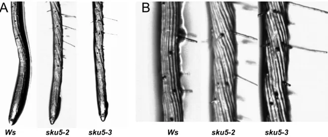
SKU5 was firstly described via the characterisation of a null sku5 mutant in Arabidopsis, it was reported to affect two directional growth processes, especially in root growth (Sedbrook et al., 2002).
I-2-1 SKU5 gene family in Arabidopsis
18 genes were identified as homologous genes of SKU5 in Arabidopsis thaliana. They are named SKU5-similar (SKS) genes and form the SKU5/SKS gene family. Members of this family encode proteins showing high identities. They were compared to SKU5 by BLAST analysis, and in which SKS1, SKS2 and SKS3 encode proteins with the highest identities with SKU5 (Table I-2).
CBP1 SKU5 CBP1 SKU5 CBP1 SKU5 CBP1 SKU5 CBP1 SKU5 CBP1 SKU5 CBP1 SKU5 CBP1 SKU5
Table I-2. SKS genes products are highly similar to SKU5 at the protein level.
SKS genes are found in all Arabidopsis chromosomes, however chromosomes 1 and 4 exhibit most of them with 7 and 6 SKS genes, respectively. Several genes localize as neighbors in the same chromosome, such as SKS7 and SKS8, SKS11 and SKS13, SKS12 and SKS14 (Figure I-5). SKS7 and SKS8 genes have the same transcriptional direction, but SKS11 and SKS13, SKS12 and SKS14 have the opposite transcriptional direction. These pairs exhibit similar gene structures with 8 exons for SKS7 and SKS8, whereas SKS11, 12, 13, 14 have only 3 exons. Such distribution and high similarity between these pairs suggest rather recent events of duplication. Within the SKU5/SKS family, gene structure varies in a large range as shown table I-3, with 3 to 10 exons according to the member of the family.
93 45 AT1G75790 (SKS18) 89 48 AT5G66920 (SKS17) 90 49 AT2G23630 (SKS16) 93 49 AT4G37160 (SKS15) 94 46 AT1G55560 (SKS14) 93 46 AT3G13400 (SKS13) 93 47 AT1G55570 (SKS12) 91 49 AT3G13390 (SKS11) 91 48 AT4G28090 (SKS10) 92 48 AT4G38420 (SKS9) 92 49 AT1G21850 (SKS8) 93 49 AT1G21860 (SKS7) 92 48 AT1G41830 (SKS6) 92 49 AT1G76160 (SKS5) 92 51 AT4G22010 (SKS4) 98 60 AT5G48450 (SKS3) 96 66 AT5G51480 (SKS2) 95 67 AT4G25240 (SKS1) 100 100 AT4G12420 (SKU5) Percent identity
Table I-3. Heterogeneity in gene structure within the SKU5/SKS family
Number of exons Gene name
10 SKS3 9 SKU5, SKS1, SKS2 8 SKS5, SKS6, SKS7, SKS8, SKS15, SKS16, SKS18 7 SKS9, SKS10, SKS17 6 SKS4 3 SKS11, SKS12, SKS13, SKS14
cDNA phylogenetic tree calculated by DNA maximum likelihook program with molecular clock confirmed the short evolutional distance between SKS7 and SKS8, SKS11, SKS12, SKS13 and SKS14 (Figure I-6), which suggests that SKS7 and SKS8, SKS11, SKS12, SKS13 and SKS14 could be the duplications to each other respectively. Then the duplicated SKS11, SKS13 in chromosome 3 duplicated in chromosome 1 as SKS12, SKS14.
SKS8 SKS7
SKS14 SKS12
SKS13 SKS11
Figure I-5. Some genes are neighbors in the same chromosome.
SKS7 and SKS8, SKS11 and SKS13, SKS12 and SKS14 are close to each other in the same chromosome. SKS7 and SKS8 genes have the same transcriptional direction, but SKS11 and SKS1, SKS12 and SKS14 have the opposite transcriptional direction. Genes are indicated in the figure; Yellow: exons; Red: UTR; white: introns.
Figure I-6. cDNA un rooted phylogenetic tree calculated by DNA maximum likelihook program with molecular clock.
Gene names are indicated. The length of the lines means the evolutional distance between different cDNA. Bar is 0.1 billion years.
I-2-2 In Silico Expression Data
AtGenExpress Visualization Tool (AVT) analysis shows that about half of these genes (SKS2, SKS7, SKS8, SKS10, SKS11, SKS12, SKS13, SKS14, and SKS18) are highly expressed in reproductive organs whereas they are not significantly detected in other organs (Figure I-7A). Six members, including SKU5 (SKU5, SKS3, SKS4, SKS5, SKS6 and SKS17) are expressed broadly all over the whole plant except in senescing leaves and mature seeds (Figure I-7B). SKS15 and SKS16 exhibit a far more specific pattern of expression as they were found to be only expressed in roots (Figure I-7C). SKS1 is also strongly expressed in roots but is also found in the shoot apex (before and after boilting) and at the early stages of silique and seed development (Figure I-7D). This gene is not significantly expressed in leaves, floral organs and mature seeds. Interestingly, SKS9 exhibits a complementary profile of expression as it is mostly expressed in leaves and floral organs and not significantly expressed in roots, shoot apex, siliques and seeds (Figure I-7) (Schmid et al., 2005).
A B
Figure I-7. AtGenExpress Visualization Tool (AVT) analysis of SKS family members.
Genes exhibiting rather similar profiles of expression were grouped within the same graphs (A-C); SKS1 and SKS9 showed nearly opposite patterns as illustrated in D. These data were extracted from the developmental set of microarray data (Schmid et al, 2005)
The values are absolute values from micro array. And the organs are indicated in X-axis. Genes’ IDs are indicated in figures.
I-2-3 SKU5/SKS proteins
All SKS gene members encoded proteins consist of 3 conserved domains identified as Cu-oxidase type 3, type 1 and type 2 (from N-ter to C-ter). According to multi-domain
predictions, SKU5, SKS1, SKS2 and SKS3 are predicted to be multi-copper oxidase-related proteins whereas the others are predicted more generally to belong to oxidoreductases and pectinesterase (Table I-4). Further domain analysis of the distinct proteins using NCBI Conserved Domain Database prediction revealed however the presence of the Cu-oxidase domains of type 3, 1 and 2 in all members of the family (Figure I-8).
When comparing SKU5/SKS sequences to the ascorbate oxidase from Zucchini, which has the copper binding activity, none of SKS members was found to have enough conservation for maintenance of an active copper binding center .Within the SKS members, critical residues of
SKS18 PLN02168 (copper ion binding / pectinesterase)
SKS15, SKS16, SKS17 PLN02835 (oxidoreductase)
SKS11, SKS12, SKS13, SKS14 PLN02354 (copper ion binding / oxidoreductase)
SKS9, SKS10 PLN02792 (oxidoreductase)
SKS4, SKS5, SKS6, SKS7, SKS8 PLN02991 (oxidoreductase)
SKU5, SKS1, SKS2, SKS3 PLN00044 (multi-copper oxidase-related protein)
Multi-Domains
Figure I-8. General structure of predicted SKS proteins.
6 proteins were selected as examples. They all include the highly conserved Cu-oxidases 3, 1 and 2 (from N-ter to C-ter) which are indicated in the figure.
Table I-4. SKS genes encode proteins with different multi-domains
SKU5 SKS4 SKS9 SKS11 SKS15 SKS18
copper binding center motifs are absent or mutated suggesting that copper binding to these centers is very unlikely as previously observed for maize CBP1 (Figure I-2).
Whereas gene structure was varying in a large range, SKU5 and SKS proteins share common features. In addition to the above mentioned multi-domains, they all include a leader peptide of rather divergent sequences at the N-terminus that is cleaved after biosynthesis of the protein (predicted by SignalP4.1 tool (Emanuelsson et al., 2007)). ClustalW multiple
alignment analysis of amino acid sequences (Thompson et al., 1994) shows the conservation between members of the SKU5/SKS family (ANNEXE 1). The major difference within the SKU5/SKS members is observed at the C-terminus as only 3 members, including SKU5, exhibit a hydrophobic region preceded by an ω-domain as in CBP1. This C-terminal domain is susceptible to be cleaved and replaced by a GPI anchor for SKU5, SKS1 and SKS2 using various GPI anchor prediction tools (Eisenhaber et al., 2004; Poisson et al., 2007; Pierleoni et al., 2008). (Figure I-9).
SKU5 515 FGHPDNVLYC GALSKLQKPQ ----KVSSSA -SKSIGFTSL SMVVMALVMM MMLQH * SKS1 518 MDPPDNVLYC GALKNLQKEQ ----HHSAAT -SILNGHLKL ML--LMVLLA SVFRFC * SKS2 518 MDPPENVMYC GALQAMQKEQ ----HHSSAT KSMTNGQLIL IFSMMMVLLS SFSSFC * SKS3 509 NSVPKNSIYC GRLSPLQKDQ AQRVNFSGSQ RSIFVTSRGI LLALFAILVN -INRLCNTKK ILDSTAKAQT DLSLVETDRI SEKNRDRISY SKS4 498 YPPPKNALMC GRAKGRHTRP F SKS5 498 YPIPKNALLC GRASGRSTRP L SKS6 499 YPIPKNSRLC GRARGRHTRP L SKS7 498 YLIPKNALLC GRASSSHR SKS8 498 YLIPKNALLC GRATGHHTTT PGPLSEGSER F SKS9 507 YLLPKNALLC GRASNKHT-- -TP SKS10 502 YPLPKNALLC GRASNKNMSI ITP SKS11 507 YNMPETSLQC GLVKGTPKPN PYAGA SKS12 508 YNMPETSLQC GLVKGKPKVN PYAGA SKS13 505 YNIPLNTNLC GIVKGLPLPT PYTI SKS14 504 YNIPLNTNLC GIVKGLPLPA HYS SKS15 502 AEPPLNVLYC GKAKRPL SKS16 500 SEPPVNVLFC GKAKHPRLI SKS17 506 YNPPDNLQLC GKAVGRHV SKS18 503 NPIPGNVIRC GKVR
Figure I-9. Comparison of SKU5/SKS C-terminus.
Only SKU5, SKS1 and SKS2 are predicted to have an ω-site at C-terminus, (pointed with *) and an hydrophobic C-terminus.
SKS3 has additional amino acid residues at the C-terminus but cannot be predicted to be further modified by GPI-tail because of the good water solubility of the C-terminus.
The other members missed these additional peptide, are not predicted to have an ω-site and to be modified with a GPI anchor.
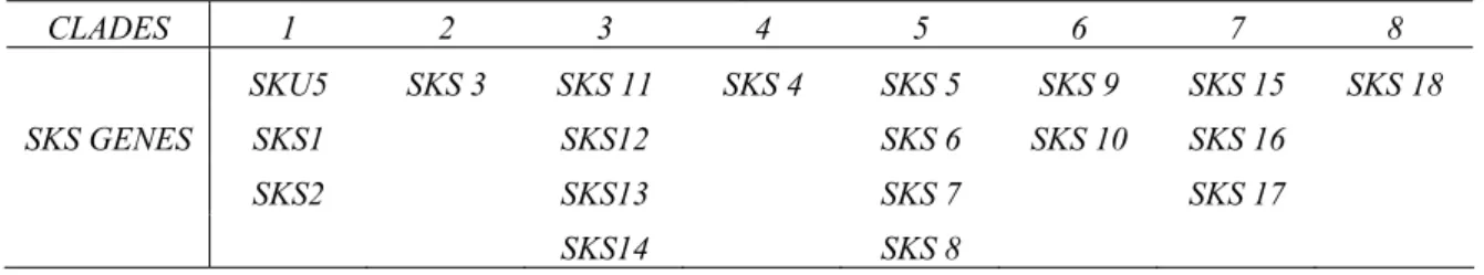
In addition to the C-terminus differences, short sequences inserted or deleted in few
sequences contribute to increase the divergences, as for example at positions 210-223 or 313-317 (ANNEXE 1). These differences support identification of various clades (Table I-5).
Table I-5. SKS members exist in different clades of phylogenetic tree.
CLADES 1 2 3 4 5 6 7 8 SKU5 SKS 3 SKS 11 SKS 4 SKS 5 SKS 9 SKS 15 SKS 18 SKS GENES SKS1 SKS12 SKS 6 SKS 10 SKS 16 SKS2 SKS13 SKS 7 SKS 17 SKS14 SKS 8
By proteomic and genomic analysis, SKU5 and SKS1 were confirmed to be associated with plasma membrane fraction and to have the GPI anchor in Arabidopsis (Borner et al., 2003). And by searching the Plant Proteome Database, SKS2 is also identified to be localized at the plasma membrane as a GPI-anchored protein. Considering the low expression level of SKS2 in most plant organs or cells in culture, it is possible that SKS2 was not confirmed by
proteomic analysis because it was under the detection level. SKS2 has the required domains to be a GPI protein as SKU5 and SKS1. SKU5 is the sole member for which the presence of the GPI was established; accordingly, by using translational fusion with the GFP (Green
Fluorescent protein), SKU5 was found to be localized at the plasma membrane and at least partly in the cell wall (Sedbrook et al, 2002). It is however difficult to know whether this dual localization reflects the distribution of the endogenous protein or results from the
overexpression of the transgene (as a 35SCaMv promoter was used). Cleavage of the GPI anchor for a SKU5 protein associated at the plasma membrane might be a mechanism regulating the relative abundance of the protein at the surface of the PM.
I-2-4 SKU5/SKS proteins through evolution
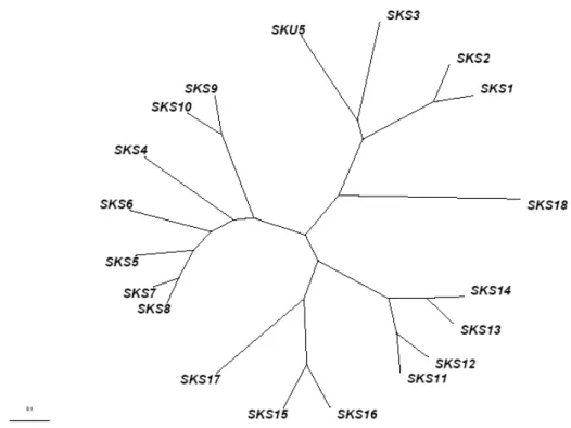
Comparative analysis of the SKU5/SKS sequences by using BLAST tools against available genome sequence databases of land plants, allowed to identify genes exhibiting SKS-like sequences in mosses, club-mosses, conifers, monocot plants and eudicot plants. The phylogenetic tree of the deduced proteins was calculated by COBALT tool (Figure I-10). Clades for the SKS members are labelled by their numbers accordingly toTable.I-5. The distance between different SKS genes in Arabidopis is even larger than that of homologous genes between different divisions. It suggests that members of the SKS family is a group of ancient genes which might have diverged very early, that they existed before the diversity of
higher plants and evolved in their own ways to acquire specific functions and potentially play different roles in plants.
Figure I-10. COBALT phylogenetic analysis of proteins encoded by homologous genes of the SKU5/SKS family.
250 proteins encoded by SKS putative homologous genes were identified by BLAST analysis (BLASTP 2.2.27+) (Altschul et al., 1997; Altschul et al., 2005) and phylogenetic tree was calculated by COBALT tool. In this phylogenetic tree, genes were classified by divisions as mosses, club-mosses, conifers, monocot plants and eudicot plants in different colour codes. The size of triangles depends on numbers of homologous proteins (labelled as leaves) inside one clade.
Data shows that SKS genes exist through all higher plants. The distance between different members of SKS gene family in Arabidopsis is even larger than that of homologous genes between different divisions.
Parameter utilized: Fast Minimum Evolution, Max Seq different is 0.85. Bar is 0.1 billion years. . 1 2 3 4 5 6 7 8
Conclusion and Discussion
CBP1 is a GPI-anchored maize protein that was identified as a possible interactor of ABP1. SKU5 and SKS are homologous genes of CBP1 in Arabidopsis. They belong to a rather ancient gene family. In Arabidopsis, this family contains 19 members. They encode proteins with conserved amino acid sequences and domains but include mutations impairing copper-binding and oxidase activity. SKU5, SKS1, SKS2 and SKS3 encode proteins with the highest similarities with CBP1. SKU5, SKS1, SKS2 are the only three members predicted to be modified at their C-terminus by substitution of the hydrophobic domain by a GPI anchor as CBP1. Other members are unlikely to get this post-translational modification and thus to be associated with the plasma membrane. I started to be interested into this protein family as CBP1 was shown to be interacting with ABP1 C-terminus. It was hypothesized that CBP1 could act as a carrier helping ABP1 to reach and be positioned at the outer surface of the plasma membrane or to be directly part of an auxin receptor complex. The output of the in silico analysis performed on the SKU5/SKS gene family in Arabidopsis is that despite the relatively high homology between the members, only 3 genes are putative GPI-anchored proteins. Further work was mainly focused on SKU5, SKS1 and SKS2 genes.
ANNEXE 1. Alignment of SKS proteins. 10 20 30 40 50 60 ....|....| ....|....| ....|....| ....|....| ....|....| ....|....| 1 --- ----MDLFKI LLLVFFVNIS FCFAADPYSF YNFEVSYITA SPLGVPQQVI SKU5 1 --- MAATCSLLAS FLLCFALLSA VSFAADPFVS YDFRVSYLTA SPLGVPQQVI SKS1 1 --- MAAT-DFFFA FVFSFALIFG FSFAGDPYVS YDFTLSYITA SPLGVPQQVI SKS2 1 --- MRCFPPPLWC TSLVVFLSVT GALAADPYVF FDWTVSYLSA SPLGTRQQVI SKS3 1 --- ---MRGSCKV SIVLLLVLIN GVLGDNPYRF FTWKITYGDI YPLGVKQQGI SKS4 1 --- -MAGSASFAA ALFIGLSLLF AVTAEDPYRF FEWNITYGDI YPLGVRQQGI SKS5 1 --- MMAVGRSGGT ILLFCLSFFA AVTAESPYRF FDWNVTYGDI YPLGVRQQGI SKS6 1 --- MKVKSMNTRA MITTLLFLIS LAFAEDPYRF FEWHVTYGNI SPLGVAQQGI SKS7 1 --- MEVKSVNTTA MILGLFFLIS FVAAEDPYKF FEWHVTYGNI SPLKVAQQGI SKS8 1 ---M CWWLNGAVWT MMMMTISIIS FVQADDPYRF FDWRVTYGNI SPLGIPQRGI SKS9 1 ---M EWWLNGGVW- MMMMTTTIIS FVKAEDTL-F YNWRVTYGKI ALDTLPRRGI SKS10 1 --- MRG-VKLLAA CLYLAAAATV VVRAEDPYFH HVWNVTYGTV SPLGVPQQVI SKS11 1 --- MKGGVKLLAV CLCVATATVM MVQAEDPYFH HVWNVTYGTA SPLGVPQQVI SKS12 1 --- -MQGGRLLTV LVCLAS-TVA LVSAGDPYFY YTWNVTYGTA APLGIPQQVI SKS13 1 --- -MEG-RLLTV LVCLVS-TVA IVNAGDPYFF HTWNVTYGTA SPLGVPQKVI SKS14 1 ---MKQTN LLVCKLFIGA LFWLG---SV LVNAEDPYMF YTWTVTYGTR SPLGVPQQVI SKS15 1 ---MKQKH LLLLGFLL-A YCFSS---VF VINAEDPYLF FTWTVTYGTR SPLGVPQQVI SKS16 1 MKMASRKTTS LLNHLLLLGA LTLLSS--LV IVKGESPYKF YTWTVTYGII SPLGVPQQVI SKS17 1 --- --MRHVFVEV LVLISLVILE LSYAFAPISS YQWVVSYSQR FILGGNKQVI SKS18 70 80 90 100 110 120 ....|....| ....|....| ....|....| ....|....| ....|....| ....|....| 47 AINGKFPGPT INVTTNENLV VNVRNKLDEG LLLHWNGIQQ RRVSWQDGVL GTNCPIPPKW SKU5 51 AVNGQFPGPL LNATTNYNVV VNVFNHLDEP LLLTWPGIQM RRNSWQDGVL GTNCPIPPRW SKS1 50 AVNGKFPGPV INATTNYNVH VNVLNHLDEP LLLTWPGVQM RRNSWQDGVL GTNCPIPPNW SKS2 51 GINGQFPGPI LNVTTNWNVV MNVKNNLDEP LLLTWNGIQH RKNSWQDGVL GTNCPIPSGW SKS3 48 LINGQFPGPH IDAITNDNII ISVFNYLKEP FLISWNGVQQ RKNSWQDGVV GTTCPIPPGK SKS4 50 LINGAFPGPD IHSVTNDNLI INVYNSLDEP FLLSWNGIQQ RRNSFVDGVY GTTCPIPPGK SKS5 51 LINGQFPGPD IHSVTNDNLI INVHNSLDEP FLISWNGVQN RRNSYVDGMY GTTCPIPPRS SKS6 51 LINGKFPGPD IISITNDNLI INVFNHLDEP FLLSWNGIRN WKNSFQDGVY GTMCPIPPGK SKS7 51 LINGKFPGPD IAAVTNDNLI INVFNHLDEP FLISWSGIRN WRNSYQDGVY GTTCPIPPGK SKS8 52 LINGQYPGPD IYSVTNDNLI INVHNDLDEP FLLSWNGVQL RKNSYQDGVY GTTCPIPPGK SKS9 50 LINGQFPGPE IRSLTNDNLV INVQNDLDDP FLLSWNGVHM RKNSYQDGVY GTNCPIPPGK SKS10 50 LINGQFPGPN VNSTSNNNVI INVFNNLDEP FLLTWNGIQH RKNCWQDGTP GTMCPIMPGT SKS11 51 LINGQFPGPN INSTSNNNVI VNVFNNLDEP FLITWAGIQH RKNCWQDGTA GTMCPIPPGQ SKS12 49 LINGQFPGPN LNSTSNNNVV INVFNNLDEP FLLTWSGLQH RKNSWQDGVT GTSCPIPAGT SKS13 48 LINGQFPGPN LNSTSNNNVV INVFNHLDEP FLLTWSGIQH RKNCWQDGVA GTSCPIPAGQ SKS14 53 LINGQFPGPA IEAVTNNNIV VNLINKLDEP FLITWNGVKQ RRTSWQDGVL GTNCPIQPNS SKS15 52 LINGQFPGPP IEGVTNNNIV VNVINKLDEP FLITWNGIKQ RKMSWQDGVL GTNCPIQPKS SKS16 59 LINGQFPGPK LEVVTNDNII LNLINKLDQP FLLTWNGIKQ RKNSWQDGVL GTNCPIQPNS SKS17 49 VINDMFPGPI LNATANDIIV VNIFNNLPEP FLMTWNGLQL RKNSWQDGVR GTNCPILPGT SKS18 130 140 150 160 170 180 ....|....| ....|....| ....|....| ....|....| ....|....| ....|....| 107 NWTYEFQVKD QIGSFFYFPS LHFQRASGGF GSFVVNPRAI IPVPFSTPDG DITVTIGDWY SKU5 111 NFTYQFQVKD QIGSFFYSPS LNFQRASGGF GPIVINNRDI IPIPFPQPDG ELIFIIGDWY SKS1 110 NFTYDFQLKD QIGSYFYSPS LNFQRASGGF GALIINNRDL VPIPFTEPDG EIIFIIGDWY SKS2 111 NWTYEFQVKD QIGSFFYFPS TNFQRASGGY GGIIVNNRAI IPVPFALPDG DVTLFISDWY SKS3 108 NFTYVIQVKD QIGSFYYFPS LAFHKAAGAF GAIRVWSRPR IPVPFSPPDG DFWLLAGDWY SKS4 110 NYTYILQMKD QIGSFYYFPS LGFHKAAGGF GGIRILSRPR IPVPFPDPAG DTTVLIGDWY SKS5 111 NYTYILQVKD QIGSFYYFPS LAFHKAAGGF GGIRILSRPG IPVPFADPAG DYTVLIGDWY SKS6 111 NYTYALQVKD QIGSFYYFPS LGFHKAAGGF GGIRISSRAL IPVPFPTPAD DYTLLVGDWY SKS7 111 NYTYALQVKD QIGSFYYFPS LGFHKAAGGF GAIRISSRPR IPVPFPAPAG DYTVLIGDWY SKS8 112 NYTYAIQVKD QIGSFFYFPS LAVHKAAGGF GGFRILSRPR IPVPFPEPAG DFTFLIGDWF SKS9 110 NYTYDFQVKD QVGSYFYFPS LAVQKAAGGY GSLRIYSLPR IPVPFPEPAE DFTFLVNDWY SKS10 110 NYTYHFQPKD QIGSYFYYPS TAMHRSAGGF GGLRVNSRLL IPVPYADPED DYTVLIGDWY SKS11 111 NFTYHFQPKD QIGSYFYYPT TAMHRAAGGF GGLRVNSRLL IPVPYADPED DYTILINDWY SKS12 109 NFTYHFQPKD QIGSYFYYPS TALHRFAGGF GGLRVNSRLL IPVPYADPED DRTILINDWY SKS13 108 NFTYHFQPKD QIGSYFYYPT TSLHRFAGGF GGLRVNSRLL IPVPYADPED DYTVLLGDWY SKS14 113 NWTYQFQLKD QIGTYTYFAS TSLHRASGAF GALNINQRSV ITTPYPTPDG DFTLLVSDWF SKS15 112 SWTYHFQLKD QIGTYAYFAS TSMHRASGAF GALNVNQRSV IFVPYPKPDA DFTLLVSDWY SKS16 119 NFTYKFQTKD QIGTFNYFPS TAFHKAAGGF GAINVYARPG IPIPYPLPTA DFTLLVGDWF SKS17 109 NWTYRFQVKD QIGSYFYFPT LLLQKAAGGY GAIRIYPPEL VPVPFPKPDE EYDILIGDWF SKS18