SHORT COMMUNICATION
Phylogenetic evidence for a new genotype of
Acanthamoeba
(Amoebozoa, Acanthamoebida)
Daniele Corsaro&Danielle VendittiReceived: 11 August 2009 / Accepted: 1 April 2010 / Published online: 22 April 2010 # Springer-Verlag 2010
Abstract Acanthamoeba are widespread free-living amoe-bae, able to cause infection in animals, with keratitis and granulomatous encephalitis as major diseases in humans. Recent developments in the subgenus classification are based on the determination of the nucleotide sequence of the 18S rDNA. By this mean, Acanthamoeba have been clustered into 15 sequence types or genotypes, called T1 to T15. In this study, we analysed near full 18S rDNA of an Acanthamoeba recovered from an environmental sample and various unidentified Acanthamoeba sequences retrieved from GenBank. We provided phylogenetic evidence for a new genotype, which we proposed to name T16.
Introduction
The genus Acanthamoeba comprises several species of free-living amoebae, widespread in many types of habitats, playing a role of major microbial predators. Many strains naturally harbour endosymbionts or may act as vector/ reservoir for various microbial pathogens. Furthermore, trophozoites can be used as host cells to isolate intracellular organisms (Horn and Wagner 2004; Corsaro and Greub2006).
Acanthamoeba spp. may cause diseases in animals, including humans, like keratitis, encephalitis and dissemi-nated tissue infections; thus, great interest is brought to identification of pathogenic species or strains (Marciano-Cabral and (Marciano-Cabral2003; Visvesvara et al2007).
Acanthamoeba species have been traditionally clustered into three morphological groups, on the basis of cyst features (Pussard and Pons 1977). However, successive studies based on biochemical and molecular methods have pointed out in many cases incoherences in strain/species clustering, proving also that some species, e.g. Acantha-moeba polyphaga, are polyphyletic. Gast et al. (1996) and Stothard et al. (1998) proposed to classify Acanthamoeba strains on the basis of 18S ribosomal RNA gene (18S rDNA) sequence similarities. By analysing more than 50 strains from the three morphogroups, they identified 12 sequence types or genotypes, named T1 to T12, where each genotype should correspond to natural species or species complex (Gast et al. 1996; Stothard et al. 1998). This approach has been largely adopted, and three new geno-types have been established, called T13 to T15 (Horn et al.
1999; Gast2001; Hewett et al. 2003).
D. Corsaro (*)
:
D. VendittiCHLAREAS, Chlamydia Research Association, 12 rue du Maconnais,
54500 Vandoeuvre-lès-Nancy, France e-mail: [email protected]
D. Corsaro
Swiss Federal Research Institute WSL, Wetlands Research Group, Station 2,
1015 Lausanne, Switzerland D. Corsaro
École Polytechnique Fédérale de Lausanne (EPFL), Laboratory of Ecological Systems, Station 2, 1015 Lausanne, Switzerland
D. Corsaro
Laboratory of Soil Biology, University of Neuchâtel, rue Emile Argand 11,
This study focuses on the phylogenetic analysis of an Acanthamoeba strain, isolated from a freshwater pond, and of other Acanthamoeba sequences, identified as candidates for novel genotypes.
Materials and methods
Sample origin and DNA extraction
Amoebae were recovered from a freshwater pond contain-ing decaycontain-ing vegetable matter (South Italy) onto bacterised 1.5% non-nutritive agar during a study focusing on the search for chlamydiae in the environment (Corsaro and Venditti,2009). Fungal overgrowth persisted in subcultures even after 24-h HCl treatment of cysts; thus, agar plates were eliminated. Prior to discharge plates, a small quantity of amoebae was recovered from the least contaminated agar and centrifuged in Page’s amoeba saline buffer (three times at 200×g, 5 min). Total DNA was extracted by repeated passages through a 24-gauge needle after freezing–thawing. PCR for the amoeba 18S rDNA was performed by using several primer sets: CAT1 (5′-CAT GCA TGT CTA AGT ATA AGC-3′) with GSPr (5′-TTC AC <G/A> GTA AAC <G/A> ATC TGG GC-3′) (modified from Stothard et al.
1999), or 1137R (5′-GTG CCC TTC CGT TCA AT-3′), and
892cF (5′-GTC AGA GGT GAA ATT CTT GG-3′) with Br (5′-GAT CCT TCT GCA GGT TCA C-3′). Primers 1137R and 892cF were from Schroeder et al. (2001), and Br was modified from Gast (2001). Reaction conditions were 5 min at 94°C, followed by 40 cycles of 94°C 45 s, 56°C 30 s, 72°C 1 min, with final extension of 72°C 5 min. PCR products were purified and sequenced by using the same PCR primers, and screened at BLAST for their identity. Phylogenetic analysis
The obtained sequence was aligned with closest relatives, retrieved after BLAST (www.ncbi.nlm.nih.gov), and with representatives of each Acanthamoeba genotype. Multiple alignments were obtained with clustalX and edited with BioEdit. Introns were excluded from the analysis. Phylo-genetic reconstructions were made on a partial∼1,450-bp diagnostic region of the 18S rDNA, in order to include also the genotype T15 (Hewett et al. 2003; Schroeder et al.
2001), and on full sequences (T15 excluded). Treeing methods used were neighbour-joining, minimum evolution (NJ, ME; p-distance) and maximum parsimony (MP), with MEGA3 (Kumar et al. 2004), and maximum likelihood (ML, GTR+gamma+I:4 model), with TREEFINDER (Jobb et al. 2004), with bootstrap test of 1,000. Following Stothard et al. (1998), genetic similarity between full sequences (∼2,200 bp) was calculated with BioEdit by
pairwise comparison, under optimal global alignment, using all sites and indels but excluding introns, and by removing common and terminal gaps.
Results and discussion
Heavy fungal overgrowth prevented subculture and isola-tion of the strain cvX. Cysts onto agar appeared wrinkled, with a polygonal endocyst; therefore, we considered this strain as belonging to group II. DNA analyses were then performed on extracts of amoebae scraped directly from a contaminated agar plate. Amoebae resulted negative for chlamydiae.
Amplicons obtained with CAT1/1137R (∼1,400 bp) and 892cF/Br (∼1,120 bp), starting from both DNA extracts and after PCR with CAT1/Br (∼2,200 bp), give identical sequences. Thus, a unique Acanthamoeba intronless se-quence of 2,157 bp was obtained. At BLAST, our sese-quence revealed almost identical (99.2) to the strain U/HC1, isolated from a human keratitis in Brazil, of undefined genotype (Alves et al. 2000). Other closest relatives were strains of genotype T13, showing sequence identity of 94%, while following entries in the BLAST all showed sequence identity values <93%.
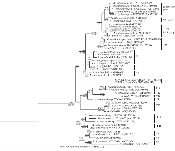
Figure 1 shows a phylogenetic tree based on a partial ∼1,450-bp portion of the 18S rDNA, in order to include also the genotype T15, corresponding to the species Acanthamoeba jacobsi (Hewett et al.2003), for which only this part of the gene is available. However, this gene portion is a diagnostic region comprising eight variable sequence regions able to differentiate among all the 15 genotypes (Schroeder et al. 2001). In this partial tree, our sequence emerged in a highly supported holophyletic clade with the strains of genotype T13 and clustered unambiguously with the strain U/HC1. The genotype T15, presented as the sister group of the genotype T13 in the original report of Hewett et al. (2003), emerged as an independent clade in the present analysis.
To identify genotypes, pairwise similarity values were determined by considering available full sequences, i.e. >2,100 bp. Pairwise similarity value between the strains U/HC1 and cvX was of 99.17% (dissimilarity 0.8%). Sequence from cvX strain showed 16 point mutations from that of U/HC1, 10 of which are in the diagnostic region, and two nt insertions. T13 genotype was represented by three strains, showing values of 96.0–97.7% (dissimilarity 2.3–4%): UWC9 isolated from contact lens case (Fritsche et al.1993), and TUMK-23 and UWET39 from soils (Horn et al. 1999; Hewett et al. 2003). Pairwise similarity value between the group U/HC1 and cvX and the T13 strains was 93.6–94.6% (dissimilarity 5.4–6.4%). Following the crite-rion of a 5% of dissimilarity (Stothard et al. 1998), the
group U/HC1 and cvX represents a new genotype, from keratitis sample (Alves et al. 2000) and freshwater (this study), for which we proposed the label T16. The T13 is closely related to but clearly distinct from T16, and comprises both ocular (Fritsche et al. 1993) and environ-mental strains (Horn et al.1999; Edagawa et al.2009).
In our study, we retrieved from GenBank other five sequences of environmental strains of undefined genotype: A.‘polyphaga’ ATCC 30872, from freshwater (Alves et al.
2000); Acanthamoeba sp. strains SE2-6F, E-5C and OB3b-3A, from rice soil (Murase and Frenzel 2008); and Acanthamoeba sp. strain MSG27, from marine sediment (Liu et al.2006).
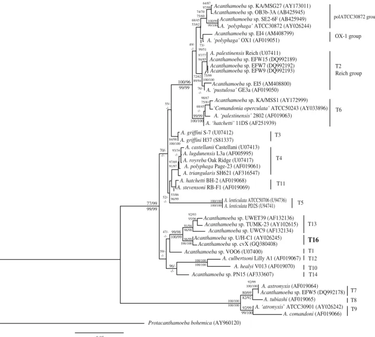
These five strains, ‘pol30872 group’, emerged as a relatively robust holophyletic clade in phylogeny performed on the 1,450-bp diagnostic region (Fig. 1), very weakly linked in ML tree to a paraphyletic T2 lineage, and sister group to a holophyletic T6 lineage. In NJ and ME, T2 emerged as holophyletic and separated from these five strains, while under MP significant bootstrap values (>65%) support independent emergence for each ‘pol30872’, ‘OX-1’ and ‘Reich’ groups in a large T2–T6 clade, but with ‘pol30872’ sister to T6. In full gene (>2,000 bp, Fig.2) trees, the five‘pol30872 group’ strains clustered together, but with lower bootstrap values (%BV 74/70 and 75/60 for ML/MP and NJ/ME), and the T2 split 0.02
Acanthamoeba sp. E-5C (AB425955)
AcaVNAK01 (GQ905495) 85/90 100/100 91/73 86/88 84/76 93/93 78/55 77/92 99/92 97/98 98/63 86/84 -/-37/- -/- -/- -/-94/81 92/94 84/95 100/99 -/-83/70 96/96 -/-82/43 81/82 99/96 99/99 67/- 25/- 81/- 100/99 100/100 86/91 89/90 87/68 95/95 95/97 99/99 97/99 100/100 58/--/41 40/44 90/93 48/20 51/66 100/99 100/100 74/99 99/99 92/82 91/92 99/99 100/100 94/98 100/100 74/98 99/99 93/71 64/59 -/-89/62 79/87 83/65 84/92 61/-69/79
A. culbertsoni Lilly A1 (AF019067) Acanthamoeba sp. VOO6 (U07400)
Acanthamoeba sp. PN15 (AF333607) A. healyi V013 (AF019070) Acanthamoeba sp. EFW5 (DQ992178) A. astronyxis (AF019064) A. tubiashi (AF019065) A. comandoni (AF019066) A. ‘astronyxis’ ATCC30901 (AY026242)
T T
T
Protacanthamoeba bohemica (AY960120)
T9 T8 T7 T5 T16 T15 T13 T1 T10 T12 T14 A. ‘palestinensis’ 2802 (AF019063) Acanthamoeba sp. KA/MSS1 (AY172999) ‘Comandonia operculata’ ATCC50243 (AY033896) A. ‘hatchetti’ 11DS (AF251939)
T6
A. royreba Oak Ridge (U07417) A. castellanii Castellani (U07413) A. polyphaga Page-23 (AF019061)
A. stevensoni RB-F1 (AF019069) A. lugdunensis L3a (AF005995)
A. griffini H37 (S81337) A. triangularis SH621 (AF316547) A. griffini S-7 (U07412) A. hatchetti BH-2 (AF019068) T T T T11 T4 T3
A. palestinensis Reich (U07411)
A. ‘pustulosa’ GE3a (AF019050) Acanthamoeba sp. EI5 (AM408800)
Acanthamoeba sp. EFW15 (DQ992189)
Acanthamoeba sp. EFW9 (DQ992193)
Reich group T2
A. ‘polyphaga’ OX1 (AF019051) Acanthamoeba sp. EI4 (AM408799)
OX1 group
A. ‘polyphaga’ ATCC30872 (AY026244) Acanthamoeba sp. KA/MSG27 (AY173011)
Acanthamoeba sp. SE2-6F (AB425949)
Acanthamoeba sp. OB3b-3A (AB425945) polATCC30872
group
A. lenticulata ATCC50706 (U94736) A. lenticulata PD2S (U94741)
Acanthamoeba sp. TUMK-23 (AY102615) Acanthamoeba sp. cvX (GQ380408)
A. jacobsi ATCC30732 (AY262360)
Acanthamoeba sp. UWC9 (AF132134) A. jacobsi AC194 (AY262362) A. jacobsi AC080 (AY262361) Acanthamoeba sp. UWET39 (AF132136)
Acanthamoeba sp. U/H-C1 (AY026245)
Acanthamoeba sp. PN13 (AF333609)
Acanthamoeba sp. EFW7 (DQ992192)
Fig. 1 Maximum likelihood 18S rDNA tree of the genus Acantha-moeba based on the diagnostic region of 1,450 bp including all the genotypes. Bootstrap values (1,000 replications) for ML/MP and NJ/ ME are shown above and below the nodes, respectively. GenBank
accession numbers are in parentheses. Upper T after GenBank indicates type strain for the species. T1–T15 recognised Acantha-moeba genotypes; T16, in bold, new genotype proposed in this study. Protacanthamoeba bohemica (Acanthamoebida) is used as outgroup
into apparently distinct ‘OX-1’ and ‘Reich’ lineages, with the‘OX-1’ as sister to the ‘pol30872’.
Intra-group pairwise dissimilarity values, based on full sequences (>2,000 bp), were of 0.6–4.8% for T2, 0.7–4.4% for the‘pol30872’ group and 1.5–2.5% for the T6. For T2, dissimilarity values were 0.6–3.5% and 2.3% within the Reich and the OX-1 groups, respectively, and 3.4–4.8% between the groups.
Mean inter-group dissimilarity values were >4.5%. T6 strains differed in mean by 5.2% (4.6–5.6%) from the ‘pol30872’ group, and by 5.3% (4.6–6.5%) from T2. Mean
dissimilarity value between the‘pol30872’ group and T2 was 4.6% (3.5–5.9%), with slightly higher value for the Reich group, 4.9% (3.9–5.9%), with respect to OX-1 group, 4.1% (3.5–4.7%).
T2 genotype seemed to be of environmental source (Stothard et al. 1998; Lorenzo-Morales et al. 2006); however, recently, strains of this genotype have been recovered in human cases of keratitis and encephalitis (Maghsood et al.2005; Walochnik et al.2008). Pathogenic strains seems more closely related to the strain OX-1, and Maghsood et al. (2005) proposed to split the genotype T2 0.05 100/96 99/99 74/70 75/60 72/62 89/94 75/99 100/100 98/67 75/81 84/98 100/100 91/94 96/98 -/-68/45 -/- -/-100/100 100/100 -/- -/-93/34 -/- -/-97/77 94/85 92/99 99/100 100/100 100/100 100/99 99/100 68/45 53/62 77/99 99/99 92/93 95/96 99/98 100/99 96/99 100/100 53/86 96/99 73/-99/51 99/99 100/100 80/69 82/92 97/69 91/97 -/-100/100 100/100 92/99 100/100 64/87 97/98
Acanthamoeba sp. TUMK-23 (AY102615)
Acanthamoeba sp. cvX (GQ380408) Acanthamoeba sp. UWC9 (AF132134) Acanthamoeba sp. UWET39 (AF132136)
Acanthamoeba sp. U/H-C1 (AY026245)
A. culbertsoni Lilly A1 (AF019067) Acanthamoeba sp. VOO6 (U07400)
Acanthamoeba sp. PN15 (AF333607) A. healyi V013 (AF019070) A. ‘polyphaga’ ATCC30872 (AY026244)
A. ‘palestinensis’ 2802 (AF019063) A. ‘polyphaga’ OX1 (AF019051)
Acanthamoeba sp. KA/MSS1 (AY172999) ‘Comandonia operculata’ ATCC50243 (AY033896) A. palestinensis Reich (U07411)
A. ‘pustulosa’ GE3a (AF019050) Acanthamoeba sp. EI5 (AM408800)
Acanthamoeba sp. KA/MSG27 (AY173011)
Acanthamoeba sp. EFW15 (DQ992189) Acanthamoeba sp. SE2-6F (AB425949)
A. lenticulata ATCC50706 (U94736) Acanthamoeba sp. EFW9 (DQ992193)
Acanthamoeba sp. EI4 (AM408799)
Acanthamoeba sp. EFW7 (DQ992192) Acanthamoeba sp. OB3b-3A (AB425945)
A. lenticulata PD2S (U94741) A. ‘hatchetti’ 11DS (AF251939)
A. royreba Oak Ridge (U07417) A. castellanii Castellani (U07413)
A. polyphaga Page-23 (AF019061)
A. stevensoni RB-F1 (AF019069) A. lugdunensis L3a (AF005995) A. griffini H37 (S81337) A. triangularis SH621 (AF316547) A. griffini S-7 (U07412) A. hatchetti BH-2 (AF019068) Acanthamoeba sp. EFW5 (DQ992178) A. astronyxis (AF019064) A. tubiashi (AF019065) A. comandoni (AF019066) A. ‘atronyxis’ ATCC30901 (AY026242) Protacanthamoeba bohemica (AY960120)
-/-T3 T4 T11 T6 T5 T2 Reich group OX-1 group T13 T16 T1 T12 T10 T14 T8 T9 T7 polATCC30872 group
Fig. 2 Maximum likelihood 18S rDNA tree of the genus Acanthamoeba based on full sequences. Bootstrap values (1,000 replications) for ML/ MP and NJ/ME are shown above and below the nodes, respectively.
GenBank accession numbers are in parentheses. T1–T14 recognised
Acanthamoeba genotypes; T16, in bold, new genotype proposed in this study. Protacanthamoeba bohemica (Acanthamoebida) is used as outgroup
into two groups T2A and T2B, on the basis of 4% dissimilarity, and eventually to differentiate between patho-genic and non-pathopatho-genic strains.
The ‘pol30872’ group could represent an additional subclade of the genotype T2; however, in phylogenetic reconstructions, this group appeared distinct from both Reich and OX-1 groups. The sister group relationship between the‘pol30872’ and the OX-1 group found in full sequence tree is very weakly supported (BV 68% and 62% in ML and ME trees, respectively), and there is no support unifying all‘pol30872’, OX-1 and Reich groups in a large clade T2 (Fig.2). In phylogenetic reconstructions using the genotypes T7–T8–T9 as outgroup instead of Protacantha-moeba (not shown), the ‘pol30872’ emerged as holophy-letic (70% in ML tree) but as sister to T6 (52%), and OX-1 and Reich groups followed as paraphyletic well-supported (89% and 79%, respectively) clades. All putative T2 subgoups (Reich, OX-1 and ‘pol30872’) and T6 were supported by 100%.
Stothard et al. (1998) proposed a minimum dissimilarity value of 5% for new genotypes in Acanthamoeba. However, programmes used for sequence alignment and analysis as well as number and variety of strains analysed influenced the results, explaining slight discrepancies between studies. We analysed eight T2 and four T6 strains, for which only three and one, respectively, were available on the study of Stothard et al., as well as other five closely related strains. However, position of OX-1 group was unclear, and we consider only the Reich group as true members of the T2.
Molecular phylogeny shows the ‘pol30872’ group emerging as a distinct clade within the T2/T6 lineage. A future redefinition of the arbitrary 5% dissimilarity value, or its less rigid application, would possibly place this clade as a new genotype. Acanthamoeba sp. ATCC 30872 could be the reference strain, without the specific name‘polyphaga’. This strain is sometimes used as lab organism, e.g. to study interactions with bacteria or pathogenicity on human cell cultures (Steinert et al.1998; Rocha-Azevedo et al.2006), but always with the confusing name A. polyphaga, showing significant differences in its properties, as compared to other A.‘polyphaga’ belonging to other genotypes like T4. In conclusion, we presented genetic and phylogenetic data supporting the establishment of a new genotype T16, with representatives from clinical and environmental samples, and proposed to consider a cluster of five environmental strains as a distinct group within the T2/T6 lineage.
References
Alves JMP, Gusmão CX, Teixeira MM, Freitas D, Foronda AS, Affonso HT (2000) Random amplified polymorphic DNA profiles as a tool
for the characterization of Brazilian keratitis isolates of the genus
Acanthamoeba. Braz J Med Biol Res 33:19–26
Corsaro D, Greub G (2006) Pathogenic potential of novel Chlamydiae and diagnostic approaches to infections due to these obligate
intracellular bacteria. Clin Microbiol Rev 19:283–297
Corsaro D, Venditti D (2009) Detection of Chlamydiae from freshwater environments by PCR, amoeba coculture and mixed
coculture. Res Microbiol 160:547–552
Edagawa A, Kimura A, Kawabuchi-Kurata T, Kusuhara Y, Karanis P (2009) Isolation and genotyping of potentially pathogenic Acanthamoeba and Naegleria species from tap-water sources in
Osaka, Japan. Parasitol Res 105:1109–1117
Fritsche TR, Gautom RK, Seyerdirshti S, Bergeron DL, Lindquist TD (1993) Occurrence of bacterial endosymbionts in Acanthamoeba spp. isolated from corneal and environmental specimens and contact lenses. J Clin Microbiol 31:1122–1126
Gast RJ (2001) Development of an Acanthamoeba-specific reverse dot-blot and the discovery of a new ribotype. J Eukaryot
Microbiol 48:609–615
Gast RJ, Ledee DR, Fuerst PA, Byers TJ (1996) Subgenus systematics of Acanthamoeba: four nuclear 18S rDNA sequence types. J
Eukaryot Microbiol 43:498–504
Hewett MK, Robinson BS, Monis PT, Saint CP (2003) Identification of a new Acanthamoeba 18S rRNA gene sequence type, corresponding to the species Acanthamoeba jacobsi Sawyer, Nerad and Visvesvara, 1992 (Lobosea: Acanthamoebidae). Acta
Protozool 42:325–329
Horn M, Wagner M (2004) Bacterial endosymbionts of free-living
amoebae. J Eukaryot Microbiol 51:509–514
Horn M, Fritsche TR, Gautom RK, Schleifer KH, Wagner M (1999) Novel bacterial endosymbionts of Acanthamoeba spp. related to the Paramecium caudatum symbiont Caedibacter caryophilus. Environ Microbiol 1:357–367
Jobb G, von Haeseler A, Strimmer K (2004) TREEFINDER: a powerful graphical analysis environment for molecular phyloge-netics. BMC Evol Biol 4:18
Kumar S, Tamura K, Nei M (2004) MEGA3: integrated software for molecular evolutionary genetics analysis and sequence
align-ment. Brief Bioinformatics 5:150–163
Liu H, Ha YR, Lee ST, Hong YC, Kong HH, Chung DD (2006) Genetic diversity of Acanthamoeba isolates from ocean
sedi-ments. Korean J Parasitol 44:117–125
Lorenzo-Morales J, Ortega-Rivas A, Martinez E, Khoubbane M, Artigas P, Periago MV, Foronda P, Abreu-Acosta N, Valladares B, Mas-Coma S (2006) Acanthamoeba isolates belonging to T1, T2, T3, T4 and T7 genotypes from environmental freshwater
samples in the Nile Delta region, Egypt. Acta Trop 100:63–69
Maghsood AH, Sissons J, Rezaian M, Nolder D, Warhurst D, Khan NA (2005) Acanthamoeba genotype T4 from the UK and Iran and isolation of the T2 genotype from clinical isolates. J Med Microbiol 54:755–759
Marciano-Cabral F, Cabral G (2003) Acanthamoeba spp. as agents of disease in humans. Clin Microbiol Rev 16:273–307
Murase J, Frenzel P (2008) Selective grazing of methanotrophs by
protozoa in a rice field soil. FEMS Microbiol Ecol 65:408–414
Pussard M, Pons R (1977) Morphologie de la paroi kystique et taxonomie du genre Acanthamoeba (Protozoa, Amoebida).
Protistologica 8:557–598
Rocha-Azevedo B, Menezes GC, Silva-Filho FC (2006) The interac-tion between Acanthamoeba polyphaga and human osteoblastic
cells in vitro. Microb Pathogen 40:8–14
Schroeder JM, Booton GC, Hay J, Niszl IA, Seal DV, Markus MB, Fuerst PA, Byers TJ (2001) Use of subgenic 18S ribosomal DNA PCR and sequencing for genus and genotype identification of Acanthamoebae from humans with keratitis and from sewage
Steinert M, Birkness K, White E, Fields B, Quinn F (1998) Mycobacterium avium bacilli grow saprozoically in coculture with Acanthamoeba polyphaga and survive within cyst walls.
Appl Environ Microbiol 64:2256–2261
Stothard DR, Hay J, Schroeder-Diedrich JM, Seal DV, Byers TJ (1999) Fluorescent oligonucleotide probes for clinical and environmental detection of Acanthamoeba and the T4 18S rRNA gene sequence type. J Clin Microbiol 37:2687–2693
Stothard DR, Schroeder-Diedrich JM, Awwad MH, Gast RJ, Ledee DR, Rodriguez-Zaragoza S, Dean CL, Fuerst PA, Byers TJ (1998) The evolutionary history of the genus Acanthamoeba and
the identification of eight new 18S rRNA gene sequence types. J
Eukaryot Microbiol 45:45–54
Visvesvara GS, Moura H, Schuster FL (2007) Pathogenic and opportunistic free-living amoebae: Acanthamoeba spp., Balamu-thia mandrillaris, Naegleria fowleri, and Sappinia diploidea. FEMS Immunol Med Microbiol 50:1–26
Walochnik J, Aichelburg A, Assadian O, Steuer A, Visvesvara G, Vetter N, Aspöck H (2008) Granulomatous amoebic encephalitis caused by Acanthamoeba amoebae of genotype T2 in a human immunodeficiency virus-negative patient. J Clin Microbiol