CORRESPONDENCE
Nonmelanomatous Skin
Cancer Following Cervical,
Vaginal, and Vulvar
Neoplasms: Etiologic
Association
Human papillomavirus infection is the major cause of cancers of the cervix, vagina, and vulva (1). Nonmelanoma-tous skin cancers have been associated with human papillomavirus infection in patients with epidermodysplasia ver-ruciformis and in patients who are im-munosuppressed or nonimmunosup-pressed, although the data are scant
(1,2).
We used the cancer registry of the Swiss Canton of Vaud (with a popula-tion of approximately 600 000 in 1990) for the period from 1974 through 1994 to obtain additional quantitative infor-mation on this topic, which has patho-genic and public health implications. Data were collected for women who had
in situ or invasive neoplasms of the
cer-vix, vagina, or vulva and for women who had nonmelanomatous skin cancer. These data were then used to calculate the incidence of nonmelanomatous skin cancer in women who had been regis-tered with an in situ or invasive neo-plasm of the cervix, vagina, or vulva (3). The registry is tumor based, and mul-tiple primary tumors in the same person are entered separately. The basic infor-mation available consists of sociodemo-graphic characteristics of the patient, the primary site of the tumor, the histologic type of the tumor according to the stan-dard International Classification of Dis-eases (ICD) for Oncology (4), and the time of diagnostic confirmation. Passive and active follow-ups are recorded, and each subsequent item of information concerning a registered cancer is used to complete the record of that patient.
Since 1974, a registration scheme that applies the standardized rules used for incident cancers has been used for carcinoma in situ and severe dysplasia (CIN III, cervical intraepithelial
neopla-sia III) of the uterine cervix (ICD code: 180.0–180.9), vagina (ICD: 184.0), and vulva (ICD: 184.1–184.3) (4).
In the present study, when all syn-chronous neoplasms were excluded, there were 2339 histologically con-firmed cases of carcinoma in situ of the cervix uteri, nine cases of carcinoma in
situ of the vagina, and 85 cases of
car-cinoma in situ of the vulva. The study also included 789 cases of invasive neo-plasms of the cervix, 69 cases of inva-sive neoplasms of the vagina, and 153 cases of invasive neoplasms of the vulva. These cases were followed to the end of 1996 for the occurrence of can-cer, migration, or death.
We calculated the expected numbers of individuals with nonmelanomatous skin cancer based on site-, age-, and cal-endar-period-specific incidence rates, multiplied by the observed number of person-years at risk. The statistical sig-nificance of the observed/expected ra-tios (standardized incidence ratio [SIR]) and the corresponding 95% confidence interval (CI) were based on the Poisson distribution.
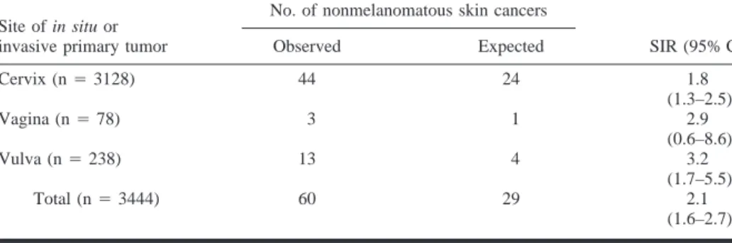
Table 1 gives the observed and ex-pected numbers of nonmelanomatous skin neoplasms after diagnosis of in
situ or invasive neoplasms of the
cer-vix, vagina, and vulva. A statistically significant excess of skin cancer was registered after cervical neoplasms (44 observed and 24 expected; SIR ⳱ 1.8; 95% CI ⳱ 1.3–2.5) and vulvar neoplasms (13 observed and four ex-pected; SIR⳱ 3.2; 95% CI ⳱ 1.7–5.5). Likewise, three nonmelanomatous skin cancers were observed after vaginal neoplasms versus one expected (SIR⳱ 2.9; 95% CI ⳱ 0.6–8.6).
Oall, 60 skin cancers were observed ver-sus 29 expected (SIR⳱ 2.1; 95% CI ⳱ 1.6–2.7).
An excess of nonmelanomatous skin cancer after diagnosis of carcinoma
in situ of the cervix has been reported (5,6). The present data extend this
observation to other neoplasms of the lower female genital tract and, there-fore, provide epidemiologic support to the suggestion of a possible role of human papillomavirus infection in the etiology of nonmelanomatous skin cancer (7).
FABIOLEVI LALAO RANDIMBISON CARLOLAVECCHIA
R
EFERENCES(1) International Agency for Research on Cancer (IARC). Monographs on the evaluation of car-cinogenic risks to humans No. 64, human pap-illomaviruses. Lyon: IARC; 1995.
(2) Shamanin V, zur Hausen H, Lavergne D, Proby CM, Leigh IM, Neumann C. Human papillomavirus infections in nonmelanoma skin cancers from renal transplant recipients and nonimmunosuppressed patients. J Natl Cancer Inst 1996;88:802–11.
(3) Levi F, Te VC, Randimbison L, La Vecchia C. Statistics from the Registry of the Canton of Vaud, Switzerland, 1988–1992. In: Parkin DM, Whelan S, Ferlay J, Raymond L, Young J, editors. Cancer incidence in five continents, vol VII. Lyon: IARC Sci Publ No. 143; 1997. p. 674–7.
(4) World Health Organization. International Clas-sification of Diseases for Oncology (ICD-O). Geneva: World Health Organization; 1976. (5) Bjorge T, Hennig EM, Skare GB, Soreide O,
Thorensen SO. Second primary cancers in patients with carcinoma in situ of the uterine cervix. The Norwegian experience 1970–1992. Int J Cancer 1995;62:29–33. (6) Levi F, Randimbison L, Vecchia CL,
Fran-ceschi S. Incidence of invasive cancers follow-ing carcinoma in situ of the cervix. Br J Can-cer 1996;74:1321–3.
Table 1. Observed and expected cases, in Vaud, Switzerland, from 1974 through 1994, and standardized incidence ratios (SIRs) of subsequent nonmelanomatous skin cancer after an initial
diagnosis of in situ or invasive neoplasms of the cervix, vagina, and vulva, as well as the corresponding 95% confidence intervals (CIs)
Site of in situ or invasive primary tumor
No. of nonmelanomatous skin cancers
SIR (95% CI) Observed Expected Cervix (n⳱ 3128) 44 24 1.8 (1.3–2.5) Vagina (n⳱ 78) 3 1 2.9 (0.6–8.6) Vulva (n⳱ 238) 13 4 3.2 (1.7–5.5) Total (n⳱ 3444) 60 29 2.1 (1.6–2.7)
(7) Burk RD, Kadish AS. Treasure hunt for human papillomaviruses in nonmelanomatous skin can-cers. J Natl Cancer Inst 1996;88:781–2.
N
OTESAffiliations of authors: F. Levi, L. Randimbi-son, Registre Vaudois des Tumeurs, Institut Uni-versitaire de Me´decine Sociale et Pre´ventive, Lau-sanne, Switzerland; C. La Vecchia Istituto di Ricerche Farmacologiche ‘‘Mario Negri,’’ and Is-tituto di Statistica Medica e Biometria, Universita` degli Studi di Milano, Italy.
Correspondence to: Fabio Levi, Registre Vau-dois des Tumeurs, CHUV-Falaises 1, CH-1011 Lausanne, Switzerland (e-mail: fabio.levi@ inst.hospvd.ch).
We gratefully acknowledge the contribution of the Vaud Cancer Registry’s staff and the collabo-ration of the pathological laboratories operating in the Canton of Vaud.
Re: Distinguishing Second
Primary Tumors From Lung
Metastases in Patients With
Head and Neck Squamous
Cell Carcinoma
The recent paper by Leong et al. (1) highlights progress regarding a long-standing oncology dilemma in distin-guishing a solitary metastatic deposit from a new cancer in patients with known prior cancer. The specific clini-cal scenario is familiar to those who fre-quent head and neck tumor boards— specifically, the patient with known head and neck cancer (squamous cell carcinoma) who simultaneously or sub-sequently manifests a solitary pulmo-nary nodule, which is similarly con-firmed as squamous cell carcinoma.
Many head and neck oncologists have turned wistfully toward their tumor board pathologist with the simple ques-tion, ‘‘Is this a metastasis or a new pri-mary tumor?’’ The promise of this pub-lished work by Leong et al. (1) is that we are moving closer to providing the cli-nician with molecular diagnostic tools to answer the question more precisely.
Judicious application of molecular techniques to complement clinical judg-ment in the ‘‘metastasis versus primary tumor’’ scenario will clearly prove ben-eficial in selected circumstances. Never-theless, maximizing clinical thinking be-fore soliciting molecular ‘‘truth telling’’ will be important. In their abstract, Leong et al. state ‘‘. . . a solitary SCC [squamous cell carcinoma] in the lung
more likely represents a metastasis than an independent lung cancer.’’ However, this is largely dependent on the patient cohort selected. The study group in the paper by Leong et al. is dominated by patients with advanced, lymph node-positive, and/or recurrent head and neck cancers. Of the 16 patients studied, 13 presented with stage IV tumors and 15 were lymph node positive at presenta-tion. These represent compelling prog-nostic features for locoregional disease recurrence and eventual distant metasta-ses. Thus, it is not surprising that 12 of 16 lung tumors appeared to represent metastases in this group of patients with highly advanced-stage disease for whom clinical judgment would largely dictate the same. This is by no means meant to detract from the importance of this work. Rather, it is suggested that such molecular analysis may prove far more important in patients with earlier stage disease for whom the clinical likelihood of distant metastasis is deemed far lower.
Approximately one quarter to one third of the patients with head and neck cancer present with stage I or stage II disease (lymph node negative); in these patients, lung metastases would be dis-tinctly unusual. For these patients, the cost of mistakenly assuming a meta-static process could be tragic, and the value of confirming a molecular distinc-tion may be critical to optimizing therapy recommendations.
Leong et al. state in the ‘‘Discus-sion’’ section, ‘‘Most solitary lung nod-ules in patients with HNSCC [head and neck squamous cell carcinoma] may ac-tually reflect advanced tumor spread.’’ These authors would not wish to inad-vertently mislead the general oncologist into thinking that this is true for all pa-tients with head and neck cancer. This conclusion is strongly influenced by the clinical staging of the original tumors. A molecular examination of 16 patients with early stage head and neck tumors who manifest solitary pulmonary nod-ules might well lead others to draw the opposite conclusion. Nevertheless, these advances in tumor fingerprinting will surely provide tangible benefits to se-lected cancer patients in whom the judi-cious application of molecular data will complement and clarify clinical judgment. PAUL M. HARARI
R
EFERENCE(1) Leong PP, Rezai B, Koch WM, Reed A, Eisele D, Lee DJ, et. al. Distinguishing second pri-mary tumors from lung metastases in patients with head and neck squamous cell carcinoma. J Natl Cancer Inst 1998;90:972–7.
N
OTECorrespondence to: Paul M. Harari, M.D., De-partment of Human Oncology, 600 Highland Ave. K4/B100, Madison, WI 53792-0600.
R
ESPONSEWe welcome Dr. Harari’s refreshing embrace of a novel strategy for the reso-lution of a long-standing oncologic impasse. Comparative microsatellite analysis is a highly effective tool in dis-tinguishing second lung tumors from lung metastasis. No doubt, similar ge-netic strategies addressing equally rel-evant clinical issues will play an in-creasing role in the integrated multidisci-plinary approach to patients with head and neck squamous cell carcinoma (HNSCC) and cancers at other sites.
As always, caution and discretion must be exercised when generalizing study results to the individual patient. Our study reflects the experience of a large tertiary care center where patients are often referred for management of ad-vanced HNSCC. As Dr. Harari points out, the incidence of solitary lung me-tastases will probably be lower in pa-tient populations over represented by low-stage HNSCCs. For the individual patient with HNSCC, however, micro-satellite analysis remains a valid and valuable tool for discerning the nature of a solitary lung tumor. The use of such molecular approaches is not intended to replace sound clinical judgment but to facilitate it. WILLIAMH. WESTRA WAYNE M. KOCH DAVIDSIDRANSKY JINJEN
N
OTESAffiliations of authors: W. H. Westra (Depart-ment of Otolaryngology–Head and Neck Surgery and Pathology), W. M. Koch, D. Sidransky, J. Jen (Department of Otolaryngology–Head and Neck Surgery), The Johns Hopkins Medical Institutions, Baltimore, MD.
Correspondence to: William H. Westra, M.D., The Johns Hopkins Medical Institutions, Depart-ment of Pathology, 600 N. Wolfe St., Baltimore, MD 21287-6417.