Influence of coronary artery bypass grafting on ventricular
late potentials as a predictive factor for ventricular
arrhythmias during short- and long-term follow-up
*J. FRIELINGSDORF, A. E. GERBER, A. LASKE AND O. BERTELFrom the Cardiology and Cardiovascular Surgery Division, Triemli Hospital Zurich and the ^Cardiology Division, University Hospital Zurich, Switzerland
KEY WORDS: Ventricular late potentials, signal-averaged electrocardiogram, coronary artery bypass grafting, ventricular arrhythmias, coronary heart disease.
Ventricular late potentials have been identified as a prognostic factor in the prediction of ventricular arrhythmias in patients after myocardial infarction. In this prospective study the possible impact of late potentials on the prediction of ventricular arrhythmias in the short- and long-term follow-up after coronary artery bypass grafting was evaluated. In 188 patients (165 men, 23 women, age 57 ±8 years) with chronic coronary heart disease 48 (26%) had late potentials before bypass grafting; after the procedure this was reduced to 39 (21%) (ns). In 16 (33%) of the 48 patients with late potentials before bypass grafting, late potentials were no longer present in the short-term follow-up (9 ±6 days).
Conversely, seven (5%) of the 140 patients without late potentials before bypass grafting had late potentials in the short-term follow-up after grafting. Nine (19%) of the 48 patients with late potentials before bypass grafting had ventricular arrhythmias in the peri-operative phase, which had to be treated with antiarrhythmic agents. In contrast, only three (2%>) of the 140 patients without late potentials before bypass grafting had to be treated for ventricular arrhythmias (P<0001). In the long-term follow-up of 29 ± 3 months, there were no events in the group of 149 patients without late potentials after grafting. In the 39 patients with late potentials after grafting, there were two (5%) events (two patients with arrhythmic syncope).
Conclusions: (1) Patients with late potentials before bypass grafting have a markedly higher risk of developing serious
ventricular arrhythmias in the peri-operative period than patients without late potentials. (2) Patients without late potentials have a very low risk of developing serious ventricular arrhythmias in the peri-operative period. (3) During long-term follow-up there was only a low probability of developing symptomatic ventricular arrhythmias in patients with or without late potentials.
Introduction potentials1'8 20', the incidence of inducible ventricular
,, . • , , , . .• , , • , r , tachycardia121 "221 and mortality independent from left
Ventricular late potentials are high-frequency, low- J. , . r2124i ~, , ,,
„, ,v , • , • ., . , ° • c ./ r\nc ventricular ejection fraction1 '. There are hardly any
amplitude signals in the terminal portion of the QRS .J „ . , e
-„ , , • . u A * * A *u c i . reports on the effect of aortocoronary bypass grafting on complex, which can be detected on the surface electro- ,y . . . . , • • , . •
cardiogram (ECG) by computer signal-averaging and lJe o c c u i; re n c e o f;e" ^r'c" > a r arrhythmia in relation to band-pass filtering techniques"-3' Late potentials are the signal-averaged ECG' • >
,, ., , . , , , . • , The aim of the present study was to predict the generally thought to represent delayed and inhomo- . . *\ u u • u • i i
, .. • , . ,. occurrence of ventncuar arrhythmias by ventricular ate
geneous conduction in regions with normal myocardium • , • , , . , c ..
<-and patchy scar tissue, which is considered to be the Po t e n t i a l s I n t h e u s h o r t- and long-term follow-up after electrophysiological substrate of reentrant ventricular c o r o n a ry a r t e ry by Pa s s gr a f t i n
§-tachycardia14"61. In recent years late potentials have been
identified as a strong and independent prognostic factor Methods in the prediction of spontaneous and inducible
ventricu-lar tachycardia and sudden death in patients with coron- STUDY PATIENTS
ary heart disease after myocardial infarction1'-37"151. In The study group consisted of 188 consecutive patients several studies'16"20' a correlation between patency of (165 men, 23 women, mean age 57 ± 8 years) admitted infarct-related arteries and the prevalence of late poten- for coronary artery bypass grafting. The extent of coron-tials was found. Early reperfusion by thrombolytic ary heart disease was determined by counting the num-therapy seems to reduce the prevalence of late ber of major vessels (left main, anterior descending, circumflex and right coronary artery) with a diameter Revision submitted 23 Augusi 1994. and accepted 5 September 1994. stenosis >70%. Left ventricular ejection fraction was
Correspondence O. Bertel. MD. Cardiology Division. Sladtspital Triemli. determined by ventriculography in all patients, and the
Birmensdorferstrasse 496. CH-8063 Zurich. Switzerland diagnosis of a previous myocardial infarction was based
on a history of documented infarction with enzyme increase or significant Q waves in the resting ECG. A peri-operative myocardial infarction was diagnosed if there were either new pathological Q waves on the ECG or an at least two-fold increase of creatinine phospho-kinase, with a significant myocardial fraction of more than 10%. Patients with a history of syncope or sus-tained ventricular tachycardia before bypass surgery were not included.
ECG SIGNAL-AVERAGING
Signal-averaged ECGs were registered on admission and at discharge (9 ± 6 days later). Patients with bundle branch block, atrial fibrillation, pre-excitation, pace-maker rhythm and patients with a history of myocardial infarction 3 months prior to coronary bypass grafting were excluded from analysis.
RECORDINGS
The signal-averaged ECGs were performed with a marquette MAC 15 HiRes ECG recorder. The ECG was recorded during sinus rhythm using standard bipolar orthogonal X, Y and Z leads. Signals were amplified, averaged (250 complexes) and filtered with a 40-250 Hz filter. To avoid the problems with filtering often seen with bidirectionally multi-pole time domain filters, the system used a Fourier transform filtering technique. For analysis, the filtered leads were com-bined into a vector magnitude plot. A computer algo-rithm determined the duration of the filtered QRS complex, the duration of low amplitude signals <40 uV and the root mean square voltage in the terminal 40 ms of the QRS complex. Late potentials were defined as present if two of following three criteria were fulfilled: (1) filtered QRS duration >120ms; (2) duration of low amplitude signals (<40uV) >38 ms; (3) root mean square voltage in the terminal 40 ms of the QRS complex <25 uV. The end-point and onset of the filtered QRS complex were verified visually. We mainly followed the standards of the ACC policy statements1'5271.
SHORT-TERM FOLLOW-UP
After coronary bypass grafting patients were permanently monitored in the intensive care unit by a Hewlett Packard system (Orion arrhythmia detection and analysis system). The patients' charts were reviewed for the occurrence of ventricular arrhythmias in the monitored period and thereafter, when clinically indi-cated, by means of 24-h ECG recordings. Ventricular arrhythmias were judged as serious and all were treated with an antiarrhythmic therapy when haemodynami-cally relevant (if there were more than 8-10 premature ventricular beats per min, R on T and ventricular tachycardia). The cardiac surgeons who assigned the antiarrhythmic therapy were not aware of the results of the signal-averaged ECG.
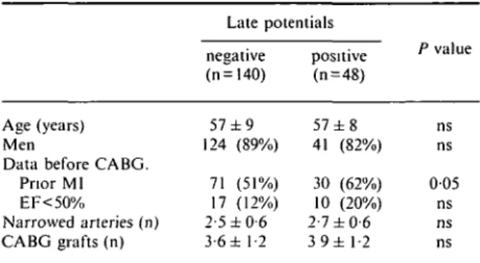
Table 1 Clinical data before coronary artery bypass grafting
Age (years) Men
Data before CABG. Prior MI EF<50% Narrowed arteries (n) CABG grafts (n) Late potentials negative (n=140) 57 ±9 124 (89%) 71 (51%) 17 (12%) 2-5 ±0-6 3-6 ± 1-2 positive (n=48) 57 ± 8 41 (82%) 30 (62%) 10 (20%) 2-7 ±0-6 3 9 ± 1-2 P value ns ns 0 0 5 ns ns ns CAB = coronary artery bypass. CABG=coronary artery bypass grafting: EF=ejection fraction: MI = myocardial infarction.
LONG-TERM FOLLOW-UP
All patients were seen for an outpatient examination with exercise testing 3 months after coronary bypass surgery. Thereafter, follow-up data were acquired by means of telephone communication with the referring physicians, who all followed the patients regularly. No physician was aware of the results of the signal-averaged ECG. End-points were syncope or sudden cardiac death.
STATISTICAL ANALYSIS
Values were expressed as mean ± standard deviation. Chi-square analysis was used for the comparison of proportions from two independent groups. Student's paired and unpaired t-test was used to compare vari-ables with normal distribution. Statistical significance was set at the 005 level.
Results
CLINICAL FINDINGS
Of the 188 patients 101 (54%) had a history or ECG signs of a previous myocardial infarction, whereas 87 (46%) had none. Twenty-one (11%) were in NYHA class I, 153 (81%) in class II or III and 14 (8%) in class IV. Fourteen (7%) had single-vessel, 54 (29%) two-vessel and 120 (64%) three-vessel disease. Twenty-seven (14%) had a left ventricular ejection fraction <50%. Ninety-seven patients (52%) had one or more occluded vessels whereas 91 (48%) had only stenosis. Except for myo-cardial infarction, clinical data were comparable for patients with and without late potentials (Table 1).
SIGNAL-AVERAGED ECG AND LATE POTENTIALS
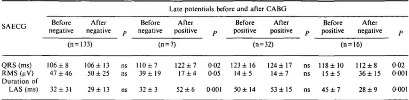
Before coronary artery bypass grafting there were 48 (26%) patients with late potentials, whereas after coron-ary artery bypass grafting 39 (21%) patients had late potentials (ns), but the mean pre- and postoperative parameters of the signal-averaged ECG of the entire study population did not show any significant difference: filtered QRS duration 1 1 0 ± l l m s and 110± 15ms,
Table 2 Pre- and postoperative findings of late potentials and signal-averaged ECG in 188 patients
Late potentials before and after CABG SAECG QRS (ms) RMS (uV) Duration of LAS (ms) Before negative (n = 106 ± 8 47 ± 4 6 32 ±31 After negative 133) 106 ± 13 50 ± 2 5 2 9 ± 1 3 P ns ns ns Before negative After positive (n = 7) 110±7 39 ± 19 32 ± 3 122 ± 7 17 ± 4 52 ± 6 P 002 005 0001 Before positive After positive (n = 32) 123 ± 16 14 ± 5 50 ± 14 124 ±17 14±7 53 ±15 P ns ns ns Before positive After negative (n=16) 118 ± 10 15±5 45 ± 7 112±8 36±15 28 ± 9 P 002 0001 0001 CABG=coronary artery bypass grafting. LAS = low amplitude signals, RMS = root mean square voltage, SAECG = signal averaged ECG.
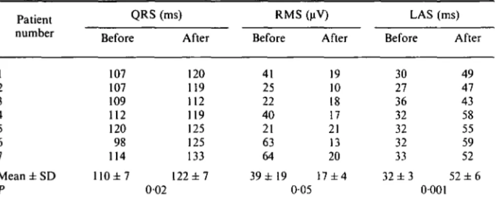
Table 3 Individual parameters of the signal-averaged ECG before and after CABG in 16 patients whose pre-operative late potentials became negative postoperatively
Patient number 1 2 3 4 5 6 7 8 9 10 11 12 13 14 15 16 M e a n ± S D P QRS i Before 119 102 122 119 121 123 115 135 122 111 118 109 108 125 134 102 118±10 (ms) After 112 104 117 102 115 114 110 127 124 101 103 115 115 115 110 104 I12±8 002 RSM Before 16 17 18 12 19 13 8 13 23 18 18 17 15 15 4 14 15±5 (uV) After 38 21 23 67 23 45 23 31 25 68 42 32 25 43 35 37 36 ± 15 0001 LAS Before 43 44 49 45 37 42 49 41 42 40 44 44 42 39 69 44 45 ± 7 (ms) After 30 37 49 34 31 33 36 24 27 32 17 29 19 22 12 21 28 ± 9 0001 CABG=coronary artery bypass grafting, LAS = low amplitude signals, RMS = root mean square voltage.
respectively, (ns); duration of low amplitude signals 36 ± 29 ms and 33 ± 15 ms, respectively, (ns); and root mean square voltage 39 ± 43 uV and 43 ± 26 uV, respec-tively, (ns). In 16 (33%) of the 48 patients with late potentials before coronary artery bypass grafting, late potentials were no longer present in the short-term follow-up (9 ± 6 days), which was accompanied by a significant change of all three criteria of the signal-averaged ECG in these 16 patients (Tables 2 and 3). On the other hand, seven (5%) of the 140 patients without late potentials before coronary artery bypass grafting had late potentials in the short-term follow-up after coronary artery bypass grafting, which was again re-flected by a significant change in the signal-averaged ECG (Tables 2 and 4). Two (29%) of these seven patients had a documented peri-operative myocardial infarction, whereas of the remaining five (71%) patients two had three-vessel disease, a pre-operative history of inferior myocardial infarction and one occluded vessel,
one patient had three-vessel disease and one occluded vessel, and two patients had three-vessel disease with-out occluded arteries and no history of myocardial infarction.
PERI-OPERATIVE MORTALITY
Five patients died in the peri-operative period, which is a mortality of 2-6%. They were excluded from analysis. Only one of these patients had late potentials. He died of low output syndrome. Two of the remaining four patients died of sepsis and two of low output syndrome.
VENTRICULAR ARRHYTHMIA IN THE PERI-OPERATIVE PERIOD (TABLE 5)
Nine (19%) of the 48 patients with late potentials before bypass surgery had haemodynamically relevant
Table 4 Individual parameters of the signal-averaged ECG before and after CA BG in seven patients with newly acquired late potentials postoperatively
Patient number 1 2 3 4 5 6 7 Mean ± SD P QRS Before 107 107 109 112 120 98 114 110±7 (ms) After 120 119 112 119 125 125 133 122 ± 7 002 RMS Before 41 25 22 40 21 63 64 39 ±19 (uV) After 19 10 18 17 21 13 20 17 ±4 0 0 5 LAS Before 30 27 36 32 32 32 33 32 ± 3 (ms) After 49 47 43 58 55 59 52 52 ± 6 0001
CABG = coronary artery bypass grafting, LAS = low amplitude signals, RMS = root mean square voltage.
ventricular arrhythmias in the peri-operative phase, such as frequent premature ventricular beats associated with non-sustained ventricular tachycardias in eight patients and persistent ventricular bigeminus in one patient, which had to be treated with intravenous antiarrhythmic agents (lidocaine or amiodarone). Six of these patients had a history of inferior or posterior myocardial infarc-tion. Another two late potential-positive patients with non-sustained ventricular tachycardias were shown to have a peri-operative myocardial infarction. In con-trast, only three (2%) of the 140 patients without late potentials before revascularization had to be treated for non-sustained ventricular tachycardia (P<0001). One of them had a history of inferior myocardial infarction. A fourth late potential-negative patient showed non-sustained ventricular tachycardia during peri-operative myocardial infarction. There was no dif-ference between the arrhythmias in patients with and without peri-operative myocardial infarction.
EVENTS AT LONG-TERM FOLLOW-UP (TABLE 5)
The mean follow-up was 29 ± 3 months. One patient left the district and could not be followed. No patient died during this period. There were no serious ventricu-lar arrhythmias during exercise testing. In patients who remained late potential-negative there were no events after coronary revascularization. In two (5%) patients, who remained late potential-positive and who had already been treated peri-operatively with
intra-Table 5 Pre-operative findings of signal-averaged ECG and arrhythmic events peri-operative and during long-term follow-up in 188 patients Late potentials pre-operative Number Arrhythmic events Peri-operative Follow-up Negative Positive 140 48
venous lidocaine because of ventricular arrhythmia, arrhythmic events reoccurred within 6 months of bypass surgery. Four months after coronary revascularization the first patient suffered several cardiac syncopes and on Holter recordings several episodes of dizziness corre-lated with ventricular tachycardias. He objected to antiarrhythmic therapy and his follow-up showed non-sustained asymptomatic ventricular tachycardias. The second patient had a cardiac syncope 5 months after bypass surgery. He was monitored in the intensive care unit and intravenous lidocaine was administered, again because of ventricular tachycardias. He remained event-free on amiodarone. Both patients had a history of pre-operative inferior and posterior myocardial infarc-tion and an occluded right coronary artery. There were too few patients with arrhythmic events in the long-term follow-up to allow meaningful statistical comparison.
Discussion PREVIOUS STUDIES
Based on prospective studies, 14-29% of post-infarction patients with late potentials will experience sustained ventricular tachycardias within the first year19-281 and 4-40% will die suddenly19-28'. Gomes
el a/.'29' showed that the signal-averaged ECG was the most powerful of different variables to predict ventricu-lar tachycardia and sudden death after myocardial infarction.
Early successful reperfusion with thrombolytic therapy or coronary angioplasty of an infarct-related artery in acute myocardial infarction was associated with a reduction in late potentials, ventricular arrhyth-mias and mortality119"22'30"32'. The limited data on the effect of late successful reperfusion on the signal-averaged ECG with coronary angioplasty or coronary revascularization has shown that approximately two-thirds of the patients remained late potential-positive and the other third lost their positivity125-26-33'. The effect of coronary revascularization on ventricular
arrhythmias has remained controversial. Some reports have described no effect'34"361, while others revealed a beneficial effect I37-38'. Manolis et al.li9] evidenced less
inducible ventricular tachycardias after coronary re-vascularization. Rasmussen et a/.'401 showed a favour-able follow-up of patients with coronary bypass surgery in exercise-induced ventricular tachycardia. Positive observations on the effect of bypass surgery to reduce sudden death have been made by several investigators'41"441.
PRE-OPERATIVE FINDINGS
Using the previous described definition 26% of our patients with chronic coronary heart disease had late potentials before coronary bypass grafting. The preva-lence corresponds well with the data found in the other studies'1"3'7"151, especially when we consider the differ-ences in study population and the diverging definitions of an abnormal signal-averaged ECG.
SHORT-TERM EFFECT OF CORONARY ARTERY BYPASS GRAFTING ON LATE POTENTIALS AND VENTRICULAR ARRHYTHMIAS
In the present study, late potentials disappeared after coronary artery bypass grafting in 33% of the patients with late potentials before surgery, which is comparable to 18%-36% in the available data'2526'331. Previous studies showed that early patency of the infarct-related artery is the main independent predictor of late potentials'16"20451, but the exact mechanism of late re-perfusion of occluded arteries on the signal-averaged ECG remains unclear. Late potentials originate from a zone of electrically abnormal ventricular myocardium in an area of previous myocardial infarction and are re-lated to the degree of slow conduction" 6-9-46"48J. There is some evidence that even late reperfusion improves the function of viable myocardium that is present within the infarct zone'4950', which might influence the substrate for ventricular reentry. In seven (5%) patients without late potentials before coronary artery bypass grafting, late potentials were evident for the first time. In two of these patients a new myocardial infarction was docu-mented, which may explain the appearance of new late potentials. The remaining five patients showed advanced coronary artery disease; a myocardial infarction might have occurred unnoticed.
There was a strikingly higher percentage of peri-operative ventricular arrhythmias (19%) in patients with late potentials than in patients without late potentials (2%), while the long-term follow-up showed a low occurrence of symptomatic ventricular arrhythmias. This suggests that the increase in peri-operative ventricu-lar arrhythmias is probably associated with temporary changes in the arrhythmogenic milieu which may trigger the arrhythmogenic substrate. The initiation of ventricu-lar arrhythmias by triggering events is favoured by the bypass operation through additional factors, such as increased sympathetic nerve activity, acute withdrawal
of beta-blockers, myocardial ischaemia, hypoxemia, acidosis, electrolyte dysbalance and hypothermia.
LONG-TERM FOLLOW-UP AFTER CORONARY ARTERY BYPASS GRAFTING
In the current study there was a low complication rate due to ventricular arrhythmia after bypass surgery for the whole population (1%) and the few events only occurred in patients with positive late potentials. In two patients with positive late potentials after aortocoronary bypass grafting, there was a documented arrhythmic event within the first half year of follow-up. Both patients had a syncope during follow-up and episodes of dizziness correlated to ventricular tachycardias on Holter-recordings as well as a history of pre-operative non-anterior myocardial infarction with an occluded right coronary artery. Their follow-up had been event-free for more than 2 years. Kuchar et a/.'5'1 and Gang
et a/.'52' revealed that in patients with syncope the signal-averaged ECG is a sensitive and specific non-invasive screening test for detecting serious ventricular arrhyth-mias, especially in patients with coronary heart disease.
Results of multivariate analysis in patients with myo-cardial infarction have indicated that risk stratification based on late potentials is independent of more tradi-tional determinants of risk that include left ventricular ejection fraction or the presence and complexity of ventricular ectopy'10"141. However, the influence of cor-onary artery bypass grafting on the risk of ventricular arrhythmias, based on late potentials, has not been subject to intensive investigation. Only Borbola et a/.'251 and Lacroix et alP6] followed patients with
signal-averaged ECG recorded before and after coronary artery bypass grafting. They found that after coronary revascularization late potential-positive patients did not show a higher complication rate due to ventricular arrhythmias than late potential-negative patients.
LIMITATIONS OF THE STUDY
(1) A possible limitation of the study is the varied medication used before and after coronary bypass graft-ing. Before revascularization almost all patients were on beta-blockers, calcium channel blockers, nitrates and platelet inhibitors, whereas after bypass grafting most patients had only platelet inhibitors. From earlier studies we know that heart rate alone, which was sig-nificantly higher after revascularization, does not affect the parameters of signal-averaged ECG'53"55'. Prelimi-nary data on the effect of beta-blockers showed no significant change to the parameters of signal-averaged ECG'561. (2) Patients who were included in this study probably have a lower risk profile because none had known severe ventricular arrhythmias before coronary bypass surgery.
CLINICAL IMPLICATIONS
Peri-operative ventricular arrhythmias appear par-ticularly in patients with positive late potentials. These
patients should be monitored more intensely in the peri-operative period to avoid unfavourable additional factors which might trigger the arrhythmogenic sub-strate. During long-term follow-up there was only a low probability of developing symptomatic ventricular arrhythmias in patients with or without late potentials.
References
[1] Simson MB. Use of signals in the terminal QRS complex to identify patients with ventricular tachycardia after myocardial infarction. Circulation 1981; 64: 235-42.
[2] Rozanski JJ, Mortara D. Robert PD. Meyerburg RJ. Castellanos A. Body surface detection of delayed depolariza-tions in patients with recurrent ventricular tachycardia and left ventricular aneurysm. Circulation 1981; 63: 1172-8. [3] Breithardt G, Becker R. Seipel L, Abendroth RR. Ostermeyer
J. Noninvasive detection of late potentials in man — a new marker for ventricular tachycardia. Eur Heart J 1981, 2: 1-11.
[4] El-Sherif N, Scherlag BJ. Lazzara R. Hope RR. Reentrant ventricular arrhythmias in the late myocardial infarction period. 1. Conduction characteristics in the infarction zone Circulation 1977; 55: 686-702.
[5] Josephson ME, Wit AL. Fractionated electrical activity: fact or artefact? Circulation 1984; 70: 529-32.
[6] Gardner PI. Ursel PC, Fenoglio JJ, Wit AL. Electrophysi-ologic and anatomic basis for fractionated electrograms recorded from healed myocardial infarcts. Circulation 1985: 72: 596-611.
[7] Zimmermann M, Adamec R, Simonin P, Richez J. Prognostic significance of ventricular late potentials in coronary artery disease. Am Heart J 1985; 109: 725-32.
[8] Nalos PC. Gang ES, Mandel WJ, Ladenheim ML. Lass Y. Peter T. The signal-averaged electrocardiogram as a screening test for inducibility of sustained ventricular tachycardia in high risk patients' A prospective study. J Am Coll Cardiol 1987; 9: 539-48.
[9] Breithardt G, Borggrefe M. Recent advances in the identifica-tion of patients at risk of ventricular tachyarrhythmias: role of ventricular late potentials. Circulation 1987: 75: 1091-6. [10] Denniss AR, Richards DA, Cody DV et al Prognostic
significance of ventricular tachycardia and fibrillation induced at programmed stimulation and delayed potentials detected on the signal-averaged electrocardiograms of survivors of acute myocardial infarction. Circulation 1986; 74: 731-45. [11] Gomes JA, Winters SL. Martinson M, Machac J, Stewart D.
Targonski A. The prognostic significance of quantitative signal-averaged variables relative to clinical variables, site of myocardial infarction, ejection fraction and ventricular pre-mature beats: a prospective study. J Am Coll Cardiol 1989; 13: 377-84.
[12] Engel TR. High-frequency electrocardiography: Diagnosis of arrhythmia risk. Am Heart J 1989; 118: 1302-16.
[13] Borggrefe M, Schafer J, Breithardt G. Postinfarction late potential study (PILP-study): prognostic significance of ven-tricular late potentials (abstr). Circulation 1990: 82 (Suppl III): III-356.
[14] Richards DAB, Byth K, Ross DL, Uther JB. What is the best predictor of spontaneous ventricular tachycardia and sudden death after myocardial infarction? Circulation 1991: 83: 756-63.
[15] Task Force Committee of the European Society of Cardiol-ogy, the American Heart Association, and the American College of Cardiology. Standards for analysis of ventricular late potentials using high-resolution or signal-averaged electrocardiography. Circulation 1991; 83: 1481-8.
[16] Lew AS, Hong M, Xu YX, Peter T, Gang E. The relation of ventricular late potentials to patency of the infarct artery: possible implications for late reperfusion (abstr). Circulation 1988; 78 (Suppl II): 11-578.
[17] Lange RA. Cigarroa RG. Wells PJ, Kremers MS. Hillis LD. Influence of anterograde flow in the infarct artery on the incidence of late potentials after acute myocardial infarction. Am J Cardiol 1990: 65: 554-8.
[18] Eldar M. Leor J. Hod H et al. Effect of thrombolysis on the evolution of late potentials within 10 days of infarction. Br Heart J 1990: 63: 273-6.
[19] Gang ES, Lew AS. Hong M. Wang FZ. Siebert CA. Peter T. Decreased incidence of ventricular late potentials after suc-cessful thrombolytic therapy for acute myocardial infarction. N Engl J Med 1989; 321: 712-6.
[20] Vatterott PJ. Hammill SC. Bailey KR. Wilgten CM. Gersh BJ. Late potentials on signal-averaged electrocardiograms and patency of the infarct-related artery in survivors of acute myocardial infarction. J Am Coll Cardiol 1991; 17: 330-7 [21] Bourke JP, Young AA. Richards DAB. Uther JB. Reduction
in incidence of inducible ventricular tachycardia after myocar-dial infarction by treatment with streptokinase during infarct evolution. J Am Coll Cardiol 1990: 16: 1703-10.
[22] Kersschot IE, Brugada P, Ramentol M el al. Effects of early reperfusion in acute myocardial infarction on arrhythmias induced by programmed stimulation: a prospective, random-ized study. J Am Coll Cardiol 1986; 7: 1234-42.
[23] Cigarroa RG, Lange RA, Hillis LD. Prognosis after acute myocardial infarction in patients with and without residual anterograde coronary blood flow. Am J Cardiol 1989: 64:
155-60.
[24] ISIS-2 (Second International Study of Infarct Survival) Col-laborative Group. Randomised trial of intravenous strepto-kinase, oral aspirin,both, or neither among 17 187 cases of suspected acute myocardial infarction. ISIS-2. Lancet 1988: 2: 349-60.
[25] Borbola J, Serry C. Goldin M, Denes P Short-term effect of coronary artery bypass grafting on the signal-averaged electrocardiogram. Am J Cardiol 1988; 61: 1001-5.
[26] Lacroix D. Kacet S. Dagano J et al. Signification prog-nostique et evolution des potentiels tardif apres pontage aortocoronaire. Arch Mai Coeur 1991: 84: 71-6
[27] Breithardt G, Cain ME. El-Sherif N et al. Standards for analysis of ventricular late potentials using high-resolution or signal-averaged electrocardiography: A statement by a task force committee of the European Society of Cardiology and the American Heart Association, and the American College of Cardiology. J Am Coll Cardiol 1991; 17: 999-1006. [28] Blomstom-Lundqvist C. Late potentials — a clinical update.
Clin Physiol 1992; 12: 319-23.
[29] Gomes JA, Winters SL, Stewart D. Horowitz S, Milner M, Barreca P. A new noninvasive index to predict sustained ventricular tachycardia and sudden death in the first year after myocardial infarction: based on signal-averaged electrocardio-gram, radionuclide ejection fraction and Holter monitoring J Am Coll Cardiol 1987; 10. 349-57.
[30] Sager PT. Perlmutter RA, Rosenfeld LE. McPherson CA. Wackers FJT, Batsford WP. Electrophysiologic effects of thrombolytic therapy in patients with a transmural anterior myocardial infarction complicated by left ventricular aneurysm formation. J Am Coll Cardiol 1988: 12: 19-24. [31] Moreno FL, Karagounis L, Marshall H, Menlove RL, Ipsen
S, Anderson JL. Thrombolysis-related early patency reduces ECG late potentials after acute myocardial infarction. Am Heart J 1992: 124: 557-64
[32] Aguirre FV, Kern MJ. Hsia J el al. Importance of myocardial infarct artery patency on the prevalence of ventricular arrhythmia and late potentials after thrombolysis in acute myocardial infarction. Am J Cardiol 1991; 68: 1410-6. [33] Manolis AS, Katsaros C. Foussas S. Olympios C, Fakiolas C,
Cokkinos DV. Effect of successful coronary angioplasty on the signal-averaged electrocardiogram. PACE 1992; 15:950-6. [34] Huikuri HV, Korhonen UR. Takkunen JT. Ventricular arrhythmias induced by dynamic and static exercise in relation to coronary artery bypass grafting. Am J Cardiol 1985; 55: 948-51.
[35] Michelson EL, Morganroth J, MacVaugh H. Postoperative arrhythmias after coronary artery and cardiac valvular sur-gery detected by long-term electrocardiographic monitoring. Am Heart J 1979; 97: 442-8.
[36] DeSoyza N, Thenabadu PN, Murphy ML, Kane JJ, Doherty JE. Ventricular arrhythmia before and after aorto-coronary bypass surgery. Int J Cardiol 1981; 1: 123-30.
[37] Bryson AL, Pansi AF, Schechter E, Wolfson S. Life-threatening ventricular arrhythmias induced by exercise: Cessation after coronary bypass surgery. Am J Cardiol 1973; 32: 995-9.
[38] Nordstrom LA, Lillehei JP. Adicoff A, Sako Y, Gobel FL. Coronary artery surgery for recurrent ventricular arrhythmias in patients with variant angina. Am Heart J 1975; 89: 236-41. [39] Manohs AS, Rastegar H, Estes III M. Effects of coronary artery bypass grafting on ventricular arrhythmias: Results with electrophysiological testing and long-term follow-up. PACE 1993; 16. 984-91.
[40] Rasmussen K, Lunde PI, Lie M. Coronary bypass surgery in exercise-induced ventricular tachycardia. Eur Heart J 1987; 8. 444-8.
[41] Vismara LA. Miller RR, Price JE, Karem R, DeMaria AN. Mason DT. Improved longevity due to reduction of sudden death by aortocoronary bypass in coronary atherosclerosis. Prospective evaluation of medical versus surgical therapy in matched patients with multivessel disease. Am J Cardiol 1977: 39: 919-26.
[42] Hammermeister LE, Derouen TA, Murray JA. Dodge HT. Effect of aortocoronary saphenous vein bypass grafting on death and sudden death. Comparison of non-randomized medically and surgically treated cohorts in comparable coro-nary disease and left ventricular function. Am J Cardiol 1977; 39: 925-31.
[43] Tresch DD, Wetherbee JN, Siegel R. Long-term survivors of prehospital sudden cardiac death treated with coronary by-pass surgery. Am Heart J 1985, 110- 1139-45.
[44] Kaiser GA, Ghahramani A, Bolooki H Role of coronary artery surgery in patients surviving unexpected cardiac arrest. Surgery 1975; 78: 749-54.
[45] De Chillou C, Sadoul N. Briancon S, Aliot E. Factors determining the occurrence of late potentials on the
signal-averaged electrocardiogram after a first myocardial infarction a multivariate analysis. J Am Coll Cardiol 1991: 18: 1638-42. [46] Simson MB Signal averaging. Circulation 1987; 75 (Suppl
III): 69-73.
[47] Denniss AR, Ross DL, Richards DA, Uther JB. Changes in ventricular activation time on the signal-averaged electro-cardiogram in the first year after acute myocardial infarction. Am J Cardiol 1987; 60: 580-3.
[48] Simson MB, Untereker WJ, Spielman SR et al. Relation between late potentials on the body surface and directly recorded fragmented electrocardiograms in patients with ventricular tachycardia. Am J Cardiol 1983, 51. 105-12. [49] Sabia PJ, Powers ER. Ragosta M, Sarembock IJ. Burwell LR.
Kaul S. An association between collateral blood flow and myocardial viability in patients with recent myocardial infarc-tion. N Engl J Med 1992; 327: 1825-31.
[50] Topol EJ, CaliffRM, Vandormael M. A randomized trial of late reperfusion therapy for acute myocardial infarction Circulation 1992, 85- 2090-9.
[51] Kuchar DL. Thorbum CW. Sammel NL. Signal-averaged electrocardiogram for evaluation of recurrent syncope. Am J Cardiol 1986; 58: 949-53.
[52] Gang ES, Peter T, Rosenthal ME, Mandel WJ, Lass Y. Detection of late potentials on the surface electrocardiogram in unexplained syncope. Am J Cardiol 1986; 58: 1014-20. [53] Grogan EW. Does heart rate affect late potentials? Effects of
atrial pacing and isoprotenerol on the signal averaged electro-cardiogram (abstr). Circulation 1990: 82 (Suppl III). 111-752. [54] Caref EB, Goldberg N. Mendelson L el al. Effect of exercise on the signal-averaged electrocardiogram in coronary artery disease. Am J Cardiol 1990: 66. 54-8.
[55] Kremers MS. Black WH. Lange R, Wells PJ. Solo M. Electrocardiographic signal-averaging during atrial pacing and effect of cycle length on the terminal QRS in patients with and without inducible ventricular tachycardia Am J Cardiol 1990; 66: 1095-8.
[56] Denniss AR, Ross DL, Cody DV, Russell PA. Young AA, Uther JB. Effect of antiarrhythmic therapy on delayed poten-tials in patients with ventricular tachycardia (abstr). J Am Coll Cardiol 1984, 3: 495