1159
Immunization of Noncolonized Cystic Fibrosis Patients against
Pseudomonas
aeruginosa
Stanley J. Cryz, Jr., Joanna Wedgwood, Alois B. Lang,
Anna Ruedeberg, J. U. Que, Emil Furer,
and Urs B. Schaad*
Swiss Serum and Vaccine Institute and Department of Pediatrics. Inselspital. University ofBerne. Berne. Switzerland
The long-term safety and immunogenicity of a polyvalentPseudomonas aeruginosaconjugate vaccine was evaluated in 30 noncolonized cystic fibrosis patients. Four doses were administered over 3 years, and patients were followed for a mean of 38 months. No acute or long-term adverse effects were noted. Immunization engendered a significant antibody response to all vaccine com-ponents. A decline in titers during year 3 of observation was associated with a marked rise in the isolation ofP. aeruginosa.This organism was isolated repeatedly from the respiratory tract of 4 patients and only once from 7 patients. The remaining patients were repeatedly culture-negative. Only 1 patient showed clinical deterioration associated with multiple isolations ofP.aeruginosa.
Pseudomonas aeruginosa is the leading cause of broncho-pulmonary infection in patients with cystic fibrosis (CF) [1]. A substantial proportion of morbidity and mortality in this patient population is the result of recurrent pulmonary exac-erbations subsequent to colonization. Rarely can P. aerugin-osabe eradicated through antibiotic treatment. Further com-plicating treatment is the fact that multiply resistant strains often arise after repeated or prolonged drug therapy or pro-phylaxis.
Colonization of the lower respiratory tract by mucoid vari-ants ofP.aeruginosa is preceded by infection of the upper respiratory tract by smooth strains [2]. Such natural exposure toP.aeruginosastimulates a vigorous humoral antibody re-sponse to many somatic and extracellular antigens [3, 4]. However, such antibodies do not prevent or even ameliorate subsequent infections. In fact, several laboratories [5, 6] have noted a positive correlation between elevatedanti-P
aeruginosaantibody titers and a more severe disease course. This may be the result of immune complexes being deposited in the lungs and other organs.
Anti-P. aeruginosa lipopolysaccharide (LPS) antibodies acquired after natural exposure are not effective at promot-ing opsonophagocytic killpromot-ing [7]. We have found that
anti-P.aeruginosaLPS antibodies acquired subsequent to coloni-zation had very low average affinity constants, which
Received 26 July 1993; revised 14 December 1993.
Written informed consent was obtained from patients or their guardians prior to their enrollment in this study and subsequent to the review and acceptance of the study by the Institutional Review Board of the Inselspital, Berne.
Reprints or correspondence: Dr. S. J. Cryz, Jr., Swiss Serum and Vaccine Institute. P.O. Box 2707. CH-3001 Berne. Switzerland.
*Present affiliation: Department of Pediatrics. University of Basel. Basler Kinderspital, Basel, Switzerland.
The Journal of Infectious Diseases 1994;169:1159-62
© 1994 by The University of Chicago. All rights reserved. 0022-1899/94/6905-0040$01.00
correlate with poor opsonic activity [8]. In contrast, vaccine-induced antibodies are opsonic and of high affinity [8].
We have recently described the safety and immunogenic-ity of aP.aeruginosaO-polysaccharide (O-PS)-toxin A con-jugate vaccine in 22 young. noncolonized CF patients [9]. Vaccination resulted in a good antibody response and ex-erted no obvious harmful effects on clinical status. However. the comparatively short length of follow-up (mean, 18 months) did not allow firm conclusions regarding the effect of vaccination on the acquisition ofP.aeruginosa, long-term
clinical ramifications. and duration of antibody response. These studies have been expanded to include 30 noncolon-ized patients followed for an average of38 months after vacci-nation. Various clinical. serologic, and bacteriologic parame-ters were analyzed.
Materials and Methods
Over 31 months, 30 CF patients (13 males, 17 females) aged 1.6-24.0 years (mean, 8.3) were enrolled. All were in good health and clinically stable and had no history of infection with
P.aeruginosa. On entry. sputum or throat swabs (or both) were
negative forP.aeruginosa.
Patients were immunized intramuscularly atO. 2, 12, and 36 months with a low (12.5 J.lgofO-PS per serotype,n = 19) or a high (25 J.lg of O-PS per serotype,n= 11) dose of octavalent O-PS-toxin A conjugate vaccine [9] that contained the follow-ing International Antigen Typfollow-ing System (IATS) LPS serotypes: 1.2/5.3.4,6,7. 10, and 16. Patients were observed for 60 min after immunization. For assessment of adverse reactions. report forms were completed by the patient or parent for 96 h after vaccination. Each patient was examined and interviewed ,...., 1 week after immunization. Venous blood samples were taken at the time of each immunization and at 1- and 3-month intervals thereafter. Blood chemistry profiles were analyzed at baseline and 1 week after the first two immunizations. Disease severity, recorded at the time of the initial immunization and at 12-month intervals thereafter, was determined on the basis of spu-tum and throat cultures collected at 3- to 6-month intervals and
1160 Concise Communications JID 1994:169 (May)
Figure 1. Lipopolysaccharide IgG antibody response to A, IATS-6: B. IATS-lI;C,IATS-I 0; and D, toxin A. Each data point represents geometric mean titer for all patients. Arrows indicate immunization times. 35
t
D 35 0 J2t tt
t
Time
(Months) ut
ott
o 10 IS 10 .JS IS 10 40 Athroat swabs or sputum cultures from 7 patients (23%) during year I, from 2 during year 2, and from 4 during year 3. All 7 patients were culture-negative (6 by BAL) on subsequent culturing (6-24 months of follow-up). Four patients had P. aeruginosa isolated on two or more occasions. In the first patient, P. aeruginosawas first isolated from sputum 33 months after primary immunization. A BAL sample taken 5 months later was also positive as were 7 subsequent sputum or throat cultures taken over 10 months. This patient was hospitalized (without exacerbation) and treated with intrave-nous antibiotics in an attempt to eradicate P. aeruginosa,
which was ineffective. Changes in clinical scores were asso-ciated with colonization (Chrispin-Norman score of5 at time
o
and 7 at 36 months; Bernese score of24 at time 0 and 21 at 36 months). Significantly. only a 2- to 4-fold transient rise in anti-LPS antibody titer was engendered by vaccination.The second patient hadP. aeruginosaisolated (BAL and sputum) 13 months after immunization; four subsequent . . 10
1:+---+---\
j ..
c
tr.
35....
ResultsAbout 43% of vaccinations were associated with local reac-tions characterized by pain, swelling, erythema, or a combi-nation thereof. Such reactions were predominantly mild and transient. Severity of reactions was not age-dependent and did not increase with booster doses of vaccine. There were no systemic reactions of note nor any clinically significant changes in blood chemistry parameters that could be attrib-uted to immunization.
Because the magnitude and kinetics of the immune re-sponse did not differ substantially between the two vaccine dosages, the results were combined for ease of analysis. Rep-resentative IgG antibody responses to three LPS serotypes and to toxin A are shown in figure I. The two-dose primary immunization schedule elicited a highly significant(P< .0 I) rise in antibody titers to all nine vaccine components. Al-though antibody levels declined, they were still significantly
(P
<
.05) higher than baseline at 12 months. A single booster dose at this time evoked a pronounced rise in IgG antibody levels. Anti-LPS titers subsequently declined so that by 24 months, they were no longer significantly above baseline. Although antitoxin A antibody titers also fell, they remained significantly(P < .01) above preimmunization levels at all time points. A second booster dose at 36 months stimulated a vigorous anti-LPS and antitoxin A antibody response with peak titers achieved I month later. Again, antibody levels rapidly declined over the subsequent 4 months but remained significantly(P<
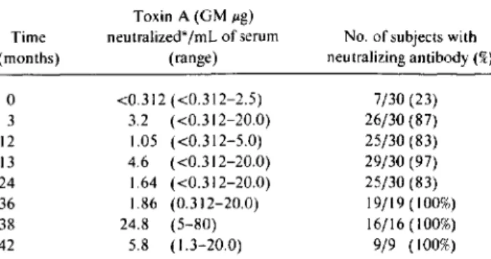
.05) above baseline.Before immunization, only 23% of patients possessed toxin A-neutralizing antibodies (table I). Primary vaccina-tion resulted in a
>
IO-fold rise in mean toxin A neutralizing levels with 87% of patients seropositive. Neutralizing antibod-ies were maintained by most patients throughout the study with periodic boosters causing a significant(P< .0 I) rise in neutralizing activity.P.aeruginosawas never isolated from 19 (61%) of the im-munized patients.P.aeruginosawas isolated only once from by two scoring systems, the clinic Bernese CF score and the Chrispin-Norman radiographic score [9]. Patients were followed for a mean of 37.8 months (range, 20-46).
Total serum anti-LPS immunoglobulin G (IgG) antibody was measured by ELISA [9]. The average affinity constants for such antibodies were determined [8]. The ability of sera to neutralize the cytotoxicity of toxin A was determined in a HEp-2 cell assay
[9]. P. aeruginosaclinical isolates were serotyped by agglutina-tion of whole bacteria using polyclonal typing sera (Public Health Laboratory Service, London). Bronchoalveolar lavage (BAL) was done on selected patients using an Olympus (Lake Success, NY) bronchoscope. BAL fluid was plated directly onto blood agar plates, and each bacterial colony type was identified by standard laboratory techniques.
Significance of differences between antibody concentrations at various time points was assessed with a pairedttest.
lID 1994; 169(May) Concise Communications 1161
Discussion
Table1. Toxin A-neutralizing antibody engendered by immuni-zation.
NOTE. Patients were immunized atO.2.12.and36months. GM. geo-metric mean.
*Limit of detection was0.312JIg neutralized/ml. of serum.
Colonization of the lower respiratory tract of CF patients with P.aeruginosa may be viewed as a gradual, multistep process influenced by several factors, including initial colon-ization of the upper respiratory tract, translocation to the lower respiratory tract, decreased mucociliary clearance, con-comitant infection with other bacteria or viral pathogens, CF transmembrane conductance regulator mutation genotype [ 10], and a switch from a smooth to a mucoid phenotype. Given these circumstances, preventing or even delaying the acquisition ofP.aeruginosacould provide a substantial
bene-References
I. Marks MI. The pathogenesis and treatment of pulmonary infections in patients with cystic fibrosis. J Pediatr1981;98:173-9.
2. Pier GR Des Jardin D. Aquilar T. Barnard M. Speert DP. Polysaccha-ride surface antigens expressed by non mucoid isolates of Pseudo-monas aeruginosafrom cystic fibrosis patients. J Clin Microbiol
1986;24:189-96.
3. Pedersen S5. EspersenF.HeibyN. Jensen T. Immunoglobulin A and immunoglobulin G antibody response to alginates from Pseudo-monas aeruginosain patients with cystic fibrosis. J C1in Microbiol
1990;28:747-55.
4. FomsgaardA.HeibyN. ShandGH.Conrad RS. GalanosC. Longitu-dinal study of antibody response to lipopolysaccharides during chronicPseudomonas aeruginosalung infection in cystic fibrosis. In-fect Immun1988;56:2270-8.
5. SchaadVB.Lang AB. Wedgwood J. Beuhlmann U. Hirer E. Serotype-specific serum IgG antibodies to Iipopolysaccharides ofPseudomonas aeruginosain cystic fibrosis: correlation to disease. subclass distribu-tion. and experimental protective capacity. Pediatr Res 1990;27: 508-13.
fit to the patient. Attempts to achieve this goal with prophy-lactically administered antibiotics have had a modest degree of success [II].
A previous attempt to immunize noncolonized CF pa-tients againstP.aeruginosadid not influence the rate of colo-nization [12]. Furthermore, there appeared to be a predispo-sition to a more severe disease course once infection occurred among the vaccinated group [12]. In contrast, we have found that multiple immunizations with a polyvalentP.
aeruginosavaccine over 3 years was not associated with any identifiable adverse events other than local reactions at the site of administration. The disease course did not appear to be affected by vaccination as monitored by two scoring sys-tems nor was there any evidence ofan immune complex-like syndrome. Furthermore, while 4 immunized patients ap-peared to become colonized or infected withP.aeruginosa
during the course of this study, they did not undergo an un-usually rapid clinical deterioration.
Immunization engendered a significant rise in IgG anti-body levels to all vaccine constituents. Interestingly, there was a pronounced anamnestic response to booster doses ad-ministered at 12 and 36 months, a phenomenon not seen in healthy adult subjects [13]. However, there was a more rapid drop-off in antibody titers after booster doses compared with primary immunization. The kinetics and longevity of the im-mune response would indicate that booster vaccinations would be required at 12- to 18-month intervals to maintain potentially protective antibody levels.
The current findings demonstrate that vaccination of non-colonized CF patients with a polyvalent O-PS-toxin A con-jugate vaccine was not associated with any identifiable risks. We are now attempting to increase the immunogenicity of this vaccine with novel adjuvants. On the basisofthese prom-ising results, a double-blind placebo-controlled trial to evalu-ate vaccine efficacy in noncolonized CF patients may be warranted. 7/30 (23) 26/30 (87) 25/30 (83) 29/30 (97) 25/30 (83) 19/19(100%) 16/16 (100%) 9/9 (100%)
No. of subjects with neutralizing antibody(%) <0.312 «0.312-2.5) 3.2 «0.312-20.0) 1.05 «0.312-5.0) 4.6 «0.312-20.0) 1.64 «0.312-20.0) 1.86 (0.312-20.0) 24.8 (5-80) 5.8 (1.3-20.0) Toxin A (GMJIg) neutralized*ImLof serum (range)
o
3 12 13 24 36 38 42 Time (months)cultures were negative. However, P. aeruginosa was again cultured at 37 months from BAL fluid. This patient was hos-pitalized (no exacerbation) after the second isolation ofP. aeruginosa for intravenous antibiotic treatment. Clinical scores remained unchanged throughout the 42 months of follow-up. After vaccination, a good (-- 10-fold rise) anti-body response was mounted and maintained to the LPS sero-type expressed by the infecting strain.
P.aeruginosa was first isolated from sputum of the third patient 21 months after immunization, with a second isola-tion at 24 months. Vaccinaisola-tion elicited a minimal «2-fold) response to this serotype. Clinical scores remained essen-tially unchanged.
P.aeruginosawas first isolated from the fourth patient 29 months after vaccination with a second isolation at 35 months. This patient mounted an excellent immune re-sponse after vaccination and maintained titers significantly above baseline levels. P. aeruginosawas not isolated from this patient at a 6-month follow-up.
None of the P.aeruginosastrains isolated had a mucoid phenotype. Four serotypes (IATS-2/5/16, IATS-4, IATS-5, and IATS-ll) were identified. One strain was nontypeable. IATS-il was not represented in the vaccine.
1162 Concise Communications lID 1994; 169 (May)
6. van Bever HP, Gigase PL, DeClerck LS. Bridts CH. Franckx H. Stevens WJ. Immune complexes and Pseudomonas aeruginosa antibodies in cystic fibrosis. Arch Dis Child 1988;63: 1222-8.
7. EichlerI,Joris L. Hsu YP. van Wye J. Bran R, Moss R. Nonopsonic antibodies in cystic fibrosis. Pseudomonas aeruginosa lipopolysaccha-ride-specific immunoglobulin G antibodies from infected patient sera inhibit neutrophil oxidative response. J Clin Invest 1989;84: 1794-804.
8. Bruderer U. Cryz SJ Jr. Schaad UB. Deusinger M, Que JU. Lang AB. Affinity constants of naturally acquired and vaccine-induced
anti-Pseudomonas aeruginosa antibodies in healthy adults and cystic
fi-brosis patients. J Infect Dis 1992; 166:344-9.
9. Schaad UB. Lang AB. Wedgwood J, et al. Safety and irnmunogenicity of Pseudomonas aeruginosa conjugate A vaccine in cystic fibrosis. Lancet 1991;338:1236-7.
10. Kubesch P, Dark T, Wulbrand U, et al. Genetic determinants airways' colonization with Pseudomonas aeruginosa in cystic fibrosis. Lancet 1993;341: 189-93.
II. Valerius NH. Koch C, Hoiby N. Prevention of chronic Pseudomonas aeruginosacolonization in cystic fibrosis by early treatment. Lancet 1991 :338:725-6.
12. Longford DT. Hiller J. Prospective. controlled study of a polyvalent
Pseudomonas vaccine in cystic fibrosis: three year results. Arch Dis
Child 1984;59:J13J-4.
13. Cryz SJ Jr, Sadoff JC, Fiirer E. Immunization with a Pseudomonas aeruginosaimmunotype 50-polysaccharide-toxin A conjugate vac-cine: effect of a booster dose as antibody levels in humans. Infect Immun 1988;56:1829-30.
Mutations in the Catalase-Peroxidase Gene from Isoniazid-Resistant
Mycobacterium tuberculosis
Isolates
Manuel Altamirano, Johanna Marostenmaki, Alfred Wong, Mark FitzGerald, William A. Black, and John A. Smith
Medical Microbiology. Department of Pathology. and Division of Respiratory Medicine. Department of Medicine. University of British Columbia; Vancouver General Hospital; and British Columbia Centre for Disease Control. Vancouver. Canada
Isoniazid resistance in Mycobacterium tuberculosis has been associated with total deletion of the katG gene, which codes for catalase-peroxidase production. To determine whether this is a common mechanism of drug resistance, 9 isolates of isoniazid-resistant and1of isoniazid-sensi-tive M. tuberculosis were analyzed by polymerase chain reaction amplification of a 237-bp se-quence of the katG gene. Amplification was observed in the isoniazid-sensitive isolate and in 8 resistant isolates; in only 1 isoniazid-resistant isolate was there no amplification of the expected band, suggesting gene deletion. DNA sequencing showed that 8 of the 9 isolates had point mutations, deletions, or insertions of 1-3 bases. Evidence corroborating the presence of muta-tions in the katG gene was obtained by single-strand conformation polymorphism analysis in these 8 isolates. Thus, mutations as well as insertions and deletions in the katG gene can account for inactive catalase peroxidase, leading to isoniazid resistance; gene deletion occurs only infre-quently, in-11 %of cases.
Tuberculosis is still a problem worldwide.Itis estimated that one-third of the world's population is infected by
Myco-bacterium tuberculosis.Tuberculosis is among the most im-portant infectious diseases, with 3 million deaths per year [ 1].Tuberculosis is a problem not only in developing coun-tries: Industrialized countries have experienced a decline in
Received 27 October 1993; revised 10 January 1994.
Presented in part: Molecular Mechanisms of Drug Resistance, 1993 Al-bany Conference, AlAl-bany. New York. 9-12 September 1993 (abstract 25). Reprints or correspondence: Dr. Manuel Altamirano, Medical Microbiol-ogy. Dept. of Pathology, University of British Columbia, 2733 Heather St., Vancouver, B.C.. Canada, V5Z IM9.
The Journal of Infectious Diseases 1994;169:1162-5 © 1994 by The University of Chicago, All rights reserved. 0022-1899{94{6905-004\ $01.00
tuberculosis, but since 1985 this trend has been reversed, with increases of18.4%in the United States to 33%in Swit-zerland [2]. The increase in tuberculosis frequency in the United States can be correlated with the AIDS epidemic, the influx of infected immigrants. outbreaks of tuberculosis in congregative facilities [3], and numbers of homeless [4]. To complicate the problem, outbreaks of multi drug-resistant tu-berculosis have been reported lately in 14 hospitals and in the prison system, with documented cases of multidrug-res is-tant tuberculosis in 241 patients and 15 health workers [5]. Drug resistance has been found in 60% of homeless persons with tuberculosis in Boston, 21% in New York, and 27% in San Antonio [4]. Also, there is a rapid spread of multidrug-resistant tuberculosis, especially in human immunodefi-ciency virus-infected patients. [6], who have a mortality rate of~89%[3]. This situation is reminiscent of the prechemo-therapy age[7].