For Hepatocyte Adhesion
by
Stephanie Therese Lopina
B.S., Chemical Engineering, University of Notre Dame, 1986 M.S., Chemical Engineering, Lehigh University, 1990
Submitted to the Department of Chemical Engineering in Partial Fulfillment of the Requirements for the Degree of
Doctor Of Philosophy at the
Massachusetts Institute Of Technology February, 1996
© 1996 Massachusetts Institute of Technology. All rights reserved.
Signature of Author
Stephanie T. Lopina
Certified by
P06ssor Linda G. Cima Thesis Supervisor
Accepted by
;AA';SACHUsrrsT IrS TU.E Professor Robert E. Cohen
OF TECHNOLOGY Chairman, Committee for Graduate Students
MAR
2
2
1996
LIBRARIESCarbohydrate-Derivatized Poly(Ethylene Oxide) Hydrogels
For Hepatocyte Adhesion
by
Stephanie Therese Lopina
Submitted to the Department of Chemical Engineering on 28 September 1995 in partial fulfillment of the requirements for the degree of
Doctor Of Philosophy
Abstract
Poly(ethylene oxide) creates an scaffold for engineered biomaterials. PEO has been shown to be resistant to nonspecific protein and cell adhesion; its hydroxyl endgroups provide a site for ligand attachment to promote specific hepatocyte adhesion. A series of radiation-crosslinked poly(ethylene oxide) (PEO) hydrogels which span a range of free chain end concentrations and tether lengths were synthesized, characterized and modified with bioactive ligands to create a scaffold for hepatocyte adhesion. The derivatized materials were seeded with hepatocytes to determine the cellular response.
Covalent crosslinks between polymer backbone chains are produced in aqueous PEO solutions through high energy electron beam irradiation, preserving endgroup
functionality for further derivatization. Star PEO, with PEO chains emanating from poly(divinyl benzene) cores, was used to produce biomaterials with a large range of endgroup concentrations and tether arm lengths. Swelling and compression
measurements confirmed that highly overlapping solutions of star PEO behave in a similar fashion to solutions of linear molecules. Irradiated solutions of high molecular weight linear PEO for hydrogel macrostructure and low molecular weight PEO oligomers for derivatizable endgroups produced hydrogels with many short ligand tethers.
Irradiation crosslinked PEO hydrogels were shown to be biocompatible in that they are not functionalized by irradiation, they permit minimal protein adsorption, they do not allow nonspecific cell adhesion and they exhibit minimal inflammatory response in vivo.
The PEO hydroxyl endgroups were activated with tresyl chloride chemistry to provide a leaving group for efficient ligand coupling through amines or thiols. Bioactive ligands 1-amino-1-deoxy-BD-galactose (ADGal) and arginine-glycine-aspartic acid (RGD) were uniformly coupled to activated hydrogels through terminal amino groups.
Hepatocytes adhere and spread on ADGal-modified PEO hydrogels in a
concentration/tether length dependent manner, suggesting that spreading is achieved when multiple binding sites are activated on the asialoglycoprotein receptor (ASGP-R), unique to hepatocytes. Hepatocytes did not adhere to unmodified hydrogels or to glucose-modified hydrogels (a negative control). Cells cultured on ADGal-modified PEO hydrogels exhibit normal morphology and function. Cytometry experiments confirm that mechanical forces are transferred to the cytoskeleton through the ASGP-R.
Acknowledgments
While working on my thesis, I have been aided and influenced by many people. I could not possibly thank everyone by name, but will use this space to acknowledge some of the contributions.
The work was funded in part by the National Institute of Health. PEO stars, the base material for most of the substratum produced for this thesis, were kindly donated by Paul Rempp, Pierre Lutz and Bertrand Schmidt of the Institut C Sadron, Strausbourg, France
and Professor Edward Merrill of MIT. I enjoyed the many hours spent with Kenneth Wright of MIT's High Voltage Research Lab, who not only ran the 1950s vintage van de
Graaf electron beam accelerator with expert skill, but also imparted bits of wisdom along the way. Hepatocyte isolations were completed by others who generously provided
hepatocytes for our lab, including Kristen O'Neill of the Shriner's Burn Center (Cambridge, MA); Magali Fountaine, Matthias Kauffman, and Kaoru Sano of the Children's Research Hospital (Boston, MA); Mitsuo Miazawa and George Wu of the Cima lab.
I had the pleasure of working with many undergraduates, whose efforts greatly increased the quantity and quality of data I was able to generate. Thanks to Cynthia Blake-Powell, Scott Cohen, Stephanie DeWeese, Sooyoung Kim, James Quirk, and David Weisburg for the dedicated hours they spent on my projects. Special thanks to Son Nguyen, Raul Rodriguez and Karen Zee whose undying enthusiasm and probing
questions kept the intellectual spark alive.
I appreciate the many conversations I had with graduate students Ed Perez, Sue Sofia Allgor, Philip Kuhl, Ann Park and George Wu where ideas were generated and bounced around. Tim Royappa also gave invaluable advice and assistance during his Post-Doctoral appointment in the lab. Working together in a synergistic effort paid large dividends for me.
Special effort was required this past year as I completed my thesis without a home base in Cambridge. I thank the many people who provided a bed or couch for me to sleep
on including Sue and Russ Allgor, my former roommates Marie-Jose Bl61anger and Heidi Wald, Mrs. Francis Elliot, Jaqui Lynch and Peter Nelson, Ann Park, Rae Simpson, and
Stefan Winkler. I also appreciate the leg work carried out by Sue Sofia Allgor, Wendy Koegler, Ann Park and especially Liz Webb (and undoubtedly others).
Thanks to my lab buddies, Scott Borland, Sue Hobbs and Ann Park, who would always lend an ear when I needed to chat and would occasionally drag me out of the lab for a beer or Tosci's cone. Marie-Jos6 and Heidi were awesome roommates. They certainly made our little place on Harvard Street a home. We started out acquaintances
and became good friends.
Most of all, I owe a huge debt of gratitude to my husband Tom. He was always at my side through the good and bad; my emotional support all the way. Thanks for believing in me no matter what.
Table of Contents
List of Tables 8
List of Figures 9
Chapter 1: Introduction and Literature Review 11
1.1 Background and Motivation 11
1.2 Cell Transplantation 15
1.3 Poly(Ethylene Oxide) (PEO) as Biomaterial 16
1.3.1 Grafted PEO 17
1.3.2 PEO Networks 19
1.4 Cell Receptors 21
1.4.1 Integrins and Cell Adhesion 22
1.4.2 Hepatic Asialoglycoprotein Receptor 24
1.5 Hepatocyte Adhesion to Materials with Bound Glycosides 26
Chapter 2: Materials Development and Characterization 30
2.1 PEO Characteristics 30
2.2 PEO Hydrogel Formation 31
2.2.1 Radiation Chemistry 33
2.2.2 Van De Graaf Electron Generator 34
2.2.3 Sample Preparation 34
2.2.4 Hydrogel Functionalization 36
2.3 Molecular Weight Between Crosslinks 36
2.3.1 Equilibrium Swelling 38
2.3.1.2 Linear Hydrogels 41
2.3.1.3 Star Hydrogels 42
2.3.1.3.1 Theoretical Development 42
2.3.1.3.2 Swelling Analysis 49
2.3.1.4 Linear/Oligomer Mixed Hydrogels 53
2.3.2 Uniaxial Deformation 62
Chapter 3: Substrate Derivatization and Characterization 70
3.1 Derivatization Chemistry 70
3.2 Grafted PEO 71
3.2.1 PEO Activation 72
3.2.2 PEO Coupling to Surfaces 73
3.2.3 Grafted PEO Reactivation 75
3.2.4 Ligand Coupling 76
3.3. PEO Hydrogels 77
3.3.1 Activation of PEO Hydrogels 77
3.3.2 Ligand Coupling 78
3.3.2.1 Coupling Procedure 78
3.3.2.2 Fluorescent Lectin Binding Analysis 80 3.3.2.3 Colorimetric Analysis of Carbohydrate Concentration 81 3.3.2.4 Galactose Partitioning Into Hydrogels 87
3.3.3 Nonspecific Protein Adsorption 88
3.4 Derivatization Induced Polymer Degradation 91
Chapter 4: Hepatocyte Interactions 92
4.1 Cell Culture Materials and Methods 92
4.1.3 Hepatocyte Isolation 93
4.1.4 Hepatocyte Seeding 94
4.1.4.1 Harsh vs. Gentle Seeding 94
4.1.4.2 Materials Transfer 95
4.1.4.3 Tight Fit Wells 98
4.2 Hepatocyte Adhesion and Spreading 98
4.2.1 PEO Grafted Surfaces 98
4.2.2 PEO Hydrogels 100
4.2.2.1 Non-Specific Interactions 100
4.2.2.2. Amino-Galactose Derivatized Hydrogels 101 4.2.2.3. RGD Derivatized Linear/Oligomer Hydrogels 106
4.3 Hepatocyte Viability 106
4.4 Hepatocyte Culture 107
4.4.1 Morphology 107
4.4.2 Differentiated Function 109
4.5 Characterization of the Nature of Adhesion 112
4.5.1 Trypsin Detachment 112
4.5.2 Competition with Galactose 113
4.6 Cytoskeletal Interactions 114
Chapter 5: Relationship Between Ligand Tether Length and Concentration for
Hepatocyte Spreading 121
5.1 Asialoglycoprotein Receptor Subunit Clustering 121
5.2 Ligand Distribution and Tether Length 122
Chapter 6: Conclusions 131
6.1 PEO Hydrogels 131
6.2 Hydrogel Derivatization 132
6.3 Hepatocyte Adhesion 133
6.4 Star PEO Applications 134
Chapter 7: References 136
Appendices
A. Alternate Example: Bound EGF Promotes Cell Growth 154 A. 1 Motivation: Hepatocyte Response to Epidermal Growth Factor 154
A.2 EGF Coupling to PEO Hydrogels 155
A.3 Hepatocyte Response 155
B. Protocols 157
B. 1. Carbohydrate Measurement with Acidic Anthrone 158
B.2. ELISA 159
B.3. Fluorescein-Labeled Lectin Binding 162
B.4. Live/Dead Viability Assay 163
B.5. Scanning Electron Microscopy 164
B.6 Tresyl Chloride Activation -PEO Hydrogels 165 B.7 Tresyl Chloride Activation -PEO in Solution 167
1-1. Biomaterials 11
1-2. PEO Grafting Techniques 18
1-3. Ligand Dissociation Constants 27
2-1. Polymer Characteristics 31
2-2. Radiation Chemistry 35
2-3. Star Solution Overlap Radius 47
2-4. Crosslink Density of Star PEO Hydrogels 52
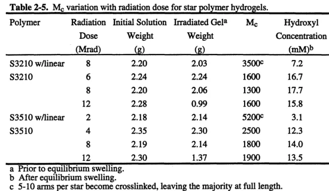
2-5. Me Variation with Radiation Dose for Star Polymer Hydrogels 54
2-6. Oligomer Mobility Factor 59
2-7. Oligomer Endgroup Concentration 61
2-8. Hydrogel Elastic Moduli 62
2-9. Network Structures Derived from Elastic Moduli 68 3-1. Linear PEO Characteristics for Activation Analysis 73
3-2. Coupling Optimization 74
3-3. Coupling Yield 80
3-4. RGD Derivatized Linear/Oligomer PEO Hydrogel Characteristics 80 3-5. Hydrogels used in Fluorescent Lectin Binding Analysis 81
3-6. Mass Balance Closure 82
3-7. Ligand-Coupled Star 3510 85
3-8. Hydrogel Carbohydrate Ligand Concentration 86
3-9. Hydrogel Mesh Size 89
4-1. Hepatocyte Area on PEO Grafted Slides 99
4-2. Inflammatory Response 102
4-3. Hepatocyte Behavior on ADGal-Derivatized Hydrogels 104
4-4. ELISA Results 112
4-5. Galactose Competition 114
5-1. Tether Extension 126
5-2. Hepatocyte Area 128
List of Figures
1-1. Hydrogel Variations 14
1-2. PEO Networks 21
1-3. Integrin Receptor 23
1-4. Asialoglycoprotein Receptor Endocytosis and Recycling 25
1-5. Asialoglycoprotein Receptor 26
1-6. Polyacrylamide Gel-Bound Cluster Glycoside 27
2-1. Radiation Induced Crosslinks in Star PEO 32
2-2. FTIR Spectra of PEO 37
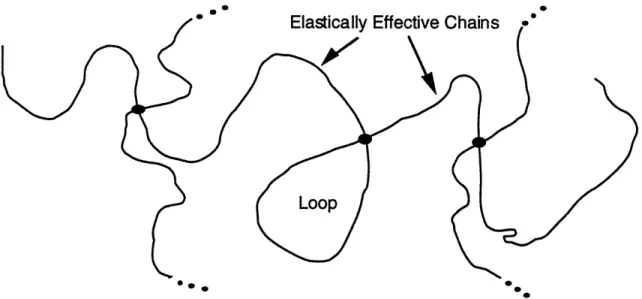
2-3. Elastically Effective Chains 39
2-4. Crosslink Concentration as a Function of Polymer Concentration 43
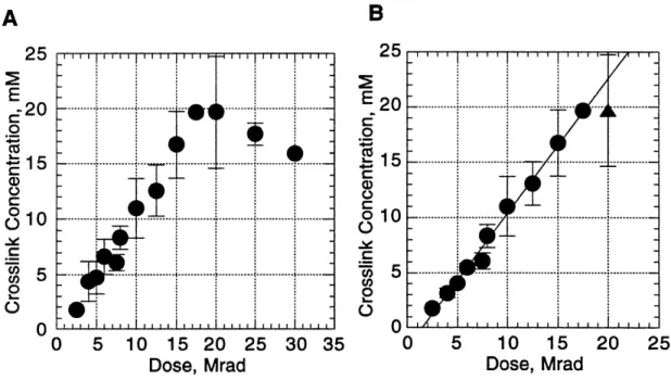
2-5. Crosslink Concentration as a Function of Dose 43
2-6. Star Polymer Conformation in Solution 44
2-7. Star Overlap 46
2-8. Crosslinks per Star Arm 53
2-9. A Priori Prediction of Oligomer Incorporation 58
2-10. Weighted Probability Prediction of Oligomer Incorporation 60 2-11. Weighted Probability Prediction of Linear/Oligomer Hydrogel Crosslinks 61
2-12. Hydrogel Stress-Strain Curves 63
2-13. Molecular Weight Between Crosslinks 69
3-1. Derivatization Chemistry 71
3-2. Vacuum Effect 76
3-3. Bioactive Ligands 79
3-4. Background Absorbance from Thermally Degraded PEO 83 3-5. Thermally Degraded Carbohydrate Calibration Curves 83 3-6. Fibronectin Adsorption onto Underivatized PEO Hydrogels 90 3-7. Fibronectin Adsorption onto ADGal-PEO Hydrogels 90 3-8. Activation and Coupling Induced PEO Degradation 91 4-1. Lactate Dehydrogenase Activity: Harsh vs. Gentle Seeding 96
4-2. Cell Counts: Harsh vs. Gentle Seeding 96
4-3. Lactate Dehydrogenase Activity: Substrate Transfer 97
4-4. Cell Counts: Substrate Transfer 97
4-5. Hepatocytes on ADGal-Star PEO 103
4-6. Hepatocytes on ADGal-Linear/Oligomer PEO 105
4-9. ELISA Technique 110
4-10. ELISA Results 111
4-11. Magnetic Twisting Device 115
4-12. Ligand-Coated Ferromagnetic Beads 116
4-13. Stress Induced Angular Strain 119
4-14. Cytoskeletal Stiffness 120
5-1. En-Face View of Asialoglycoprotein Receptor 122
5-2. Tether Stretch 124
5-3. Tether Stretching Distance 124
5-4. Hepatocyte Area 129
5-5. Tether Length vs. Tether Stretch 129
1. Introduction and Literature Review
1.1 Background and Motivation
Biomaterials -substances other than food or drugs contained in therapeutic or diagnostic systems that are in contact with tissue or biological fluids (Peppas, 1994) -have been used for many decades. The development of polymeric biomaterials has traditionally evolved on a trial and error basis as need arose. Existing materials which
exhibit properties suitable for a biomedical need have been implanted and the resulting biological response observed. Optimization has proceeded along the same trial and error
scheme, with some success. Table 1-1 lists some such biomaterials presently in clinical use.
Table 1-1. Biomaterials
Polymer Application
Cellulose Dialysis membranes
Poly(methyl methacrylate) Bone cement
Polyurethanes Catheters, pacemakers
Polytetrafluoroethylene (Teflon) Catheters, vascular grafts
Polyester (Dacron) Vascular grafts
Silicone rubber Catheters, tubing
The ability to specifically engineer a material to meet the precise needs of a
biomedical application has obvious benefits. The explosion of knowledge in molecular biology in recent years, coupled with advances in surface chemistry and materials science, provides the foundation to achieve the goal of developing ideal biomaterials designed to meet specific needs. Researchers in the biomaterials community recognize the potential for combining disciplines to develop materials of the future (Peppas, 1994; Ratner, 1993).
One of the goals in the field of biomaterials is development of synthetic implantable materials with surfaces which remain bland and inert towards the adsorption of proteins and cells. Such materials are sought for a wide range of applications including vascular grafts, drug delivery devices, and cell encapsulation membranes. Elucidation of
immobilizing ligands which foster highly specific cell interactions and precise control of cell behavior from the substrate. Such ligand-modified materials have the potential to enable or facilitate selective migration and growth of cells in vivo, thus expanding therapeutic options for regenerating tissues as diverse as bone, nerve, and liver. The biomaterial model for this thesis is the development of a scaffold for hepatocyte (liver cell) transplantation, a proposed alternative to whole organ transplantation.
Presently, whole organ liver transplantation is the only viable option for end-stage liver disease. The need for liver donors continues to grow and far exceeds the available supply. From 1987 to 1992 the number of people on the liver transplant waiting list grew more than 268%, with approximately one quarter of these patients being children under
10 years of age (UNOS, 1992). If hepatocytes can be made to grow on an implantable scaffold while maintaining differentiated function, the available organs could be
expanded since only a portion of the liver would be used for each transplant. Increasing the number of transplants would lead to earlier intervention and better tissue matches.
A common theme in the development of inert materials is the incorporation of polyethylene oxide (PEO) as a bulk or surface component. In such composite materials, accumulating experimental evidence supports a positive correlation between the amount of PEO at the surface and resistance to interaction with the biological milieu. The question for implantable biomaterials becomes how to achieve sufficient ethylene oxide (EO) density at the surface of a device. For simple abhesion, only EO segment density matters. The constraints on materials used for ligand immobilization, though, are greater - in addition to high EO segment density, the surfaces must possess a sufficient surface density of free chain ends for ligand attachment, and the bond of the PEO chain to the bulk material must be strong enough to withstand forces exerted by the cells pulling the free end bearing the ligand. A host of considerations - including the number of receptors and their affinity for the ligand, the quantity of receptors which must be occupied to obtain the desired cell response, the spacing between receptors once bound to ligand, and the mobility of the ligand when bound to the substrate - govern the optimal ligand
concentration for any given cell type and receptor-ligand system. Ligand surface densities ranging from 50-100,000 ligands/pm2 (or 10-20,000 nm2/ligand) have been reported as necessary for achieving appropriate interactions between cells and
immobilized ligands in various systems (Weigel, 1978; 1979; Schnaar, 1978; 1989; Guarnaccia 1982a,b; Oka, 1986; Raja, 1986; Kobayashi, 1988; Brandley, 1990; Massia, 1991a,b; Weisz, 1991a,b; Ito, 1992).
One way to meet the constraints of sufficient EO density and strongly-bonded chains as well as general in vivo biocompatibility is to employ covalently crosslinked networks comprising primarily EO. Two general approaches for forming PEO networks are endlinking and backbone crosslinking via radiation-induced free radical processes. Several such networks have been described and applied for various applications in biomaterials where abhesion is desired. A major limitation of the network approach,
however, is that few chain ends remain available for ligand modification.
A guiding issue in developing inert biomaterials, where the ligands are essentially tethered via PEO chains, is that the concentration of ligand accessible at the surface of the gel is not necessarily the only determinant of cell response to the ligand. It has been demonstrated, for example, that adhesion receptors for extracellular matrix molecules generate a different signal inside the cell when they are clustered together than when free (Kornberg, 1991). If interactions between two or more ligand receptors in the cell membrane are important, then ligand mobility, which is related to the length of the PEO "tether", also becomes a variable in describing the system.
The main thrust of this work is thus to synthesize and characterize a series of
radiation-crosslinked hydrogels which spans a range of free chain end concentrations and modify these materials with bioactive ligands to create a scaffold for hepatocyte
adhesion. Hydrogels with the structures shown in Figure 1-1 have been formed to vary both the mode and concentration of ligand presented to cells on a synthetic substrate. In gels of type A and B, a gel is formed from high molecular weight linear PEO. In gels of type B, a low molecular weight PEO oligomer is co-crosslinked to the backbone of the linear PEO gel. Most of the free chain ends in the resulting gel are ends of oligomer chains; thus the ligand is presented in closely-spaced groups of two with the spacing between ligands determined by the length of oligomer used. In gels of type C, PEO stars with a high degree of functionality are used to create gels in which the chain ends are more evenly distributed and the tethers are relatively long. Gels of type C are presumed to impart significantly more mobility to the ligand; allowing lower concentrations of ligand to be effective in eliciting a biological response in cases where receptor mobility is important. Some hydrogels are modified with the synthetic peptide arginine-glycine-aspartic acid (RGD), the sequence located within many cell adhesive proteins which is recognized as a ligand for integrin cell-surface adhesion receptors (Orlando, 1991; KUihn,
1994). Other hydrogels have been modified with carbohydrate ligands which interact with liver cells via the hepatic asialoglycoprotein receptor.
cm
o I
0.
x
"D T 141.2 Cell Transplantation
Hepatocyte transplantation is a conceptually attractive alternative to whole organ liver transplants because it could allow early intervention in the treatment of liver disease and increase the donor pool to include living donors. Initial studies of hepatocyte
transplantation involved injecting a suspension of hepatocytes directly into existing host tissues such as fat (Jirtle, 1980), liver (Matas, 1976), and spleen (Mito, 1979). The efficiency of this approach is generally low because the hepatocytes must adhere and form new tissue structures within the confines of existing mature tissue. Given these constraints, hepatocyte mass is unable to increase significantly from the original injected mass. Providing an artificial scaffold for the hepatocytes may permit greater hepatocyte growth. Demetriou and coworkers intraperitoneally injected hepatocytes attached to collagen-coated micro-carrier dextran beads (Demetriou, 1987; 1988; Moscioni, 1989). They were able to replace hepatic function in rats with 90% partial hepatectomy for 60 days. Others have examined use of a three-dimensional polymer scaffold on which to anchor cells (Thompson, 1989a; Cima, 1991).
Although some cell survival is seen, one of the key obstacles in obtaining survival and growth of the mass of cells required to have a metabolic impact is the bioreactivity of the material. Polymeric materials which have been employed in transplant devices include collagen-coated dextran beads (Demetriou, 1987; 1988; Moscioni, 1989), polylactic and polyglycolic acids (Cima, 1991), polytetrafluoroethylene (Gore-Tex) (Thompson, 1989a; 1989b), and polyvinyl alcohol (Ivalon) (Soni, 1975; Jackson, 1984). While these materials are all biocompatible in the sense that they are nontoxic and are associated with a mild tissue response, they permit nonspecific protein adsorption and cell adhesion. Numerous studies have been undertaken to measure protein adsorption on various surfaces and identify the cause (Ratner, 1976; 1981; Mori, 1982; Castillo, 1985; Merrill, 1987; Lee, 1988; Jeon, 1991; Andrade, 1992; Norde, 1992; Prime, 1993).
Protein adsorption is primarily due to hydrophobic and electrostatic interactions between proteins and the material surface (Andrade, 1992). A case study which carefully
measured albumin adsorption and desorption under varying conditions onto surfaces with a range of hydrophilicities found adsorption to be entropically driven (Lee, 1988).
Surfaces exhibiting minimal protein binding are amorphous, solvated by water, nonelectrolytic, and H-bond accepting (Merrill, 1987). When materials which permit nonspecific protein adsorption and cell adhesion are implanted, the hepatocytes seeded onto the transplant must compete with connective tissue cells to occupy the surface. Over time, the connective tissue encapsulates the device preventing adherent hepatocytes from
problem and the hepatocytes may dominate the surface. But clinically, it is more likely that a small number of cells will be implanted to grow and cover the surface, forming a new organ. This would not be possible if the device becomes overgrown with other cell types.
1.3 Poly(Ethylene Oxide) (PEO) as Biomaterial
Poly(ethylene oxide) (PEO) is a recognized biomaterial which has been approved by the Food and Drug Administration (FDA) for implantation. A common theme in the development of inert materials is the incorporation of poly(ethylene oxide) as a bulk or surface component. In such composite materials, accumulating experimental evidence supports a positive correlation between the amount of PEO at the surface and resistance to interaction with the biological milieu. The hydrophilicity and solubility properties of PEO produce surfaces which are in a liquid-like state with extremely flexible polymer chains. It is believed that the rapid movement of these chains influences the
thermodynamics at the solution/polymer interface. A repulsive force may develop due to a loss of configurational entropy of the surface-bound PEO upon approach of a protein or other particle (Andrade, 1987; Jeon, 1991). Adsorption of proteins is actively prevented as the PEO chains sweep out a large excluded volume (Atha, 1981). The PEO-water interface also has a low interfacial free energy and thus provides a low driving force for protein adsorption - the proteins will not feel any greater effects from the surface than they do from the bulk solution (Andrade, 1973). Proteins adhere much more readily to materials with high surface free energy (Schakenraad, 1989). Andrade describes PEO as the ideal polymer from the perspective of low protein adsorption:
(PEO) is a neutral molecule, and its weak hydrogen bonding
characteristics can be easily satisfied by surrounding water molecules. Its low refractive index suggests that van der Waals interactions will be
relatively low. Its stereochemical structure suggests that it tends to fit into the tetrahedral water lattice with minimal perturbation of water structure. It is perturbation of water structure, and particularly its enhanced
structuring in the presence of hydrophobic solutes, that leads to the hydrophobic interaction....
PEO is infinitely soluble in water and has exceptionally low chain rigidity. It is a dynamic and mobile molecule in solution. It fits almost all of the criteria for a surface that has minimal interactions of any kind with protein and for a surface that would be of intrinsically high mobility and rapid dynamics, and is therefore optimum for steric exclusion.
1.3.1. Grafted PEO
To take advantage of its inert characteristics, PEO is grafted onto potential
implantation materials as well as enzymes, drugs and liposomes for use in drug delivery systems. The reticuloendothelial system (RES) rapidly clears drugs and liposomes from the blood, but therapeutic enzymes and liposomes with covalently grafted PEO chains have extended stability in blood circulation (Davis, 1980; Yoshioka, 1991; Needham,
1992). PEO-grafted lipids used in liposomes produced for drug delivery systems have been termed Stealth® lipids to describe their ability to avoid the RES (Needham, 1992).
Many different techniques have been used to graft PEO onto various materials in order to minimize the biological response (Table 1-2). A general trend is observed where an increase in the EO concentration on the surface is inversely proportional to the protein adsorption or platelet adhesion onto the material, but complete resistance to protein adsorption remains difficult to obtain due to constraints on segment densities which can be achieved by grafting. In fact, a low segment density can actually enhance platelet deposition and activation by solvating what would otherwise be a prohibitively
hydrophobic substrate, making functional groups on the base material accessible to the biological milieu (Chaikof, 1992). The relationship between EO surface concentration and protein resistance has recently been cast in quantitative terms using an elegant experimental system comprising self-assembled monolayers (SAMs) of alkyl chains modified with EO oligomers of systematically varied length, where very high surface chain densities (and thus complete resistance to protein adsorption) can be achieved (Prime, 1993). SAMs are created when thiol-terminated alkyl or PEO chains adsorb in a tightly packed monolayer onto gold-coated substratum creating densely packed,
pseudocrystalline arrays of predominantly trans-extended chains oriented with their sulfur termini at the gold-SAM interface (Nuzzo, 1990). The critical density of grafted chains required for complete protein resistance scaled as n-0 .4 (n is the number of monomers in
the grafted chain), a scaling remarkably consistent with a power law for polymers
attached to a surface by one end where the average number of monomer groups a distance R from the surface scales as n0-4 (de Gennes, 1980). While SAMs did not spontaneously
desorb from gold over several days in water at room temperature (Pale-Grosdemange, 1991) biological shear forces and forces exerted by cells may pull adsorbed moieties from the surface, making SAMs unsuitable for implantation. These studies suggest that
implantable substratum could be made protein resistant given a sufficient PEO segment density.
Table 1-2. PEO Grafting Techniques Grafting Technique adsorption Substrate glass mica quartz Reference Hiatt, 1971 Luckham, 1985 Gasanov, 1991 di/tri-block adsorption PEO-co-PVC PEO-co-PPO, PEO-co-PBO carbodiimide coupling
cyanuric chloride activation
dithio-carbamate photo-grafting
epoxide reaction
hexamethylene diisocyanate coupling photo-polymerization
radiation
surface-physical-interpenetrating-network
2,4,6-trichloro-s-triazine activation PBO -poly(butylene oxide)
PEO -poly(ethylene oxide)
PET -poly(ethylene terephthalate)
PVC polyethylene poly(L-lysine) cellulose proteins PET PVC polystyrene polyurethane glass silastic film PET PET, PMMA, polyurethane
enzymes
PMMA poly(r PPO - poly(pr PVC -poly(vi Nagaoka, 1990 JH Lee, 1989 Sawhney, 1992 Kishida, 1992 Abuchowski, 1977 Gombotz, 1989; Desai, 1991b Mori, 1982; Nagaoka, 1984; Nakao, 1986 Bergstrim, 1992 Han, 1989 Tseng, 1992 Sun, 1987 Merrill, 1990 Desai, 1991a Davis, 1980 nethyl methacrylate) opylene oxide) nyl chloride) I m m1.3.2. PEO Networks
Given the difficulty in obtaining a pure PEO surface by covalently grafting PEO chains to implantable materials (Chaikof, 1986; Gombotz, 1989; JH Lee, 1989; Merrill,
1990; Desai, 1991a,b; Andrade, 1992; Prime, 1993; Drumheller, 1995), attention has turned to the formation of networks with PEO as an intrinsic component. The PEO component of non-covalent networks formed with block copolymers of PEO and poly(L-lactide) (PLLA) (Hu, 1993) or PEO and polysiloxane (Pekala, 1986a,b; Verdon, 1990) act to reduce protein adsorption and platelet activation from that observed with the base (PLLA or polysiloxane) material. Methoxy poly(ethylene glycol) monomethacrylates with PEO side chains of various lengths (MnG, where n equals the number of EO repeat units), can be polymerized with methylmethacrylate (MMA) to create covalently
crosslinked hydrogels with inherent PEO characteristics (Nagaoka, 1984; JH Lee, 1990). In a similar manner, PEO-diacrylates photopolymerize with trimethylolpropane
triacrylate (TMPTA) to create grafted semi-interpenetrating networks with a significant PEO component (Drumheller, 1995). Once again, enough PEO must be incorporated into the copolymer to mask the MMA or TMPTA. Platelet and protein adsorption were eliminated in a MMA-PEO copolymer with 35 wt% MnG of n > 50 (Nagaoka, 1984). A similar degree of PEO incorporation was required to prevent the adhesion of human foreskin fibroblasts (HFF) to TMPTA-PEO networks (Drumheller, 1995).
Networks consisting solely of PEO would presumably be exempt from concerns of biological interactions with copolymers or substratum. PEO networks can be prepared by the crosslinking reactions of PEO with plurifunctional isocyanates (Graham, 1984;
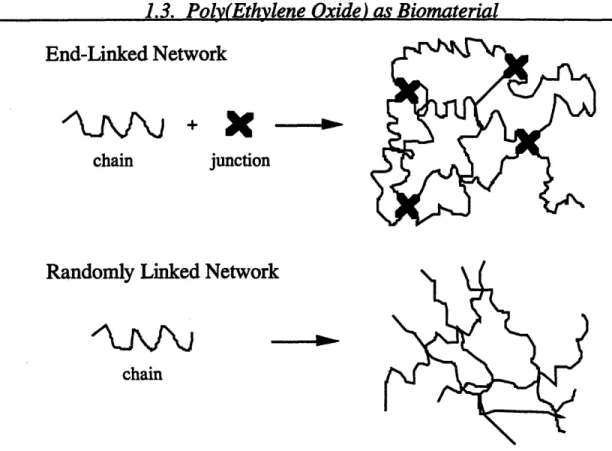
Gnanou, 1987; Yoshikawa, 1989). The isocyanates become network junctions with PEO chains endlinked to consecutive junctions to form the macrostructure (Figure 1-2). While such a network produces a purely PEO material, few free chains are available for further derivatization and attachment of bioactive moieties. Although the isocyanates are extremely reactive and unlikely to be free after crosslinking, their presence in the hydrogel will create added concerns for FDA approval of a device manufactured in this manner.
A novel biodegradable PEO hydrogel which is formed by in situ photopolymerization was shown to eliminate thrombosis and preserve long-term patency after crushing rat carotid arteries. It was also shown to inhibit thrombosis and reduce long-term intimal thickening in rabbit arteries injured by balloon angioplasty (Hill-West, 1994). The polymer precursor combines the inert character of PEO (center chain) with the water lability of poly(lactic acid) (adjacent segments); and is capped with tetraacrylate termini
hydrogel barriers, but no chain ends remain for further derivatization with bioactive moieties.
PEO networks with random links distributed along the polymer backbone, as opposed to being restricted to the chain ends, can be achieved through radiation crosslinking (Merrill, 1983; 1990; Minkova, 1989). This preserves hydroxyl endgroups for derivatization and ligand attachment while eliminating reagents needed for chemical crosslinking which may produce undesired reactions within the biological system (Figure 1-2). Irradiation of PEO in aqueous solution breaks down the water into hydrogen and hydroxyl radicals. PEO crosslinks are formed when the hydrogen radicals abstract hydrogens from the polymer backbone creating macroradicals. These macroradicals diffusion terminate by coupling to each other, forming the covalent carbon-carbon crosslink. Either gamma irradiation (Minkova, 1989), or high energy electron beam irradiation (Merrill, 1983; 1990) are effective in producing PEO hydrogels. Since PEO crosslink formation from macroradicals is a bimolecular reaction, the reaction rate should be proportional to the second power of macroradical concentration. Maximizing the radiation dose rate maximizes the concentration of macroradicals and favors crosslinking over degradation. Therefore, electron radiation, with a dose rate three orders of
magnitude greater than gamma radiation, is the preferred source to optimize radiation crosslinking of PEO solutions (Merrill, 1990).
The hydroxyl groups present on the end of each PEO chain provide the only site for ligand coupling chemistry. A hydrogel of linear PEO (Figure 1-1 A) would have very few hydroxyl groups available for derivatization. The endgroup concentration in the gel can be increased by irradiating a solution which includes both high MW PEO to provide hydrogel macrostructure and low MW PEO oligomers which will increase the number of endgroups (Figure 1-1 B). Another option is to produce hydrogels from high
functionality PEO stars. Rempp and coworkers have developed a star form of the polymer in which PEO "arms" are grafted onto an anionically polymerized divinyl benzene core, typically 10-60 arms per star (Gnanou, 1988; Merrill, 1990; Rempp, 1990). Radiation crosslinking of the stars into hydrogels provides a 5-30 fold increase in
available hydroxyl groups over linear polymer hydrogels (Figure 1-1 C). Preliminary studies indicate that a "pure" PEO surface is presented, completely masking the divinyl benzene core (Merrill, 1990; Rempp, 1990).
End-Linked Network
chain
junction
Randomly Linked Network
chain
Figure 1-2. PEO Networks. End-linked networks have chemical crosslinking
agents and few chain ends available for ligand derivatization. Randomly linked
networks are crosslinked along the polymer backbone.
1.4. Cell Receptors
One of the ways cells interact with their environment is through receptors, proteins on the surface of cells which have binding sites with high affinity for particular substances, referred to as "ligands". Receptors fall into several classes, depending on their primary functions. Some receptors serve to transduce signals which control cell growth, govern protein synthesis and secretion, and regulate the composition of intracellular fluids. Selective binding of ligands such as hormones, pheromones or neurotransmitters initiates a sequence of reactions that changes the cell function. Other receptors are involved in the selective uptake of extracellular proteins, growth factors and hormones via receptor-mediated endocytosis. Two examples include the low-density lipoprotein (LDL) receptor which removes LDL cholesterol from the blood stream and the asialoglycoprotein
receptor (ASGP-R) which removes abnormal serum glycoproteins to be destroyed in the cell lysosomes. Receptors also play a role in cell adhesion, both cell-cell adhesion (e.g.
involved in receptor-mediated cell interactions. Thus, there is an emerging rational basis for design of biomaterials with immobilized ligands which control cell behavior via receptor-mediated events. To create a transplantation scaffold for the treatment of liver disease, the polymer matrix should interact in a highly selective manner with hepatocytes while remaining bland to other cells.
1.4.1. Integrins and Cell Adhesion
Integrins form a major class of cell receptors which interact with proteins of the extracellular matrix (ECM) leading to cell adhesion. Integrins are transmembrane
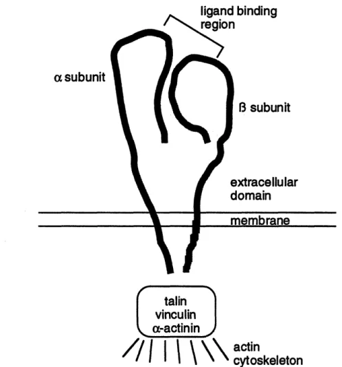
heterodimeric glycoproteins composed of noncovalently associated a and 8 subunits with a single ligand binding site (Figure 1-3). At least 14 a subunits and 8 3 subunits have been identified, but the B 1 and 83 subfamilies are primarily involved in interactions with
the ECM. Integrin subunit structure, binding characteristics, and function have been described in detail in several reviews (Ruoslahti, 1987; Buck, 1990; Kiihn, 1994). Once the integrin binds ligand, conformational changes lead to an association between the cytoskeletal portion of the receptor and the attachment proteins vinculin, talin, and a-actinin forming focal adhesion patches (Stamatoglou, 1990; Massia, 1991; Wang, 1993). These attachment proteins then bind to actin and microtubules in the cytoskeleton to physically link actin-associated proteins with the extracellular matrix (Beckerle, 1990;
Otey, 1990; Turner, 1990). Through this direct link, bound integrins are able to transmit mechanical forces from the extracellular matrix to the cytoskeleton, affecting cell
adhesion, shape and function (Ingber, 1991, Wang, 1993; 1994).
Cell adhesion to foreign surfaces in the body is typically mediated by proteins adsorbed from extracellular fluids. Specific amino acid sequences found in extracellular matrix proteins form the ligands which bind to integrins. The RGD sequence (found in fibrinogen, fibronectin, vitronectin, collagen, thrombospondin, and von Willebrand factor) was one of the first to be identified as an integrin-recognition motif; its binding to various integrins has been well documented (Buck, 1990; Orlando, 1991; Kiihn, 1994). Surfaces which present immobilized RGD become cell adhesive as the peptide is bound by the integrins, transferring forces to the cell cytoskeleton (Massia, 1991a,b; Lin, 1992). Other amino acid sequences have been found to have similar effects, including arginine-glutamic acid-aspartic acid-valine (REDV) found in fibronectin (Hubbell, 1991) and tyrosine-isoleucine-glycine-serine-arginine (YIGSR) found in laminin (Hubbel, 1991; Massia, 1991b). Nonspecific adhesion occurs when proteins adsorb onto a surface,
linand hindinn
a subunit
talin
vinculin
a-actinin
actin
cytoskeleton
Figure 1-3. The integrin receptor is composed of an a and B subunit with a single ligand binding site. Ligand-bound integrins interact with the cytoskeleton through attachment proteins talin, vinculin and a-actinin. (Redrawn from Buck, 1990.)
thereby presenting these and other cell adhesion moieties to cell adhesion receptors (van Wachem, 1987). One approach to promote cell adhesion to synthetic scaffolds is to incorporate these natural peptides into the polymer matrix. However many cells, including endothelial cells, fibroblasts and connective tissue, have receptors for these peptide sequences, limiting the usefulness of an inert base. The integrin receptor for REDV seems to be unique to endothelial cells since REDV modified surfaces do not support the adhesion of human foreskin fibroblasts, human vascular smooth muscle cells or platelets while promoting human umbilical vein endothelial cell adhesion (Hubbell,
the adhesion of hepatocytes while remaining bland to the rest of the biological milieu would incorporate a ligand which interacts with a receptor unique to hepatocytes.
1.4.2. Hepatic Asialoglycoprotein Receptor
Hepatocytes have a unique receptor for asialoglycoproteins (ASGP) which may be exploited for development of a cell transplantation device. Unlike integrins, the ASGP receptor is not known to be attached to the cytoskeleton, but it may still provide a mechanism for hepatocyte adhesion. The ASGP receptor has been studied extensively both in the solubilized form (Sarkar, 1979; Baenziger, 1980; Lee, 1987; Drickamer,
1988) and intact within the hepatocyte membrane (Hubbard, 1979; Connolly, 1982; Lee, 1983; Townsend, 1986) and the results have been summarized in several excellent
reviews (Schwartz, 1984; Spiess, 1990; Lodish, 1991; Geffen, 1992). Long researched as a model for receptor-mediated endocytosis, the ASGP-R binds asialoglycoproteins -proteins which have lost sialic acid endgroups exposing a terminal galactose residue which is bound by a B-bond to the next residue. The ASGP-R has specific affinity for galactose residues, functioning to clear these damaged proteins from the circulation. Ligand-bound receptors migrate to clathrin-coated pits which are endocytosed, forming vesicles which transport the damaged proteins to lysosomes for degradation (Wall, 1981; Drikamer, 1988; Fuhrer, 1991). Once the ligand has separated from the receptor, the receptor is recycled to the cell surface with a cycle time of 60-150 minutes (Bridges,
1982; Schwartz, 1982; Weigel, 1984). (Figure 1-4)
The hepatic ASGP-R is a member of the class of mammalian lectins designated C-type lectins, which exhibit an absolute requirement for Ca++ to bind carbohydrate ligands (Tolleshaug, 1980). Affinity studies indicate that while mono-, bi-, and triantennary galactose-terminated oligosaccharides are all taken up by the ASGP-R, receptor binding affinity in intact hepatocytes increases by five orders of magnitude as the ligand valency increases from a single galactose to a tri-branched form (Connolly, 1982; Rice, 1993). Binding affinity for the isolated receptor does not vary with ligand valency, suggesting an organizational or structural difference between the isolated receptor and the intact
receptor in the cell membrane. The human ASGP-R is a hetero-oligomer composed of two related polypeptide chains designated HHL-1 and HHL-2 (HHL for Human Hepatic Lectin) (Lodish, 1991), each with its own galactose binding site (Figure 1-5). The rat ASGP-R is composed of three subunits, RHL-1, RHL-2 and RHL-3. The only difference
Galactose Ligand
Figure 1-4. Asialoglycoprotein receptor endocytosis and recycling.
between RHL-2 and RHL-3 is the glycosylation pattern, which does not affect ligand binding or receptor oligomerization (Sawyer, 1988; Rice, 1993). These two receptor subunits can be considered as one, RHL-2/3, equivalent to the HHL-2. Both HHL-1 and HHL-2 receptor subunits have been cloned and expressed in fibroblasts and hepatoma cells; from these studies it has been demonstrated that the subunits HL-1 and HL-2
cooperate to enable high-affinity binding of triantennary ligands. HL-1 by itself is unable to bind and internalize triantennary ligands, while HL-2 functions to enable high affinity binding of triantennary ligands without affecting endocytosis of bound ligand-receptor complexes (Shia, 1989). Evidence points to a structure of the fully functional ASGP-R as comprising at least one HL-2 chain and at least three HL-1 chains (Lodish, 1991, Rice
1993). The optimal spacing of sugar moieties in tri-branched ligands (i.e., the spacing giving the highest affinity binding) is consistent with the spacing between the binding sites of a [(HL-1)3(HL-2)] receptor structure as described by Lodish (Lodish, 1991) and
shown in Figure 1-5. Molecular dynamics simulations provides a carbohydrate
conformational theoretical basis for the observed relationship between galactose spacing and binding affinity (Balaji, 1993). The great affinity of binding triantennary ligands suggests a conformational change on triantennary binding which induces cytoskeletal interactions (Baenziger, 1980; Connolly, 1982).
polymer scaffold. It is unique to the hepatocyte and exhibits selective binding to galactose-terminated oligosaccharides, which can be immobilized on PEO tethers. The binding affinity for di- and triantennary ligands are greater than the binding affinity between integrins and ECM proteins (Table 1-3). The long flexible PEO tethers may allow multifunctional receptor binding, even with monoantennary ligands. Achieving trivalent binding may induce a conformational change which leads to cytoskeletal interactions (Baenziger, 1980; Connolly, 1982) and cell spreading.
lactose ading
es
Clathrir
(HL-1)3HL-2 En Face View
Figure 1-5. Asialoglycoprotein Receptor
1.5. Hepatocyte Adhesion to Materials with Bound Glycosides
Hepatocyte adhesion to bound glycosides was first investigated to study the role carbohydrates may play in cell-cell adhesion (Weigel, 1978; 1979; Guarnaccia, 1982a,b). Flat synthetic polyacrylamide gels which had been derivatized with various carbohydrates were used as a model system. Researchers found a specificity of rat hepatocyte adhesion to immobilized galactose-terminated ligands, matching observations made with soluble ligands. Adhesion was found to be a threshold phenomena with a critical ligand
concentration of 3 mM (mmoles ligand per liter of gel) (Weigel, 1979). Immobilization of a cluster glycoside which contains three glycosidically linked sugars (Figure 1-6) did not alter the critical ligand concentration (Weigel, 1979). This result may be expected as the cluster glycoside used does not have the galactose spacing which soluble ligand affinity studies have shown to be critical for high affinity binding (YC Lee, 1984;1989;
Table 1-3. Ligand dissociation constants.
Receptor Ligand Kd (nM) Ref.
Integrin fibronectin fibrinogen vitronectin ASGP-R Gal(B1-4)GlcNAc(B1-2)Man Gal(B1-4)GlcNAc(B1-2)Man(a 1-6) Man Gal(B 1-4)GlcNAc(8 1-2)Man(al-3)/ Gal(B1-4)GlcNAc(B1-6)\ Man Gal(8 1-4)GlcNAc(B1-2)/ Gal(81-4)GlcNAc(1-6)\ Gal(31-4)GlcNAc(B1-2)Man(a l-6\ Man Gal(B 1-4)GlcNAc(B 1-2)Man(a 1l-3)/ Gal(B 1-4)GlcNAc(B1-2)Man(al-6)\ Man Gal(B1-4)GlcNAc(B 1-2)Man(a 1-3)/ Gal(B1-4)GlcNAc(B1-4)/ 1500-3000 2200-2300 Hubbell, 1991 Altieri, 1991 900-1000 Orlando, 1991 283,000 YC Lee, 1984; 1989 13,200 YC Lee, 1984; 1989
1,750
YC Lee, 1984;
1989 81.3 1.85 YC Lee, 1984; 1989 YC Lee, 1984; 1989 Gal -galactose GlcNAc -N-acetylglucosamine Man -mannose Gal-O-CH2 I Gal-O-CH2-C-NH-CO-(CH2)5 -Gal-O-CH2adherent hepatocytes to spread, 10 fold greater than that required for hepatocyte adhesion (Oka, 1986). One theory put forward to account for the difference is that the force
generated between the substratum and the cell surface must attain a minimum value per unit surface area of contact in order for spreading to occur (Oka, 1986). This force could be increased by increasing the total number of hepatocyte-substratum interactions
(increasing the ligand concentration) or by increasing the association constant of a constant number of interactions (using high affinity ligands).
Polyacrylamide surfaces containing covalently bound carbohydrates were used further to study ASGP-R function intact within the cell membrane. Because the hepatic ASGP-R is a C-type lectin, Ca++ is required to achieve ligand binding (Geffen, 1992).
Asialoglycoproteins bound to the ASGP-R from the solution state are rapidly released from the receptor in the presence of a divalent cation-free buffer containing a chelating agent (Tolleshaug, 1980; Weigel, 1984; Schwartz, 1984). Similar behavior might be expected of hepatocytes adherent to surfaces through the ASGP-R. However,
hepatocytes are difficult to remove from galactose-modified polyacrylamide gels using a chelating agent (Schnaar, 1978; Weisz, 1991a). This inability to remove adherent cells depends on the time of incubation on the substrate; for incubation times less than about 30 minutes cells can be released readily by a chelating agent but they become
progressively more difficult to remove as the incubation time is increased (Schnaar, 1978; Weigel, 1978). Receptor-ligand binding is a noncovalent, reversible phenomena. While free ligand was successful in blocking hepatocyte adhesion, once bound the cells
exhibited sugar-resistant adhesion on the same time scale as resistance to chelating agents (Guarnaccia, 1982b). Concomitant with increasing difficulty of disrupting the bonds between the ASGP-R and immobilized ligands using chelating agents or competitive ligand, the strength of adhesion between the cells and the substrate, as assayed by centrifugal detachment, also increases over the first 45 minutes (Guarnaccia, 1982a; Weisz, 199 lb). Adhesion-strengthening requires the concerted interactions of cytoskeletal components, actin and microtubules, and clathrin, a major component of coated pits. All of these elements had to be disrupted to counteract the time dependent adhesion strengthening (Weisz, 1991b). Based on these observations, Weisz and Schnaar proposed an interaction between the ligand-bound receptor and the cytoskeleton which required the participation of coated pit proteins (Weisz, 199 lb) but did not describe a mechanism.
Oligosaccharide-substituted styrene-type macromers and their polymerizates have been produced as a coating agent to enhance hepatocyte adhesion and viability on
polystyrene culture dishes (Kobayashi, 1988). Galactose-specific adhesion with a critical threshold concentration required for adhesion was observed, similar to the behavior of
hepatocytes seeded onto galactose-modified polyacrylamide gel. Hepatocytes adherent to polystyrene culture dishes which had been precoated with polystyrene-co-lactose were better able to maintain differentiated function over seven days in culture as compared to
hepatocytes cultured on standard collagen-coated tissue culture dishes (Gutsche, 1993). In the context of biomaterials design, the ultimate goal of understanding a particular receptor-ligand interaction is an ability to control cell and tissue behavior in vivo. Ideally, an in vitro assay should mimic the in vivo situation as closely as possible. Thus, the material used to present ligands in the in vitro assay should also be suitable for
physiological implant. Polyacrylamide exhibits unsatisfactory biocompatibility in vivo (Gin, 1990) and is thus not an ideal material for such applications. Any adsorbed modifications, such as the polystyrene-co-lactose, will wear away in vitro, proving unsuitable over time.
The ideal biomaterial, as described by Ratner and Hoffman (Ratner, 1976) "does not cause thrombosis, destruction of cellular elements, alteration of plasma proteins,
destruction of enzymes, depletion of electrolytes, adverse immune responses, damage to adjacent tissue, cancer and/or toxic or allergic reactions." PEO meets these criteria: it has been shown to minimize protein and platelet adsorption and activation, has been
approved by the FDA, and can be made into hydrogels with the desired mechanical properties to mimic soft tissue. Derivatization with galactose ligands should produce a hepatocyte specific material, and the long flexible PEO arms may permit multi-valent ligand binding on the ASGP-R.
The polymer scaffolds produced for this thesis are based on poly(ethylene oxide), a biomaterial which is generally recognized as exhibiting minimal nonspecific protein
adsorption. The majority of the work focuses on PEO hydrogels, which present a pure PEO surface to cells in culture. A complementary system of PEO chains grafted to glass was also considered.
2.1. PEO Characteristics
Poly(ethylene oxide) stars produced by living anionic polymerization from divinyl benzene (DVB) cores were provided by P. Rempp and B. Schmidtt of Strasbourg, France. Star PEO was obtained in the form of a powder which contained traces of the initiator, naphthalene, and other impurities. Analysis of a 5% (w/v) solution of the polymer in deuterated chloroform by proton NMR revealed, in addition to the expected peaks at 8 = 3.7 (ether hydrogens) and 8 = 7.3 (aromatic hydrogens), several broad peaks in the range 8 = 0.9 - 2.6. Impurities were removed by adsorption on activated charcoal from a 5% (w/v) solution of the polymer in MilliQ-purified water. The charcoal was precipitated from the solution by repeated centrifugation at 1200xg and the resulting clear polymer solution was dried by rotary evaporation. NMR analysis of the purified polymer revealed only the ether hydrogens and a peak at 8 = 7.3 arising from the hydrogens on the DVB core of the stars. A peak due to hydrogens in the terminal hydroxyl position was not detected above the noise; this peak would be < 50% of the area of the aromatic
hydrogens. The unassigned peaks which had been observed prior to purification were completely removed. Star characteristics have been studied by light scattering (Allgor, unpublished). While the polydispersity of star arm molecular weight (Ma) is very
narrow, the number of arms attached per star (f) is quite broad leading to a large range of star sizes in a give batch.
Linear PEO (nominal MW 100K) obtained from Polysciences as a powder containing approximately 1% silica added as a flowing agent was purified in a similar fashion. The molecular weight and polydispersity of linear PEO was determined by GPC in
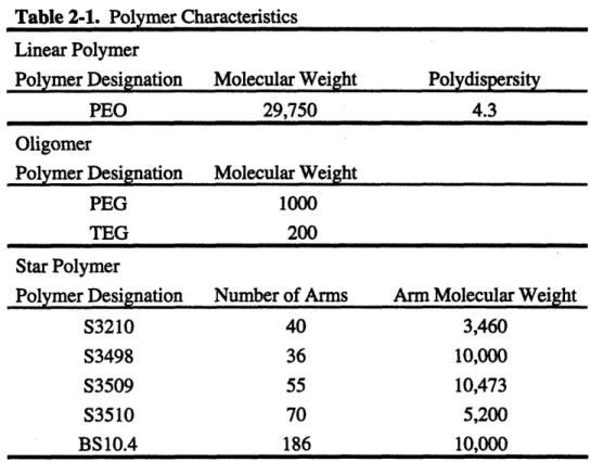
chloroform using a 3-column sequence (PL-gel guard, linear, and 1000-A) with RI detection. Data were analyzed with Perkin Elmer Turbochrome 3 software. Values of Mn = 29,750 and Mw/Mn = 4.3 were obtained. Characteristics of the polymer precursors used in forming the gels are shown in Table 2-1.
Table 2-1. Polymer Characteristics Linear Polymer
Polymer Designation Molecular Weight Polydispersity
PEO 29,750 4.3
Oligomer
Polymer Designation Molecular Weight
PEG 1000
TEG 200
Star Polymer
Polymer Designation Number of Arms Arm Molecular Weight
S3210 40 3,460
S3498 36 10,000
S3509 55 10,473
S3510 70 5,200
BS10.4 186 10,000
2.2. PEO Hydrogel Formation
PEO hydrogels present a pure PEO surface to the cells, eliminating any chance of interaction with an underlying surface which may be present in a grafted system. Irradiation crosslinking provides two advantages over chemical crosslinking: (1) Crosslinks formed via irradiation occur along the polymer backbone, preserving the hydroxyl endgroups for derivatization and ligand attachment. (2) Irradiation crosslinking does not incorporate reagents such as those needed for chemical crosslinking which may produce undesired functionalization, affecting the cell response.
Many of the technologically important properties of gels, such as the permeability to solutes, elastic modulus, and the average length of dangling chain ends which can be functionalized with specialized chemical groups, are functions of the network structure. The structure of a gel network formed by radiation-induced crosslinking of star molecules will fall between two extremes depicted in Figure 2-1. At one extreme, crosslinks are formed primarily between arms of the same star molecule, with few star-star crosslinks. This behavior is expected if the stars essentially repel each other in solution. At the other extreme, the dominant mode of crosslinking is star-star, resulting from free
of neighboring stars interpenetrate freely at concentrations above a critical concentration, it should be possible to obtain both extremes of network behavior by varying the weight concentration op of a particular star molecule in the crosslinking solution or,
alternatively, by varying the arm molecular weight, Ma, at constant star functionality, f, and cOp. For identical crosslink concentrations, these two structures are expected to yield different properties. Dominance of star-star crosslinking should result in a higher elastic modulus and lower permeability to solutes.
Crosslinked star networks may have a fundamental application in helping elucidate the solution properties of star polymers. Free-radical mediated crosslinking of polymer chains in solution serves to "freeze" neighboring segments; the resulting network structure should reflect aspects of the relative proximity of interchain and intrachain segments. In solutions of star molecules, analysis of network structures formed by radiation crosslinking may allow corroboration of light- and neutron-scattering evidence which suggest that in the semi-dilute regime the arms of star polymers freely
interpenetrate and behave essentially as solutions of linear molecules (Adam, 1991; Willner, 1994).
Case 1: intra-star crosslinks dominate Case 2: inter-star crosslinks dominate
Figure 2-1. Radiation induced crosslinks in star PEO will fall between the extremes of intra-star and inter-star crosslink dominance.
2.2.1. Radiation Chemistry
Polymers will either crosslink or degrade when subjected to ionizing radiation. (Here ionizing radiation may be either high energy electron or gamma radiation.) While the distinction between which polymers will crosslink vs. degrade is not clear, some trends have been observed. Radiation effects can be considered a kinetic phenomena with crosslinking and chain scission (degradation) as competing reactions. Crosslinking is favored between polymers with an easily abstracted hydrogen or methyl group while large side groups off the polymer backbone lead to degradation (Peppas, 1986). Chapiro notes a correlation between the heat of polymerization and the tendency to crosslink, reporting that polymers with a heat of polymerization greater than 16 kcal/mol tend to crosslink (Chapiro, 1962).
Specifically considering poly(ethylene oxide), crosslinks are formed when hydrogens are abstracted from the polymer backbone creating macroradicals. These macroradicals diffusion terminate by coupling to each other, forming the covalent carbon-carbon crosslink. The competing reaction of main chain scission leaves a carbon or oxygen radical at the end of a polymer chain which will degrade to an inert chain end by various mechanisms. When crystalline PEO is irradiated in the bulk, chain scission dominates over crosslinking (King, 1967). Crosslinking is slightly favored when irradiation is carried out in the melt, likely due to the increased mobility of the polymer chains (Graham, 1960).
Increased chain mobility was the original objective when researchers started to irradiate polymers in solution (King, 1970). Indeed, crosslinking greatly dominates when PEO is irradiated in solution. However, the preference for crosslinking is not solely due to increased chain mobility, but rather to a change in the route towards the formation of crosslinks. PEO crosslinks in solution are formed by an indirect interaction with water radiolysis products as the mediator which increases the efficiency of formation of main chain carbon radicals and thus increases the efficiency of crosslinking (King, 1970). Chemical reactions involved in water radiolysis and PEO crosslinking are outlined in Table 2-2. Since PEO crosslink formation from macroradicals is a bimolecular reaction, the reaction rate should be proportional to the second power of macroradical
concentration. Maximizing the radiation dose rate would maximize the concentration of macroradicals and favor crosslinking over degradation. Therefore, electron radiation, with a dose rate three orders of magnitude greater than gamma radiation, is the preferred radiation source to optimize radiation crosslinking of PEO solutions (Merrill, 1990).
process, leading to polymer degradation, particularly at low dose rates (Peppas, 1986). Degradation may be initiated by the formation of weak peroxidic bonds in the polymer backbone which decompose causing oxidative degradation of the main chain (Table 2-2) (Dennison, 1986). The degradation creates carbonyls in the network, an undesirable functionality for "inert" biomaterials. The strong polarity of carbonyl groups imparts a charge to the polymer, which increases nonspecific protein adsorption and cell adhesion (Andrade, 1992; van Wachem, 1987). The increased dose rate of electron beam
irradiation reduces the oxygen effect (Peppas, 1986).
2.2.2. Van de Graaf Electron Generator
PEO crosslinking was achieved using high energy electrons produced from a 3 MeV van de Graaf electron generator at the MIT High Voltage Research Laboratory. Mr. Kenneth Wright operated the generator at all times. The generator can provide up to 10 Mrad/s to a sample up to 7.5 cm wide, with a uniform penetration depth of about 1 cm in aqueous PEO solutions. Samples pass through the beam on a conveyer belt, with the dose rate delivered varying with electron beam current and belt speed. The total dose received depends on the dose rate and number of passes through the beam.
Radiation was delivered to samples at a rate of 250,000 rad/s with total dose delivered ranging from 2 to 25 Mrad. In a few instances, samples were packed in ice, but in
general no attempt was made to dissipate the heat generated during irradiation. Hydrogen gas produced during radical recombination (Table 2-2) may become trapped and form bubbles in the finished gel. Bubble formation was prevented by staging the irradiation in 2 Mrad doses separated by a minimum of 20 hours, allowing the hydrogen to diffuse from the gel.
2.2.3. Sample Preparation
After purification, PEO was dissolved in sterile MilliQ water to the desired concentration (2-20 wt%). All linear/oligomer hydrogels were formed from solutions containing a 4:1 weight ratio of linear PEO to oligomer. Radiation-induced chain scission and formation of side products (carbonyls, unsaturation) was minimized by degassing the polymer solutions to remove oxygen. Solutions were deaerated under vacuum for approximately 1 hour and then backflushed with nitrogen. Samples were irradiated in 2-g aliquots weighed into clean 60-mm glass petri dishes, keeping the depth of PEO solution exposed to the beam below 3 mm to insure that the dose received
_hble 2-2. Radiation Chemistry water radiolysis H20 + eaq H + *OH (1) hydrogen abstraction -C H2-C H2 O- + *OH - -CH -C H -O- + H20 (2) PEO crosslinking 0 --C H-C H -O--C H2-C H -0- + -C H2-C H-O- (3) -C H2-C H-O-radical recombination H + H H2 (4) OH + *OH • H202 (5) chain scission --CH 2-C H -O-C H2C H2-0- + H202 --C H 2-C=0 + HO-C H-C H2-O- +H OH H -- O-CH2-CH-O-CHCH2 - + eaq -O-C H2=C H2 + -- C H2-C H2- + H20 oxygen reactions -CH 2-CH-O- +02 -CH 2C 2-O-C H2-C H -O- C H CH2-- (8)
8
--C Hz-C H - + H2C=O + HC-O-C
H-CH2-O---CH CH2 -O + -CH 2 -CH O -CH -C H + -C H-•H2 (9)
petri dish covers and the edges were sealed with parafilm. The high radiation dose effectively sterilizes the gels and they were handled as sterile after crosslinking. Samples were stored in a 40C refrigerator between staged irradiation doses and after the final irradiation.
2.2.4. Hydrogel Functionalization
The main reaction incurred during radiation crosslinking is recombination of main chain carbon radicals to form crosslinks. However, other minor reactions can occur which lead to formation of carbonyl groups and unsaturated linkages, which are
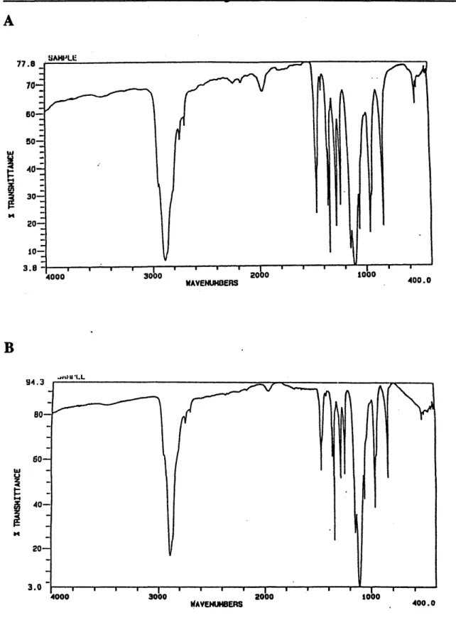
undesirable because these groups are often reactive in a biological environment (Table 2-2). FTIR spectroscopy analysis was used to verify that the crosslinking conditions used in these experiments did not result in formation of undesired reactive groups. The FTIR spectrum of purified PEO in Figure 2-2a shows the canonical C-H stretch at 2950 cm-1,
the C-O-C ether band at 1125 cm-1, and various C-H modes between 600 and 1500 cm-1. There is a small peak at 2000 cm-1 which is traceable to residual bound water present in the polymer. This bound water is notoriously difficult to remove entirely from the polymer (Pedley, 1979). The spectrum of the crosslinked polymer shown in Figure 2-2b is substantially similar to that of the parent material, which indicates that there is no gross alteration in the chemistry of the base material from irradiation, either by oxidation, conjugated n-bond formation or other degradation pathways. There is a notable absence of any peaks in the 1500-1900 cm-1 region, which clearly indicates the lack of any carbonyl functionalities in the gel. There are also no unsaturated bonds in the gels, judging by the absence of any significant peaks in the 2000-2600 cm-1 region. Finally,
the absence of conjugated linkages is also borne out by the colorlessness of the gels. Irradiation of PEO to form hydrogels does not appear to alter the properties important for inert characteristics.
2.3. Molecular Weight Between Crosslinks
The network structure of PEO hydrogels produced by electron beam irradiation influences the accessibility of the gel-linked ligand to cell surface receptors and governs the permeability of the gel to nutrients and to molecules (such as extracellular matrix proteins) secreted by the cells. The average molecular weight between crosslinks, Mc, is a significant measure of the hydrogel network structure. The average molecular weight between crosslinks is also the average molecular weight between a terminal crosslink and
WAVENU4I3ERS "qu*.
.i" UI.L
WAVENUM1BERS 400.0
Figure 2-2. FTIR spectra of (a) pure 100,000 MW PEO and (b) dried PEO gel formed by crosslinking a 20% solution with a radiation dose of 8 Mrad.
B
w
U
z
length. For linear/oligomer gels, 70-90% of ligand is linked to the ends of the oligomers, and thus the tether length for these gels is closer to 0.5 MWoligomer. Me can be
determined from rubber elasticity theory using either equilibrium swelling measurements or stress-strain curves obtained from uniaxial deformation.
2.3.1. Equilibrium Swelling
Once irradiated, a hydrogel will swell in solvent to an equilibrium swollen volume (or deswell in the case of syneresis). This swelling behavior of hydrogel networks has been investigated for many years. Theoretical and experimental investigations support the use of Flory's theory based on Gaussian chain statistics for predicting the crosslink density in networks which possess a low volume fraction of polymer, homogeneous F-functional junctions, and low or moderate degrees of crosslinking (Flory, 1950; Gnanou, 1987).
Bray, Peppas and Merrill modified the original swelling equation to account for networks formed in the presence of solvent (Bray, 1973; Peppas, 1976).
The theoretical development is based on the balance of elastic forces of the chains holding the network together and the mixing forces trying to pull the chains apart as solvent enters the hydrogel. Mathematically, equilibrium is reached when the chemical potential of the solvent in the network is equal to the chemical potential of the solvent in the bulk.
0 0 -AG
an
(2-1)
where gI1 and g10 are the chemical potentials of solvent in the network and in the bulk, respectively, AG is the change in free energy, and n I is the number of moles of solvent. As noted above, the change in free energy includes a mixing term and an elastic term:
AG = AGmix + AGel
(2-2)
The standard Flory-Huggins relationship is used to determine the free energy of mixing polymer and solvent, yielding: