CASE REPORT
Preoperative Embolization of Hypervascular CastlemanÕs Disease of
the Mediastinum
John Henri Robert,
1Georgios Sgourdos,
2Neoklis Kritikos,
1Dominique Didier,
2Sylvain Terraz
21Department of Surgery, Geneva University Hospital, 1211 Geneva 14, Switzerland 2Department of Radiology, Geneva University Hospital, 1211 Geneva 14, Switzerland
Abstract
We report the case of a 31-year-old woman with mediastinal
CastlemanÕs disease of the hyaline vascular type. This large tumor
was safely resected after arterial embolization. We describe the
steps of this interventional procedure and discuss related necessary
precautions.
Key words:
CastlemanÕs disease—Hypervasular
tumors—Medi-astinum—Arterial embolization
CastlemanÕs disease (CD) is a rare form of lymph node hyperplasia,
most commonly presenting as a solitary mediastinal mass.
Com-puted tomography (CT) and angiographic features of CD are
nonspecific [1] and a definite diagnosis is only based on histology
findings. The surgical approach is usually curative when the
dis-ease is unicentric [2], but it is frequently associated with profuse
bleeding. Only a few previous reports have suggested arterial
embolization in the preoperative management of CD [3, 4]. We
report a case of localized CD presenting as a voluminous mass in
the posterior mediastinum. Preoperative arterial embolization of
this tumor minimized intraoperative bleeding.
Case Report
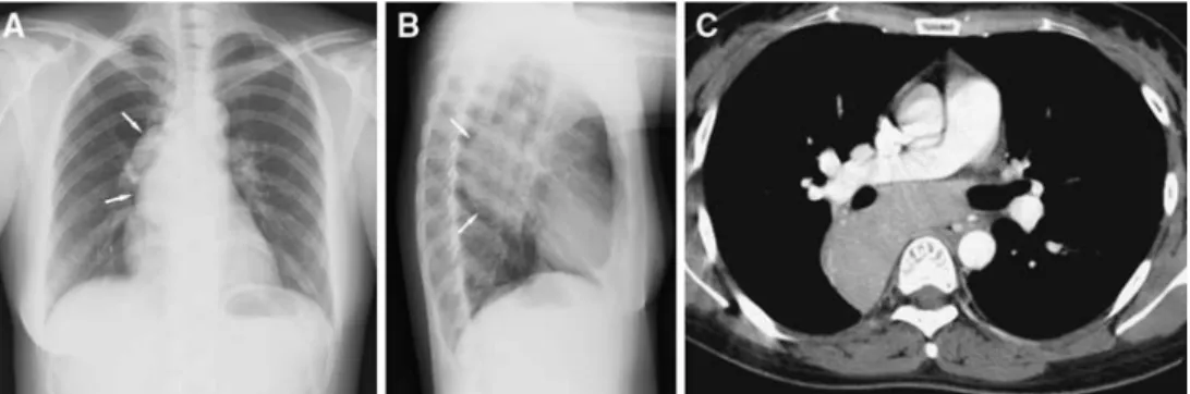
A 31-year-old nonsmoking Asian woman with no medical history presented with a dry cough for 6 months without any other symptoms. An abnormal opacity in the posterior mediastinum was found on chest X-ray (Figs. 1A and 1B). CT confirmed the presence of a 12-cm homogeneous, well-delineated hypervascular right paravertebral mass of the posterior medias-tinum (Fig. 1C). There was no evidence of pleural fluid or bone invasion. Laboratory investigations were within the normal range. Fine-needle aspi-ration or needle-core biopsy was not performed considering the risk of bleeding from this hypervascular mass and because of its location.
A preoperative descending thoracic aortogram was obtained by means of a 5F pigtail catheter (Cordis, Roden, The Netherlands), in order to visualize the origin of the bronchial and extrabronchial arteries from the aorta. A selective bronchial arteriography by means of a 5F steam-modified Cobra catheter (Cordis, Roden, The Netherlands) supported the presence of a hypervascular mediastinal mass. The lesion was exclusively supplied by two arterial branches of a broncho-intercostal trunk originating from the
descending thoracic aorta; it drained into large uphill inferior esophageal varices (Fig. 2). In a second step, embolization of these two feeding arteries was performed, in order to minimize intraoperative bleeding. The superior arterial branch, which supplied the superior and lateral portions of the mass, was selectively catheterized with a 2.5F microcatheter (Tracker 18; Boston Scientific, Cork, Ireland) and embolized by means of gelatin cross-linked tris-acryl microspheres 500–750 lm in diameter (Embospheres; Biosphere Medical, Roissy-Charles-de-Gaulle, France). Occlusion of the proximal segment was completed by three 3-0 silk threads (Silkam; Aesculap, Tuttlingen, Germany). The second arterial branch, which supplied the inferior and medial portions of the mass, was superselectively catheterized beyond the left bronchial artery and embolized in the same manner. Final postembolization angiography showed complete occlusion of these feeding arteries and the absence of vascularization of the mass (Fig. 2). After this procedure, no adverse effects were observed.
A right anterior thoracotomy was performed 7 days after embolization. Despite tight adhesions with the lung, the esophagus, and the subcarinal area, the tumor could be completely removed. Blood loss during surgery did not exceed 200 mL. A single chest tube was placed and removed on the third postoperative day. The postoperative course was uneventful and the patient was discharged on the fifth postoperative day. Thirty-six months after surgery, she is doing well without any sign of recurrence of her disease. The definite histological diagnosis was CastlemanÕs disease of the hyaline vascular type. Macroscopically, the tumor was surrounded by a fibrous capsule and its internal organisation was similar to that of a lymph node.
Discussion
Hypervascular mediastinal tumors are rare entities, benign or
malignant, occasionally found in children [5]. They include
he-mangiopericytomas [6], hemangioendothelioma [7], soft-tissue
angiosarcomas [8], pheochromocytomas [9], and some benign
tu-mors [10]; for practical purposes, arteriovenous malformations [11]
can also be included in this group. CD is another type of these
hypervascular tumors. First described in the mid-1950Õs [12], this
rare entity still causes diagnostic and therapeutic dilemmas and its
etiology is poorly understood (viruses and autoimmunity and
immunodeficiency states have been suggested). There are three
histological variants of CD: the hyaline vascular type, which
ac-counts for approximately 90% of cases, the plasma cell type, and
an intermediary ‘‘mixed’’ type. However, clinical significance is
determined by another classification, based on the distribution of
the disease. The localized form is generally benign and is cured by
surgical resection, whereas the multicentric form is more
aggres-Correspondence to: Sylvain Terraz, MD; email: [email protected]
ª Springer Science+Business Media, Inc. 2006 Published Online: 2 February 2007
C
ardio
V
ascular
and
I
nterventional
R
adiology
Cardiovasc Intervent Radiol (2008) 31:186–188 DOI: 10.1007/s00270-006-0156-y
sive and tends to be accompanied by various immunologic
abnormalities and systemic constitutional symptoms. CD is usually
seen in the chest, but it has also been reported in the neck,
retro-peritoneum, pelvis, and wherever lymph nodes are present [2].
Hypervascularity of these tumors entails the propensity to bleed
and accounts for the difficulty in managing them from the start [13].
Spontaneous hemothoraces have been described [6]; biopsies, so
commonly resorted to in other solid thoracic tumors, are rightly
Fig. 1. Chest X-ray and CT of a 31-year-old woman presenting a dry cough for 6 months. (A) Posteroanterior chest radiograph shows a large, homogeneous, and well-marginated opacity (white arrows) that causes mediastinal widening, but does not blur the right border of the heart. (B) Lateral chest radiograph confirms its posterior location in the mediastinum. (C) Contrast-enhanced chest CT
(mediastinal window) at the level of the right pulmonary artery shows a homogeneous and hypervascular mass with regard to soft tissues. The close relationship with the mass effect on the esophagus, right main bronchus and right pulmonary artery is particularly well dem-onstrated.
Fig. 2. Angiography prior to and following embolization. (A) Arterial phase of aortography shows the hypervascular mediastinal mass with two feeding arteries coming from a broncho-intercostal trunk: a superior and lateral branch (black arrow) and an inferior and medial branch (white arrow). (B) Venous phase of aortography shows the hypervascular mass draining in large uphill varices (black arrows). (C) Selective arteriography of the superior branch after embolization shows an occlusion of this vessel with small intraluminal defects (black arrow) corresponding to silk threads. (D) Selective arteriography of the inferior branch after embolization shows o an occlusion of this vessel with preservation of mediastinal branches (black arrow). After embolization, the mediastinal mass is completely devascularized.
feared and avoided [14]. Needless to say, the risk of bleeding is
even greater when surgery is contemplated. Although occasionally
reported successful [15, 16], thoracoscopy should be avoided in CD.
One case of hemangiopericytoma, in which diagnosis was not
known preoperatively, had to be operated on twice. Intractable
hemorrhage (2.1 L blood loss) occurred during the first operation,
which was only exploratory. Arterial embolization was successful
before the second surgical attempt—curative this time [17]. Indeed,
in all cases of tumors known or suspected to be hypervascular,
preoperative arteriography with embolization, which was first
re-ported in the 1970Õs [3], has made their management safer,
partic-ularly when surgery is warranted. This sophisticated technique,
however, is not devoid of risks and implies certain precautions.
Nonabsorbable microspheres might prevent the recurrence of
tumor hypervascularity due to recanalization of the embolized
ar-tery, as frequently observed with absorbable gelatin sponges. It is
essential to avoid material smaller than 325 lm in diameter, as it
can pass through bronchopulmonary anastomoses and lead to
pulmonary infarction or systemic arterial embolism (via
anasto-moses between bronchial arteries and pulmonary artery or vein,
respectively) [18]. By the same token, it is as important to avoid
embolic agents that cause distal occlusion to such an extent that
normal peripheral branches supplying the bronchi, esophagus,
aorta, and pulmonary artery might be equally occluded, leading to
obviously catastrophic complications [19]. For all of these reasons,
when bronchial artery embolization is contemplated, we
recom-mend the use of microparticles ranging in diameter from 350 to
700 lm [20]. Proximal vessel occlusion might preclude repeat
embolization after bronchial artery embolization for hemoptysis
[21]. In the case described, proximal embolization with
inexpen-sive silk threads was added to the procedure. This was done to
decrease the risk of new collateral vessels prior to surgery.
The most disastrous complication after bronchial artery
embo-lization is spinal cord ischemia due to the occlusion of spinal
arteries, which has been reported in 0–6.5% of cases [20–23].
Visualization of radicular branches on bronchial or intercostal
angiograms is not an absolute contraindication to embolization.
However, when the anterior spinal artery (artery of Adamkiewicz)
is identified at arteriography, embolization should not be
per-formed. Consequently, in order to decrease the risk of this dreaded
complication, it is of utmost importance to visualize all bronchial
and nonbronchial arteries prior to embolization.
References
1. McAdams HP, Rosado-de-Christenson M, Fishback NF, et al. (1998) Castleman disease of the thorax: radiologic features with clinical and histopathologic correlation. Radiology 209:221–228
2. Bowne WB, Lewis JJ, Filippa DA, et al. (1999) The management of unicentric and multicentric CastlemanÕs disease: A report of 16 cases and a review of the literature. Cancer 85:706–717
3. Walter JF, Rottenberg RW, Cannon WB, et al. (1978) Giant medias-tinal lymph node hyperplasia (CastlemanÕs disease): angiographic and clinical features. Am J Roentgenol 130:447–450
4. Safford SD, Lagoo AS, Mahaffey SA (2003) Preoperative embolization as an adjunct to the operative management of mediastinal Castleman disease. J Pediatr Surg 38:1–3
5. Parez N, Bader-Meunier B, Roy CC, et al. (1999) Paediatric Castleman disease: Report of seven cases and review of the literature. Eur J Pediatr 158:631–637
6. Mori M, Nakanishio N, Furuya K (1999) Hemangiopericytoma of the mediastinum causing spontaneous hemothoprax. Ann Thorac Surg 598:1525–1527
7. Rehring TF, Deutchman A, Cross JS (1999) Polymorphous heman-gioendothelioma. Ann Thorac Surg 68:1396–1397
8. Meis-Kindblom JM, Kindblom LG (1998) Angiosarcoma of soft tissue: A study of 80 cases. Am J Surg Pathol 22:683–697
9. Montana E, Montana X, Morera R, et al. (1990) Functioning para-ganglioma (pheochromocytoma) of the thorax: preoperative emboli-zation (letter). J Cardiothorac Surg 100:626–627
10. Mineo TC, Biancari F, Cristino B, et al. (1995) Benign vascular tumours of the mediastinum: Presentation of 3 cases and review of the literature. Thorac Cardiovasc Surg 43:361–364
11. Cheng SL, Wang HC, Yang PC (2003) Posterior mediastinal arterio-venous malformation with atypical color Doppler sonographic findings. J Clin Ultrasound 31:44–47
12. Castleman B, Iverson L, Menendez VP (1956) Localized mediastinal lymph-node hyperplasia resembling thymoma. Cancer 9:822–830 13. Iyoda A, Yusa T, Hiroshima K, et al. (2000) CastlemanÕs disease in the
posterior mediastinum: Report of a case. Surg Today 30:473–476 14. Kim JH, Jun TG, Sung SW, et al. (1995) Giant lymph node hyperplasia
(CastlemanÕs disease) in the chest. Ann Thorac Surg 59:1162–1165 15. Sica GS, Di Lorenzo N, Sileri PP, et al. (1999) Thoracoscopic approach
to giant lymph node hyperplasia (CastlemanÕs disease). Surg Laparosc Endosc Percutan Tech 55:519–522
16. Seirafi PA, Ferguson E, Edwards FH (2003) Thoracoscopic resection of CastlemanÕs disease: Case report and review. Chest 123:280–282 17. Morandi U, Stefani A, De Santis M, et al. (2000) Preoperative
embo-lization in surgical treatment of mediastinal hemangiopericytoma. Ann Thorac Surg 69:937–939
18. Pump K (1972) Distribution of bronchial arteries in human lung. Chest 62:447–551
19. Marshall TJ, Jackson JE (1997) Vascular intervention in the thorax: bronchial artery embolization for hemoptysis. Eur Radiol 7:1221–1227 20. Corr PD (2005) Bronchial artery embolization for life-threatening hemoptysis using tris-acryl microspheres: short-term result. Cardiovasc Intervent Radiol 28:439–441
21. Kato A, Kudo S, Matsumoto K, et al. (2000) Bronchial artery embo-lization for hemoptysis due to benign diseases: Immediate and long-term results. Cardiovasc Intervent Radiol 23:351–357
22. Mal H, Rullon I, Mellot F, et al. (1999) Immediate and long-term results of bronchial artery embolization for life-threatening hemoptysis. Chest 115:996–1001
23. Wong ML, Szkup P, Hopley MJ (2002) Percutaneous embolotherapy for life-threatening hemoptysis. Chest 121:95–102