Vacuum-Assisted Closure (V.A.C.®) for
Temporary Coverage of Soft-Tissue Injury in
Type III Open Fracture of Lower Extremities
Ludwig Labler, Marius Keel, Otmar Trentz
1Ab stract
Background and Purpose: The difficulty in the treat-ment of severe open fractures is a high infection rate and the problem of an adequate temporary cover-age of the soft-tissue damcover-age between successive second-look operations. The vacuum-assisted closure (V.A.C.®) offers good temporary soft-tissue coverage with a proven bacterial clearance and protects, at the same time, the soft tissue against secondary damage. The retrospective study reports the soft-tissue man-agement of severe open fractures of Gustilo type IIIA and IIIB with V.A.C.® or Epigard®.
Patients and Methods: All open fractures were in the lower extremity and a result of a nonpenetrating trau-ma. V.A.C.® was used as a temporary dressing in 14 fractures and an Epigard® in twelve fractures. Results: One early amputation was observed in each group. In the group with the soft-tissue coverage by Epigard®, in spite of less type IIIB fractures and less polytraumatized patients, the rate of infections (6/11) was substantially higher compared with patients man-aged by V.A.C.® therapy (infection: 2/13).
Conclusion: V.A.C.®, a temporary soft-tissue substitute, reduces the rate of infection and is an alternative of choice for the management of type III open fractures.
Key Words
Vacuum-assisted closure · V.A.C.® · Open fracture · Soft-tissue injury
Eur J Trau ma 2004;30:305–12
DOI 10.1007/s00068-004-1389-6
Introduction
The management of high-energy open fractures contin-ues to be a difficult problem confronting the surgeon. The essential part of an initial soft-tissue damage treat-ment is copious irrigation and thorough debridetreat-ment [1, 2], which can cause significant soft-tissue defects. Then it must be decided whether the wound should be closed primarily or left open for delayed suture or reconstruc-tive surgery. Different surgical methods have been de-veloped to perform these difficult closures or temporary coverage. These include closure devices [3], skin grafts, local flaps, and free flap transfers. It was recommended to carry out the definitive soft-tissue coverage as soon as possible, preferably within the first 72 h after trauma [4], by a delayed primary closure of the wound [5] to prevent flap failures, infection and delayed bone heal-ing. This, however, is often impracticable because of a complicated fracture, wound contamination or already existent infection or the associated injuries accompa-nied by hemodynamic instability and microcirculatory dysfunctions based on systemic inflammation or edema [6, 7] which prevent an early long-lasting reconstructive surgical intervention. Although skin grafts are readily obtainable, they depend on the vascularity of the recipi-ent bed and may be contraindicated by exposed bone, cartilage, tendons or surgical implants. Consequently, one or more “second-look” operations are neces-sary prior to the definitive closure and an appropriate wound dressing is needed as a temporary coverage be-tween repeated debridements. The essential request for such a temporary closure is to prevent the exposed vital structures from desiccation and bacterial
contamina-1 Division of Trauma Surgery, Department of Surgery, University Hos-pital Zurich, Zurich, Switzerland.
tion and to induce a locally normal circulatory situation and proliferation of the wound granulation tissue. The traditional dressings for open wounds are wet-to-dry closures [1, 2, 8–17], antibiotic-impregnated bead pouch [18] and skin substitutes including xenografts, human allografts or synthetic membranes such as, Epigard®
(Biovision GmbH, Ilmenau, Germany) [19–23]. The vacuum-assisted closure (V.A.C.®, Kinetic Concepts,
Inc., San Antonio, TX, USA), a new, efficient system for the treatment of problematic wounds [24–29], be-came a generally accepted technique and was applied as a temporary wound closure also in the case of open fractures [30–32]. Epigard® treatment was the standard
dressing for soft-tissue management in our clinic until the late 1990s. Since 2000 it was substituted stepwise by V.A.C.®, which finally became the standard temporary
closure of problematic wounds of various types. The purpose of this case control report is to compare ret-rospectively two standard techniques of a temporary wound coverage and to evaluate the results of the use of these therapies in the management of patients with high-energy soft-tissue injuries of Gustilo type III open fractures [2] of lower extremities.
Patients and Methods
All patients had severe open fractures of the lower extremity classified as type IIIA or IIIB according to Gustilo et al. [2] and were admitted as an emergency to our clinic. In this study we excluded all type IIIC fractures because of the associated vascular injuries, which compromise the circulatory situation of the local soft-tissue injury. The patients not included in the study were treated during the interim when we stepwise intro-duced V.A.C.®, and both techniques were used
simul-taneously and partly consecutively in the same patient. All fractures were a result of a high-energy trauma, and the partly polytraumatized patients were treated ac-cording to the Advanced Trauma Life Support Guide-lines [33] and our standard trauma protocol [34, 35]. Af-ter control of the airway, ventilation and cardiovascular functions, life-saving procedures were performed. Af-terwards, hemorrhage control, radical wound debride-ment, decompression of compartments, and primary stabilization of major fractures (“day 1 surgery”) were carried out [34, 35]. Debridement of open fractures was performed in a stepwise manner, i.e., one debridement in the emergency room and two further debridements with copious irrigation in the operating room on the ad-mission day, and primary immobilization of the fracture
was carried out preferably by external skeletal fixation. “Second-look” operations included subsequent thor-ough debridements, fracture redislocation and repeated irrigation with normal saline solution and were carried out, as far as possible, every 48 h in dependence of the wound and patients condition. The V.A.C.® system or
Epigard® was applied at the end of each surgical second
look. This procedure was repeated until the soft-tissue defect became macroscopically clean and was free of necrotic tissue.
The V.A.C.® system was applied in a group of
twelve patients with 14 open fractures treated between June 2001 and December 2002 (group I). The V.A.C.®
system consists of polyurethane soft sponge shaped to fit the wound and placed into the cavity. The 400–600 mm pore size of the sponge maximizes tissue growth [36]. The suction tube with side ports was embedded in the center of the sponge allowing communication of its lumen to all spaces of the foam and attached to an ad-justable suction pump. A transparent occlusive gas- and fluid-impermeable plastic film was applied over the foam to seal it airtight and the pump was started to pro-duce a continuous negative pressure of 125 mmHg.
A group of eleven patients with twelve open frac-tures, hospitalized between March 1998 and November 1999, was treated with Epigard® as the temporary closure
(group II). Epigard® dressing, reticulated polyurethane
foam laminated to a microporous polypropylene film, is applied in a manner similar to skin grafting, i.e., it is stapled to the wound edges and afterwards covered with a sterile dressing. Both groups of our patients belong to time periods when Epigard® and V.A.C.®, respectively,
were used as a standard technique. The dressing change in both groups was performed under general anesthe-sia during each “second look” in the operating room. Bacterial cultures were obtained routinely on admission and during each “second look”. A cephalosporin of the third generation was systemically applied in therapeu-tic doses on admission and continued or adapted to the bacterial culture results.
Clinical data in Table 1 include the study population, the cause of the injury, the Injury Severity Score (ISS) [37], associated injuries, and hospital and intensive care unit (ICU) stay. Fracture localization, classification ac-cording to Gustilo et al. [2] and Mangled Extremity Se-verity Score (MESS) [38–40] are summarized in Table 2. Evaluation of both techniques includes the duration of the temporary coverage use, the final wound closure type and its outcome, and the fracture management and
its complications (Table 2). Bacterial characteristics and soft-tissue complications are summarized in Table 3. All patients were followed for a minimum of 12 months af-ter definitive soft-tissue coverage to verify healing or complications.
Results
The temporary wound closure changes averaged 4.3 in group I (range 1–10) and 2.2 in group II (range 1–3). Epigard® was used for a total of 4.9 days on average
(range 2–9 days), whereas V.A.C.® covered the wound
for 11.3 days (range 2–35 days). Figure 1 shows a severe soft-tissue defect of IIIB fracture with luxation of the right ankle (patient #2), which was temporarily closed by V.A.C.®. Only four wounds out of both groups (group I:
patient #9; group II: patients #7, 8, and 10) did not need any further treatment, and the delayed primary clo-sure could be directly performed. All other soft-tissue defects were covered mostly by microvascular flaps or mesh grafts (Table 2). The clinical result of a type IIIB fracture (patient #2) after soft-tissue reconstruction with microvascular flap is shown in Figure 2. Soft-tissue reconstruction was performed after an average of 12.3
days in group I (range 2–35 days) and 4.1 days (range 2–8 days) in group II. An amputation in group I had to be performed 10 days after admission (patient #5) due to complete necrosis of the soft tissues of the foot. We had two serious soft-tissue complications in group I with infections (patients #6 and 11) resulting in a nonunion situation of the bone (one intraarticular fracture of the distal femur and one tibial shaft fracture) where the soft-tissue defect was closed by a microvascular flap 12 and 5 days after trauma, respectively. In both cases the plate implants were removed and a debridement of the soft tissue and of the nonhealing fracture was performed. Both fractures were stabilized by plates and bone graft-ing from iliac crest. Both fractures consolidated after 6 and 4 months, respectively. One minor complication, also in group I, with an infection in the former bed of the microvascular flap used for reconstruction of the tibial soft-tissue defect occurred in patient #8. After debride-ment the wound healed uneventfully without any signs of infection. In group II, six complications were severe soft-tissue infections, combined with a nonunion in two cases, and one complication was a nonunion without in-fection (Tables 2 and 3). In three patients (#1, 2, and 6)
Table 1. Clinical characteristics of patients. a: head; b: thorax; c: abdomen; d: extremities: e: skin; F: female; ICU: intensive care unit; ISS: Injury
Se-verity Score [37]; M: male.
Patient Gender Age Cause of injury Multiply injured Associated Hospital ICU stay (years) Traffic Impaction Fall from height (ISS) injury stay (days) (days)
Group I 1 M 56 + No 17 – (V.A.C.®) 2 F 64 + Yes (41) a, b, c, d, e 61 15 3 F 18 + Yes (41) a, b, c, d, e 32 32 4 F 45 + No 34 – 5 M 56 + Yes (34) a, b, c, d, e 67 37 6 M 22 + Yes (57) a, b, c, d 158 27 7 M 19 + Yes (19) a, b, d 37 10 8 M 54 + Yes (34) a, b, d, e 63 27 9 M 33 + No 30 – 10 F 68 + Yes (34) c, d, e 62 20 11 M 31 + No 35 – 12 M 35 + Yes (34) b, c, d, e 43 5 Group II 1 F 30 + No b, d 29 6 (Epigard®) 2 M 43 + No 27 3 M 75 + No 38 – 4 M 40 + No 41 2 5 F 89 + Yes (20) d, e 95 – 6 M 62 + No 35 – 7 M 20 + No 25 – 8 F 43 + Yes (18) b, d 21 – 9 M 49 + No 20 – 10 M 49 + No 27 – 11 M 46 + Yes (38) b, c, d, e 104 48
the soft-tissue injury was closed by a microvascular flap and in two patients (#8 and 10) by a delayed primary closure. The free flap reconstructions were performed on the 2nd, 3rd and 7th day and the delayed primary closures on the 4th and 8th day after admission. An am-putation had to be performed 5 days after admission on the sixth patient (#5) due to an uncontrollable infection situation. The patient died 2 days afterwards due to car-diovascular instability. The three observed nonunions of tibial fractures in group II were caused by a local in-fection 43 and 125 days after trauma in two cases, and in one case the nonunion was seen after 75 days. The local infection of the tibial fracture in patient #1 resulted in a septic complication of the uncontrollable infection un-der the free flap, and as a consequence, an amputation
was necessary on the 8th day. Of the remaining five pa-tients, four (#2, 6, 8, and 10) had a debridement 43, 157, 33, and 125 days after trauma with loss of the primary free flap in two cases (#2 and 10) and replacement by a soleus flap. Implants were removed in three patients (#2, 6, and 10) during debridement. In patient #6 the frac-ture consolidated. After reaming, a new intramedullary nail was used in patient #2, and the tibial nonunion was treated by a new plate and a bone graft from iliac crest in patient #10. Both tibial fractures healed 4 months later. After treatment of the local infection, the fracture of patient #8 healed uneventfully after 5 months. In pa-tient #9 the nonunion situation after bone debridement was treated by a bone graft from the iliac crest and the fracture consolidated after 12 weeks. All seven serious
Table 2. Open fracture pattern. MESS: Mangled Extremity Severity Score [38–40].
Patient Injury pattern and fracture classification Fracture management Soft-tissue manage-
Compli-Localization Gustilo ment cations
classification MESS Primary Secondary Definitive
stabilization stabilization stabilization
(admission day) (days after
admission)
Group I 1 + + 5 External fixator Plate 5 + (V.A.C.®) 2 + + 7 External fixator Plate 6 + 3 + + 4 External fixator – – + 4 + + 4 External fixator Plate 3 + 5 + + 5 External fixator – 10 +
+ + 7 External fixator K-wire 2 +
6 + + 5 External fixator Plate 6 + + + + 5 External fixator Screw 6 +
7 + + 2 External fixator Screw 7 + 8 + + 6 External fixator Plate 18 + 9 + + 4 External fixator – – + 10 + + 6 External fixator Plate 12 + 11 + + 3 External fixator Plate 5 + + 12 + + 3 Plate – – + Group II 1 + + 5 Plate – – + + (Epigard®) 2 + + 2 External fixator Nail 3 + + 3 + + 4 Plate – – + 4 + + 3 Plate – – + 5 + + 5 External fixator – – + 6 + + 4 External fixator Plate 7 + 7 + + 4 External fixator Plate 5 + 8 + + 6 External fixator Plate 9 + 9 + + 3 Nail – – + + 10 + + 3 External fixator Plate 8 + +
11 + + 9 External fixator Plate 2 + + + 5 External fixator Cerclage 2 +
Fem ur Kn ee Tibi a
Foot IIIA IIIB Delayed prim
ary closur e M esh Mi cr ovascular flap Amputati on No nu n io n Ankle
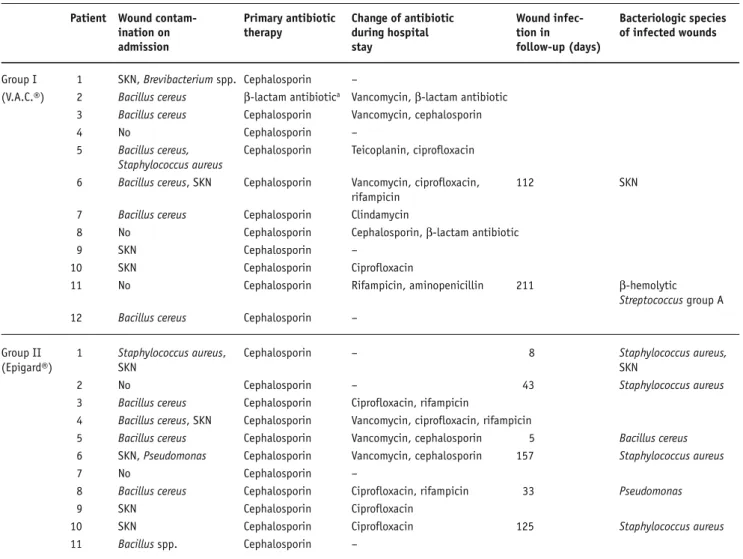
Table 3. Bacteriologic characteristics of soft-tissue defects. No: no bacterial growth; SKN: Staphylococcus coagulase-negative.
Patient Wound contam- Primary antibiotic Change of antibiotic Wound infec- Bacteriologic species
ination on therapy during hospital tion in of infected wounds
admission stay follow-up (days)
Group I 1 SKN, Brevibacterium spp. Cephalosporin – (V.A.C.®) 2 Bacillus cereus β-lactam antibiotica Vancomycin, β-lactam antibiotic
3 Bacillus cereus Cephalosporin Vancomycin, cephalosporin 4 No Cephalosporin – 5 Bacillus cereus, Cephalosporin Teicoplanin, ciprofloxacin
Staphylococcus aureus
6 Bacillus cereus, SKN Cephalosporin Vancomycin, ciprofloxacin, 112 SKN
rifampicin
7 Bacillus cereus Cephalosporin Clindamycin
8 No Cephalosporin Cephalosporin, β-lactam antibiotic 9 SKN Cephalosporin –
10 SKN Cephalosporin Ciprofloxacin 11 No Cephalosporin Rifampicin, aminopenicillin 211 β-hemolytic
Streptococcus group A
12 Bacillus cereus Cephalosporin –
Group II 1 Staphylococcus aureus, Cephalosporin – 8 Staphylococcus aureus,
(Epigard®) SKN SKN
2 No Cephalosporin – 43 Staphylococcus aureus
3 Bacillus cereus Cephalosporin Ciprofloxacin, rifampicin
4 Bacillus cereus, SKN Cephalosporin Vancomycin, ciprofloxacin, rifampicin 5 Bacillus cereus Cephalosporin Vancomycin, cephalosporin 5 Bacillus cereus
6 SKN, Pseudomonas Cephalosporin Vancomycin, cephalosporin 157 Staphylococcus aureus
7 No Cephalosporin –
8 Bacillus cereus Cephalosporin Ciprofloxacin, rifampicin 33 Pseudomonas 9 SKN Cephalosporin Ciprofloxacin
10 SKN Cephalosporin Ciprofloxacin 125 Staphylococcus aureus
11 Bacillus spp. Cephalosporin –
aMeronem® used because of severe lung injury
Figures 1a and 1b. a) Severe soft-tissue defect of a IIIB fracture with luxation of the right ankle (patient #2). b) External fixation of the right ankle
and coverage of soft-tissue defect by V.A.C.® after debridement of the soft tissue and fracture.
complications in group II occurred in seven out of nine tibial fractures, whereas in group I only one of seven tibial fractures was involved in bac-terial infection (Tables 2 and 3). The MESS of the tibal fractures averaged 5.4 in group I (range 3–7) and 4.1 in group II (range 2–7).
All wounds except three ampu-tations (group I: patient #5; group II: patients #1 and 5) were available for follow-up examination. During follow-up, local soft tissue healed uneventfully in 11/13 and 5/10 lower extremities in groups I and II, respec-tively.
Discussion
Severe soft-tissue damage, wound contamination, compromised vascu-larity, and fracture instability are the four factors determining the outcome of severe type III open fractures and are predictive of later complications [2]. The fracture location and its con-figuration are of less importance [2]. The main obstacle always is the man-agement of the wound injury, and the bone mostly is not a problem when the soft tissue is in order [41]. The studies dealing with the management of open fractures and their delayed definitive stabilization concentrate on skeletal fixation and definitive wound closure. The problem of the open wound mostly is not discussed in detail. In our study, we concen-trated on the healing process of the wound before a definitive closure and on the temporary wound dress-ing. The definitive wound closure as well as the skeletal location and stabilization are not the topic of this study and are listed in Table 2.
The V.A.C.® management has
not yet been compared with any
of the classic methods of temporary closure of open fractures except one case report where poor results of a wet-to-dry dressing were fairly improved by a
successful V.A.C.® application [32]. Although the
number of our patients is small for firm conclusions to be reached, a drawback of all studies reporting
Figures 2a to 2d. a, b) Clinical result of the type IIIB fracture of Figure 1 after soft-tissue
recon-struction with a microvascular flap. c, d) X-rays of the IIIB fracture 8 months after trauma with documented bone healing.
a b
open fractures of lower extremities with 16 patients at best [30–32, 42–44], we believe that there is still a higher tendency to an infection when Epigard® is
ap-plied. This follows not only from a lower infection rate in group I (Table 2), but also from the fact that the group in question includes more IIIB fractures, more severely injured patients (ISS > 17) with SIRS (systemic inflammatory response syndrome) accom-panied with immune deficiency and requiring some-times a multitude of dressing changes with sur gical debridements in the operating room before defini-tive soft-tissue reconstruction. This also is due to the fact that in many of the severely injured patients (ISS > 17) in group I, the reconstructive surgery by free flap could be performed only when the patients were under stable conditions and this exceeded mostly the time recommended for an early covering of soft-tissue de-fects in open fractures [4, 5]. The nearly twofold num-ber of surgical debridements in group I, compared with group II, might be responsible for the lower infection rate in the follow-up [45]. This as well might explain that one infection only was observed, and that in the case of debridement after the shortest time of 5 days in group I, where the wounds were generally covered later by a surgical intervention after 6.9 days on aver-age, in comparison with group II where the interven-tion was carried out earlier after 4.4 days and the num-ber of infections was higher. The low infection rate of the V.A.C.® system corresponds with the results in the
literature [30–32, 42, 43]. The MESS values of ≥ 4 are sensitive and indicate an amputation [40], and in this connection it is worth mentioning that no amputations were necessary in the tibial fractures of group I despite MESS values of 5.4 (Table 2). In group II, on the other hand, two amputations in this fracture localization by values of 4.1 had to be carried out as a consequence of an uncontrollable infection situation.
Although the difference between both groups is not statistically significant and a striking proof could not yet be supplied, important properties of V.A.C.®,
miss-ing in the Epigard® system, speak in favor of the
for-mer one. The negative pressure continuously removes the interstitial fluid from the wound together with the factors suppressing the proliferation of keratinocytes, fibroblasts and vascular endothelial cells. This simulta-neously improves the capillary blood flow and produces a rapid formation of granulation tissue. The bacterial colonization is substantially reduced after few days. The
dressing completely isolates the wound and decreases a secondary contamination from the environment. The mechanical pressure seems to promote cell mitosis and new blood vessel formation and the suction draws the wound edges slowly together.
Conclusion
In spite of the small number of patients, the results show the efficiency of the V.A.C.® system, and its use in the
wound care of severe open fractures can be recom-mended as an alternative to Epigard®.
References
1. Gustilo RB, Anderson JT. Prevention of infection in the treatment of one thousand and twenty-five open fractures of long bones: retrospective and prospective analyses. J Bone Joint Surg Am 1976;58:453–8.
2. Gustilo RB, Mendoza RM, Williams DN. Problems in the manage-ment of type III (severe) open fractures: a new classification of type III open fractures. J Trauma 1984;24:742–6.
3. Narayanan K, Furtrell JW, Bentz M, et al. Comparative clinical study of sure-closure device with conventional wound closure techniques. Ann Plast Surg 1995;35:485–91.
4. Godina M. Early microsurgical reconstruction of complex trauma of the extremities. Plast Reconstr Surg 1986;78:285–92.
5. Gorman PW, Barnes CL, Fischer TJ, et al. Soft-tissue reconstruc-tion in severe lower extremity trauma. A review. Clin Orthop 1989;243:57–64.
6. Shackford SR, Mackersie RC, Holbrook TL, et al. The epidemiol-ogy of traumatic death. A population-based analysis. Arch Surg 1993;128:571–5.
7. Schimpff SC, Miller RM, Polkavetz S, et al. Infection in the severely traumatized patient. Ann Surg 1974;179:352–7.
8. Howe HR Jr, Poole GV Jr, Hansen KJ, et al. Salvage of lower ex-tremities following combined orthopaedic and vascular trauma. A predictive salvage index. Am Surg 1987;53:205–8.
9. Lange RH. Limb reconstruction versus amputation decision mak-ing in massive lower extremity trauma. Clin Orthop 1989;243: 92–9.
10. Chapman MW, Mahoney M. The role of early internal fixation in the management of open fractures. Clin Orthop 1979;138: 120–31.
11. Clancey GJ, Hansen ST Jr. Open fractures of the tibia: a review of one hundred and two cases. J Bone Joint Surg Am 1978;60:118–22. 12. Rittmann WW, Schibli M, Matter P, et al. Open fractures.
Long-term results in 200 consecutive cases. Clin Orthop 1979;138:132–40.
13. Caudle RJ, Stern PJ. Severe open fractures of the tibia. J Bone Joint Surg Am 1987;69:801–7.
14. Velazco A, Whitesides TE Jr, Fleming LL. Open fractures of the tibia with the Lotte nail. J Bone Joint Surg Am 1983;65:879–85. 15. Holbrook JL, Swiontkowski MF, Sanders R. Treatment of open
frac-tures of the tibial shaft: Ender nailing versus external fixation. A randomized, prospective comparison. J Bone Joint Surg Am 1989;71:1231–8.
16. Helfet DL, Howey T, Dipasquale T, et al. The treatment of open and/or unstable tibial fractures with an unreamed double-locked tibial nail. Orthop Rev 1994;23:Suppl:9–17.
17. Whittle AP, Russell TA, Taylor JC, et al. Treatment of open fractures of the tibial shaft with the use of interlocking nailing without roaming. J Bone Joint Surg Am 1992;74:1162–71.
18. Henry SL, Ostermann PAW, Seligson D. The antibiotic bead pouch technique. The management of severe compound fractures. Clin Orthop 1993;295:54–62.
19. Kiffner E, Bohmert H. Experiences with Epigard, a synthetic skin substitute, in the treatment of skin defects. Fortschr Med 1976;94:861–4.
20. Alexander JW, Wheeler LM, Rooney RC, et al. Clinical evaluation of Epigard, a new synthetic substitute for homograft and hetero-graft skin. J Trauma 1973;13:374–83.
21. Roth RR, Winton GB. A synthetic skin substitute as a temporary dressing in Mohr surgery. J Dermatol Surg Oncol 1989;15:670–2. 22. Weise K, Weller S. Treatment results of comparative study using
polyurethane synthografts. Akt Traumatol 1981;11:1–6. 23. Knapp U, Weller S. Soft tissue injuries in compound fractures.
Zentralbl Chir 1979;104:154–60.
24. Morykwas MJ, Argenta LC, Shelton-Brown EI, et al. Vacuum-assist-ed closure: a new method for wound control and treatment: ani-mal studies and basic foundation. Ann Plast Surg 1997;38:553–62. 25. Argenta LC, Morykwas MJ. Vacuum-assisted closure: a new
meth-od for wound control and treatment: clinical experience. Ann Plast Surg 1997;38:563–77.
26. Morykwas MJ, Argenta LC. Nonsurgical modalities to enhance healing and care of soft tissue wounds. J South Orthop Assoc 1997;6:279–88.
27. Fabian TS, Kaufman HJ, Lett ED, et al. The evaluation of subatmo-spheric pressure and hyperbaric oxygen in ischemic full-thickness wound healing. Am Surg 2000;66:1136–43.
28. Philbeck TE Jr, Whittington KT, Millsap MH, et al. The clinical and cost effectiveness of externally applied negative pressure wound therapy in the treatment of wounds in home healthcare Medi-care patients. Ostomy Wound Manage 1999;45:41–50.
29. Labler L, Oehy K. Vacuum sealing of problem wounds. Swiss Surg 2002;8:266–72.
30. Fleischmann W, Strecker W, Bombelli M, et al. Vacuum sealing as treatment of soft tissue damage in open fractures. Unfallchirurg 1993;96:488–92.
31. Mooney JF 3rd, Argenta LC, Marks MW, et al. Treatment of soft tis-sue defects in paediatric patients using the V.A.C.™ System. Clin Orthop 2000;376:26–31.
32. Greer S, Kasabian A, Thorne C, et al. The use of a subatmospheric pressure dressing to salvage a Gustilo grade IIIB open tibial fracture with concomitant osteomyelitis to avert a free flap. Ann Plast Surg 1998;41:68.
33. Collicott PE, Hughes I. Training in advanced trauma support. JAMA 1980;293:1156–9.
34. Ertel W, Trentz O. Causes of shock in severely traumatized patient: emergency treatment. In: Goris RJA, Trentz O, eds. The integrated approach to trauma care, the first 24 hours. Heidelberg: Springer, 1995:78–87.
35. Trentz O, Friedl HP. Therapeutic sequences in the acute period in unstable patients. In: Goris RJA, Trentz O, eds. The integrated ap-proach to trauma care, the first 24 hours. Heidelberg: Springer, 1995:172–8.
36. Wake MC, Patrick CW, Mikos AG. Pore morphology effects on the fibrovascular tissue growth in porous polymer substrates. Cell Transplant 1994;3:339–43.
37. Baker SP, O’Neill B, Haddon W Jr, et al. The Injury Severity Score: a method for describing patients with multiple injuries and evalu-ating emergency care. J Trauma 1974;14:187–96.
38. Gregory RT, Gould RM, Peclet M, et al. The mangled extremity syn-drome (M.E.S.): a severity grading system for multisystem injury of the extremity. J Trauma 1985;25:1147–50.
39. Johansen K, Daines M, Howey T, et al. Trauma. Objective criteria accurately predict amputation following lower extremity. J Trauma 1990;30:568–73.
40. McNamara MG, Heckman JD, Corley FG. Severe open fractures of the lower extremity: a retrospective evaluation of the Mangled Extremity Severity Score (MESS). J Orthop Trauma 1994;8:81–7. 41. Allgöwer M. Weichteilprobleme und Infektionsrisiko der
Osteo-synthese. Langenbecks Arch Chir 1971;329:1127–36.
42. Prokuski L. Negative pressure dressing for open fracture wounds. Iowa Orthop J 2002;22:20–4.
43. DeFranzo AJ, Argenta LC, Marks MW, et al. The use of vacuum-as-sisted closure therapy for treatment of lower-extremity wounds with exposed bone. Plast Reconstr Surg 2001;108:1184–91. 44. Hersovici D Jr, Sanders RW, Scaduto JM, et al. Vacuum assisted
wound closure (VAC therapy) for the management of pa-tients with high-energy soft tissue injuries. J Orthop Trauma 2003;17:683–8.
45. Merritt K. Factors increasing the risk of infection in patients with open fractures. J Trauma 1988;28:823–7.
Address for Correspondence
Ludwig Labler, MD Division of Trauma Surgery Department of Surgery University Hospital Zurich Rämistraße 100
8091 Zürich Switzerland
Phone (+41/1) 255-1111, Fax -4406 e-mail: [email protected]
![Table 1. Clinical characteristics of patients. a: head; b: thorax; c: abdomen; d: extremities: e: skin; F: female; ICU: intensive care unit; ISS: Injury Se- Se-verity Score [37]; M: male.](https://thumb-eu.123doks.com/thumbv2/123doknet/14851743.630119/3.892.70.807.176.587/table-clinical-characteristics-patients-abdomen-extremities-intensive-injury.webp)
![Table 2. Open fracture pattern. MESS: Mangled Extremity Severity Score [38–40].](https://thumb-eu.123doks.com/thumbv2/123doknet/14851743.630119/4.892.87.831.158.757/table-open-fracture-pattern-mangled-extremity-severity-score.webp)