HAL Id: hal-02381183
https://hal.archives-ouvertes.fr/hal-02381183
Submitted on 26 Nov 2019
HAL is a multi-disciplinary open access
archive for the deposit and dissemination of
sci-entific research documents, whether they are
pub-lished or not. The documents may come from
teaching and research institutions in France or
abroad, or from public or private research centers.
L’archive ouverte pluridisciplinaire HAL, est
destinée au dépôt et à la diffusion de documents
scientifiques de niveau recherche, publiés ou non,
émanant des établissements d’enseignement et de
recherche français ou étrangers, des laboratoires
publics ou privés.
merotely during anaphase in fission yeast.
Thibault Courtheoux, Guillaume Gay, Yannick Gachet, Sylvie Tournier
To cite this version:
Thibault Courtheoux, Guillaume Gay, Yannick Gachet, Sylvie Tournier. Ase1/Prc1-dependent spindle
elongation corrects merotely during anaphase in fission yeast.. Journal of Cell Biology, Rockefeller
University Press, 2009, 187 (3), pp.399-412. �10.1083/jcb.200902093�. �hal-02381183�
The Rockefeller University Press $30.00
T. Courtheoux and G. Gay contributed equally to this paper.
Correspondence to Sylvie Tournier: tournier@cict.fr; or Yannick Gachet: gachet@cict.fr
Abbreviations used in this paper: Kt, kinetochore; MT, microtubule; SAC, spindle assembly checkpoint; SPB, spindle pole body; wt, wild type.
Introduction
The fidelity of kinetochore (Kt) attachment during mito
sis is essential to maintain genetic integrity in all eukary
otes. Studies in mammalian tissue culture cells suggest that
Kt–microtubule (MT) misattachments are a major cause of
chromosome missegregation and aneuploidy (Cimini et al.,
2001, 2003). Merotelic Kt orientation is a Kt–MT mis
attachment in which a single Kt binds MTs from both spindle
poles rather than just one. Such misattachments occur fre
quently during vertebrate early development (Cimini et al.,
2003) and are not detected by the spindle assembly check
point (SAC; Khodjakov et al., 1997; Wise and Brinkley,
1997; Cimini et al., 2004). Rather, merotelic attachments are
corrected before anaphase onset by an aurora B–dependent
mechanism that promotes MT destabilization (Cimini et al.,
2006). Therefore, promoting Kt–MT turnover appears to be
the key to correcting merotelic attachment. A recent study
established a direct link between the presence of extra cen
trosomes, the accumulation of lagging chromosomes, and
chromosomal instability (Ganem et al., 2009). However, the
precise mechanisms leading to the appearance of aneuploidy
in cancer cells are still unknown.
Model organisms such as yeast have proven to be power
ful tools in which to study the mechanisms that control chromo
some attachment. In budding yeast, a single MT attachment site
is present on each Kt (O’Toole et al., 1999), making it impossi
ble to study merotelic attachment directly. However, spindle
elongation was shown to be delayed in the presence of dicentric
chromosomes (Gardner et al., 2008). Fission yeast, like higher
eukaryotes, possesses multiple MT attachment sites on each Kt
(Ding et al., 1993). Sister chromatid cohesion is mediated by a
conserved protein complex known as cohesin (Uhlmann et al.,
1999). Centromeric heterochromatin attracts cohesin, thereby
ensuring sister centromere cohesion and proper chromosome
segregation (Sakuno et al., 2009). The Pcs1/Mde4 monopolin
complex proteins maintain MTbinding sites together in the
central Kt domain (Gregan et al. 2007), whereas the centromeric
heterochromatin coating the flanking domains provides rigidity,
both systems contributing to the prevention of merotely (Gregan
et al., 2007). Surprisingly, the monopolin complex also appears
to be required to promote spindle elongation and integrity
F
aithful segregation of sister chromatids requires
the attachment of each kinetochore (Kt) to
micro-tubules (MTs) that extend from opposite spindle
poles. Merotelic Kt orientation is a Kt–MT misattachment
in which a single Kt binds MTs from both spindle poles
rather than just one. Genetic induction of merotelic Kt
attachment during anaphase in fission yeast resulted
in intra-Kt stretching followed by either correction or
Kt disruption. Laser ablation of spindle MTs revealed
that intra-Kt stretching and merotelic correction were
dependent on MT forces. The presence of multiple
mero-telic chromosomes linearly antagonized the spindle
elongation rate, and this phenomenon could be solved
numerically using a simple force balance model. Based
on the predictions of our mechanical model, we
pro-vide in vivo epro-vidence that correction of merotelic
attach-ment in anaphase is tension dependent and requires an
Ase1/Prc1-dependent mechanism that prevents
spin-dle collapse and thus asymmetric division and/or the
appearance of the cut phenotype.
Ase1/Prc1-dependent spindle elongation corrects
merotely during anaphase in fission yeast
Thibault Courtheoux,
1,2Guillaume Gay,
1,2Yannick Gachet,
1,2and Sylvie Tournier
1,21Université de Toulouse and 2Centre National de la Recherche Scientifique, Laboratoire de Biologie Cellulaire et Moléculaire du Controle de la Prolifération UMR5088, F-31062 Toulouse, France
© 2009 Courtheoux et al. This article is distributed under the terms of an Attribution– Noncommercial–Share Alike–No Mirror Sites license for the first six months after the publica-tion date (see http://www.jcb.org/misc/terms.shtml). After six months it is available under a Creative Commons License (Attribution–Noncommercial–Share Alike 3.0 Unported license, as described at http://creativecommons.org/licenses/by-nc-sa/3.0/).
THE
JOURNAL
OF
CELL
BIOLOGY
on May 31, 2011
jcb.rupress.org
Downloaded from
Original image data can be found at:
http://jcb.rupress.org/content/suppl/2009/11/02/jcb.200902093.DC1.html Supplemental Material can be found at:
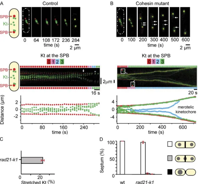
By filming a rad21-K1 ndc80-gfp cdc11-cfp strain to simultane
ously image both poles and Kts from metaphase to anaphase,
we confirmed that the stretched Kts in this strain were indeed
single sister chromatids. In the kymograph representation in
Fig. 1 B (middle), it is possible to visualize individual Kts
(a maximum of six in fission yeast) at different time points be
fore Kt stretching (Video 2). At the 90s time point, three Kts
are located at the top pole, whereas 40 s later, two others have
arrived at the bottom pole (Fig. 1 B, middle), leaving a single,
stretched merotelic Kt in the middle. We confirmed that stretched
Kts were single chromatids using a strain that allows simultane
ous observation of both the centromere of chromosome I (two
red dots after chromosome replication) and the Kts (six green
dots;
Fig. S1
). This finding was further confirmed by filming
cells in the presence of Hoechst and quantifying the fluores
cence intensity (Fig. S1, D and E).
22% of mitotic cells (n = 79) showed stretched merotelic
Kts (Fig. 1 C). By following the merotelic Kt with time, we
found that at least 75% (n = 17) of these relaxed into a single
dot, which segregated to one of the poles (see Fig. 5 B). In no
case did the presence of a merotelic attachment lead to the cut
phenotype, suggesting that most merotelic Kts are corrected
(Fig. 1 D). This was confirmed in fixed mitotic cells in which
only 3% of cells (n = 1,966) showed a “cutlike” phenotype (as
judged by the presence of DAPI signal under calcofluorstained
septum). The 25% of stretched merotelic Kts that were not cor
rected instead disrupted into two pieces that regained the two
poles (Fig. 1 B, 450 s). The mean maximum stretching distance
of a Kt before its disruption was 1.56 ± 0.28 µm (n = 7).
Collectively, these observations demonstrate that the
stretched lagging chromosomes, which are often observed dur
ing mitosis in rad21-K1 cells at the permissive temperature, are
merotelic. Most importantly, such merotelic attachments are
corrected during anaphase.
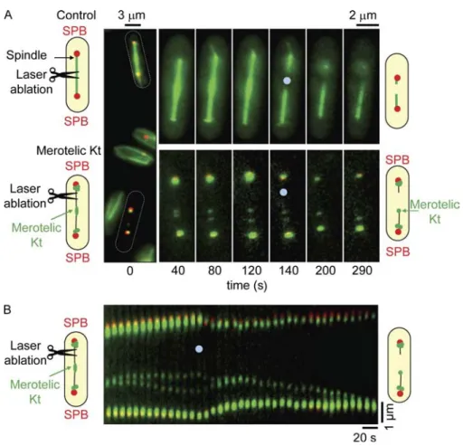
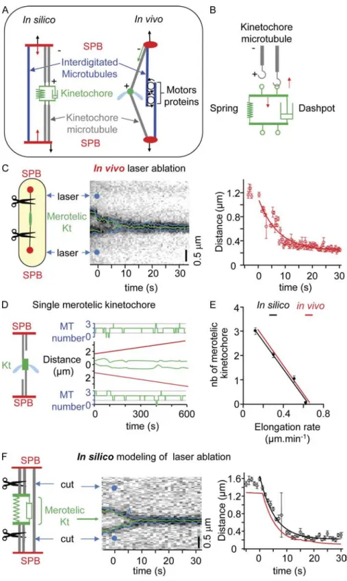
Kt stretching during merotelic attachment is imposed by MT-dependent forces
To investigate whether Kt stretching was dependent on forces im
posed by spindle MTs on the merotelic Kt, we performed laser
ablation experiments on spindles showing a single, stretched Kt
(Botvinick et al., 2004). We used a 2Dguided pulsed Nd:YAG
laser ( = 532 nm) to perform multiple spindle ablation simul
taneously (see Materials and methods) on cells expressing an
2GFP tubulin (Fig. S1 F). To control for lasercutting effi
ciency, we mixed an 2GFP tubulin strain (Fig. 2 A, control) and
a rad21-K1 ndc80-gfp cdc11-cfp strain (Fig. 2 A, merotelic Kt)
and simultaneously performed laser ablation in both populations
(
Video 3
). In control cells, laser ablation resulted in the collapse
of decorated spindles (Fig. 2 A, top), suggesting that the settings
used were sufficient to disrupt spindle MTs. When laser abla
tion was performed on spindles with merotelic chromosomes,
the stretched Kt immediately relaxed and moved toward the pole
opposite to the cut (Fig. 2 A, bottom). Kymographic analysis of
Kt dynamics during laser ablation is presented in Fig. 2 B. In all
cases (n = 37), the stretched Kt relaxed to a single dot which then
moved, in 75% of cases, to the pole opposite to the cut. The speed
at which Kts regained the pole (1.15 ± 0.36 µm/min; n = 11)
(Choi et al., 2009). It is currently accepted that in
Schizosaccha-romyces pombe
, spindle elongation is determined by the speed
of tubulin incorporation at the spindle midzone (Mallavarapu
et al., 1999) rather than by the pushing forces of astral MTs in
anaphase B (TolićNørrelykke et al., 2004). The spindle MT
midzone is organized by the MTbundling protein Ase1 (also
known as Prc1; Pellman et al., 1995; Loïodice et al., 2005;
Yamashita et al., 2005). It is presently unknown whether the
spindle elongation defect seen in monopolin mutants is caused
by the presence of lagging chromosomes as previously sug
gested in cohesion mutants (Pidoux et al., 2000).
Because of the complexity of the mechanisms controlling
spindle morphogenesis, mathematical and computational mod
els that integrate experimental results have been developed
over the past few years (Gardner and Odde, 2006; Mogilner
et al., 2006). These can be used to predict mitotic phenotypes
or to simply verify biological assumptions. In our study, we
genetically induced merotely in fission yeast and quantitatively
characterized its impact on mitotic progression from metaphase
to anaphase, cytokinesis, and cell abscission. Using live cell
video microscopy and laser surgery of merotelic Kts, we for
mally demonstrate that merotelic Kt stretching is specifically
mediated by a tensiondependent mechanism exerted by MTs.
Using a mathematical model of merotelic attachment, we find
that mitotic progression can be mechanically blocked by Kt
misattachment independently of the SAC. Based on the predic
tions of our mechanical model, we provide in vivo evidence
that the function of the interdigitated pole–pole spindle MTs,
which support spindle structure and provide tension across the
spindle, participate in the correction of merotelic attachment in
anaphase and prevent the appearance of the cut phenotype and
asymmetric division.
Results
Merotelic attachment can be corrected during anaphase B in fission yeast
Based on previous data suggesting that centromeric cohesion is
required to prevent merotelic attachment (Gregan et al., 2007),
we characterized the segregation defects seen in cells mutated
for the Rad21 cohesin subunit at the permissive temperature of
25°C. We analyzed Kt dynamics during metaphase in either
wildtype (wt; Fig. 1 A) or rad21-K1 cells (Fig. 1 B). The move
ments of the six Kts were recorded (Fig. 1, A and B, top; and
Videos 1
and
2
), and automated analysis was used to map the
position of each Kt relative to the spindle pole bodies (SPBs)
for each frame of the video (Fig. 1, A and B, bottom; see Mate
rials and methods). Using this methodology, it was possible to
determine the number of Kts present at each SPB for each frame
of the video. This number is reported along the top of the kymo
graph using a color code (Fig. 1, A and B, middle). In wt cells,
the six Kts make rapid oscillatory movements between the spin
dle poles during metaphase and congress into two bunches of
three on either side of the spindle midzone before the onset of
anaphase A as previously described (Fig. 1 A; Tournier et al.,
2004; Courtheoux et al., 2007). Migration of the Kts to the poles
is coordinated; i.e., all of the Kts arrive at the poles within 30 s.
on May 31, 2011
jcb.rupress.org
attached to a single pole) attachment. Nevertheless, Pidoux et al.
(2000) suggested that the negative effect of merotelic attachment
on spindle elongation was caused by the activation of a check
point rather than the result of mechanical restraint. Therefore,
we investigated whether the presence of additional merotelic
chromosomes affected the rate of anaphase B. Automated analy
sis of videos of rad21-K1 cells containing one (Fig. 1 B and
Fig. 3 A) to three merotelic attachments (
Fig. S2, A and B
) re
vealed a linear reduction of spindle elongation rate from 1.3 ±
0.09 µm/min (n = 12) in control cells to 0.123 ± 0.06 µm/min
(n = 5) in the presence of three merotelic attachments (Fig. 3 B).
To confirm that merotelic attachment directly antagonizes
anaphase B, we followed the changes in spindle elongation rate
in cells exhibiting Kt disruption. As shown in Fig. 3 (A and C),
was similar to that observed for the poleward movement of Kts
during anaphase (Fig. 2 B). These experiments demonstrate that
merotelic Kts are subjected to opposed forces generated by spin
dle MTs and that merotelic Kt correction during anaphase is de
pendent on the destabilization of MTs.
Merotelic attachment linearly reduces spindle elongation rate
It was previously reported that the presence of lagging chro
mosomes in fission yeast affects the rate of spindle elongation
during anaphase B (Pidoux et al., 2000). Because several cat
egories of lagging chromosomes were observed in this study,
it was unclear whether the effect on spindle elongation was
imposed by merotelic as opposed to monotelic (chromosome
Figure 1. A defect in centromeric cohesion affects Kt biorientation. (A and B, top) Image series showing an ndc80-gfp cdc11-cfp cell (A) or a rad21-K1
ndc80-gfp cdc11-cfp cell (B) during mitosis. The six Kts (green) are located between the two SPBs (red). Schematic representations of fission yeast cell
shape are shown (dashed ovals). (middle) Kymograph of the cell shown above. In control cells, anaphase A (arrows) takes place 45 s before anaphase B (asterisk). The number of Kts at each SPB is indicated for each time point on both sides of the kymograph. (bottom) Automated tracking analysis of SPB (red) and Kt (green) positions in the same cell (see Materials and methods). (C) Percentage of mitotic cells with stretched merotelic Kts in rad21-K1 ndc80-gfp
cdc11-cfp cells (rad21-K1). (D) Percentage of cut-like phenotype and asymmetric cells in wt (n = 2,466) and rad21-K1 cells (n = 1,966) stained with DAPI
calcofluor. White, correct segregation with a septum; gray, cut cells; black, asymmetrically dividing cells. Error bars indicate SD.
on May 31, 2011
jcb.rupress.org
positioned within the cell (Fig. 4 B, left; and
Video 4
). In the
presence of a merotelic Kt, however, a general plus end MT de
polymerization lead to spindle collapse at a rapid rate (Fig. 4 B,
right; and
Video 5
). These experiments demonstrate that mero
telic attachment can lead to spindle collapse when the structure
of the spindle is compromised.
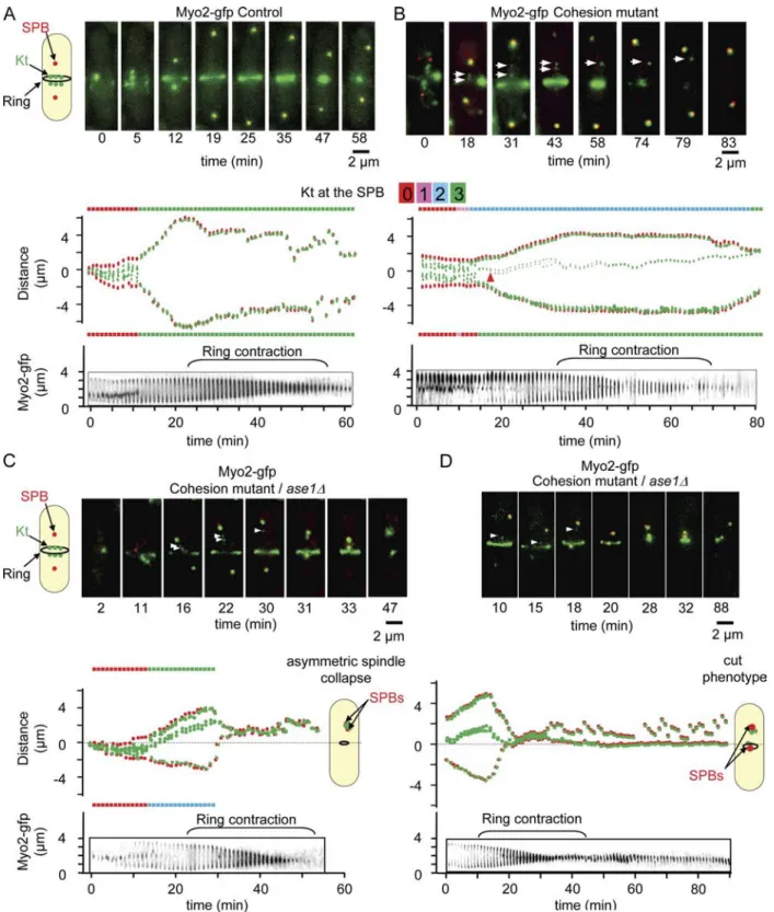
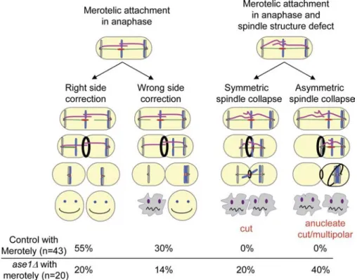
Ase1/Prc1/Map65 participates in the correction of merotelic attachment in anaphase and prevents the appearance of cut phenotype and asymmetric division
It has previously been reported that lagging chromosomes can
affect cell abscission (completion of cytokinesis; Norden et al.,
2006). However, experiments in fission yeast suggest that no
coordination exists between chromosome segregation and cyto
kinesis, as cut mutants complete cytokinesis in the absence of
chromosome segregation (Yanagida, 1998). If this is the case,
it is likely that correction of merotelic attachment in anaphase
will be essential to prevent the appearance of a cut phenotype
and subsequent cell death. To investigate this point further, we
performed live cell imaging and followed merotelic attachment
from early mitosis to cytokinesis by observing Kt dynamics
and cytokinetic actin ring contraction simultaneously within
the same cell using the strain ndc80-gfp (Kt) cdc11-cfp (SPBs)
myo2-gfp
(actomyosin ring; Fig. 5; Mulvihill et al., 2001). In
control cells, Kt segregation and spindle elongation were com
pleted before myosin II ring contraction (Fig. 5 A and
Video 6
).
The same experiment performed in a cohesion mutant back
ground revealed that the majority of stretched merotelic Kts
anaphase B increased more than twofold (0.95 ± 0.03 to
2.19 µm/min; n = 7) after the disruption of a single, stretched Kt.
These experiments show that merotelic attachments directly and
incrementally reduce spindle elongation.
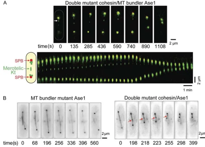
Merotelic attachment leads to spindle collapse in the absence of the bundling factor Ase1
Spindle elongation requires the activity of motor proteins at the
interdigitated MT midzone, the organization of which involves
the antiparallel bundling protein Ase1 (also known as Prc1;
Pellman et al., 1995; Loïodice et al., 2005; Yamashita et al.,
2005). In fission yeast, ase1∆ cells enter anaphase but fail to
complete anaphase B (Loïodice et al., 2005). To investigate the
role of overlapping MTs on mitotic progression in the presence
of merotelic attachment, we analyzed ase1∆ cells with a mero
telic attachment (Fig. 4 A). The double mutant ase1∆ rad21-K1
was inviable at the semipermissive temperature (31–33°C),
although both single mutants were alive at these temperatures
(
Fig. S4 B
). At the permissive temperature (25°C), however,
cells entered mitosis in the presence of a stretched merotelic Kt.
However, during midanaphase B, the spindle collapsed, and
the distance between the two SPBs rapidly diminished (from 7
to 2 µm; rate = 1.05 ± 0.1 µm/min; n = 6; Fig. 4 A). Spindle
collapse at this rate was never observed in the two single mu
tants. We confirmed this result by visualizing simultaneously
both MTs (tubulin GFP) and the merotelic Kt (Ndc80GFP).
Initiation of anaphase B in ase1∆ was followed by the dis
assembly of the spindle and the two SPBs becoming symmetrically
Figure 2. Kt stretching is imposed by MT-dependent forces. (A) A mixed population of
atb2-gfp cdc11-cfp (control) and rad21-K1 ndc80gfp cdc11-cfp cells (merotelic Kt) was
treated simultaneously by laser ablation. Image series showing the two cells before (time 0) and at the moment of laser ablation (see Ma-terials and methods). Schematic representa-tions of fission yeast cell shape are shown (dashed ovals). (B) Kymograph of the rad21-K1 cell shown in A illustrating the relaxation of the merotelic Kt after laser ablation. Blue dots indicate the point of laser impact.
on May 31, 2011
jcb.rupress.org
Fig. 5, C and D; and
Videos 8
and
9
), only 34% (n = 20) of
merotelic attachments were corrected and 6% of stretched
merotelic Kts (n = 20) disrupted into two pieces. Instead, we found
that merotelic attachment in the absence of Ase1 lead to either
(85%; n = 43) were corrected during anaphase B before com
pletion of actomyosin ring contraction (Fig. 5 B and
Video 7
),
whereas the remaining 15% were disrupted into two pieces.
Oppositely, when tension across the spindle was defective (ase1∆;
Figure 3. Merotelic attachment influences the rate of spindle elongation. (A) Kymograph of a rad21-K1 ndc80-gfp cdc11-cfp cell showing a single, stretched Kt. (B) Spindle elongation rate of rad21-K1 cdc11-cfp ndc80-gfp cells in the absence of merotelic attachment (zero) or in presence of one, two, or three merotelic Kts. (C) Graphic representation of spindle elonga-tion in the rad21-K1 ndc80-gfp cdc11-gfp cell shown in A (red dots). The rate of spindle elongation was calculated before (red) or after (blue) disruption of the merotelic Kt. Error bars indicate SD.
Figure 4. Merotelic attachment leads to abortive anaphase when interdigitated MTs are defective. (A, top) Image series of a rad21-K1 ase1∆ ndc80-gfp
cdc11-cfp cell during mitosis showing two lagging Kts (one of them stretched; white arrow). (bottom) Kymograph showing a rapid collapse of the spindle.
A schematic representation of a fission yeast cell shape is shown (dashed oval). (B, left) Image series of ase1∆ atb2-gfp (MTs) ndc80-gfp (Kts) during mitosis. (right) Image series of rad21-K1 ase1∆ SV40-gfp-atb2 (MTs) ndc80-gfp (Kts) cell during mitosis in the presence of a merotelic attachment (red arrows).
on May 31, 2011
jcb.rupress.org
Figure 5. Merotelic attachment leads to the cut phenotype and asymmetric division when interdigitated MTs are defective. (A–D, top) Image series showing an ndc80-gfp cdc11-cfp myo2-gfp cell (A), a rad21-K1 ndc80-gfp cdc11-cfp myo2-gfp cell (B), or an ase1∆ rad21-K1 ndc80-gfp cdc11-cfp myo2-gfp cell (C and D) during mitosis from metaphase to anaphase up to cytokinesis. The six Kts (green) are located between the two SPBs (red) with the actomyosin ring located at the midzone. (middle) Automated tracking analysis of the cell shown above. The number of Kts at each SPB is indicated for each time point on both sides of the kymograph. Kt dynamics (middle) and cytokinesis (bottom, ring contraction) were filmed simultaneously. Arrowheads indicate the positions of the stretched merotelic Kts. (B) After correction (43 min), the Kt moves to the SPB, and cytokinesis takes place. (C) Example of an asymmetric spindle collapse in an ase1∆ mutant in the presence of a merotelic attachment. After completion of cytokinesis, the formation of an anucleate cell and a diploid cell is observed. (D) Example of a symmetric spindle collapse seen in an ase1 mutant in the presence of merotelic attachment. After the execution of cytokinesis, the formation of a cut phenotype is observed.
on May 31, 2011
jcb.rupress.org
could be used to predict complex phenotypes associated with
merotelic attachment.
In conclusion, our results showed that in presence of
merotelic attachment, the spindle behaves as a nanomachine,
whose behavior can be numerically described using a simple
force balance model. We further demonstrate that merotelic at
tachment can mechanically block spindle elongation and cause
spindle regression.
In silico evidence that merotelic
attachment leads to spindle collapse in the absence of interdigitated MTs
To validate our in vivo observations, we abrogated the push
ing force present in between the two SPBs in the mechanical
model at the 80s time point (Fig. 7 A, right). In this situation,
merotelic Kts were still able to stretch but failed to disrupt as
a result of the lack of tension exerted on the Kt (unpublished
data). Instead, we observed the immediate collapse of the spin
dle at a rapid negative rate of 1.47 ± 0.35 µm/min (n = 10;
Fig. 7 B). Thus, our mechanical model predicts that the mitotic
spindle will rapidly collapse in the presence of a merotelic Kt
when interdigitated MTs are absent or weak and that proper
tension across the spindle underlies Kt correction and disrup
tion during anaphase B. An alternative mathematical model to
reproduce the ase1 mutant phenotype would be to reduce the
pushing force as the spindle elongates until a critical length of
7 µm in which the force is reduced to zero. A similar result was
obtained in this type of simulation (Fig. S5, C and D). These ex
periments validate the prediction of our merotelic model, dem
onstrate that merotelic attachment can lead to spindle collapse
when the structure of the spindle is compromised, and confirm
that correction of merotelic Kts during anaphase B is triggered
by a tensiondependent mechanism.
Discussion
The maintenance of genetic stability is essential to prevent
the development of human cancers. Genetic instability can be
caused by several mechanisms such as a defective DNA dam
age checkpoint or a defect in chromosome segregation, which is
also called aneuploidy. Although aneuploidy is a welldescribed
phenotype of solid tumors, the molecular mechanisms involved
in this process are controversial. Some evidence suggests that
aneuploidy can be induced by a failure in mitotic control (Cahill
et al., 1998). Indeed, it is well established that defects in MT–Kt
attachment activate the SAC (Cleveland et al., 2003). However,
recent studies suggest that aneuploidy can be induced even in the
presence of a robust checkpoint (Tighe et al., 2001; Thompson
and Compton, 2008). Perhaps the SAC does not detect all types
of Kt misattachment. Indeed, several studies suggest that the
SAC is not activated in the presence of merotelic attachment
and that, consequently, this misattachment does not affect mi
totic progression (Cimini et al., 2001, 2004).
Cohesion between sister chromosomes is mediated by a
protein complex degraded in mitosis by a protease called sepa
rase (Tatebayashi et al., 1998; Tomonaga et al., 2000; Nasmyth
and Schleiffer, 2004). Recent work in S. pombe revealed that
asymmetric (40%; n = 20; Fig. 5 C and Video 8) or symmetric
spindle collapse (20%; n = 20; Fig. 6 D and Video 9). In such
situations, ring contraction and cell abscission took place nor
mally, leading to the appearance of either cut cells (Samejima
et al., 1993) or asymmetric division followed by cell death (Fig. 5,
C and D;
Fig. S3
; and see Fig. 8). These findings were confirmed
in fixed cells (Fig. S4 A). In agreement with these results, ase1∆
was synthetically lethal with rad21-K1 at the semipermissive
temperature (31–33°C), although the single mutants were alive
at these temperatures, confirming that Ase1/Prc1 participates
in the correction of merotelic attachment in anaphase cells
(Fig. S4 B). Collectively, these experiments demonstrate that
correction of merotelic Kt during anaphase B is triggered by an
Ase1/Prc1dependent tension mechanism.
A minimal mechanical model
predicts that merotelic attachments mechanically block spindle elongation and cause mitotic regression
To validate our in vivo observations, we established a simple
mathematical model based on a framework previously described
(BrustMascher et al., 2004; Gardner et al., 2005; Civelekoglu
Scholey et al., 2006). In our model, the mitotic spindle is defined
by four objects, the two faces of the merotelic Kt and the two
SPBs (Fig. 6 A). Because fission yeast possesses up to four Kt–MT
attachment sites per chromosome (Ding et al., 1993), our model
assumes a maximum of four MTs attached per Kt distributed on
each side of the spindle. The set of 10 parameters required to gen
erate this model was obtained by direct measurements of in vivo
characteristics such as Kt speed at anaphase onset, spindle elonga
tion rate, and merotelic Kt mean stretching length. To determine
the stiffness and friction coefficient of the merotelic Kt, we per
formed a doublelaser ablation experiment in which both sides
of the merotelic Kt were cut (Fig. 6 C, left). From the observed
relaxation of the system, we were able to determine precisely
the stiffness and friction coefficient of the merotelic Kt (Fig. 6 C,
right). This set of values was then maintained throughout the
study (Table I).
Fig. 6 D shows a simulation of a single merotelic attach
ment that is undergoing stretching. The number of MTs attached
to the merotelic Kt is indicated on each side of the graph. This
model was sufficient to reproduce the in vivo dynamics of the
merotelic Kt and could be applied to both two (
Fig. S5 A
) and
three (Fig. S5 B) merotelic attachments. The final setting of the
model reproduced the relationship between spindle elongation
rate and the number of merotelic Kts present within the spindle
(Fig. 6 E). We were able to validate our model by determining
the effect of doublelaser ablation in silico on both sides of the
merotelic Kt (Fig. 6 F, left). Kymograph analysis of this experi
ment, coupled with convolution of the signal, is presented in
Fig. 6 F (middle and right), revealing that the friction coefficient
of the merotelic Kt determined in silico is similar to that ob
served in vivo. We tested the effect of multiple merotelic attach
ments on mitotic progression. Simulations predicted that more
than three merotelic attachments would result in a negative
spindle elongation rate (i.e., spindle collapse) at a speed of 0.2 ±
0.11 µm/min (n = 18). These findings suggest that our model
on May 31, 2011
jcb.rupress.org
as previously reported in cohesindepleted Xenopus laevis ex
tracts (Kenney and Heald, 2006). Several other studies suggest
that a lack of cohesion induces segregation defects compatible
with merotelic attachment (Kenney and Heald, 2006; Gregan
et al., 2007), suggesting that cohesin may play a structural role in
Kt–MT attachment. In our study, we formally demonstrate that
the cohesin Rad21 is required to prevent the formation of mero
telic attachment. We also show that merotelic attachment leads
to intraKt stretching, which is abolished by laser Mt ablation.
cohesins are key elements, which determine the type of MT–Kt
attachment (Toyoda et al., 2002; Yokobayashi et al., 2003;
Sakuno et al., 2009). Indeed, alteration of mitotic cohesin with
a thermosensitive mutant (rad21-K1) results in severe chromo
some segregation defects and leads to activation of the SAC
at the nonpermissive temperature (Tatebayashi et al., 1998;
Toyoda et al., 2002). However, at the permissive temperature
of 25°C, the rad21-K1 mutant is able to form a bipolar spindle
and initiates mitosis in the absence of chromosome alignment,
Figure 6. A minimal model for the description of merotelic attachment. (A, left) Schematic representation of the fission yeast spindle. MTs and associated proteins are symbolized by force generators fixed to the two SPBs; Kt force generators (red arrows) apply forces between the Kt and the poles, whereas spindle force generators (black arrows) apply forces be-tween both SPBs. (B) In the mechanical model, the Kt is composed of a spring and a dash-pot, and each Kt provides a site for MT (gray) attachment (green loop). Red arrows indicate the forces applied by the attached MT (upward arrow) and the Kt elastic structure (downward arrow). (C) Characterization of the merotelic Kt spring constant using laser ablation. (left) Laser ablation on both sides of the merotelic Kt is per-formed. (middle) Kymograph of the merotelic Kt after laser ablation (blue dots). (right) Graphic representation showing Kt relaxation after laser ablation (time 0 = time of laser impact). (D) Simulation of mitotic progression in the presence of a single merotelic Kt (green), with SPBs shown in red. The number of MTs attached to the Kt is indicated for each SPB (green). (E) Comparison of spindle elongation rates between simulated (black) and in vivo data (red) as a function of the number (nb) of merotelic Kts. (F, left) Kt and spindle force gen-erators are shut off (virtual laser ablation) dur-ing a merotelic simulation. (middle) Synthetic kymograph of a merotelic Kt after the elimina-tion of the two force generators (blue dots). (right) Comparison of Kt relaxation between simulated (black) and in vivo data (red). Error bars indicate SD.
on May 31, 2011
jcb.rupress.org
aurora independent. An alternative mechanism is that unbal
anced forces across the merotelic Kt participate in the correction
mechanism in anaphase. Further experiments will be necessary
to clarify the participation of aurora in anaphase B.
Although it was previously suggested that merotelic at
tachment does not affect mitotic progression, we demonstrate in
this study that spindle elongation rate is linearly dependent on
the number of merotelic Kts. This effect is independent of the
SAC, as deletion of either Mad2 or Bub1 does not prevent the
appearance of this phenotype (unpublished data). In agreement
with this finding, we found that Mad2 was never recruited onto
merotelic Kts in anaphase (unpublished data). In many respects,
our in vivo results are similar to what is seen in vitro in Xenopus
egg extracts where bipolar spindles assemble in the absence of
either chromosomes or centrosomes, suggesting that spindle
morphogenesis is solely dependent on the presence of motor
proteins (Heald et al., 1996).
Multiple proteins are involved in the structure of the Kt,
and several of them are known to influence MT dynamics. An
advanced mathematical model reproducing the biochemical
properties of each Kt component is the ultimate goal, although
Most importantly, we also observed stretching and disruption of
merotelic Kt in cohesinplus background strains, although these
events were rare (unpublished data). Recent work has high
lighted the importance of intra as opposed to interKt stretch
ing in satisfying the SAC (Maresca and Salmon, 2009; Uchida
et al., 2009). It is likely that motor proteins are required in fis
sion yeast to stretch merotelic Kts, and our model provides a
unique system to identify which mitotic players are required for
the establishment of tension across the merotelic Kt.
The aurora B kinase plays a critical role in correcting Kt
misattachment by phosphorylating key substrates at the Kt and
promoting the turnover of Kt MTs (Tanaka et al., 2002; Cimini
et al., 2006). It has been proposed that the physical distance be
tween aurora B and its Kt substrates determines whether MT–Kt
connections are maintained (Tanaka et al., 2002; Andrews et al.,
2004). We found that the unique S. pombe aurora kinase, Ark1,
was present on the outer faces of stretched merotelic Kts during
anaphase, although the signal seen in anaphase was consider
ably weaker than during metaphase (unpublished data). Based
on these observations, the simplest interpretation of our find
ings is that the anaphase correction of merotelic attachment is
Table I. Parameters used in the merotelic modelNotation Definition Adimensionalized value Value
General
N Number of merotelic Kts 1–4 NA
Mk Maximum number of Kt–MTs per side 4 NA
Geometry
L0 Spindle initial length 2–6 2–6 µm
do Equilibrium distance between attachment sites 0.01 0.01 µm
Mechanical
Centromere spring constant 2 20 pN/µm
µs SPB friction coefficient 1.4 420 pN.s/µm
µk Centromere friction coefficient 1 300 pN.s/µm
µc Sister chromatid friction coefficient 0.2 60 pN.s/µm
Motors
Fk Kt force generators stall force 1 10 pN
Vk Kt force generators maximum speed 1 3102 µm/s
Fmz Midzone force generators stall force 7 70 pN
Vmz Midzone force generators maximum speed 0.4 1.2102 µm/s
Dynamic instability
fa Attachment frequency 0.04 0.04 1/s
fd Detachment frequency 0.1 0.1 1/s
NA, not applicable. Value is determined with Fk at 10 pN and Vk at 0.03 µm/s.
Figure 7. Model predicts spindle collapse in the absence of interdigitated MTs. (A) Sche-matic representation of wt and ase1∆ models in which spindle force generation is turned off. (B) Simulation of mitotic progression and Kt dynamics in the ase1 model. Red, SPBs; green, merotelic Kt. The spindle force generator is turned off at the 80-s time point.
on May 31, 2011
jcb.rupress.org
collapse is a consequence of a different number of MTs link
ing the merotelic Kt to the two SPBs (see previous paragraph),
but we cannot exclude the possibility that astral MTs may also
play a role in anchoring one of the two poles to the cell cortex
(Gachet et al., 2004; TolićNørrelykke et al., 2004). In budding
yeast, the kinesin Kar3 provides, like Ase1, the structural stabil
ity of the spindle in the presence of a dicentric chromosome
(Gardner et al., 2008). We found no such role for Klp2 (Kar3
homologue; unpublished data), although this kinesin has been
implicated in Kt capture by MTs in both yeasts (Tanaka et al.,
2007; Gachet et al., 2008). The function of Kar3 in budding
yeast during dicentric attachment could be explained by a role
for this kinesin in chromosome cohesion (Mayer et al., 2004)
rather than at the interdigitated MT midzone. Alternatively,
because in the case of a dicentric chromosome the forces are
applied on the chromosome arms in between the two dicentric
chromosomes rather than on the Kt structure itself, it is pos
sible that the motor proteins involved in dicentric chromosome
stretching or merotelic Kt stretching are different.
In conclusion, our study provides a unique biological model
illustrating the impact of merotelic attachment on mitotic pro
gression, starting from metaphase to anaphase up to the execu
tion of cytokinesis. Our findings suggest that a disbalance of
forces exerted by MTs on the merotelic Kt create a brute force
mechanism that is essential for the correction of merotelic attach
ments in anaphase. When the structure of the spindle is affected,
merotelic attachments are not corrected, and cells undergo asym
metric division or cut phenotype, leading to cell death (Fig. 8).
This mechanism is likely to be conserved in higher eukaryotes,
as our mathematical model is simply based on force balance gen
erated by molecular motors characterized by linear force–
velocity relationships. Additionally, Ase1 (Prc1) is evolutionarily
conserved, and correction of merotelic attachment is known to be
such a model is far from being established because the biochem
ical constants involved in MT dynamics within the Kt sleeve are
unknown. We chose a macroscopic approach to study the bio
logical mechanics at the Kt. In our model, the Kt is seen as an
object that can move MTs either forward or backward. All of the
undefined biochemical constants are described by a single force
applied to the Kt. The model faithfully reproduces the in vivo
observations, including Kt stretching, relaxation, and correction.
It confirms that in the presence of merotelic attachment, the
spindle is constrained by simple mechanical links. This simple
model could also explain the phenotype of spindle collapse re
cently reported in several fission yeast mutants (Alexandru et al.,
2001; Griffiths et al., 2008). The model also predicts that the bias
seen toward correction, and not random segregation, is likely to
be the consequence of a different number of MTs linking the
merotelic Kt to the two SPBs (e.g., three MTs to one SPB and
one to the other). The molecular mechanisms that explain this
bias are unknown. A more general mitotic model that takes into
account the state of attachment of all the Kts (merotelic or syn
thelic) from metaphase to anaphase needs to be established.
Comparison of such a model and observed Kt dynamics in wt
and mutant strains will highlight the mechanisms of MT dynam
ics at the Kt that promote successful biorientation.
In fission yeast, the spindle is positioned in the middle of
the cell and is encircled by an actin ring that is detectable early
in metaphase (Arai and Mabuchi, 2002; Wu et al., 2003). Dur
ing anaphase B, spindle structure is stabilized by the presence of
overlapping antiparallel MTs (Ding et al., 1993), the destruction
of which results in the collapse of the mitotic apparatus when a
merotelic Kt is present. This can either happen symmetrically
or asymmetrically in respect to the actomyosin ring, leading to a
cut phenotype or to the formation of aneuploid cells after com
pletion of cytokinesis. In our mathematical model, asymmetric
Figure 8. Model summarizing the different effects of merotelic attachment on mitotic pro-gression. Schematic representation illustrating the impact of merotelic attachment in control cells (left) and in cells with a defective spindle structure (right). In the control cells, the correc-tion of merotelic attachment is coordinated with the onset of cytokinesis to avoid the forma-tion of cut/anucleate. In cells with a defective spindle structure (ase1 mutant), this coordina-tion is lost, leading to the appearance of a cut/anucleate phenotype and cell death. The different categories of cells were scored by live cell imaging using either a rad21-K1
ndc80-gfp cdc11-cfp myo2-ndc80-gfp strain or an ase1∆ rad21-K1 ndc80-gfp cdc11-cfp myo2-gfp
during mitosis from metaphase to cytokinesis and cell abscission. For clarity, the model does not include the percentage of cells showing merotelic Kt disruption (15% for control and 6% for ase1∆).
on May 31, 2011
jcb.rupress.org
Mathematical model of merotelic attachment
The mathematical model used in this study is essentially an adaptation and a simplification of the framework developed by Civelekoglu-Scholey et al. (2006). Indeed, merotelic attachment at anaphase only differs from proper attachment in metaphase in that the pair of sister chromatids linked by chromatin is replaced by two sides of the Kt linked by the Kt structure.
In our model, the mitotic spindle is described as a one-dimensional organization of 2N + 2 elements, where N is the number of merotelic Kts: 2N Kt sides and two poles. All of the positions are given with respect to an axis with its origin at the spindle center. In the following, the positions of
the top and bottom poles are denoted as xs and xs*, respectively. By
defi-nition of the coordinate system, xs*=− ∀ .xs t Speeds are denoted as xs
and xs*. Similarly, the positions of the nth Kt sides are denoted as xn and
xn*, and their speeds are denoted as xn and xn*, respectively, for the top
and bottom sides of chromosome n with n∈[1, ].N At each time, the
num-ber of kt–MTs plugged to the top and bottom Kt sides of chromosome n is
denoted as Mn and Mn*.
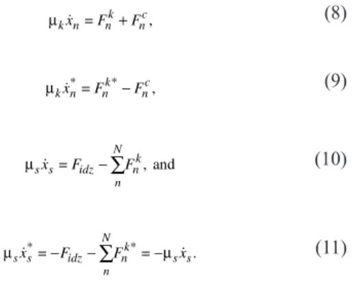
Force balance model
Three interactions are considered: (1) the structural link between xs the
two sides of the Kt, represented by a damped oscillator (Fig. 7 B), (2) an attractive force linking the spindle pole and its respective attachment site, and (3) the spindle elongation force, pushing poles outward.
Although the actual centromere structure is composed of multiple layers and different materials, we assume for the sake of simplicity that it
can be described as the combination of a spring of stiffness κ and rest
length d0 and a dashpot of friction coefficient µk. Elastic to plastic transition
(Marko, 2008), as observed during stretching, is accounted for by a
criti-cal length dc over which the material breaks, with κ and µk being set to 0.
The first force, exerted by the centromeric material on the Kt top attachment site is thus expressed by
Fnc=−κ
(
xn−xn*−d0)
−µ k(
xn−x .*n)
(1)
Civelekoglu-Scholey et al. (2006) and Cheeseman and Desai (2008) assume that Kt movement is the result of a combination of molecular motors and polymerization/depolymerization of the Kt MTs. Polymerizing (Kolomeisky and Fisher, 2001) and depolymerizing MTs coupled to the Kts (McIntosh et al., 2008) and molecular motors (Toba et al., 2006) can be faithfully described by a linear force–velocity relationship. Considering the net force linking the Kt attachment site and the pole as a linear combina-tion of those force generators, it can also be described as a force–velocity relationship. We hypothesize that this force is always directed toward the spindle pole:
F M t F x x V nk n k n s k = ( ) 1− − ,
(2)
F M t F x x V nk nk k s n k *=− *( ) 1− *− * ,
(3)
=M t F*( ) 1 x x* V n k s n k + + .
(4)
Fk and Vk are the stall force and the maximum speed, respectively,
for the Kt–spindle pole interaction. Motor proteins and cross-linkers attach to the overlap between interpolar MTs. Those elements are considered to exert an overall outward force on the SPBs, which again is described by a force–velocity relationship:
F F x x V idz mz s s mz = 1 * − − ,
(5)
=F 1 2x V mz s mz − ,
(6)
Fidz*=−Fidz.
(7)
a key process in maintaining genomic stability in eukaryotic
cells. Knowing that asymmetric cell division has in the past only
been attributed to defects in spindle positioning, our work opens
a new field of investigation in this domain.
Materials and methods
Cell culture
Media, growth, maintenance of strains, and genetic methods were per-formed as described previously by Moreno et al. (1991). rad21-K1 mutant was provided by M. Yanagida (Kyoto University, Kyoto, Japan), and ase1 mutant was provided by P. Tran (Institut Curie, Paris, France). Cells were grown at 25°C (Fig. S4) in yeast extract and centrifuged for 30 s at 3,000 g before mounting onto an imaging chamber. The different strains used in
this study are listed in Table S1.
Cell fixation
To determine the percentage of aneuploidy and cut phenotype, cells were fixed in 3.7% formaldehyde for 7 min at room temperature, washed once in PBS, and observed in the presence of DAPI/calcofluor.
Live cell imaging
Live cell analysis was performed in an imaging chamber (CoverWell PCI-2.5; Grace Bio-Laboratories) filled with 1 ml 1% agarose in mini-mal medium and sealed with a 22 × 22–mm glass coverslip. Time-lapse images of z stacks (maximum of five stacks of 0.3–0.4-µm steps to avoid photobleaching) were taken at 15-s intervals at 25°C. Exposure times were 300–500 ms using a light source (HIGHlite; Roper Industries) re-duced to 30% to avoid phototoxicity and photobleaching. Either the image with the best focal plane or projected images was prepared for each time point. Images were visualized with a charge-coupled device camera (CoolSNAP HQ; Roper Industries) fitted to an upright microscope (DM6000; Leica) with a 100× or 63× 1.4 NA objective and SEMROCK filters for GFP, CFP, or RFP and were recorded using the MetaMorph software package (MDS Analytical Technologies). Intensity adjust-ments (threshold adjustadjust-ments) were made using the MetaMorph, ImageJ (National Institutes of Health), and Photoshop (Adobe) packages. Meta-Morph was used to create line scans.
Analysis of Kt dynamics
The position of the SPBs and Kts was determined by the visualization of the Ndc80–GFP and Cdc11–CFP signals and was captured using Meta-Morph. Maximum intensity projections were prepared for each time point, with the images from each channel being combined into a single RGB image. These images were cropped around the cell of interest, and optional contrast enhancement was performed in MetaMorph where necessary. The cropped images were exported to IGOR (version Pro6; http://www .wavemetrics.com) as 8-bit RGB-stacked TIFF files, with each frame corre-sponding to one image of the time-lapse series. For both channels, custom peak detection was performed. The successive positions of the SPBs and Kts were determined, and eventual detection errors were manually cor-rected. The data generated were used to calculate the mean speed at which the Kts reached the SPBs, the rate of spindle elongation, the rate of spindle retraction, and the number of Kts at the SPBs. A stretched merotelic Kt is defined as a single Kt within an anaphase spindle (i.e., all of the other Kts have permanently regained the poles), which undergoes splitting and separates by at least 0.5 µm.
Laser ablation of mitotic cells
The system used to perform laser ablation is composed of a conventional inverted microscope (DMI6000B; Leica) equipped with a heated stage and covered with an incubation system including a temperature controller. Mitotic spindle photo ablation was achieved with a frequency-doubled Nd:YAG-pulsed laser at a wavelength of 532 nm; the pulse was estimated to have a duration of 600 ps with a repetition rate of 10 kHz (532-nm Sealed Green Microchip; JDS Uniphase). The guiding of the beam was performed using an L5D head (Roper Industries) coupled to the microscope through the epifluorescence port. In brief, the L5D head constituted of a laser shutter and a galvanometer pair mirror that guides the laser beam within the field of view of the camera. The beam is focused with a 100× NA 1.4 Plan Apo oil immersion objective lens (HCS; Leica). Images were acquired with a cooled charge-coupled device camera (CoolSNAP HQ2). The system is driven by MetaMorph software.
on May 31, 2011
jcb.rupress.org
respectively. The position-dependent forces are calculated, and the algebraic system of equations is solved using the linear algebra PACKage. The position of each element is updated according to the resulting speeds. It is possible to simulate several events: (1) chroma-tid elastic to plastic transition; if the distance between the two attach-ment sites exceeds a critical size (1.6 µm according to experiattach-mental measurements), the spring constant is set to 0, allowing the two sites to move freely toward the poles. (2) A laser can be simulated; in this case, the link between a Kt side and its respective pole is broken.
(3) In the system, this corresponds to setting Fk and Fidz to 0. (4) ase1∆
strains are simulated by setting Fidz to 0 at a given spindle critical length
(L = 7 µm; Tanaka et al., 2007; this study). Online supplemental material
Fig. S1 shows that stretched merotelic Kts are single chromatids and show simultaneous laser ablation of cells within the same field. Fig. S2 shows kymographs of multiple merotelic attachments. Figs. S3 and S4 show that merotelic attachment leads to cut/asymmetric division/multipolar spindles and cell death in the absence of Ase1. Fig. S5 shows simulations of mul-tiple merotelic attachments and of a reduction in the spindle force generator during spindle elongation. Videos 1 and 2 show time-lapse imaging of wt or rad21-K1 with both Kt and SPB signals imaged simultaneously. Video 3 shows time-lapse imaging of laser ablation of a merotelic attachment. Videos 4 and 5 show time-lapse imaging of ase1∆ or ase1∆ rad21-K1 cells expressing SV40-GFP-atb2 as a marker of -tubulin (spindle) and Ndc80-GFP as a marker of the Kts. Videos 6–9 show time-lapse imaging of wt,
rad21-K1, or ase1∆ rad21-K1 (Videos 8 and 9) cells with the actomyosin
ring, with the Kts and the SPBs imaged simultaneously. Table S1 lists the
S. pombe strains used in this study. Online supplemental material is
avail-able at http://www.jcb.org/cgi/content/full/jcb.200902093/DC1. We thank T. Toda, P. Tran, K. Hardwick, J. Millar, R. McIntosh, M. Yanagida, and J.P. Javerzat for supplying strains, J. Hyams for his support and critical read-ing of the manuscript, T. Toda, Y. Watanabe, J.P. Javerzat, A. Merdes, and F. Payre for helpful discussions, Corinne Lorenzo for assistance with micros-copy, and B. Ducommun and the members of the Laboratoire de Biologie Cellulaire et Moléculaire du Controle de la Prolifération for their support.
T. Courtheoux is supported by a fellowship from La Ligue contre le cancer. G. Gay is supported by the Centre National de la Recherche Scien-tifique. The microscopy equipment was funded by the Centre National de la Recherche Scientifique, l’Université de Toulouse, l’Association de la Recherche sur le Cancer, and GlaxoSmithKline.
Submitted: 18 February 2009 Accepted: 7 October 2009
References
Alexandru, G., F. Uhlmann, K. Mechtler, M.A. Poupart, and K. Nasmyth. 2001. Phosphorylation of the cohesin subunit Scc1 by Polo/Cdc5 kinase regulates sister chromatid separation in yeast. Cell. 105:459–472. doi:10.1016/S00928674(01)003622
Andrews, P.D., Y. Ovechkina, N. Morrice, M. Wagenbach, K. Duncan, L. Wordeman, and J.R. Swedlow. 2004. Aurora B regulates MCAK at the mitotic centro mere. Dev. Cell. 6:253–268. doi:10.1016/S15345807(04)000255 Arai, R., and I. Mabuchi. 2002. Factin ring formation and the role of Factin
cables in the fission yeast Schizosaccharomyces pombe. J. Cell Sci. 115:887–898.
Botvinick, E.L., V. Venugopalan, J.V. Shah, L.H. Liaw, and M.W. Berns. 2004. Controlled ablation of microtubules using a picosecond laser. Biophys. J. 87:4203–4212. doi:10.1529/biophysj.104.049528
BrustMascher, I., G. CivelekogluScholey, M. Kwon, A. Mogilner, and J.M. Scholey. 2004. Model for anaphase B: role of three mitotic motors in a switch from poleward flux to spindle elongation. Proc. Natl. Acad. Sci.
USA. 101:15938–15943. doi:10.1073/pnas.0407044101
Cahill, D.P., C. Lengauer, J. Yu, G.J. Riggins, J.K. Willson, S.D. Markowitz, K.W. Kinzler, and B. Vogelstein. 1998. Mutations of mitotic checkpoint genes in human cancers. Nature. 392:300–303. doi:10.1038/32688 Cheeseman, I.M., and A. Desai. 2008. Molecular architecture of the kinetochore
microtubule interface. Nat. Rev. Mol. Cell Biol. 9:33–46. doi:10.1038/ nrm2310
Choi, S.H., M.P. PeliGulli, I. McLeod, A. Sarkeshik, J.R. Yates III, V. Simanis, and D. McCollum. 2009. Phosphorylation state defines discrete roles for monopolin in chromosome attachment and spindle elongation. Curr. Biol. 19:985–995.
The stall force Fmz and maximum speed Vmz are considered constant
throughout the simulation, assuming that the structure and function of the midzone does not change during the cycle.
In the highly viscous nucleoplasm, inertia can be neglected with re-spect to drag force. Eqs. 1–7 can thus be combined in the following set of coupled first order differential equations:
µk n nk nc x =F +F ,
(8)
µk n nk nc x*=F *−F ,
(9)
µs s idz n N nk x =F −
∑
F , and(10)
µs s idz µ n N nk s s x*=−F −
∑
F *=− x.(11)
Kt–MT attachmentDuring mitosis, dynamic instability of MTs is highly regulated, and, as already described in Saccharomyces cerevisiae (Gardner et al., 2005), polymerization and depolymerization rates may vary within the spindle. A rigorous molecular or mesoscopic description of MT dynamics is thus a problem on its own, and published models often require numerous param-eters. MT–Kt interaction strength might depend on molecular motor-binding rates, MT penetration depth in the Kt structure (Joglekar and Hunt, 2002), and other factors such as length-dependent regulations. In our model, we
account for MT behavior by the factor Mn in the Kt–spindle pole interaction.
We only account for MT dynamic instability as attachment and detachment stochastic events. That is, at each time step, attached kt–MTs have a
prob-ability Pd to detach, and unattached kt–MTs have a probability Pa to attach.
These probabilities are characterized by attachment and detachment
fre-quencies fa and fd, respectively, and the time interval dt between two steps,
according to a Poisson distribution:
Pd= 1−exp
(
−f dtd)
Pa= 1−exp(
−f dta)
.(12)
In this study, we assume that those frequencies are constants. Parameter setting
The system of Eqs. 8–12 is first expressed in dimensionless quantities, with
Vk as the unit of speed and Fk as the unit of force, and 1 µm and 1 s as
the units of length and time, respectively. The remaining parameters were chosen to fit with the experimental measurements as depicted in Fig. 6. Those parameters are kept constant for all the simulations unless specified otherwise. In the case of Kt disruption, the Kt–MT force generators will work near their maximum velocity, with friction being small compared with
those forces. The maximum velocity Vk will thus be slightly higher than the
maximum observed poleward speed of the Kt fragment. To comply with
direct experimental measurement of this speed, Vk is thus set to 0.03 µm/s.
We cannot directly quantify the force exerted by the MTs at the Kt. Yet, the literature suggests that this must be on the order of 10 pN for a single
MT. Thus, we chose Fmaxk = 10 pN to estimate the magnitudes of the other
parameters (Table I). The values of the friction coefficients of the Kt and SPB are higher than reported elsewhere (Nicklas, 1988) by about a factor of 10. This discrepancy comes essentially from the relative roughness of the dynamic instability description. A higher friction coefficient allows smooth-ing out of the abrupt, nonphysiological changes induced by the plugged/ unplugged events, thus capturing the more gentle changes, such as single motor association to or dissociation from MT.
Numerical solution
The system is solved numerically using Python (version 2.5) with the Numpy (version 1.1.0) and Scipy (version 0.6.0) packages among others. The system is initialized with random position and attachment of the merotelic Kts and with a fixed spindle length. At each time step, a random number between 0 and 1 is first attributed to each MT from a uniform distribution. For attached and unattached MTs, if this number is
inferior to Pa (attached) or Pe (unattached), the MT detaches or attaches,