HAL Id: hal-02667512
https://hal.inrae.fr/hal-02667512
Submitted on 31 May 2020
HAL is a multi-disciplinary open access
archive for the deposit and dissemination of
sci-entific research documents, whether they are
pub-lished or not. The documents may come from
teaching and research institutions in France or
abroad, or from public or private research centers.
L’archive ouverte pluridisciplinaire HAL, est
destinée au dépôt et à la diffusion de documents
scientifiques de niveau recherche, publiés ou non,
émanant des établissements d’enseignement et de
recherche français ou étrangers, des laboratoires
publics ou privés.
Copyright
The virulence-associated twocomponent PhoP-PhoR
system controls the biosynthesis of polyketide-derived
lipids in Mycobacterium tuberculosis
Jesús Gonzalo Asensio, Catarina Maia, Nadia L. Ferrer, Nathalie Barilone,
Françoise Laval, Carlos Yesid Soto, Nathalie Winter, Mamadou Daffé, Brigitte
Gicquel, Carlos Martin, et al.
To cite this version:
Jesús Gonzalo Asensio, Catarina Maia, Nadia L. Ferrer, Nathalie Barilone, Françoise Laval, et al..
The virulence-associated twocomponent PhoP-PhoR system controls the biosynthesis of
polyketide-derived lipids in Mycobacterium tuberculosis. Journal of Biological Chemistry, American Society
for Biochemistry and Molecular Biology, 2006, 281 (3), pp.1313 - 1316. �10.1074/jbc.C500388200�.
�hal-02667512�
The Virulence-associated
Two-component PhoP-PhoR System
Controls the Biosynthesis of
Polyketide-derived Lipids in
Mycobacterium tuberculosis
*
Received for publication, September 20, 2005, and in revised form, December 1, 2005 Published, JBC Papers in Press, December 2, 2005, DOI 10.1074/jbc.C500388200
Jesu´s Gonzalo Asensio‡, Catarina Maia§1,2, Nadia L. Ferrer‡,
Nathalie Barilone§, Franc¸oise Laval¶, Carlos Yesid Soto‡,
Nathalie Winter§, Mamadou Daffe´¶, Brigitte Gicquel§,
Carlos Martı´n‡, and Mary Jackson§3
From the‡Departamento de Microbiologı´a, Medicina Preventiva y Salud
Pu´blica, Universidad de Zaragoza, C/Domingo Miral sin numero, 50009 Zaragoza, Spain, the§Unite´ de Ge´ne´tique Mycobacte´rienne, Institut
Pasteur, 25 rue du Dr. Roux, 75724 Paris Cedex 15, France, and the
¶De´partement “Me´canismes Mole´culaires des Infections
Mycobacte´riennes,” Institut de Pharmacologie et de Biologie Structurale, UMR 5089 (Universite´ Paul Sabatier/CNRS), 205 route de Narbonne, 31077 Toulouse Cedex, France
Two-component regulatory signal transduction systems are important elements of the adaptative response of prokaryotes to a variety of environ-mental stimuli. Disruption of PhoP-PhoR in Mycobacterium tuberculosis dramatically attenuates virulence, implying that this system directly and/or indirectly coordinates the expression of important virulence fac-tors whose identity remains to be established. Interestingly, in knocking-out the PhoP-PhoR two-component system in M. tuberculosis Mt103, dra-matic changes in the colonial morphology, cording properties, and reactivity of the mutant strain to the basic dye neutral red, all intrinsic properties of tubercle bacilli known to correlate with virulence, were noted. Because deficiencies in the ability of the mutant to form serpentine cords and stain with the dye are likely the results of alterations of its cell envelope composition, we undertook to analyze the lipid content of phoP and phoP-phoR mutants constructed in two different strains of M.
tuber-culosis. Our results indicate that PhoP coordinately and positively
regu-lates the synthesis of methyl-branched fatty acid-containing acyltreha-loses known to be restricted to pathogenic species of the M. tuberculosis complex, namely diacyltrehaloses, polyacyltrehaloses, and sulfolipids. Evidence is also provided that PhoP but not PhoR is required for the pro-duction of these lipids. This work represents an important step toward the functional characterization of PhoP-PhoR and the understanding of com-plex lipid synthesis in M. tuberculosis.
Mycobacterium tuberculosis, the causative agent of tuberculosis in humans, is one of the leading causes of mortality due to a single infectious agent (1). In the tubercle bacillus as in other prokaryotes, two-component signal transduc-tion systems are important elements of the adaptative response to a variety of stimuli (2). So far, of the 11 paired two-component systems, 5 unpaired
response regulators and 2 unpaired protein sensors that M. tuberculosis pos-sesses, the two-component system PhoP-PhoR is the one whose disruption was shown to affect the most dramatically the ability of M. tuberculosis to replicate in cellular and animal models (3). Interestingly, PhoP shows high similarity to the PhoP response regulator of Salmonella enterica serovar
typhi-murium, which senses Mg2⫹starvation and controls the expression of at least
40 genes, among which some encoding important virulence determinants (4). Further supporting the concept that PhoP is important for virulence and trans-missibility of tubercle bacilli, a multidrug-resistant strain of Mycobacterium
bovis(strain B) responsible for large tuberculosis outbreaks in Spain was found to carry an IS6110 insertion in the promoter region of phoP causing a strong up-regulation of the expression of this gene (5). To date, the stimuli sensed by the sensor histidine kinase PhoR and the genes controlled by the DNA-binding response regulator PhoP are not known. The identification of the environmen-tal signals regulating PhoP-PhoR and the characterization of the PhoP regulon would provide important information about the stresses encountered by the tubercle bacillus in vivo and allow the discovery of novel virulence factors.
In constructing a phoP mutant of M. tuberculosis (clinical isolate Mt103), it was noted that the mutant was smaller in size and different from the wild-type strain in its cording properties (3). Given the relationship that exists between cording properties and virulence on the one hand (6) and between the cording of tubercle bacilli and their lipid composition on the other hand (7–9), we sought to compare the cell envelope composition of M. tuberculosis phoP and
phoP-phoRmutants to those of their wild-type parental strains as a first step toward the identification of the PhoP regulon.
EXPERIMENTAL PROCEDURES
Bacterial Strains and Culture Media—Escherichia coli XL1-blue was main-tained in Luria Bertani (LB) broth (pH 7.5) (BD Biosciences). M. tuberculosis 1237 (a clinical isolate belonging to the W Beijing family) and Mt103 (10) were grown in Middlebrook 7H9 medium supplemented with ADC (0.2% dextrose, 0.5% bovine serum albumin fraction V, 0.085% NaCl, 0.0003% beef catalase) (Difco) and 0.05% Tween 80 or on solid Middlebrook 7H10 medium supplemented with OADC (0.005% oleic acid, 0.2% dextrose, 0.5% bovine serum albumin fraction V, 0.085% NaCl, 0.0003% beef catalase) (Difco). The antibiotics, kanamycin (25g ml⫺1) and
hygromycin (50g ml⫺1) were added when appropriate.
Construction of phoP and phoP-phoR Mutants of M. tuberculosis Mt103 and M. tuberculosis 1237—The plasmids used to disrupt phoP or both the phoP and
phoRgenes in M. tuberculosis were constructed as follows. For the disruption of phoP and phoR, a 3,804-bp DNA fragment carrying both genes was PCR-amplified from genomic DNA using Pfu polymerase (Stratagene) and primers PhoPF (5⬘-aatctagatcaagcatcagcc-3⬘) and PhoRR (5⬘-aatctagacgagtttgacggc-3⬘). A 774-bp EcoRV-BspEI restriction fragment encompassing part of the
phoPand phoR coding sequences was released from this PCR product and replaced with an hygromycin resistance cassette from Streptomyces
hygro-scopicus. The disrupted allele phoPR::hyg was then introduced into a non-replicative plasmid harboring the counter-selectable marker sacB and the col-ored marker xylE (11). For the disruption of the only phoP gene, a 2,057-bp DNA fragment carrying the entire phoP was PCR-amplified using Pfu polym-erase and primers PhoPF (described above) and PhoPR (5⬘-aatctagacccgaacg-tagaa-3⬘). An EcoRV-BclI restriction fragment internal to the phoP gene was cut out and replaced by an hygromycin resistance cassette, and the disrupted allele phoP::hyg was then introduced into the suicide vector described above. Mutants were obtained following a two-step homologous recombination strat-egy as described (11).
Complementation of the phoP and phoP-phoR Mutants—The kanamycin resistance cassette from pUC4K (Amersham Biosciences) was cloned into the PstI restriction site of pSO5 (a mycobacterial replicative plasmid carrying the entire coding sequence of phoP and 1 kb of its promoter region) (3) to yield pSO5K, the vector used to complement 1237⌬phoPR::hyg. A 3.8-kb DNA frag-ment carrying the phoP-phoR genes from M. tuberculosis H37Rv was PCR-amplified using the couple of primers phoPFtb (5⬘-aatctagatcaagcatcagccgag-gtac-3⬘) and phoRRtb (5⬘-aatctagacgagtttgacggcggcttta-3⬘), cut with XbaI, and cloned into the unique XbaI site of pNBV1 (12) to yield pJUZ1. The kanamycin resistance cassette from pUC4K was then cloned into the PstI restriction site of pJUZ1 to yield pJUZ1K, the vector used to complement 1237⌬phoPR::hyg.
Preparation and Analysis of Lipids and Fatty Acids—Radiolabeling of whole
M. tuberculosiscells with [1,2-14
C]acetic acid (specific activity, 113 Ci mol⫺1,
*This work was supported in part by European Commission Contract QLK2-CT-1999-01093 (”A Cluster for Tuberculosis Vaccine Development“), the GPH-05 research pro-gram (Institut Pasteur), and the Spanish MEC (BIO2002-04133). This research project has been co-financed by the European Commission, within the 6th Framework Pro-gramme, Contract LSHP-CT-2003-503367. The costs of publication of this article were defrayed in part by the payment of page charges. This article must therefore be hereby marked “advertisement” in accordance with 18 U.S.C. Section 1734 solely to indicate this fact.
1Present address: Laboratory of Microbiology and Immunology of Infection, Inst. for
Molecular and Cell Biology, University of Porto, Rua do Campo Alegre, 823, 4150-180 Porto, Portugal.
2Received a fellowship from Fundac¸a˜o para a Cieˆncia e a Tecnologia, Portugal. 3To whom correspondence should be addressed. Tel.: 68-88-77; Fax:
33-1-45-68-88-43; E-mail: mjackson@pasteur.fr.
THE JOURNAL OF BIOLOGICAL CHEMISTRY VOL. 281, NO. 3, pp. 1313–1316, January 20, 2006 © 2006 by The American Society for Biochemistry and Molecular Biology, Inc. Printed in the U.S.A.
at INRA Institut National de la Recherche Agronomique on May 28, 2019
http://www.jbc.org/
MP Biomedicals Inc.) and [1-14C]propionate (specific activity, 56.7 Ci mol⫺1,
MP Biomedicals Inc.), preparation of mycolic acids and extraction of total cellular and exocellular lipids from cold or radiolabeled bacterial cultures were performed as described (13). Lipids and fatty acid methyl esters were analyzed by TLC on silica gel 60-precoated plates F254(E. Merck, Darmstadt, Germany)
in various solvent systems (see the legend to Fig. 2). Radiolabeled lipids and fatty acids were visualized by exposure of TLC to Kodak BioMax MR films at ⫺70 °C. Analysis of mycolates by MALDI-TOF4
mass spectrometry was per-formed as described previously (14).
RESULTS
Morphological and Cytochemical Traits of phoP and phoP-phoR Deletion Mutants of M. tuberculosis—Dramatic changes in the morphological charac-teristics of a phoP allelic exchange mutant of M. tuberculosis Mt103 have been reported (3) with Mt103phoP::km bacilli aligning along their long axes in par-allel arrays instead of forming the typical braided bundles (or cords) of wild-type Mt103 and pathogenic M. tuberculosis strains in general (6). Because differences in cording properties are likely the result of changes in cell surface composition (7–9), which is known to vary among clinical isolates of M.
tuber-culosis(7, 15–17), we first sought to disrupt the PhoP-PhoR two-component system in another clinical isolate of M. tuberculosis harboring a slightly differ-ent cell envelope composition than Mt103. Isogenic mutants of 1237 (a clinical isolate belonging to the W-Beijing family), in which the phoP or both phoP and
phoRgenes were disrupted by deletion of an internal fragment and insertion of an hygromycin resistance cassette, were constructed by homologous recom-bination. Following the same strategy, a phoP-phoR and a novel phoP mutant of Mt103 were also constructed to ensure that these mutants carried exactly the same mutations as their 1237 counterparts. Recombination at the phoP or
phoP-phoRloci was confirmed in each mutant by Southern hybridization (data not shown).
As reported earlier for Mt103phoP::km (3), all of the newly constructed phoP and phoP-phoR mutants formed smaller colonies than their wild-type parental strains on 7H10 Middlebrook agar plates (data not shown). Auramine staining of cultures grown to late stationary phase in liquid broth devoid of Tween 80 also revealed that all of the mutants exhibited the same cording defect (3) (data not shown). Finally, staining of the phoP and phoP-phoR mutants with neutral red, a classical cytochemical test used to differentiate virulent from attenuated or avirulent isolates of M. tuberculosis (18), also revealed that all of the mutant strains had lost the ability to fix the dye (Fig. 1).
As observed in M. tuberculosis Mt103 earlier (3), disruption of phoP-phoR in
M. tuberculosisBeijing 1237 dramatically impaired the ability of this strain to grow inside human THP-1 monocyte-derived macrophages. While the num-ber of intracellular wild-type colony forming units increased⬃52-fold after 7 days of infection, the number of mutant colony forming units increased only about 10-fold during the same period (data not shown). Normal intracellular growth was restored in the mutant upon complementation with wild-type copies of the phoP and phoR genes carried by pJUZ1K (data not shown).
phoP and phoP-PhoR Mutants of M. tuberculosis Are Deficient in the Pro-duction of Multiple Methyl-branched Fatty Acid-containing Acyltrehaloses— We next undertook to compare the cell envelope composition of the phoP and
phoP-phoRmutants to that of their wild-type parental strains. Lipid extracts
from bacterial cells and culture filtrates were examined by TLC following metabolic labeling of all classes of lipids with [1,2-14
C]acetate and that of meth-yl-branched fatty acids with [1-14
C]propionate. In all cases, mutants were found to contain the same amounts of extractable lipids as their wild-type parents. Analyses of [1-14C]propionate-derived lipids from bacterial cells
revealed that all of the mutants clearly lacked two families of methyl-branched fatty acid-containing acyltrehaloses, namely 2,3-di-O-acyltrehaloses (DAT) (19, 20) and polyacyltrehaloses (PAT) (16, 21) (Figs. 2, A and B, and 3). In
4The abbreviations used are: MALDI-TOF, matrix-assisted laser desorption ionization
time-of-flight; DAT, diacyltrehaloses; PAT, polyacyltrehaloses; SL, sulfolipids; TMM, trehalose monomycolates; TDM, trehalose dimycolates; GC, gas chromatography; pks, polyketide synthase.
FIGURE 1. Cytochemical staining of phoP and
phoP-phoR mutants of M. tuberculosis with
neutral red. Neutral red staining was performed as described (38) on wild-type M. tuberculosis 1237, 1237⌬phoP::hyg, 1237⌬phoP-phoR::hyg, and on the complemented mutant strains 1237
⌬phoP-phoR::hyg/pSO5K and 1237⌬phoP-phoR::hyg/pJUZ1K. Similar results were obtained with the phoP mutants of Mt103.
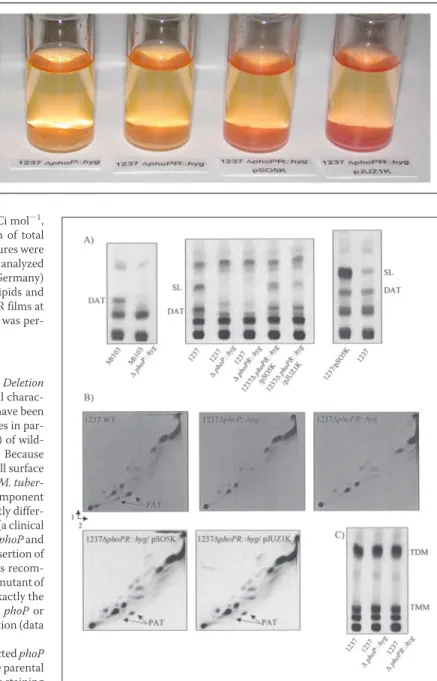
FIGURE 2. Lipid content of the wild-type, mutant, and complemented mutant strains of M. tuberculosis and effect of overexpressing phoP in wild-type M.
tuber-culosis 1237. Autoradiograms of thin-layer chromatograms of lipids derived from
[1-14C]propionate and [1,2-14C]acetic acid are shown. A, for the analysis of DAT and SL,
[1-14C]propionate-derived total lipids (30,000 total cpm) were subjected to TLC with
chloroform:methanol:water (90:10:1, v:v:v) as the solvent. B, PAT were analyzed by load-ing 40,000 cpm of [1-14C]propionate-derived total lipids on TLC plates and developing
the plates three times in petroleum ether (60/80 °C):acetone (92:8, v:v) in the first direc-tion and once in toluene:acetone (95:5, v:v) in the second direcdirec-tion. C, for the analysis of trehalose monomycolates (TMM) and trehalose dimycolates (TDM), [1,2-14
C]acetate-de-rived total lipids (20,000 total cpm) were subjected to TLC with chloroform:methanol: water (90:10:1, v:v:v) as the solvent. Only the PAT, TMM, and TDM profiles of the 1237 mutants are shown. Similar profiles were obtained with the Mt103 mutants. 1237⌬phoPR::hyg/pSO5K, phoP-phoR mutant of M. tuberculosis 1237 complemented with the phoP gene; 1237⌬phoPR::hyg/pJUZ1K, phoP-phoR mutant of M. tuberculosis 1237 complemented with the phoP and phoR genes.
ACCELERATED PUBLICATION: Complex Lipid Regulation in M. tuberculosis
at INRA Institut National de la Recherche Agronomique on May 28, 2019
http://www.jbc.org/
addition, sulfolipid (SL) production (22, 23) was abolished in 1237⌬phoP::hyg and 1237⌬phoPR::hyg (Figs. 2A and 3). M. tuberculosis Mt103 being naturally devoid of this latter family of acyltrehaloses, this phenotype was only visible in the 1237 background. All of the lipids found to be absent from the mutant cells were also absent from their culture filtrates indicating that their absence from bacterial cells was not the result of secretion into the culture medium (data not shown). Analyses of labeled lipids in a variety of solvent systems revealed no other qualitative or quantitative differences between the wild-type and the mutant strains. Likewise, a combination of MALDI-TOF mass spectrometry and TLC analyses revealed that all of the M. tuberculosis mutants produced the same types and amounts of mycolates as their parent strains (data not shown). Finally, gas chromatography (GC) analysis of short chain (up to C26) fatty acid
methyl esters from total lipids showed that the mutants generate all of the classes of fatty acids produced by the wild-type strains (data not shown).
Since the non-mycoloylated trehalose esters, DAT, PAT, and SL, mainly con-tain methyl-branched fatty acids such as mycolipenic acids, mycosanoic acids, and (hydroxy)phthioceranic acids (Fig. 3), further analyses were performed comparing wild-type and mutant strains for the presence of these fatty acids. To this aim, fatty acid methyl esters and acetylated fatty acid methyl esters were prepared from the non-radiolabeled and [1-14C]propionate-derived lipids of each of the strains
described in this study and analyzed by GC and TLC, respectively. Although the autoradiograms of the TLC plates showed bands of significantly lower intensity in the mutants as compared with the wild-type strains, it was not possible to accu-rately compare the long-chain methyl-branched fatty acid composition of the different strains due to the co-migration of the fatty acids of interest with other [1-14
C]propionate-derived compounds (mycocerosic acids in particular) (data not shown). Unfortunately, the low quantities of SL, DAT, and PAT produced by the
M. tuberculosisisolates 1237 and Mt103 also hampered the detection of the long-chain methyl-branched fatty acids esterifying these lipids by GC.
Interestingly, both the complementation of 1237⌬phoPR::hyg with the phoP and phoR genes carried by the multicopy vector pJUZ1K and with the phoP gene alone carried by pSO5K restored SL, PAT, and DAT production (Fig. 2, A and B), indicating that phoP but not phoR is required for the production of methyl-branched fatty acid-containing acyltrehaloses in M. tuberculosis. Moreover, the amounts of SL produced by 1237 (but not those of DAT and PAT) appeared to be directly correlated to the level of expression of phoP as overexpression of this gene in 1237 from the multicopy plasmid pSO5K resulted in a significant increase in the amounts of14C incorporated into SL
(Fig. 2A). Paralleling the recovery of acyltrehaloses synthesis, the ability of the complemented 1237 mutants to fix neutral red was restored (Fig. 1). DISCUSSION
A previous study had reported that disruption of phoP slightly increased the ratio of monoacylated to triacylated lipoarabinomannan in M. tuberculosis Mt103, already pointing to the existence of a relationship between PhoP and lipid metabolism (24). In this study, we provided evidence that phoP is required for the synthesis of complex acyltrehaloses restricted to pathogenic species of the M. tuberculosis complex, namely DAT, PAT, and SL. Moreover, there seems to be a direct correlation between the level of expression of phoP and the amounts of some of these lipids as overexpression of phoP from a multicopy plasmid in strain 1237 resulted in a significant increase in the amounts of SL synthesized. These results suggest that the two-component regulator PhoP-PhoR up-regulates the expression of genes involved in the biosynthesis of SL,
FIGURE 3. Structures of some trehalose-derived molecules from M. tuberculosis. The major sulfolipid SL-I is represented. In SL-I, trehalose is sulfated at the 2 position and esterified with palmitic acid and the multimethyl-branched phthioceranic and hydroxyphthioceranic acids. In DAT, trehalose is esterified with stearic acid and the multimethyl-branched mycosanoic acid. In PAT, trehalose is esterified with stearic acid and the multimethyl-branched mycolipenic acids. In TMM and TDM, trehalose is esterified with mycolic acids.
at INRA Institut National de la Recherche Agronomique on May 28, 2019
http://www.jbc.org/
DAT, and PAT. Considering that the synthesis of trehalose mono- and di-mycolates is unaffected in the mutants (Fig. 2C), it seems unlikely that PhoP-PhoR exerts its control at the level of trehalose synthesis. It is more likely that it regulates the expression of the acyltransferase, polyketide synthase (pks) or pks-associated genes involved in the synthesis or transfer of the methyl-branched fatty acyl substituents found in SL, DAT, and PAT.
Interestingly, the histidine kinase encoded by phoR does not seem to be required for the synthesis of these lipids as complementation of 1237⌬phoPR::hyg with the
phoPgene alone was sufficient to restore their synthesis. As already described in other two-component systems, phosphorylation might not be an absolute require-ment for the binding of PhoP to its DNA targets and might only serve to increase the affinity of the response regulator for certain promoters (25). Alternatively, as is the case in Salmonella, it is possible that instead of phosphorylating PhoP, PhoR catalyzes the dephosphorylation of phospho-PhoP, thereby abolishing the tran-scription of PhoP-activated genes (4). In that case, the lack of PhoR expression in 1237⌬phoP-phoR::hyg complemented with phoP would not abolish the produc-tion of acyltrehaloses.
The abolition of the synthesis of PAT, DAT, and SL probably accounts, at least in part, for the decreased ability of the phoP mutants to form cords and to fix neutral red, morphological and cytochemical traits, which have been asso-ciated with virulence attenuation of tubercle bacilli since Dubos and Middle-brook’s seed studies in the 1940s. Indeed, trehalose esters are located at or near the surface of M. tuberculosis where their fatty acid chains have been proposed to interact with the mycolic acids of the cell wall core to provide a mycobac-terial ”outer membrane“ system, while their sugar moiety interacts with the hydrophilic capsule (26). In good agreement with this model, we have found that the absence of some forms of DAT and PAT in a pks3-4 mutant of M.
tuberculosisH37Rv resulted in defects in capsule attachment causing the mutant to expose a more hydrophobic surface and, consequently, to aggregate in liquid broth (27) and to interact differently with phagocytic and non-phago-cytic cells (13). More recently, in a search for the molecular determinants underlying the different cording properties of the M. tuberculosis H37Ra and H37Rv strains using the microarray technology, Gao et al. (28) identified two polyketide synthase-associated genes, fadD21 and papA1, among the 22 genes whose expression was consistently higher in the cord-forming strain H37Rv. The pks3-4-associated gene fadD21 is expected to encode a fatty acid-activat-ing enzyme involved in DAT and PAT synthesis (27), while papA1, which is associated to pks2, is likely to encode an acyltransferase required for SL forma-tion (29). As the cording strain M. tuberculosis H37Rv is proficient in the synthesis of SL and mycolipenate-containing DAT and PAT, while the non-cording M. tuberculosis H37Ra and phoP mutants are not (7, 15–16), it is tempting to speculate that SL, DAT, and PAT are, with mycolic acids (8 –9), major determinants of cording in M. tuberculosis.
If the association between cording and virulence has been well established (6), it is at present not as clear to what extent the lack of SL, DAT, and PAT accounts for the virulence attenuation and, perhaps, reduced transmissibility (5) of the phoP knock-out mutants. The fact that, within the Mycobacterium genus, SL, PAT, and DAT are relatively restricted to the virulent strains of the M. tuberculosis complex (15, 16, 23) strongly suggests that this family of lipids serves important functions in the pathogenicity of tubercle bacilli, an assumption further supported by multiple
in vitroand in vivo studies (for a review, see Refs. 13, 22, 23, 30 and references cited therein, and 31–34). Somewhat tempering the above assumption, our recent work has indicated that M. tuberculosis isogenic mutants specifically impaired in their ability to synthesize SL or PAT and DAT did not display any marked reduction of virulence in cellular and animal models (13, 30). Nevertheless, as the mutants analyzed in both studies retained the ability to synthesize some methyl-branched fatty acid-containing acyltrehaloses, one could not exclude that the remaining families of acyltrehaloses in the mutant compensated for the missing types. The fact that the production of all of these molecules is coordinately regulated by PhoP actually tends to suggest that they act synergistically. Therefore, the effects of these lipids in virulence might only be clearly visible when the synthesis of all of them is abolished simultaneously.
It is noteworthy that this is not the first report of a two-component system regulating the synthesis of polyketides or polyketide-derived products in pro-karyotes. In Streptomyces lividans and Streptomyces coelicolor, it was shown that phosphate control of antibiotic biosynthesis is mediated by the two-com-ponent PhoP-PhoR system (35, 36). It is interesting that actinomycetes have adopted similar strategies to regulate the production of polyketide-derived products, despite the structural and functional diversity of these molecules.
Although much work remains to be done in identifying the PhoP regulon
and understanding the virulence attenuation of phoP mutants, this work rep-resents an important step toward the functional characterization of PhoP and the understanding of complex lipid synthesis in M. tuberculosis. Identifying the specific signals sensed by the histidine kinase PhoR, whether Mg2⫹as in
Sal-monella(4), phosphate as in Streptomyces (36), acetate as in E. coli (37), or otherwise, is now required to determine under which conditions and toward which aim(s) the production of methyl-branched fatty acids-containing acyl-trehaloses is regulated. Several approaches, including promoter fishing and proteomics, have been undertaken by our laboratories to complete our under-standing of this important two-component regulator.
REFERENCES
1. World Health Organization (2004) http://www.who.int/mediacentre/factsheets/ fs104/en/print.html
2. Fontan, P. A., Walters, S., and Smith, I. (2004) Curr. Sci. 86, 122–134
3. Pe´rez, E., Samper, S., Bordat, Y., Guilhot, C., Gicquel, B., and Martı´n, C. (2001) Mol.
Microbiol. 41,179 –187
4. Groisman, E. A. (2001) J. Bacteriol. 183, 1835–1842
5. Soto, C. Y., Mene´ndez, M. C., Pe´rez, E., Samper, S., Gomez, A. B., Garcia, M. J., and Martı´n, C. (2004) J. Clin. Microbiol. 42, 212–219
6. Middlebrook, G., Dubos, R. J., and Pierce, C. (1947) J. Exp. Med. 86, 175–187 7. Middlebrook, G., Coleman, C. M., and Schaefer, W. B. (1959) Proc. Natl. Acad. Sci.
U. S. A. 45,1801–1804
8. Glickman, M. S., Cox, J. S., and Jacobs, W. R., Jr. (2000) Mol. Cell 5, 717–727 9. Gao, L.-Y., Laval, F., Lawson, E. H., Groger, R. K., Woodruff, A., Morisaki, J. H., Cox,
J. S., Daffe´, M., and Brown, E. J. (2003) Mol. Microbiol. 49, 1547–1563
10. Jackson, M., Raynaud, C., Lane´elle, M.-A., Guilhot, C., Laurent-Winter, C., Enser-gueix, D., Gicquel, B., and Daffe´, M. (1999) Mol. Microbiol. 31, 1573–1587 11. Jackson, M., Camacho, L. R., Gicquel, B., and Guilhot, C. (2001) in Mycobacterium
tuberculosis Protocols(Parish, T., and Stocker, N. G., eds) Vol. 54, pp. 59 –75, Humana Press, Totowa, NJ
12. Howard, N. S., Gomez, J. E., Ko, C., and Bishai, W. R. (1995) Gene (Amst.) 166, 181–182
13. Rousseau, C., Neyrolles, O., Bordat, Y., Giroux, S., Sirakova, T. D., Prevost, M.-C., Kolattukudy, P. E., Gicquel, B., and Jackson, M. (2003) Cell. Microbiol. 5, 405– 415 14. Laval, F., Lane´elle, M.-A., De´on, C., Montsarrat, B., and Daffe´, M. (2001) Anal. Chem.
73,4537– 4544
15. Cason, J., Freeman Allen, C., DeAcetis, W., and Fonken, G. J. (1956) J. Biol. Chem. 220, 893–904
16. Daffe´, M., Lacave, C., Lane´elle, M.-A., Gillois, M., and Lane´elle, G. (1988) Eur. J.
Bio-chem. 172,579 –584
17. Reed, M. B., Domenech, P., Manca, C., Su, H., Barczak, A. K., Kreiswirth, B. N., Kaplan, G., and Barry, C. E., III (2004) Nature 431, 84 – 87
18. Dubos, R. J., and Middlebrook, G. (1948) Amer. Rev. Tuberc. 58, 698 – 699 19. Lemassu, A., Lane´elle, M.-A., and Daffe´, M. (1991) FEMS Microbiol. Lett. 78, 171–176 20. Besra, G. S., Bolton, R., McNeil, M. R., Ridell, M., Simpson, K. E., Glushka, J., van Halbeek, H., Brennan, P. J., and Minnikin, D. E. (1992) Biochemistry 31, 9832–9837 21. Minnikin, D. E., Dobson, G., Sesardic, D., and Ridell, M. (1985) J. Gen. Microbiol. 131,
1369 –1374
22. Goren, M. B., and Brennan, P. J. (1979) in Tuberculosis (Youmans, G. P., ed) pp. 63–193, W. B. Saunders Co., Philadelphia
23. Goren, M. B. (1990) in Handbook of Lipid Research. Glycolipids, Phosphoglycolipids
and Sulfoglycolipids(Kates, M., ed) Vol. 6, pp. 363– 461, Plenum Press, New York 24. Ludwiczak, P., Gilleron, M., Bordat, Y., Martı´n, C., Gicquel, B., and Puzo, G. (2002)
Microbiology 148,3029 –3037
25. Himpens, S., Locht, C., and Supply, P. (2000) Microbiology 146, 3091–3098 26. Minnikin, D. E. (1982) in The Biology of Mycobacteria (Ratledge, C., and Stanford, J.,
eds) Vol. 1, pp. 95–184, Academic Press, London
27. Dubey, V. S., Sirakova, T. D., and Kolattukudy, P. E. (2002) Mol. Microbiol. 45, 1451–1459
28. Gao, Q., Kripke, K., Arinc, Z., Voskuil, M., and Small, P. (2004) Tuberculosis 84, 188 –196
29. Sirakova, T. D., Thirumala, A. K., Dubey, V. S., Sprecher, H., and Kolattukudy, P. E. (2001) J. Biol. Chem. 276, 16833–16839
30. Rousseau, C., Turner, O. C., Rush, E., Bordat, Y., Sirakova, T. D., Kolattukudy, P. E., Ritter, S., Orme, I. M., Gicquel, B., and Jackson, M. (2003) Infect. Immun. 71, 4684 – 4690
31. Gilleron, M., Stenger, S., Mazorra, Z., Wittke, F., Mariotti, S., Bo¨hmer, G., Prandi, J., Mori, L., Puzo, G., and De Libero, G. (2004) J. Exp. Med. 199, 649 – 659
32. Converse, S. E., Mougous, J. D., Leavell, M. D., Leary, J. A., Bertozzi, C. R., and Cox, J. S. (2003) Proc. Natl. Acad. Sci. U. S. A. 100, 6121– 6126
33. Domenech, P., Reed, M. B., Dowd, C. S., Manca, C., Kaplan, G., and Barry, C. E., III (2004) J. Biol. Chem. 279, 21257–21265
34. Graham, J. E., and Clark-Curtiss, J. E. (1999) Proc. Natl. Acad. Sci. U. S. A. 96, 11554 –11559
35. Sola-Landa, A., Moura, R. S., and Martin, J. F. (2003) Proc. Natl. Acad. Sci. U. S. A. 100,6133– 6138
36. Martin, J. F. (2004) J. Bacteriol. 186, 5197–5201
37. Lesley, J. A., and Waldburger, C. D. (2003) J. Bacteriol. 185, 2563–2570
38. Soto, C. Y., Andreu, N., Gibert, I., and Luquin, M. (2002) J. Clin. Microbiol. 40, 3021–3024
ACCELERATED PUBLICATION: Complex Lipid Regulation in M. tuberculosis
at INRA Institut National de la Recherche Agronomique on May 28, 2019
http://www.jbc.org/
Martín and Mary Jackson
Laval, Carlos Yesid Soto, Nathalie Winter, Mamadou Daffé, Brigitte Gicquel, Carlos
Jesús Gonzalo Asensio, Catarina Maia, Nadia L. Ferrer, Nathalie Barilone, Françoise
Mycobacterium tuberculosis
Biosynthesis of Polyketide-derived Lipids in
doi: 10.1074/jbc.C500388200 originally published online December 2, 2005
2006, 281:1313-1316.
J. Biol. Chem.
10.1074/jbc.C500388200
Access the most updated version of this article at doi:
Alerts:
When a correction for this article is posted
•
When this article is cited
•
to choose from all of JBC's e-mail alerts
Click here
http://www.jbc.org/content/281/3/1313.full.html#ref-list-1
This article cites 36 references, 15 of which can be accessed free at
at INRA Institut National de la Recherche Agronomique on May 28, 2019http://www.jbc.org/