HAL Id: hal-03149023
https://hal-amu.archives-ouvertes.fr/hal-03149023
Submitted on 22 Feb 2021
HAL is a multi-disciplinary open access archive for the deposit and dissemination of sci-entific research documents, whether they are pub-lished or not. The documents may come from teaching and research institutions in France or abroad, or from public or private research centers.
L’archive ouverte pluridisciplinaire HAL, est destinée au dépôt et à la diffusion de documents scientifiques de niveau recherche, publiés ou non, émanant des établissements d’enseignement et de recherche français ou étrangers, des laboratoires publics ou privés.
Distributed under a Creative Commons Attribution| 4.0 International License
Management of sleep apnoea syndrome (SAS) in
patients with vasovagal syncope (VVS): a protocol for
the VVS-SAS cohort study
Vincent Puel, Isabelle Godard, Georgios Papaioannou, Philippe Gosse, Jean
Pepin, Fabrice Thoin, Jean Deharo, Frederic Roche, Naïma Zarqane, Frédéric
Gagnadoux, et al.
To cite this version:
Vincent Puel, Isabelle Godard, Georgios Papaioannou, Philippe Gosse, Jean Pepin, et al.. Management of sleep apnoea syndrome (SAS) in patients with vasovagal syncope (VVS): a protocol for the VVS-SAS cohort study. BMJ Open, BMJ Publishing Group, 2020, 10 (9), pp.e038791. �10.1136/bmjopen-2020-038791�. �hal-03149023�
Management of sleep apnoea syndrome
(SAS) in patients with vasovagal
syncope (VVS): a protocol for the VVS-
SAS cohort study
Vincent Puel,1 Isabelle Godard,1 Georgios Papaioannou,2 Philippe Gosse,2 Jean Louis Pepin,3 Fabrice Thoin,4 Jean Claude Deharo,5 Frederic Roche,6
Naïma Zarqane,7 Frédéric Gagnadoux,8 Carey Meredith Suehs ,9
Nicolas Molinari10
To cite: Puel V, Godard I, Papaioannou G, et al. Management of sleep apnoea syndrome (SAS) in patients with vasovagal syncope (VVS): a protocol for the VVS- SAS cohort study. BMJ Open
2020;10:e038791. doi:10.1136/ bmjopen-2020-038791 ►Prepublication history for this paper is available online. To view these files, please visit the journal online (http:// dx. doi. org/ 10. 1136/ bmjopen- 2020- 038791).
Received 24 March 2020 Revised 25 August 2020 Accepted 02 September 2020
For numbered affiliations see end of article.
Correspondence to Dr Vincent Puel; vpuel001@ gmail. com © Author(s) (or their employer(s)) 2020. Re- use permitted under CC BY- NC. No commercial re- use. See rights and permissions. Published by BMJ.
ABSTRACT
Introduction Recurrent vasovagal syncope (VVS) is
associated with decreased quality- of- life and frequent use of emergency services. The evidence base for causality, diagnostic procedures and potential VVS treatments is poor. Scattered observations in the literature suggest a link between respiratory disturbances during sleep and VVS. Empirical observations lead us to further hypothesise that the appropriate management of sleep apnoea syndrome (SAS) may help resolve comorbid recurrent VVS in certain patients. We therefore designed this pilot study to provide a framework for the observation of changes in outcomes accompanying the deployment of SAS treatments in patients with VVS.
Methods and analysis This is a multicentre, registry-
based study whose primary objective is to evaluate the effect of SAS management on the number of syncope/ presyncope episodes in a population suffering from both VVS and SAS. To this effect, syncope rates prior to the treatment of SAS will be compared with those occurring after the initiation of the latter. In addition, yearly assessments will collect data for echocardiography, polysomnography, Holter monitoring, table tilt tests, multiple sleep latency tests, SAS management parameters and questionnaires describing fatigue, depression and quality- of- life. Sixty patients will be included with a minimum follow- up period of 12 months. The primary analysis will use comparisons of centrality for paired data to describe the changes in syncope rates before versus after the initiation of SAS management. Longitudinal data will be analysed using mixed models with patients set as a random effect. Subgroup analyses will be performed for SAS- treatment adherence and efficacy.
Ethics and dissemination The VVS- SAS registry
was approved by an ethics committee (Comité pour la Protection des Personnes Ile- de- France VI, Reference number CPP/2-18) in accordance with French law. The princeps publication will present before–after SAS management results and longitudinal analyses.
Trial registration number NCT04294524. Pre- results.
INTRODUCTION
Vasovagal syncope (VVS) consists of an auto-matic reaction of the autonomic nervous
system (ANS) responsible for a cascade of events causing loss of consciousness. The situ-ations that result in such ANS reflex responses are described as triggers, and can include pain, unusual stress or prolonged passive orthostatism. Syncope is rare. However, certain subjects can become at a time in their lives particularly vulnerable to this type of reaction to the point that triggers become virtually absent or confused with daily activi-ties. A decrease in quality- of- life can ensue.1 2
Pinpointing a physiological explanation for recurrent syncope has been a historically elusive exercise. Beyond ruling out epilepsy, hypoglycaemia, frank hypotension or cardiac
causes,3 diagnostic testing has been reported
as highly variable and of low clinical utility.4
Current treatments for VVS are relatively
ineffective5 and the use of emergency services
for syncope episodes is frequent and costly.4 6
VVS not only occurs in upright ortho-static positions, but can also occur in supine
Strengths and limitations of this study
► This study represents the first multicentre study describing outcomes in patients suffering from both sleep apnoea and recurrent vasovagal syncope.
► A large range of variables describing sympatoex-citation, sleep apnoea and quality- of- life will be monitored over a minimum follow- up period of 12 months.
► Potential changes in syncope rates associated with sleep apnoea syndrome treatment will be quantified for the first time.
► As with all observational studies, care must be taken when inferring causality.
► The resulting dataset will provide a first evidence base required for the design of subsequent confir-matory studies.
by copyright.
on February 22, 2021 at Universite d'Aix-Marseille. Protected
Open access
positions and during sleep (often then associated with awakening accompanied by panic or a need to micturate/
defecate).7 8 Interestingly, sporadic reports have also
suggested an association between respiratory distur-bances during sleep and daytime recurrent syncope. About 23% of patients with upper airway respiratory syndrome complain of orthostatic intolerance and
simul-taneously have low blood pressure.9 Patients with VVS
have been demonstrated to have sympathetic suppres-sion during rapid eye movement sleep and increased slow
wave sleep.10 Sympathetic neural responses to induced
hypoxemia have been demonstrated to differ between
control and obstructive sleep apnoea subjects.11 However,
the latter was insufficient to explain the full range of sympathoexcitation during sleep, suggesting that other
dysfunctions in the ANS are also present.11
Based on the empirical observation of several patients without long- term follow- up, we hypothesised that the appropriate management of sleep apnoea syndrome
(SAS) may help resolve comorbid recurrent syncope.5
Continuous positive airway pressure (CPAP) for the treat-ment of SAS has also been reported by a second team to resolve repeating syncope episodes in a single case
report.12 The purpose of this protocol is therefore to
provide a first, pilot framework for the observation of changes in outcomes accompanying the deployment of SAS treatments in patients with VVS.
METHODS AND ANALYSIS Study design and objectives
This is an observational, multicentre, registry- based,
longitudinal cohort study whose primary objective is to evaluate the effect of treatment for SAS on the number of syncope/presyncope episodes in a population suffering from both idiopathic, recurrent VVS/presyncope and SAS. To this effect, ‘day 0’ in the registry corresponds to patient enrolment and the initiation of (any) SAS treat-ment, and syncope/presyncope rates prior to day 0 will be compared with those occurring afterwards. Second, this study will also describe simultaneous changes in quality- of- life and associations between syncope symptom profiles and a battery of potentially related patient
characteristics (taken from heart rhythm and blood pres-sure monitoring, polysomnography, echocardiography, table tilt tests, multiple sleep latency test (MSLT), as well
as patient- reported outcomes describing sleep- related
symptoms and fatigue).
Patient and public involvement
Patients and public were not involved in the design of this study.
The VVS-SAS registry and population
The VVS- SAS registry was initiated by collaborating
experts (Study Director: Professor Vincent Puel) to stan-dardise routine care and data collection for this popu-lation in participating centres in France. The registry includes all consecutive adult patients (thus avoiding selection bias) simultaneously suffering from recurrent, idiopathic VVS (or orthostatic intolerance) and obstruc-tive sleep apnoea. Within the framework of this protocol, VVS or orthostatic intolerance is diagnosed by cardiolo-gists with VVS expertise (most often as a function of a table tilt test, and occasionally despite a negative table tilt test if the expert finds the patient’s symptoms to be vagal in origin). The first inclusion occurred on 22 February 2017 and inclusions will continue until the required sample size is reached (inclusions are currently estimated to reach sample size in the first half of 2021). The specific
eligibility criteria are presented in table 1. The
partici-pating centres include two private clinics (Clinique Bel Air, Bordeaux, France; Clinique Bouchard, Marseille, France) and six publicly funded hospitals (University Hospitals of Angers, University Hospitals of Bordeaux, University Hospitals of Grenoble, Public Assistance— Hospitals of Marseille, University Hospitals of St Etienne, France; Princess Grace Hospital, Monaco). The single- payer social security system in France guarantees coverage to all residents, thus guaranteeing a wide representation of diverse socioeconomic and urban- versus- rural popula-tions in the participating centres.
Primary outcome
‘D0’ indicates the day on which the patient started using continuous positive airway pressure (CPAP) or wearing
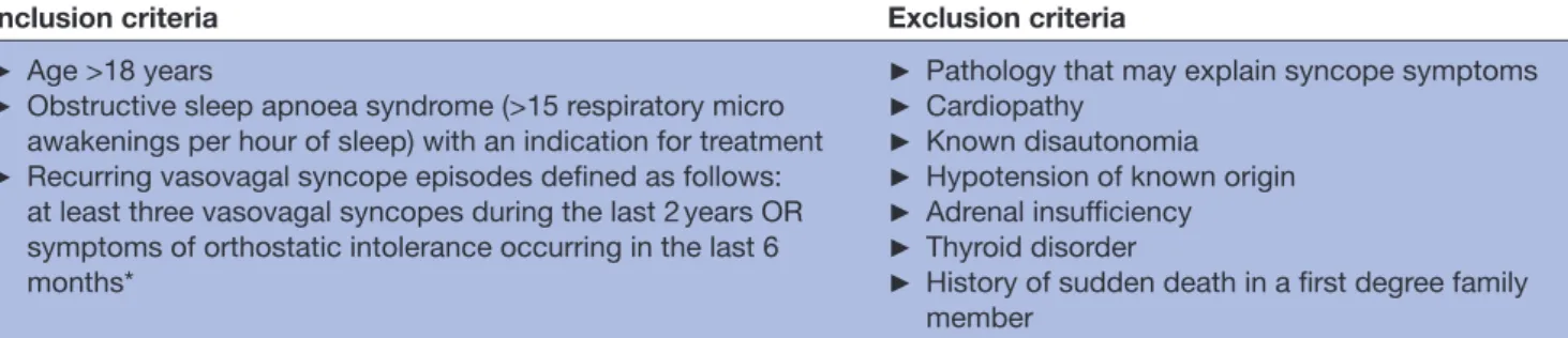
Table 1 The study eligibility criteria
Inclusion criteria Exclusion criteria
► Age >18 years
► Obstructive sleep apnoea syndrome (>15 respiratory micro awakenings per hour of sleep) with an indication for treatment
► Recurring vasovagal syncope episodes defined as follows: at least three vasovagal syncopes during the last 2 years OR symptoms of orthostatic intolerance occurring in the last 6 months*
► Pathology that may explain syncope symptoms
► Cardiopathy
► Known disautonomia
► Hypotension of known origin
► Adrenal insufficiency
► Thyroid disorder
► History of sudden death in a first degree family member
*Symptoms are triggered by orthostatism and can include presyncopes without loss of consciousness, asthenia, dizziness, visual disturbances, tinnitus, palpitations, headache, limitations of physical activity.
by copyright.
on February 22, 2021 at Universite d'Aix-Marseille. Protected
a mandibular advancement device (MAD) for the treat-ment of sleep apnoea. Changes or adjusttreat-ments to SAS treatment do not reset the D0. The number of episodes of syncope or presyncope (hereafter referred to as ‘(pre)
syncope’) occurring before SAS management (Sbefore)
is determined by a patient’s report for the time period covering months −6 to D0. The number of episodes of (pre)syncope occurring after SAS management is deter-mined by prospectively recording (pre)syncope events during the study and counting them for the time period
covered by months +6 to +12 (Safter). The primary outcome
is the per- patient difference in the number of (pre) syncope episodes occurring before and after the initia-tion of SAS treatment via CPAP or MAD: Sdiff=Sbefore–Safter.
Sample size
Our working hypothesis is based on an expected mean value of Sbefore- expected=10, an expected mean value of Safter-
expected=2, with a corresponding mean for Sdiff- expected=8. The
latter expected values are gross estimations of popula-tion means based on a handful of empirical observa-tions, and the results of this first study will greatly help to improve these estimates, a necessary step for planning future, larger studies. For this first exploratory, pilot study and given the absence of previously published data, we conservatively chose a twice- as- large SD (as compared with Sdiff- expected) of 16 for a standard effect size of 0.5,
which is considered as a ‘medium’ effect size.13 Such an
effect size also applies to any mean difference encoun-tered, as long as the corresponding pooled SD remains less than twice as large as the difference between means. To detect a standard effect size of 0.5 for paired data with a type 1 error rate of α=0.05 and power set at 1−β=0.90,
44 patients are required.14 To take into account the
possi-bility of loss to follow- up or otherwise missing data, 60 patients will be included.
Visits
The information collected during this study corresponds to routine practice in the participating centres. Recruit-ment and follow- up take place during regularly sched-uled, yearly visits for patients consulting for idiopathic recurrent syncope. Following patient information and verification of eligibility criteria, the patient is enrolled in the VVS- SAS registry and data collection then starts.
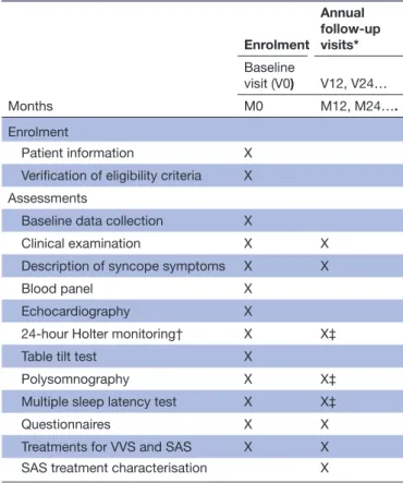
Table 2 lists the assessments that are performed at the
first baseline visit, as well as a subset of the latter that are reperformed on a yearly basis. Each assessment is described in the following paragraphs, along with which data are recorded.
Data are recorded for all yearly visits that occur during the inclusion period and then for a subsequent 12- month follow- up period. The inclusion period is not set in an a priori fashion; inclusions will proceed until 60 patients are included. The present study will remain open for follow- up until each patient has at least 1 year of follow- up. The register will continue functioning as usual for an indefinite period of time.
Assessments and variables Baseline data collection
The following baseline characteristics will be recorded once at the start of the study: age, sex and socio- professional category (agricultural, entrepreneur, senior professional, intermediate professions, employee, worker, retiree, no activity). The presence/absence of facial dysmorphia variables will be recorded (retrognathia, archival palate, mallampati score 3 or 4, anterior open bite, cervico- mental angle blunting, other otorhinolar-yngology abnormalities (lower tongue, tonsillar hyper-trophy)). The age at which the patient’s first syncope/ presyncope occurred will be recorded.
Clinical examination
A general clinical examination will be performed at each visit and the following recorded: weight (kg), height
(cm), body mass index (kg/m2), abdominal
circumfer-ence (cm). Blood pressure (systolic and diastolic in mm Hg) and pulse (beats per minute) will be measured while lying down and after having remained in a standing posi-tion for 3 min.
Table 2 The chronology of visits and assessments performed in the VVS- SAS registry
Months Enrolment Annual follow- up visits* Baseline visit (V0) V12, V24… M0 M12, M24…. Enrolment Patient information X
Verification of eligibility criteria X Assessments
Baseline data collection X
Clinical examination X X
Description of syncope symptoms X X
Blood panel X
Echocardiography X
24- hour Holter monitoring† X X‡
Table tilt test X
Polysomnography X X‡
Multiple sleep latency test X X‡
Questionnaires X X
Treatments for VVS and SAS X X
SAS treatment characterisation X
*The total number of annual follow- up visits will be determined on a per- patient basis by the time required to recruit 60 patients, plus one additional year of follow- up.
†Continuous data collection for both blood pressure and cardiac rhythm.
‡At V12 only.
by copyright.
on February 22, 2021 at Universite d'Aix-Marseille. Protected
Open access
Description of syncope symptoms
The patient is asked to maintain a journal noting the dates of both syncopes and presyncopes throughout the study. In addition, the syncope/presyncope history for each patient will be carefully described at each yearly visit, starting with the number of syncopes (and/or presyn-copes) during the last 6 months. The patient is further requested to select the best descriptor for the frequency of his/her syncope and presyncope episodes (never, rare, every 3 months, every month, every week, several times per week), the position in which they occur (standing, sitting, lying down), known triggers (heat, crowding, pain, stress, effort, lack of sleep, none) and timing (diurnal, nocturnal, postprandial, peri/post- miction/defecation). Finally, the patient is asked whether or not any signs precede a loss of consciousness (always, sometimes, never) and whether or not they have adapted their behaviour so as to avoid (pre) syncope episodes (and if so, how?).
Blood panel
The following blood work will be performed according to local, routine procedures at baseline only: haemoglobin (g/L), haematocrit (%), thyroid stimulating hormone (μU/L), sodium and potassium levels (mmol/L) and morning cortisol (mmol/L at 08:00).
Echocardiography
An echocardiography is performed at baseline only and according to routine practice. The following data are recorded: end diastolic diameter of the left ventricle (mm), left ventricular ejection fraction (%), systolic blood pressure (mm Hg), whether or not the examina-tion was normal (yes/no).
24-hour Holter monitoring
Holter monitoring is performed on a yearly basis. Data will be collected for a full 24- hour period for each patient, with separate datasets for heart rhythm data and arterial pressure data.
Hearth rhythm data will be centrally analysed under the direction of Professor Frédéric Roche at the Univer-sity Hospital of Saint- Etienne, Saint- Etienne, France. The data recorded include the total number of heartbeats analysed, and the percentage of successive RR (interbeat intervals between all successive heart beats) intervals that differ by more than 50 m. In addition, the following four variables will be recorded at three time levels (daytime defined as between 07:30 and 21:30, nighttime, and total): (1) the SD of NN (interbeat intervals from which artefacts have been removed) intervals (ms), the root mean square of successive RR interval differences (ms), the SD of successive RR interval differences and the Heart Rate Variability Index. In terms of fourier trans-formed frequency domains, the following four variables will be recorded (again for the same three time levels):
total power (ms2), very low frequency power (ms2), low
frequency power (ms2), high frequency power (ms2).
Blood pressure data are recorded every 15 min during a full 24- hour period, with notation of the times at which the patient went to sleep and got up in the morning. Blood pressure data will be analysed under the direction of Professor Philippe Gosse at the University Hospital of Bordeaux, Bordeaux, France. In addition to the number of blood pressure measurements performed, the means and SD for systolic pressures, diastolic pressure and heart rate are recorded for the daytime, nighttime and total time frames.
Table tilt test
Tilt table testing characterises the autonomic nervous response to the gravitational stress associated with a passive change from a supine position to an upright posi-tion.15 The table tilt test is performed at baseline only, and
patients must have fasted for at least 4 hours15 and start
the test with an empty bladder. The patient is placed in a decubitus dorsal position for 10 min, and then the table is tilted to 60°. If after 20 min of this orthostatic position no syncope symptoms have occurred and if systolic blood pressure remains ≥100 mm Hg, one sublingual spray of trinitrine (0.3 mg) is administered, and the patient left at a 60° tilt for another 15 min (in case of contraindica-tion for trinitrine, the orthostatic posicontraindica-tion is simply main-tained for 45 min). The orthostatic position is interrupted and stopped in case of syncope, or at the request of the patient or as decided by the investigator.
The presence of any of the following while in the ortho-static position constitutes a positive tilt table test: (1) a classic vasovagal reaction, (2) delayed orthostatic hypo-tension (DOH), (3) orthostatic hypohypo-tension in combi-nation with a vasovagal reaction (OHVVR), (4) postural orthostatic tachycardia syndrome (POTS), (5) reproduc-tion of patient- specific reacreproduc-tion. A classic vasovagal reac-tion starts with a first phase of asymptomatic physiological adaptation to the orthostatic position before the onset of a sudden second phase with a characteristic rapid drop in arterial pressure with a simultaneous decrease in cardiac frequency (true or relative bradycardia). Typical vagal symptoms are experienced with syncope or presyncope. DOH is recognised by a progressive, gradual decrease in arterial pressure, but not cardiac frequency, throughout the tilt table test accompanied by typical symptoms (for example vision disturbances, sweating, asthenia, nausea, headache). The OHVVR starts with a gradual decrease in blood pressure similar to that seen in DOH, but co- oc-curs with accelerating cardiac frequency followed by a vasovagal reaction with an abrupt drop in blood pres-sure and cardiac frequency. Symptoms usually start after the latter and can result in syncope. DOH and OHVVR are both considered as forms of orthostatic intolerance. POTS refers to sinus tachycardia reflex at the initiation of an orthostatic position and is defined by a cardiac frequency >120 beats per minute (or an acceleration in cardiac frequency of ≥30 beats per minute) associated with unstable blood pressure and poor clinical tolerance.
by copyright.
on February 22, 2021 at Universite d'Aix-Marseille. Protected
Polysomnography
Polysomnography will be performed at the initial baseline visit and at 1 year later under continuous positive pressure or MAD in order to obtain MSLT (see the Multiple sleep latency test section) according to routine practice and will be characterised by the following variables: the total period of sleep time (min), the total sleep time (min), the wake after sleep onset time (min), the time spent in sleep states 1, 2, 3 and 5 (ST1; ST2, ST3, ST5, respectively; min), the number of obstructive apnoea and hypopnoea episodes, the number of snoring episodes, the Apnoea– Hypopnoea Index (number per hour), the number and rate (number per hour) of respiratory micro awakenings, the number of limb movements, the total number of micro awakenings per hour (including both respiratory and limb movement awakenings), the percentage oxygen saturation when awake (%), the time spent with an oxygen saturation <90% (min), the number of episodes where desaturation was >3% and the number of oxygen desaturations per hour.
Multiple sleep latency test
MSLTs are performed at baseline and a second time 1 year later. For each MSLT, four or five attempts at napping will be tested throughout the day. The times at which each nap attempt started and ended will be noted, as well as the corresponding test duration, whether or not the patient actually fell asleep, and if so, the duration in minutes of sleep latency. The mean sleep latency will be calculated, as well as the number of tests where paradoxical sleep is present.
Questionnaires
A battery of patient- reported outcome tools was admin-istered at baseline and again at each yearly visit. These comported the SF-36 Questionnaire summarising
health- related quality- of- life,16 the 12- item Impact of
Syncope on Quality- of- life Questionnaire,17 the Epworth
Sleepiness Scale for assessing subjective sleepiness,18–20
Pichot’s Fatigue Scale21 and Pichot’s Depression
Inven-tory.22 Patients were also asked to rate the severity of
the following symptoms on a discrete numeric scale ranging from 0 to 10: snoring, nocturnal agitation, rest-less legs, nycturia, morning headaches, absence of libido, nocturnal sweating, gastro- oesophagien reflux, palpita-tions and lack of concentration while driving. Finally, the patient was asked whether he/she has had an accident that he/she attributes to a lack of concentration while driving.
Treatments for VVS and SAS
At baseline and each follow- up visit, the presence/absence of the following VVS treatments will be recorded: midodid-rine, fludrocortisone, betablockers, ivabradine, compres-sion stockings, hygiene/dietary rules, further measures (with explanation when required). Other concurrent treatments will be noted using the name of the molecule involved, as well as the associated pharmacotherapeutic
class. In addition, the use of CPAP or MAD will be indi-cated for SAS management.
Detailed SAS treatment characterisation
Details concerning CPAP or MAD use will be further detailed at each follow- up visit. If the first SAS treatment indicated was CPAP, the following will be recorded: the date of deployment (D0), the name of the delivery service used, the ventilator brand and type (continuous pressure or autopiloted), the type of mask used (nasal, facial or nasal pillows), the use of a humidifier (yes/no), the mean adherence for the duration of use in hours and minutes (hh:mm), the mean hours of use per night (hh:mm), the residual Apnoea–Hyponoea Index (number/hour) and the pressure used at the 95th percentile of treatment. In addition, the presence/absence of known adverse effects will be noted (stuffy nose, irritated eyes, dry mouth, sensa-tion of suffocasensa-tion, psychological discomfort, poor toler-ance of family members, aerophagia, cutaneous lesions, headaches). If it was necessary to stop CPAP treatment since the last visit, the corresponding date of treatment stopping will be recorded, as well as the reason why (inef-ficacy, intolerance, conversion to MAD, referral for maxil-lary surgery).
Concerning patients for whom the first SAS treatment indication was MAD, the D0 will be recorded. If cessation of MAD use was required, the date of stopping will be noted as well as the reason why (inefficacy, intolerance, conversion to CPAP, referral for maxillary surgery).
Analysis strategies General considerations
Statistical analyses will be performed under the direction of the study methodologist (NM). The intention- to- treat (ITT) population is defined as all persons included in the VVS- SAS registry. The per- protocol (PP) population is defined as a subset of the ITT population without major protocol deviations and with a satisfactory SAS treatment response (ie, an Apnoea–Hypopnoea Index <10). The threshold for statistical significance is set at a bilateral probability of 0.05. Prior to database freezing, a detailed statistical analysis plan will be designed. The following paragraphs present the planned general strategies for analysis; any deviations from these strategies must be approved by the study methodologist and documented as such.
Describing the study population
The number of patients included and reaching 12 months of follow- up, with associated losses to follow- up or death, will be summarised via a flow chart. To describe the study population, descriptive statistics will be computed for the ITT population. Qualitative variables will be summarised via numbers and percentages. Normally distributed quan-titative variables will be summarised using means accom-panied by SD. Non- normal, quantitative variables will be summarised using medians accompanied by IQRs. Each
by copyright.
on February 22, 2021 at Universite d'Aix-Marseille. Protected
Open access
variable is also presented with its sample size to demon-strate the extent of missing data.
Dealing with potentially missing data
As concerns the primary outcome, missing data will not be imputed. For multivariate analyses, multiple imputa-tion is the preferred method for dealing with missing data and could be considered.
Before–after SAS management comparisons
Changes in variables before versus after SAS management (including the primary outcome) will be analysed with comparisons of centrality for paired data (paired t- test or paired Wilcoxon signed rank test) at the ITT level. A sensitivity analysis involving the PP population will also be performed. Before–after SAS management compari-sons will be further summarised via their unadjusted and confounder- adjusted (see next section) effect sizes (eg, mean differences) accompanied by 95% CIs. Similar methods will also be applied to comparisons between the first and last 6 months of SAS treatment to contrast changes associated with patient adaptation during the early stages of treatment deployment.
Potential confounders and planned interaction/subgroup analyses
The following variables may affect the primary outcome:
(1) baseline disease severity (Sbefore and baseline Apnoea–
Hypopnoea Index), (2) sex and (3) centre. Linear regres-sion models will be fit for the primary outcome, with the latter set as independent variables and also within an interaction term. Significance will be determined by the p value for the interaction term, with values less than 0.10 considered suggestive of a potential interaction and values less than 0.05 considered to confirm an interaction.
Two specific subgroup analyses are planned and include comparisons between (1) levels of SAS- treatment adher-ence (for example between patients who abandon CPAP/ MAD vs those who maintain CPAP/MAD, or between differences in the mean number of hours of CPAP usage per night) and (2) levels of SAS treatment efficacy (as esti-mated via Apopnoea–Hypopnoea Indices, for example). Further subgroup analyses are exploratory. Subgroups derived from categorical variables will be displayed as a forest plot. If the presentation of data requires it, dichot-omisation of continuous variables for inclusion in a forest plot will be performed.
Repeated measures data
Longitudinal data (repeated measures) will be analysed using mixed models with patients set as a random effect. Explaining changes in repeated measures with time is of particular interest for the following: (1) variables describing sympatoexcitation, (2) variables describing sleep perturbations and (3) questionnaire data describing fatigue, depression or quality- of- life.
Recurrent event analysis
Mean cumulative functions (MCFs) for presyncopes, syncopes and (pre)syncopes will be constructed, with
their 95% CIs. MCFs are step curves that demonstrate the accumulation of target events with time; the asso-ciated analyses are similar to survival analyses and non- parametric in that they do not assume a longitudinally
constant event rate.23 24 MCFs will be plotted for the
entire study period, with a separate panel plotting the associated slope determined by a sliding window. A flat-tening- out of the MCF slope (ie, a reduction in the (pre) syncope rate) is expected to occur at the time of onset of treatment efficacy. According to patient diary availability for the ‘before’ period, the difference in MCFs will also be constructed for Sbefore versus Safter, and the time points at which the two curves differ indicated when the associ-ated 95% CI no longer includes zero.
Exploring associations
Multivariate/multivariable exploratory studies may include multiple regressions, dimension reduction, clustering and partial least squares analyses. The results of exploratory studies will be presented as hypothesis- building post- hoc analyses. Further assertions will require reproduction of results via separate confirmatory studies.
DISCUSSION
This study has certain limitations, the first of which is a potential for recall bias, as the baseline rate of (pre) syncope events is based on a patient’s report. We advise our patients to maintain a (pre)syncope diary, and such will be recovered as much as possible for the baseline period to minimise recall bias as much as possible. Further limitations are those inherent to any observational study design, namely that care must be taken to consider the associative nature of the data and to not infer causality too early. Cohort studies can also suffer from selection bias, but this is avoided in as much as possible by the consecutive- case inclusion strategy. The results should therefore be generalisable to all patients suffering from both VVS and SAS. To our knowledge, no other study specifically addresses this population.
This cohort study will also be the first to move the evidence base for a link between VVS and SAS beyond that of anecdotal, disparate, empirical observations. Key results will include changes in VVS symptoms and
asso-ciated sympatoexcitation and quality- of- life variables
following the implementation of SAS treatment. Should improvement in VVS occur subsequent to the deployment of SAS treatments, not only would treatment options be expanded for this traditionally hard- to- treat population, but diagnostic pathways may be affected by underlining a need for polysomnography screening in the idiopathic VVS population.
ETHICS AND DISSEMINATION
The registry was approved by a randomly selected inde-pendent ethics committee (Comité pour la Protection
des Personnes Ile- de- France VI, Reference number
by copyright.
on February 22, 2021 at Universite d'Aix-Marseille. Protected
CPP/2-18) and the French National Committee on Infor-matics and Liberties (CNIL reference number 2 164 019 v 0) in accordance with French law. Because the registry simply regroups existing data without changing routine care, the patient is informed about the study and the data are collected unless the patient indicates his/her opposi-tion to the study (as per French law).
One publication presenting before–after SAS manage-ment comparisons and longitudinal analyses is planned. A second publication presenting further exploratory asso-ciation results will be published as a post- hoc study. The accompanying datasets will be made available to inter-ested researchers on provision of an appropriate CNIL- approved protocol, as per current French regulations.
Author affiliations
1Pôle d'Exploration des Apnées du Sommeil (PEAS), Nouvelle Clinique Bel- Air,
Bordeaux, France
2Department of Cardiology and Hypertension, Hôpital Saint- André, Centre
Hospitalier Universitaire de Bordeaux, Bordeaux, France
3Department of Physiology, Sleep and Exercise, Univ Grenoble Alpes, CHU Grenoble,
Grenoble, France
4Sleep Centre, Bouchard Clinic, Marseille, France
5Department of Cardiology, Hôpital de la Timone, C2VN, APHM, Marseille, France 6Department of Clinical Physiology and Excercise, Hôpital Nord, Centre Hospitalier
Universitaire St Etienne, St Etienne, France
7Department of Cardiology, Princess Grace Hospital Centre, Monaco
8Department of Respiratory and Sleep Medicine, Centre Hospitalier Universitaire
Angers, Angers, France
9Departments of Respiratory Diseases and Medical Information, Centre Hospitalier
Universitaire de Montpellier, Montpellier, France
10Department of Medical Information, IMAG, CNRS, Univ Montpellier, Centre
Hospitalier Universitaire de Montpellier, Montpellier, France Twitter Carey Meredith Suehs @SuehsCarey
Contributors VP and GP originated the study hypotheses, design and choice of sympatoexcitation/sleep assessments. PG and FR provided Holter parameters and expertise. IG is responsible for study management, registry updates and regulatory activities, and participated in the response to reviewers. CMS drafted the first version of this manuscript using the CRF, synopsis and patient information materials. She also helped write the response to reviewers. NM provided expertise of quality- of- life instruments, as well as methodological and statistical oversight during the writing process. JLP, FT, JCD, NZ, FG provided expertise on assessments and feasibility, and critically reviewed the manuscript. All authors made corrections and approved the final version for submission.
Funding The authors have not declared a specific grant for this research from any funding agency in the public, commercial or not- for- profit sectors.
Competing interests VP declares personal fees (outside the submitted work) from Resmed, Lowenstein, Phillips and non- financial support (outside the submitted work) from SOS Oxygène and ISIS. FG reports personal fees (outside the submitted work) from Air Liquide Santé, Cidelec, Resmed, Sefam, and non- financial support (outside the submitted work) from Air Liquide Santé, Asten Santé, Sefam. Patient and public involvement Patients and/or the public were not involved in the design, or conduct, or reporting, or dissemination plans of this research. Patient consent for publication Not required.
Provenance and peer review Not commissioned; externally peer reviewed. Open access This is an open access article distributed in accordance with the Creative Commons Attribution Non Commercial (CC BY- NC 4.0) license, which permits others to distribute, remix, adapt, build upon this work non- commercially,
and license their derivative works on different terms, provided the original work is properly cited, appropriate credit is given, any changes made indicated, and the use is non- commercial. See: http:// creativecommons. org/ licenses/ by- nc/ 4. 0/. ORCID iD
Carey Meredith Suehs http:// orcid. org/ 0000- 0002- 2175- 3496
REFERENCES
1 Rose MS, Koshman ML, Spreng S, et al. The relationship between health- related quality of life and frequency of spells in patients with syncope. J Clin Epidemiol 2000;53:1209–16.
2 Giada F, Silvestri I, Rossillo A, et al. Psychiatric profile, quality of life and risk of syncopal recurrence in patients with tilt- induced vasovagal syncope. Europace 2005;7:465–71.
3 Peyrouse E, Franceschi F, Prévôt S, et al. Prise en charge actuelle des syncopes - Standardisation de la démarche et unités spécialisées. Propos Cardiol 2012:33–9.
4 Sun BC. Quality- Of- Life, health service use, and costs associated with syncope. Prog Cardiovasc Dis 2013;55:370–5.
5 Puel V, Pepin JL, Gosse P. Sleep related breathing disorders and vasovagal syncope, a possible causal link? Int J Cardiol 2013;168:1666–7.
6 Sun BC, Emond JA, Camargo CA. Characteristics and admission patterns of patients presenting with syncope to U.S. emergency departments, 1992-2000. Acad Emerg Med 2004;11:S4–34. 7 Glatter KA, Chiamvimonvat N, Whitcomb C, et al. Images in
cardiovascular medicine. Malignant vasovagal syncope. Circulation 2003;107:2987–8.
8 Krediet CTP, Jardine DL, Cortelli P, et al. Vasovagal syncope interrupting sleep? Heart 2004;90:e25.
9 Guilleminault C, Stoohs R, Clerk A, et al. A cause of excessive daytime sleepiness. the upper airway resistance syndrome. Chest 1993;104:781–7.
10 Cintra F, Poyares D, DO Amaral A, Amaral A DO, et al. Heart rate variability during sleep in patients with vasovagal syncope. Pacing Clin Electrophysiol 2005;28:1310–6.
11 Smith ML, Niedermaier ON, Hardy SM, et al. Role of hypoxemia in sleep apnea- induced sympathoexcitation. J Auton Nerv Syst 1996;56:184–90.
12 Willis FB, Isley AL, Geda YE, et al. Resolution of syncope with treatment of sleep apnea. J Am Board Fam Med 2008;21:466–8. 13 Cohen J. A power primer. Psychol Bull 1992;112:155–9.
14 Faul F, Erdfelder E, Lang A- G, et al. G*Power 3: a flexible statistical power analysis program for the social, behavioral, and biomedical sciences. Behav Res Methods 2007;39:175–91.
15 Blanc J- J. Clinical laboratory testing: what is the role of tilt- table testing, active standing test, carotid massage, electrophysiological testing and ATP test in the syncope evaluation? Prog Cardiovasc Dis 2013;55:418–24.
16 Perneger TV, Leplège A, Etter JF, et al. Validation of a French- language version of the mos 36- Item short form health survey (SF-36) in young healthy adults. J Clin Epidemiol 1995;48:1051–60. 17 Rose MS, Koshman M- L, Ritchie D, et al. The development and
preliminary validation of a scale measuring the impact of syncope on quality of life. Europace 2009;11:1369–74.
18 Johns MW. A new method for measuring daytime sleepiness: the Epworth Sleepiness scale. Sleep 1991;14:540–5.
19 Koutsourelakis I, Perraki E, Economou NT, et al. Predictors of residual sleepiness in adequately treated obstructive sleep apnoea patients. Eur Respir J 2009;34:687–93.
20 Pépin J- L, Viot- Blanc V, Escourrou P, et al. Prevalence of residual excessive sleepiness in CPAP- treated sleep apnoea patients: the French multicentre study. Eur Respir J 2009;33:1062–7.
21 Cardenas J. Echelles et outils d’évaluation en médecine générale. Le Généraliste 2002.
22 Pichot P, Brun JP. [Brief self- evaluation questionnaire for depressive, asthenic and anxious dimensions]. Ann Med Psychol 1984;142:862–5.
23 Nelson W. Confidence limits for recurrence Data—Applied to cost or number of product repairs. Technometrics 1995;37:147–57. 24 Nelson WB. Recurrent events data analysis for product repairs,
disease recurrences, and other applications. SIAM, 2003.
by copyright.
on February 22, 2021 at Universite d'Aix-Marseille. Protected