HAL Id: tel-01426014
https://tel.archives-ouvertes.fr/tel-01426014
Submitted on 4 Jan 2017HAL is a multi-disciplinary open access
archive for the deposit and dissemination of sci-entific research documents, whether they are pub-lished or not. The documents may come from teaching and research institutions in France or abroad, or from public or private research centers.
L’archive ouverte pluridisciplinaire HAL, est destinée au dépôt et à la diffusion de documents scientifiques de niveau recherche, publiés ou non, émanant des établissements d’enseignement et de recherche français ou étrangers, des laboratoires publics ou privés.
Internalisation mechanisms of the endoderm during
gastrulation in the zebrafish embryo
Florence Giger
To cite this version:
Florence Giger. Internalisation mechanisms of the endoderm during gastrulation in the zebrafish embryo. Development Biology. Université Pierre et Marie Curie - Paris VI, 2016. English. �NNT : 2016PA066203�. �tel-01426014�
Thèse de Doctorat de
l’Université Pierre et Marie Curie
Spécialité : Biologie moléculaire et cellulaire du développement
École doctorale 515 – Complexité du Vivant
Présentée par
Florence GIGER
Internalisation Mechanisms of the Endoderm During
Gastrulation in the Zebrafish Embryo
Dirigée par Nicolas DAVID
Présentée et soutenue le 23 septembre 2016
Devant un jury composé de :
Professeur Claire FOURNIER-THIBAULT Présidente
Docteur Pia AANSTAD Rapporteur
Docteur Maximilian FÜRTHAUER Rapporteur
Docteur Jo BEGBIE Examinateur
Docteur Estelle HIRSINGER Examinateur
Docteur Laurent KODJABACHIAN Examinateur
During development, cells are progressively separated into distinct territories, delimited by embryonic boundaries. The first segregation event occurs during gastrulation, when the embryo is organised in three germ-layers, the ectoderm, the mesoderm and the endoderm. The molecular and cellular mechanisms ensuring this segregation have not yet been elucidated. During my PhD thesis, I have focused on the endoderm internalisation in the zebrafish embryo. Based on in vitro results, it has been suggested that germ-layer progenitors would be segregated by a passive cell sorting. Combining cell transplantation, live confocal microscopy and functional analyses, I have shown that endodermal cell internalisation actually results from an active migration process dependent on Rac1 and its effector Arp2/3, a direct regulator of actin. Strikingly, endodermal cells are not attracted to their internal destination but rather appear to migrate out of their neighbouring cells. This process is dependent on the Wnt/PCP pathway and N-cadherin. Furthermore, N-cadherin is sufficient to trigger the internalisation of ectodermal cells, without affecting their fate. Overall, these results lead to a new model of germ-layer formation, in which endodermal cells actively migrate out of the epiblast to reach their internal position.
Au cours du développement, les cellules sont progressivement séparées dans des territoires distincts délimités par des frontières embryonnaires. La première ségrégation a lieu pendant la gastrulation, quand l’embryon s’organise en trois feuillets embryonnaires, l’ectoderme, le mésoderme et l’endoderme. Les mécanismes moléculaires et cellulaires assurant cette ségrégation n’ont pas encore été élucidés. Au cours de ma thèse, je me suis focalisée sur l’internalisation de l’endoderme chez le poisson-zèbre. À partir de résultats in vitro, il a été suggéré que les progéniteurs de feuillets embryonnaires soient ségrégés par un tri cellulaire passif. En combinant des expériences de transplantation de cellules, une imagerie confocale en temps réel et des analyses fonctionnelles, j’ai montré que l’internalisation des cellules endodermiques est due en réalité à un processus de migration active dépendante de Rac1 et de son effecteur Arp2/3, un régulateur direct de l’actine. De manière surprenante, les cellules endodermiques ne sont pas attirées par leur destination interne, mais semblent plutôt migrer hors de leurs voisines. Ce processus est dépendant de la voie Wnt/PCP et de la N-cadhérine. De plus, la N-cadhérine est suffisante pour induire l’internalisation de cellules ectodermiques, sans modifier leur identité. Dans leur ensemble, ces résultats conduisent à un nouveau modèle de formation des feuillets embryonnaires dans lequel les cellules endodermiques migrent activement hors de l’épiblaste pour atteindre leur position interne dans l’embryon.
Acknowledgements
I cannot start my acknowledgements otherwise than by thanking my thesis director. Nicolas, what I can thank you for most is quite a difficult question. I probably won’t elaborate on your role in my entering the École Normale Supérieure as you keep saying you don’t remember the oral examination. Obviously I do. As I remember my first one-week internship that gave me the taste for research, the advice and encouragement you provided as my school tutor, until I finally joined the lab for my M2 and PhD. Thanks for the exceptional patience with which you taught me technical skills, scientific reasoning, oral expression competences... Thanks for your constant attention and the confidence you had in me. I do hope this is not the end of us working together.
Because everyone needs an older sister, many thanks to Aline and Aurélie, you have both played this role wonderfully for me for five years. Thanks for your sensible advice for science and everyday life, provided over the screen of my computer or around the Luxembourg garden. Élodie, thanks for having been at my side during this time, because everyone also needs a twin sister, going through the same steps of academic life and fighting administrative obstacles set by the university. Thanks for your moral support during the not-too-good periods of my PhD, we have shared much more than lég’Ulm baskets during these years!
Thanks to the students who have contributed to the young life of the lab. Julien, I forgive you for having abandoned me to the claws of Nicolas, thanks for your availability whenever I needed help when I first arrived… and even after! Thanks to Marina for her smiles and complicity, to Alex for his sarcastic humour, to Patrick for his big heart behind the metal madness, and to Gaspard for his teasing spirit.
Students and postdocs are not the only contributors to a nice lab atmosphere. I thank all of the present and past members of the Rosa and Charnay labs. However I say a special thank you to Frédéric for giving me the opportunity to work in his lab, the precise scientific advice and the ironic smile when asking me about the “pourquoi du comment”; to Sylvain for being
as hindered as me by France Musique’s social movements; to Pascale for her sense of humour and everyday kindness; to Patrick for the discrete attention to young members of the lab (and next-door lab) and the witty discussions after lunch; to Marika for the scientific life advice; to
Aurélie for the words of encouragement during the tea breaks; and to Firas for the care of our fish.
Speaking of fish, thank you Estelle for your help when I needed embryos. I am really grateful to you as well as Stéphane and Alex at Jussieu’s fish facility. Many thanks also for your scientific advice as part of my thesis committee. Another colourful figure of my thesis committee was my “thesis godmother”; thank you Sonia for listening to my concerns and shouting the very personal optimism you have for scientific life. Let’s keep your motto in mind: « au pire, tout ce qui peut arriver, c’est que ça se passe très mal ! »
Let’s now cross the Chanel and spend some time in Oxford. Thanks Jo for introducing me to the second-best model in developmental biology, and offering me my first opportunity to present my work at a meeting. Thanks Lexy and Stephen for the incomparable family atmosphere you gave to this lab. And finally, Jo, thank you so much for welcoming me repeatedly, I look forward to seeing more of you during my postdoc in London.
A few words for my family, thanks for being each of you, in your own way, interested in my work, thanks for sharing my expectations, dismissing my doubts, and being always at my side. Thanks to my friends outside the lab and in particular my music partners.
Finally, many thanks to the members of the jury for accepting to be part of it, and in particular to the reviewers, Pia Aanstad and Max Fürthauer. Bonne lecture !
List of abbreviations
BMP: bone morphogenetic protein CIL: contact inhibition of locomotion DIC: differential interference contrast EC: extracellular
EMT: epithelial-to-mesenchymal transition EVL: enveloping layer
FGF: fibroblast growth factor
FGFR: fibroblast growth factor receptor GPCR: G-protein coupled receptor GPI: glycosylphosphatidylinositol hpf: hours post fertilisation
Irf: interferon regulatory factor
MAPK: mitogen-associated protein kinase MMP: matrix metalloproteinase
MLC: myosin light chain
MLCK: myosin light chain kinase MTOC: microtubule organising centre PCP: planar cell polarity
PI3K: phosphoinositide 3 kinase
PIP3: phosphatidylinositol-triphosphate
PKC: protein kinase C PLC: phospholipase C
RTK: receptor tyrosine kinase
WASP: Wiskott-Aldrich syndrome protein
WAVE: WASP-family verprolin homology protein YSL: yolk syncytial layer
Table of contents
INTRODUCTION 17
I. Establishment of Embryonic Boundaries 21
1. Tissue Segregation during Development 21
2. Cell Sorting Based on Biophysical Properties 23 3. Cell Sorting Based on Different Repertoires of Adhesion Molecules 27 4. Cell Sorting Achieved by Contact Inhibition 29 II. Mechanisms of Active Cell Migration Segregating Cell Populations 35
1. Actin Cytoskeleton 35
2. Cell Polarity 37
3. Models of Migration in Vitro 41
4. In Vivo: Integration of the Environment 45
III. Endoderm and Mesoderm Internalisation in Model Organisms 51 1. Lessons from Sea Urchin Gastrulation: Two Ways for Cell Internalisation 51 2. Internalisation of a Coherent Layer of Cells 51
3. Ingression of Individual Cells 55
IV. Zebrafish Embryo Early Development 63
1. Stages of Development 63
2. Genetic Control of Endoderm Formation 71
3. Morphogenetic Gastrulation Movements 73
EXPERIMENTAL APPROACH 81
Live Imaging of Zebrafish Gastrulation 83
Functional Analysis of Endoderm Internalisation 85
RESULTS 89
The Endodermal Germ Layer Formation Results from Active Migration Induced by
DISCUSSION 131
Work at the Animal Pole 135
Ectopic Transplantation of Nodal-Activated Cells 135 Creation of Mosaic Embryos by Plasmid DNA Injection 137 Relevance of a Passive Cell Sorting for the Formation of Germ-layers 137
Cell Sorting and Germ-layer Formation: a Long-Lasting Idea 137 Direct Observations and Functional Analyses Challenging This Model 139 Role for Cell Sorting in the Maintenance of Germ-layer Boundaries? 141
Polarisation of Cells 141
Origin of the Polarising Signal 141
Role of Par-3 145
Role of the Wnt/PCP Pathway 145
Migration Towards a “Cell-Free” Area 147
Role of the Eph/Ephrin Signalling Pathway 147
Contact Inhibition of Locomotion 149
Role of N-cadherin in Cell Internalisation 151
N-cadherin Cell-autonomous Function 151
Subcellular Localisation of N-cadherin in Migrating Cells 153 Lack of Gastrulation Defects in N-cadherin Parachute Mutants 153 Conservation of N-cadherin and Reinterpretation of EMT During Gastrulation 155
Conclusion 157
BIBLIOGRAPHIC REFERENCES 161
APPENDIX 187
Appendix 1 189
Inhibitory signalling to the Arp2/3 complex steers cell migration. 189
Appendix 2 229
Analyzing In Vivo Cell Migration using Cell Transplantations and Time-lapse Imaging in
Figure contents
Figure 1: Models for Embryonic Boundary Formation. 20
Figure 2: Liquid-like Properties of Cell Tissues and Their Cellular Basis. 22
Figure 3: Cell Sorting Based on Different Types of Cell Adhesion Molecules. 26
Figure 4: Contact Inhibition Mediated by Eph/Ephrin Signalling. 30
Figure 5: Actin Cytoskeleton. 34
Figure 6: Cell Polarity. 40
Figure 7: Membrane Protrusions and Cell Migration. 42
Figure 8: Prechordal Plate Collective Migration. 46
Figure 9: Neural Crest Cell Migration by Contact Inhibition of Locomotion. 48
Figure 10: Back to the 50s: Live Imaging of Sea Urchin Gastrulation. 50
Figure 11: Two Main Ways for Cell Internalisation. 52
Figure 12: Involution at the Level of the Blastopore in Xenopus. 54
Figure 13: EMT at the Level of the Primitive Streak in Chick. 56
Figure 14: Zebrafish Stages of Development. 62
Figure 15: Zebrafish Extra-Embryonic Layers. 66
Figure 16: Zebrafish Fate Map. 66
Figure 17: Nodal Signalling Pathway. 72
Figure 18: Zebrafish Morphogenetic Gastrulation Movements. 74
Figure 19: Germ-layer Progenitor Physical Properties and Segregation. 76
Figure 20: Transplantation of endoderm-committed cells at the animal pole. 84
Figure 21: Internalisation model. 134
Figure 22: Internalisation of an Endogenous Cell at the Margin of the Embryo. 136
During development, cells separate into tissues and organs to give rise to a functional organism. All major animal groups are characterised by the organisation of the embryo in three germ-layers: the ectoderm forms the outside-most layer, the mesoderm lays in the middle and the endoderm is located at the centre of the embryo. The ectoderm gives rise to the epidermis, and to tissues that will constitute the nervous system. The mesoderm gives rise to most internal organs: muscles, dermis, blood cells and blood vessels, the gonads, kidneys, bones and connective tissues. The endoderm gives rise to the epithelium of the digestive tract and respiratory system, and to organs associated with the digestive system, such as the liver and the pancreas.
The organisation of the embryo in three germ-layers occurs during a process called
gastrulation, derived from the Greek (gaster), meaning stomach, gut. Before
gastrulation, all cells form a uniform layer in the embryo. Large scale cell movements remodel the embryo in order to segregate germ-layer progenitors, and in particular get mesodermal and endodermal cells internalised. The general movements of this internalisation have been described in most species. However, the cellular mechanisms underlying these movements still need to be unravelled. Being optically clear, the zebrafish embryo appeared as a good model to analyse the cell movements underlying germ-layers segregation during gastrulation.
In this introduction, I first discuss the different ways to segregate cell populations and establish embryonic boundaries, from cell sorting based on biophysical properties to active migration mechanisms. I then review the different gastrulation strategies in the major model organisms, and finally I describe the early steps of zebrafish embryo development, from fertilisation to gastrulation.
. Models for Embryonic Boundary Formation (modiied from Fagotto et al., 2014).
(A) Cell sorting based on biophysical properties, achieved (A1) by differences in adhesive strength between cell types, or (A2) by differences in cell cortex contractility.
(B) Cell sorting achieved by cell-type speciic expression of different cell adhesion molecules (CAM) with stronger afinity for homotypic binding than for heterotypic interaction.
(C) Cell sorting achieved by contact inhibition, the complementary expression of repellent cell surface cues that trigger local cortex contraction and cell repulsion at heterotypic contacts.
I.
Establishment of embryonic boundaries
1. Tissue segregation during development
Development proceeds by subdivisions of a single mass of cells into progressively smaller regions, which will eventually give rise to the tissues and organs of the adult organism. The position and size of these regions are determined by the interplay between patterning signals and gene regulatory networks. The newly determined regions become rapidly physically separated by embryonic boundaries, which impede any future exchange of cells. Boundaries allow each separate region to further evolve into complex structures (Fagotto, 2015). The first segregation of cells occurs during gastrulation and results in the creation of three germ-layers, the ectoderm, the mesoderm and the endoderm.
The phenomenon of cell sorting was discovered in sponges at the beginning of the 20th
century, when it was observed that dissociated cells from different embryonic territories gradually sort into distinct populations (Wilson, 1907). Following these first observations, Townes and Holtfreter systematically analysed the behaviour of dissociated and mixed germ-layer progenitors from frog embryos. They noticed that “mixed-up individual cells first formed a single aggregate, and then performed, according to their cell type, the same kinds of directed movements as did the corresponding tissue fragments”. This indicated that all cells could adhere to each other, but were associated with different affinities. The authors distinguished two phases in the re-aggregation process: in consequence of directed movements, the different cell types were first sorted out into distinct homogeneous layers, the stratification of which corresponded to the normal germ-layer arrangement. The tissue segregation then became complete because of the emergence of a selectivity of cell adhesion, which they termed “cell affinity”: homologous cells when they meet remain permanently united, whereas a cleft develops between non-homologous tissues (Townes and Holtfreter, 1955).
Various models have been proposed to explain tissue separation and provide a mechanistic explanation for the absence of mixing across the tissue interface. Some are based solely on physical considerations, whereas others involve more regulated cellular pathways, such as cadherin expression or Eph/ephrin signalling (Figure 1; Fagotto, 2014).
zebraish ectoderm (red) and mesendoderm (green). Modiied from Foty and Steinberg, 2013.
(B1) Interfacial tension results from the balance of surface tension (blue arrows) dictated by the cortical contractility (red) that tends to minimise the cell surface area, and cell-cell adhesion (green) that produces an opposing force increasing cell-cell contact. (B2) Heterotypic contact between loosely adhering cells (yellow) and a tightly packed epithelium (blue). Modiied from Fagotto, 2014.
(b)
(d)
(c)
2. Cell sorting based on biophysical properties
a) Differential adhesion hypothesis
Following Townes and Holtfreter’s tissue re-aggregation observations, Steinberg proposed a new interpretation of cell sorting, by comparing cells within tissues with molecules in liquids. He suggested that the binding of cell to cell was achieved through the
formation of Ca2+-dependent bounds between the surfaces of adjacent cells, comparable with
the cohesion bounds between two molecules of a liquid. Cells of different identities would differ by the organisation of bounds at their surface, which would induce cells to adhere preferentially, but not exclusively, with cells of the same identity. The principle of liquid surface tension predicted many of the configurations adopted by cells and tissues. For instance, tissue explants in isolation round up, thus minimising the surface exposed to the medium, like drop of oil in water. Similarly, when two groups of cells are put into contact, they either coalesce or remain fully separated, again similar to the behaviour of immiscible liquids (Figure 2A; Steinberg, 1958).
Steinberg suggested that cells of a strongly cohesive type, when moving among cells of a more weakly cohesive type, could by their own progressive cohesion squeeze the other cells to the periphery and thereby assume an internal position, in the absence of directed migration. Differences in mutual adhesiveness among cells could thus alone account for both sorting and selective localisation of cells (Steinberg, 1958; Steinberg, 1962). The physical and biological bases of this theory have since been validated in vitro. Measurement of the surface tension of embryonic tissues demonstrated that a tissue of lower tension indeed always enveloped a tissue of higher tension (Foty et al., 1996). The surface tension was then found to be a linear function of the level of cadherin expression (Foty and Steinberg, 2005), confirming that differences in adhesion alone could account for differences in surface tension, which induce tissue separation (Figure 1A).
The relevance of differential adhesion in segregating cell populations in vivo has been tested in different morphogenetic systems, and in particular in the zebrafish gastrula. It has been shown that mesendodem and ectoderm tissues display different surface tensions, ectoderm being more cohesive than mesendoderm. Consistently, upon separation and mixing, mesendodermal cells envelop ectodermal cells, a process dependent on E-cadherin levels (Schötz et al., 2008).
b) Differential interfacial tension hypothesis
Steinberg’s differential adhesion hypothesis was first challenged by Harris, who pointed out major differences in the physical properties of liquid drops and living cells. In particular, liquid drops are thermodynamically closed systems whereas aggregates of living cells can generate metabolic energy capable of altering cell position and adhesion. Furthermore, because intercellular adhesion is generally concentrated at small foci such as desmosomes, a maximisation of intercellular adhesion does not necessarily correlate with a maximisation of intercellular contact area (Harris, 1976).
Harris proposed alternative hypotheses able to explain cell sorting behaviour, one of them being the differential surface contraction hypothesis, which explains the differences in surface tension by the contraction of acto-myosin cell cortex (Harris, 1976). This hypothesis has been later formalised by Brodland, who precisely simulated cell-cell interactions and found they could not be explained by Steinberg’s differential adhesion hypothesis. The differential interfacial tension hypothesis he proposed includes an important component of contractile tension induced by acto-myosin filaments, which tends to round up the cell and hence reduce the interface surface between two cells, while cell-cell adhesion increases this surface (Figures 1A, Figure 2B; Brodland, 2002).
In the context of germ-layer separation during gastrulation in the zebrafish embryo, atomic force microscopy has been used to determine the level of adhesion and cortical tension at the single cell level. The authors show that higher acto-myosin-dependent cell-cortex tension, but not adhesion, correlates with ectoderm progenitor sorting to the inside of a heterotypic aggregate (Krieg et al., 2008).
At least two major objections to these conclusions can be raised. The most obvious one is that in the embryo, the ectoderm envelops the mesendoderm, which does not fit with their sorting behaviour in vitro. Authors point out the presence of the enveloping layer (EVL) and yolk syncytial layer (YSL) that could modify the relative adhesions between the different layers in the embryo to explain this discrepancy. The second objection is related to the time scale of this sorting phenomenon: while the internalisation of the mesendoderm occurs in less than one hour in vivo, progenitor cell sorting took several hours in vitro. These observations suggest that differential adhesion alone may not account for tissue separation, and that other mechanisms are likely at stake for the separation of germ-layers in the fish embryo. Direct in
-strands (expanded view). Modiied from Brasch et al., 2012.
homotypic aggregates. Modiied from Katsamba et al., 2009.
B) Segregation of Cells According to Their Specific Expression of Cadherin Types
A
B
E-cadherin E-cadherin N-cadherin N-cadherin Homotypic Mixing D
E-cadherin N-cadherin Heterotypic Mixing A) Type I Cadherin Structure and Homodimerisation
(a) EC5 EC4 EC3 EC2 EC1 90º EC1 EC1 EC1 EC2 EC3 EC4 EC5 (b) 90º EC3 EC4 EC5 p120 β-catenin α-catenin Actin EC2 EC1
vivo observations are missing to assess the relevance of differential adhesion and/or
differential interfacial tension in the formation of the germ-layers during gastrulation.
3. Cell sorting based on different repertoires of adhesion molecules
a) Cadherins
Cadherins are a large family of transmembrane proteins involved primarily in cell adhesion. Three classes of cadherins have been identified, depending on their number and arrangement of extracellular domains: classical cadherins, protocadherins and atypical cadherins. I will focus here on classical cadherins, and in particular type I classical cadherins,
which were first identified as cell surface glycoproteins responsible for Ca2+-dependent
homophilic cell-cell adhesion in the pre-implantation mouse embryo, and during chick development (Yoshida and Takeichi, 1982).
The classical cadherins are composed of five extracellular (EC) cadherin repeats, a transmembrane domain and a cytoplasmic tail. Cadherins form homodimers through their N-terminal extracellular domains and thus form trans bonds between adjacent cells. The binding relies on a double hydrophobic interaction: a tryptophan residue on the EC1 is anchored into a conserved hydrophobic pocket in the body of the partnering EC1 domain. Calcium binds to
cadherins at stereotyped binding sites situated between successive EC domains. Ca2+ binding
rigidifies the extracellular domain so that it adopts a characteristic crescent shape, critical to
adhesive trans binding. The intracellular domain interacts with p120 and -catenin, which
binds to -catenin and the actin cytoskeleton (Figure 3A; Alpha S. Yap and Barry M.
Gumbiner, 1998). The recruitment of actin fibres stabilises the complex, strengthening the cellular adhesion (Brasch et al., 2012).
b) Different combinations of cadherins
Cadherins preferentially form homophilic binding. It has been shown in vitro that cells of different types expressing either E-cadherin or N-cadherin are segregated according to the cadherin type they express (Figure 1B, Figure 3B; Katsamba et al., 2009; Nose et al., 1988; Takeichi et al., 1981). Specificity of cadherin expression has then been observed in different tissues in the chick embryo, N-cadherin being specifically expressed in the inner portion of the cell layer in closing vesicular or tubular structures (Edelman et al., 1983). Furthermore,
cadherin expression was found to be initiated in cells undergoing separation from other cell layers during morphogenetic processes such as gastrulation, neurulation and lens formation. It has therefore been postulated that specificity of cadherin expression would facilitate segregating specific cells into different tissues during development (Hatta and Takeichi, 1986). The switch from E-cadherin to N-cadherin is regarded as one of the hallmarks of the process of Epithelial-to-Mesenchymal Transition (EMT), when epithelial cells lose their characteristic polarity, disassemble cell-cell junctions and become more migratory (Wheelock et al., 2008).
The functional role of tissue-specific cadherin expression in cell sorting has however been questioned with the observation of heterotypic binding between E-cadherin and N-cadherin (Volk et al., 1987). Cells expressing different types of N-cadherins have been observed to mix, revealing the formation of heterophilic cadherin adhesive interactions (Niessen and Gumbiner, 2002).
The effect of manipulating cadherin levels and functions in normal embryonic tissues has been studied directly in the Xenopus gastrula. Interference with cadherin adhesion did not affect normal tissue separation, and the artificial creation of adhesive differences failed to induce separation (Ninomiya et al., 2012), suggesting that cadherin-based cell sorting does not play a significant role in establishing embryonic boundaries in vivo. Although the authors did not test directly the effect of expressing E-cadherin in a portion of cells and N-cadherin in another portion of cells, these in vivo observations do not support differential adhesion hypothesis, and suggest that other mechanisms would be responsible for the segregation of cell populations in the embryo.
4. Cell sorting achieved by contact inhibition
a) Eph/ephrin signalling
Over the past 20 years, Eph/ephrin signalling has been involved in the formation of a number of embryonic boundaries. Eph receptors constitute a large family of receptor tyrosine kinases (RTKs). They exclusively bind ephrin ligands, which are divided in two classes: ephrins A are extracellular proteins tethered to the cell membrane by a glycosylphosphatidylinositol (GPI) anchor and ephrins B are transmembrane proteins. Both receptors and ligands being attached to the cell membrane, Eph/ephrin signalling requires cell
cell (bottom). Modiied from Kullander and Klein, 2002. embryo. Rohani et al., 2011.
contact. Eph/ephrin signalling can be bi-directional, with intracellular pathways operating downstream of both the Eph receptor (forwards signalling) and the ephrin ligand (reverse signalling), and converging to the cytoskeleton (Figure 4A). In the majority of cases, Eph forwards signalling causes cell repulsion away from the ephrin-expressing cell, although adhesive responses have also been described (Kullander and Klein, 2002).
b) Eph/ephrin signalling at embryonic boundaries
Eph/ephrin signalling has first been studied for its role in axon guidance during neural development. The role of Eph/ephrin signalling in boundaries was discovered in the mouse embryo in the hindbrain, which is segmented in seven rhombomeres, r1-r7. The Eph receptor EphA4 is regulated by Krox20 and was found to be expressed specifically in rhombomeres 3 and 5 (Gilardi-Hebenstreit et al., 1992). Further characterisation of the expression of Eph receptors has shown that three more Eph receptors have a segmented pattern of expression in the rhombencephalon: EphB2 and EphB3 are expressed in rhombomeres 3 and 5 like EphA4, while EphA2 is expressed in rhombomere 4, suggesting that Eph/ephrin signalling could play a role during hindbrain segmentation. The functional role of Eph/ephrin signalling in hindbrain boundaries formation was later demonstrated in Xenopus and zebrafish: loss of function experiments have shown that down-regulation of EphA4 indeed led to a missegregation of rhombomeres 3 and 5 (Xu et al., 1995).
Eph/ephrin signalling has then been shown to be involved in tissue separation in the forebrain in zebrafish (Xu et al., 1996), in the somites boundaries in zebrafish (Durbin et al., 1998) and later in chick (Watanabe et al., 2009), and in the ectoderm/mesoderm and notochord/presomitic mesoderm separations in Xenopus (Fagotto et al., 2013; Rohani et al., 2011).
c) Molecular mechanism of Eph/ephrin signalling
The mechanism of cell-cell repulsion has been mostly studied in the Xenopus embryo at the ectoderm/mesoderm boundary. Cycles of alternating detachments-reattachments have been observed and described as follows: tight cadherin contacts favour ephrin-Eph interactions, which activates Rho GTPase. Rho activation induces a myosin-dependent local increase in cortical tension and inhibits cadherin cluster formation, which eventually triggers repulsion and deadhesion. Once cells apart, the repulsive signal decays, contractility
decreases, and protrusions are emitted until adhesive contacts are re-established (Figure 4B; Fagotto et al., 2013; Rohani et al., 2011). These repulsive cues trigger contact inhibition at heterotypic contacts and thus control tissue separation events based on “negative affinities” (Figure 1C; Fagotto et al., 2014).
Different models have thus been proposed to explain the formation and maintenance of boundaries formation. While the Eph/ephrin signalling pathway seems to be very efficient to maintain existing boundaries in a number of different systems, the role of this pathway in establishing boundaries has not yet been addressed. Likewise, computational modelling has suggested that, while differential adhesion is efficient to maintain but not induce cell sorting, oriented migration is more efficient to segregate different cell types (Tan and Chiam, 2014). In the next part, I will discuss mechanisms of active cell migration that could play a role in segregating cell populations.
(A1) Actin dimers and trimers are less stable than monomers and ilaments. (A2) Formins initiate polymerisation from free actin monomers and remain associated with the growing barbed end. Proilin-actin binds to formin and transfers actin onto the barbed end of the ilament. Pollard and Cooper, 2009.
(B) Nucleation promoting factors such as WASP bind an actin monomer and Arp2/3 complex. Binding to the side of a ilament completes activation, and the barbed end of the daughter ilament grows from Arp2/3 complex. Modiied from Pollard and Cooper, 2009.
(C) Actin ilaments assemble into a thin network connected to the cell membrane. Myosin motors are assembled into mini-ilaments, which connect pairs of actin ilaments and slide them with respect to each other. This can result in contractile or expansile stresses, depending on the position of the motors on the actin ilaments. Modiied from Salbreux et al., 2012.
10 10 10 B P 106 102 1 on July 5, 2016 http://science.sciencemag.org/
D Nucleation and elongation by formin Formin Profilin-actin Barbed end on July 5, 2016 http://science.sciencemag.org/
E Branching nucleation by Arp2/3 complex
F Actin monomer WASP/ Scar Arp2/3 complex Mother filament Daughter filament Elongation 70° on July 5, 2016 http://science.sciencemag.org/
II.
Mechanisms of active cell migration segregating cell populations
1. Actin cytoskeleton
a) Actin filaments
The major component of the cytoskeleton is actin, an ATP-binding protein that exists in two forms in the cell: monomers (G-actin or globular-actin) and filaments (F-actin or filamentous-actin). Only polymeric F-actin is known to have a biological function. Actin filaments are assembled by the reversible polymerisation of monomers. Actin filaments are polar; the fast-growing end is called the barbed end, and the slower-growing end is called the pointed end. Polymerisation occurs mostly at the barbed end (Korn et al., 1987) and is tightly controlled by monomer- and filament-binding proteins that regulate the monomer pool, orchestrate the formation of filaments, organise filaments into arrays, and depolymerise filaments for monomer recycling (Figure 5A; Pollard and Cooper, 2009; Pollard et al., 2000). All cells contain a cortical network of actin filaments, generally oriented with their barbed ends facing towards the plasma membrane (Welch and Mullins, 2002).
b) Actin branching
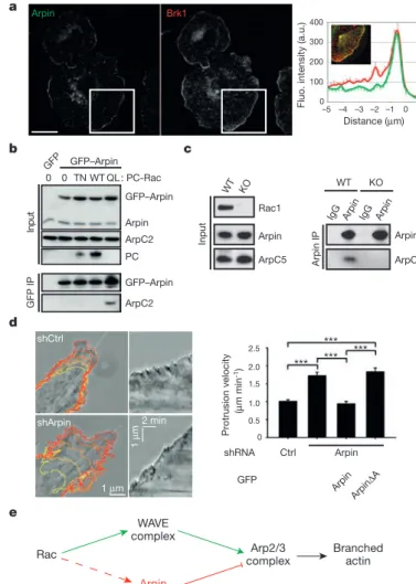
The formation of new actin filaments from actin monomers is regulated by three classes of nucleating proteins. Formins and tandem-monomer-binding nucleators form unbranched filaments (Figure 5A) while the Arp2/3 complex nucleates branched actin filaments. The Arp2/3 complex consists of seven tightly associated subunits that include the actin-related proteins Arp2 and Arp3 and five additional proteins (Machesky et al., 1994). This complex plays a dual role in actin polymerisation: it nucleates new actin filaments, and it cross-links newly formed filaments into Y-branched arrays characterised by a stereotypical branch angle of 70° (Figure 5B; Mullins et al., 1998). The Arp2/3 complex is regulated by a number of nucleation promoting factors (Derivery and Gautreau, 2010), among which proteins of the WASP (Wiskott-Aldrich syndrome protein) and WAVE (WASP-family verprolin homology protein) families. One of Arp2/3 regulators, Arpin, has recently been identified and I have participated in the characterisation of its role in in vivo cell migration during my PhD (Dang et al., 2013, APPENDIX 1).
c) Acto-myosin cortex
The cellular cortex comprises a layer of actin filaments, myosin motors, and actin-binding proteins and lies under the plasma membrane of most eukaryotic cells. Mechanically, the cortical actin mesh is the main determinant of stiffness of the cell surface and resists external mechanical stresses (Bray and White, 1988). The cortex can undergo dynamic remodelling on timescales of seconds, because of turnover of its protein constituents and network rearrangement through myosin-mediated contractions. This dynamic plasticity is a key feature of animal cell survival in a changing extracellular environment, as it allows cells to rapidly change shape, move, and exert forces (Figure 5C; Salbreux et al., 2012).
2. Cell polarity
In cell migration, polarity refers to the front-rear polarity, the molecular and functional differences between the front (closest to the direction of migration) and rear (opposite to the front) of the cell (Figure 6A). In most cases, the symmetry is broken by signals from the extracellular environment and later integrated by intracellular machineries. Cell polarity has been mostly studied in the social amoeba Dictyostelium discoideum and in neutrophils in
vitro. I will first review the different kinds of anisotropy that can break cell symmetry and
then analyse the intracellular pathways integrating these signals.
a) Breaking the symmetry; perception of the extracellular environment
The anisotropy of the extracellular environment can be of several natures. The best studied in case of cell migration have been gradients in chemical components (chemotaxis), cellular adhesion (haptotaxis), and mechanical properties (durotaxis).
Chemotaxis refers to the movement of an organism towards a chemical stimulus. Gradients of chemo-attractants like morphogens or pheromones in prospective migrating cells provide spatial cues that generate cellular asymmetry by activating specific receptors, which are distributed homogeneously in the cellular membrane. Asymmetric activation of these receptors creates a front-rear polarity that is amplified through the asymmetric recruitment and activation of signalling adaptors. This process magnifies very shallow differences in the gradient as perceived by the front and the rear of the cell (Swaney et al., 2010). The phenomenon of chemotaxis is observed in particular in wound healing. Rapid induction of
cell motility in epithelial cells and leukocytes is crucial for efficient epithelial repair and defence. Epithelial injury causes cell damage and lysis, which releases cytoplasmic molecules into the extracellular space. These damage associated molecular patterns released at the injury site, such as ATP and formylated peptides, directly mediate both cell movements and directional sensing through G-protein coupled receptor (GPCR) pathways (Enyedi and Niethammer, 2015).
In wound healing, chemotaxis is often accompanied by haptotaxis, the directional motility of cells up a gradient of cellular adhesion. In the case of haptotaxis, membrane receptors are not activated by a soluble ligand, but rather by proteins carried either by neighbouring cells or present in the extracellular matrix. This has been well characterised in mice in case of liver inflammation: chemokine ligand CXCL2 is expressed on the luminal surface of liver cells in a gradient that leads towards the injured area. Neutrophils were shown to migrate up this gradient in a chemokine receptor CXCR2-dependent manner, demonstrating the importance of chemotaxis in this system (Mcdonald et al., 2010).
Differences of mechanical properties of the environment have also been shown to play a major role in cell polarisation. Durotaxis refers to the phenomenon of cells moving according to changes in stiffness of the extracellular matrix, and has emerged as a crucial parameter controlling cell migration behaviour. A recent study has shown that the cell migration velocity doesn't have any consistency with the stiffness of the substrate, but is rather related to the stiffness gradient of the substrate. This finding suggests a new mechanism underlying the durotaxis phenomenon, highlighting the importance of the substrate stiffness gradient, rather than the stiffness itself (Joaquin et al., 2016).
b) Cellular integration of the extracellular asymmetry
The differential activation of membrane receptors such as GPCR or growth factor receptors induces diverse signalling pathways within the cells. Among the effectors that are asymmetrically recruited and activated by the membrane receptors are heterotrimeric G proteins, which activate - among other enzymes - phospholipase C (PLC) and protein kinase C (PKC), inducing the local formation of second messengers and protein phosphorylation (Van Haastert and Devreotes, 2004). G proteins also activate the phospholipid enzyme
by the Microtubule organising centre (MTOC). Modiied from Ridley et al., 2003.
ilaments (red) at the rear. Microtubules (green) originating from the centrosome (purple) are preferentially stabilised in the direction of migration allowing targeted vesicle traficking from the Golgi (brown) to the leading edge. Modiied from Jaffe and Hall, 2005. (B2) Targets of Rho GTPases. Spiering and Hodgson, 2011.
Factors Inluencing FRET Eficiency
Annu. Rev. Cell Dev. Biol. 2005.21:247-269. Downloaded from www
.annualreviews.org
by INSERM-multi-site account on 06/09/12. For personal use only
.
an important second messenger in the amplification of the response to the initial gradient of stimulus and the asymmetric activation of Rho GTPases (Garcia-Mata and Burridge, 2007).
Cdc42 is among the initial GTPases implicated in the response to polarising signals; it controls the recruitment of polarity proteins Par-3 and Par-6, atypical PKCs (aPKCs) and the
actin polymerisation machinery to the leading edge (Etienne-Manneville and Hall, 2002).
Cdc42 participates in additional polarity-related events, such as positioning the nucleus and orienting the microtubules. Cdc42 can directly promote nucleation of actin filaments via its effect on nucleation promoting factors WASP and WASP, and is essential for restricting Rac1-dependent actin polymerisation to the front of fibroblasts induced to migrate (Nobes and Hall, 1999). Rac1 is active at the front of the cell, where it activates Arp2/3 through the WAVE and WASP complex, and thereby promotes actin branching and the formation of lamellipodia (Nobes and Hall, 1999). Rho acts at the rear of the cell to generate contractile forces through Rock-mediated myosin light chain (MLC) phosphorylation, which move the cell body forwards. In addition, Rho and Rock inhibit Rac1 that also inhibits Rho, which maintains the polarity. In some situations inhibition of Rock can stimulate cell migration (Figure 6; Riento and Ridley, 2003).
3. Models of migration in vitro
Cell motility has been studied historically on two-dimensional (2D) tissue culture surfaces. In most migrating cells, a leading protrusion points in the direction of movement and is part of a polarity (Ridley et al., 2003). Cells extend three major types of membrane protrusions at the leading edge: filopodia, lamellipodia and blebs (Ridley, 2011).
a) Polymerisation-driven migration
The “crawling model” is based on actin-polymerisation-based cytoplasmic extensions, lamellipodia and filopodia. Lamellipodia are broad, sheet-like protrusions that contain a branched network of thin, short actin filaments (Ponti et al., 2004). Lamellipodia are generated by the small GTPase Rac1 and some of its effectors, as the WAVE/Scar complex and N-WASP, which control Arp2/3 (Figure 7A; Campellone and Welch, 2010; Swaney and Li, 2016). Filopodia are long, thin protrusions that emerge from the cellular membrane. They are mainly regulated by the small Rho GTPase Cdc42. They are made of long, unbranched, parallel actin bundles. Their elongation is mediated by formins (Figure 5A). Filopodia carry
out an exploratory function, enabling the cell to probe its local environment (Mattila and Lappalainen, 2008).
Polymerisation-driven migration has been described as follows: localised activation of Rac1 at the plasma membrane directs the actin nucleator Arp2/3 to form the branched filamentous actin network which drives protrusion of the lamellipodium at the front of migration (Svitkina and Borisy, 1999). Integrin receptors then form small clusters termed nascent adhesions beneath the extending lamellipodium, thereby anchoring the cellular protrusion to the underlying extracellular matrix (Swaminathan et al., 2016). The small GTPase RhoA helps to connect these nascent adhesions to acto-myosin stress fibres by activating the formin family of actin nucleators (Ridley and Hall, 1992). These force-generating machines respond to the rigidity of the surface and provide the power to enlarge and strengthen the cell-matrix adhesions needed for moving the bulk of the cell body. The cell-matrix adhesions disassemble after the nucleus passes over them, and myosin II-mediated contractility squeezes the back of the cell forwards (Figure 7A; Chen, 1981).
b) Blebbing-induced migration
First observed during primordium germ cells migration in Fundulus embryos (Trinkaus, 1973), migration by blebbing appears as an important motility mechanism and a common alternative to lamellipodia-driven migration in three-dimensional environments. Blebs are spherical protrusions formed when the plasma membrane separates from the cortex due to high cytoplasmic pressure. They differ from other cellular protrusions in that their growth is pressure-driven, rather than due to polymerising actin filaments pushing against the membrane.
The bleb life cycle can be subdivided into three phases: bleb initiation (nucleation), expansion and retraction. Bleb initiation can result from a local detachment of the acto-myosin cortex from the membrane or from a local rupture of the cortex. Hydrostatic pressure in the cytoplasm then drives membrane expansion by propelling cytoplasmic fluid through the remaining cortex or through the cortex hole. Concomitantly, the membrane detaches further from the cortex, increasing the diameter of the bleb base. As bleb expansion slows down, a new actin cortex reforms under the bleb membrane. Recruitment of myosin to the new cortex is followed by bleb retraction. In migrating cells, a new bleb forms soon after cortex re-polymerisation under the membrane (Figure 7B; Charras and Paluch, 2008).
4. In vivo: integration of the environment
a) Multiple modes of migration in vivo
Intriguingly, in addition to the well-described mode of lamellipodia-based motility, single cells can switch between several distinct 3D migration mechanisms, a phenomenon termed migratory plasticity. An early example of plasticity in the movement of cells was identified in developing Fundulus embryos (Trinkaus and Lentz, 1967). During gastrulation,
Fundulus deep cells move in the space between two confining cell layers. Non-adherent deep
cells possess large, stable blebs, which switch to flat lamellipodia or filopodia when the cells become more adhesive (Trinkaus, 1973), similar to zebrafish progenitor cells (Ruprecht et al., 2015). It is now clear that, rather than adopting one of the well described models for in vitro 2D cell migration, many cell types can use distinct mechanisms to move through diverse 3D environments (Petrie and Yamada, 2016).
b) Social behaviour of migrating cells
In multicellular organisms, cell migration is essential for development and is required throughout life for numerous processes, including wound healing and responses to infections. Dysregulation in the control of cell migration can lead to diseases such as cancer. Most migration studies have been realised in vitro, while most cells do not move as isolated entities
in vivo but rather interact with their neighbours during migration. Thus, cells must have their
locomotory machinery adapted to these constant interactions. This has prompted scientists to investigate the ‘social behaviour of cells’ (Abercrombie and Heaysman, 1953). Many models of collective migration have been described; I will here present two of them: prechordal plate collective migration and neural crest cell migration by contact inhibition of locomotion.
c) Prechordal plate progenitors collective migration
The prechordal plate is a group of cells composed of the first internalised cells on the dorsal side of the embryo. During gastrulation, the prechordal plate migrates from the embryonic organiser to the animal pole, to later give rise mainly to the hatching gland, the anterior-most structure in the zebrafish embryo (Kimmel et al., 1995; Solnica-Krezel et al., 1995). Being a cohesive group, the prechordal plate, also referred to as mesendoderm, is a very good model to study the mechanisms of oriented collective migration.
in tiles and oriented animally (arrow). Modiied from Winklbauer, 2009. (A2) The prechordal plate migrates via a distributed traction mechanism. Modiied from Weber et al., 2012.
(B1) Isolated cells (red) do not migrate towards the animal pole. When the cells are contacted by the endogenous plate (green), actin-rich protrusions are reoriented towards the animal pole and cells start migrating in this direction. (B2) Directionality is obtained by transmission of intrinsic information through cell-cell contacts. When isolated however, cells lose directionality. Modiied from Dumortier et al., 2012.
B) Zebrafish Prechordal Plate
In Xenopus, ahead of the involuting mesoderm, the leading-edge mesendodermal cells migrate as a cohesive group towards the animal pole, on the fibronectin of the blastocoele floor extracellular matrix (Winklbauer, 1990). Most studies have thus been realised on blastocoele roof explants. Prechordal plate progenitors emit frequent lamellipodia in the direction of migration, crawling underneath the cellular body of the preceding cell and thus forming a structure in tiles (Figure 8A; Winklbauer, 2009). Recent studies have shown that prechordal plate progenitors respond to local forces and migrate persistently away from the direction of the applied force (Weber et al., 2012). This response is dependent on E-cadherin. In the embryo, the traction forces that each cell exerts on the substrate must be balanced by the cell-cell adhesions that keep the cells part of a cohesive tissue. The notochord lying behind the prechordal plate is thought to exert a pulling force on the advancing prechordal plate, and thus polarise prechordal plate progenitors that migrate in the opposite direction, towards the animal pole (Figure 8A; Weber et al., 2012).
The collective behaviour of prechordal plate migration has been studied in vivo in the zebrafish embryo. These studies have revealed that all prechordal plate cells actively migrate as individuals, using actin-rich cytoplasmic extensions oriented in the direction of migration. However, prechordal plate progenitors isolated from the endogenous plate lose their orientation and do not migrate towards the animal pole. When these isolated cells are contacted by the plate, they re-orient their protrusions in the direction of movement and start migrating towards the animal pole, demonstrating that prechordal plate progenitors require a directional signal provided by contact to the endogenous plate to migrate. Prechordal plate migration is thus a true collective process, rather than the sum of individual migrations. Strikingly, groups of cells ahead of the endogenous prechordal plate do not migrate either, but resume their migration as soon as they are joined by the endogenous prechordal plate. The directionality appears to be contained in the moving group and transmitted between cells through cell-cell contacts, in an E-cadherin dependent process (Figure 8B; Dumortier et al., 2012).
d) Neural crest cell migration: contact inhibition of locomotion
The neural crest is a multipotent cell population specified at the interface between neural and non-neural ectoderms (Le Douarin and Teillet, 1973). After induction, neural crest cells undergo an epithelial-to-mesenchymal transition and delaminate from the neural tube. Neural crest cells become highly motile, colonise nearly all tissues and organs in the embryo and give
Modiied from Dupin and Le Douarin, 2014.
(B1) Contact inhibition of locomotion is represented by yellow inhibitory arrows. Collision between single cells leads to a change in the direction of migration (green arrows). Mayor and Carmona-Fontaine, 2012. (B2) Cells are polarised according to their cell-cell contacts; free edge is in green, cell contacts are in red. Théveneau and Mayor, 2011.
rise to a wide range of derivatives such as neurons, glia, bones, cartilage, endocrine cells, connective tissues and smooth muscles (Figure 9A; Dupin and Le Douarin, 2014).
Neural crest cells migrate by contact inhibition of locomotion (Carmona-Fontaine et al., 2008), a phenomenon first described in fibroblast cultures: “upon contact with another cell, a cell changes its direction and migrates in the opposite direction” (Abercrombie and Heaysman, 1954). The typical sequence of cell activities implicated in contact inhibition of locomotion are: (i) cell-cell contact, (ii) inhibition of cell protrusive activities at the site of contact, (iii) generation of a new protrusion away from the site of cell contact and (iv) migration in the direction of the new protrusion (Figure 9B; Mayor and Carmona-Fontaine, 2010). Cell-cell contact is thought to be mediated by N-cadherin, while the Wnt/PCP pathway polarises the cell by activating RhoA at the site of contact and Rac1 at the opposite site, thus inducing migration in the direction opposite to the contact (Figure 9B; Carmona-Fontaine et al., 2008; Theveneau and Mayor, 2011).
In addition to contact inhibition of locomotion, mutual cell-cell attraction (co-attraction) counterbalances the tendency of cells to disperse and allows neural crest cells to migrate collectively. Co-attraction and contact inhibition of locomotion thus act in concert to allow cells to self-organise and respond efficiently to external chemo-attractant signals (Carmona-Fontaine et al., 2011).
Active migration is omnipresent during development and can participate in the segregation of cells, and thus to the formation of embryonic boundaries that would then be stabilised as described in the first part of this introduction. One of the first separations of tissues occurs during gastrulation, when the mesoderm and the endoderm internalise to later give rise to internal tissues, while the ectoderm while give rise to the epidermis and nervous system of the embryo. In the next part, I will review the morphogenetic events that have been described for the germ-layer segregation in different model organisms, focusing on the cell movements ensuring mesoderm and endoderm internalisation.
III.
Endoderm and mesoderm internalisation in model organisms
1. Lessons from sea urchin gastrulation: two ways for cell internalisation
The first dynamic information about morphogenetic movements during gastrulation were derived from the observation and analysis of time-lapse films from 1956, taking advantage of the transparency of the sea urchin embryo (Figure 10A; Gustafson and Kinnander, 1956).
In sea urchin, gastrulation movements start after the 10th division, at 10 hours post
fertilisation. At this stage, the sea urchin embryo is a single-layered hollow blastula. The primary mesenchymal cells located at the vegetal pole internalise individually: they undergo an epithelial-to-mesenchymal transition and end up in the blastocoele. Several hours after this ingression, the vegetal plate, consisting of endomesodermal progenitors, folds inwards and initiates the archenteron. This internalisation of a coherent layer of cells has been termed invagination (Figure 10B), and results from three different mechanisms: convergent-extension movements that narrow and elongate the archenteron, involution of endodermal cells from the vegetal and lateral plate, and stretching of secondary mesenchymal cells that extend thin protrusions towards the animal pole (Miller and McClay, 1997).
These early images have enlighten two major ways for cell internalisation: either as individuals, or as a coherent layer. According to the species, mesoderm and endoderm internalisation involves either or both strategies, which I will review here for the principal model organisms (Figure 11).
2. Internalisation of a coherent layer of cells
a) Involution in Xenopus
In Xenopus, the first gastrulation movement is the indentation of the mesodermal belt at the vegetal boundary, forming the dorsal lip of the blastopore (Hardin and Keller, 1988). The blastopore groove expands laterally and dorsally and deepens, and concomitantly the internalisation of the mesoderm occurs by involution of a cohesive sheet of cells. The presumptive somitic mesoderm, the presumptive hypochord and the presumptive suprablastoporal endoderm (bottle cells) roll over the blastopore lip, thereby turning inwards.
![[PDF] Cours programmation Objet comment ca marche | Cours java](data:image/gif;base64,R0lGODlhAQABAIAAAP///wAAACH5BAEAAAAALAAAAAABAAEAAAICRAEAOw==)