HAL Id: hal-03041310
https://hal-amu.archives-ouvertes.fr/hal-03041310
Submitted on 4 Dec 2020HAL is a multi-disciplinary open access archive for the deposit and dissemination of sci-entific research documents, whether they are pub-lished or not. The documents may come from teaching and research institutions in France or abroad, or from public or private research centers.
L’archive ouverte pluridisciplinaire HAL, est destinée au dépôt et à la diffusion de documents scientifiques de niveau recherche, publiés ou non, émanant des établissements d’enseignement et de recherche français ou étrangers, des laboratoires publics ou privés.
An ionizable supramolecular dendrimer nanosystem for
effective siRNA delivery with a favorable safety profile
Dinesh Dhumal, Wenjun Lan, Ling Ding, Yifan Jiang, Zhenbin Lyu, Erik
Laurini, Domenico Marson, Aura Tintaru, Nelson Dusetti, Suzanne Giorgio,
et al.
To cite this version:
Dinesh Dhumal, Wenjun Lan, Ling Ding, Yifan Jiang, Zhenbin Lyu, et al.. An ionizable supramolecu-lar dendrimer nanosystem for effective siRNA delivery with a favorable safety profile. Nano Research, Springer, 2020, �10.1007/s12274-020-3216-8�. �hal-03041310�
Table of Contents Graphic:
An ionizable amphiphilic dendrimer self-assembles into a supramolecular dendrimer nanosystem and delivers small interfering RNA (siRNA) with potent gene silencing and effective anticancer activity, outperforming the current gold standard delivery agent while providing a favorable safety profile.
An ionizable supramolecular dendrimer nanosystem for effective siRNA delivery with a favorable safety profile
ABSTRACT
Gene therapy using small interfering RNA (siRNA) is emerging as a novel therapeutic approach to treat various diseases. However, safe and efficient siRNA delivery still constitutes the major obstacle for clinical implementation of siRNA therapeutics. Here we report an ionizable supramolecular dendrimer vector, formed via self-assembly of a small amphiphilic dendrimer, as an effective siRNA delivery system with favorable safety profile. By virtue of the ionizable tertiary amine terminals, the supramolecular dendrimer has a low positively charged surface potential and no notable cytotoxicity at physiological pH. Nonetheless, this ionizable feature imparted sufficient surface charge to the supramolecular dendrimer to enable formation of a stable complex with siRNA via electrostatic interactions. The resulting siRNA/dendrimer delivery system had a surface charge that was neither neutral, thus avoiding aggregation, nor too high, thus avoiding cytotoxicity, but was sufficient for favorable cellular uptake and endosomal release of the siRNA. When tested in different cancer cell lines and patient-derived cancer organoids, this dendrimer-mediated siRNA delivery system effectively silenced the oncogenes Myc and Akt2 with a potent antiproliferative effect, outperforming the gold standard vector, Lipofectamine 2000. Therefore, this ionizable supramolecular dendrimer represents a promising vector for siRNA delivery. The concept of supramolecular dendrimer nanovectors via self-assembly is new, yet easy to implement in practice, offering a new perspective for supramolecular chemistry in biomedical applications.
KEYWORDS
siRNA delivery, non-viral vector, dendrimer, supramolecular chemistry, ionizable vector, gene silencing
1 Introduction
Gene therapy based on small interfering RNA (siRNA) therapeutics is emerging as a novel and
promising approach to treat diseases [1, 2], particularly since the breakthrough success of the
first siRNA drug Patirisan that was approved in 2018 [3] and the recently approved second
siRNA drug Givosiran [4]. The advantages of siRNA therapeutics is their ability to potently
and specifically silence any target gene via Watson–Crick base pairing with the corresponding
complementary mRNA sequence and consequential cleavage of the mRNA. However, siRNA
therapeutics are unstable in acidic/basic conditions and in biofluids because these RNA
molecules are vulnerable to chemical and enzymatic hydrolysis. Additionally, because of their
polyanionic nature and hydrophilicity, naked siRNAs cannot easily penetrate the cell
membrane to reach the cell interior. Therefore, delivery systems that shield siRNA molecules
from hydrolytic decomposition, cover their dense negative charge and promote their transport
across cell barriers for effective gene silencing are highly sought after, which constitutes a
major challenge for clinical use [5, 6].
Identifying safe and effective systems for siRNA delivery is an actively studied topic.
Although viral vectors are generally more efficient, growing concerns over their safety and
tedious manufacturing have fostered the development of non-viral vectors which, in general,
are endowed with better safety profiles, easier manufacturing procedures and more flexible
safe-by-design options [5-9]. Lipids and polymeric systems are the two most extensively
investigated non-viral vectors [10,11]. Within the latter class, dendrimers, regularly branched
synthetic polymers with a ramified structure, are particularly promising by virtue of their
precisely controlled composition, unique cooperative multivalence and favorable high drug
loading within small nanometric volumes [12-14]. However, their translation to clinical use
To overcome the synthetic challenge of dendrimers and to capitalize on the advantages
of lipid and dendrimer vectors, we have recently proposed innovative non-covalent
supramolecular dendrimers for siRNA delivery [16-19]. The supramolecular dendrimers can
be constructed via self-assembly of small amphiphilic lipid-dendrimer conjugates through van
der Waals and hydrogen bond interactions, and combine the self-assembling feature of lipid
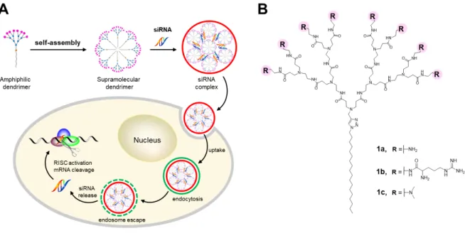
vectors with the multivalent cooperativity of dendrimer vectors for siRNA delivery (Fig. 1A).
Figure 1B shows one of our first amphiphilic lipid-dendrimer conjugates, 1a, which was
developed for siRNA delivery [19]. The structure of 1a consists of a long alkyl chain as the
hydrophobic component and a small poly(amidoamine) (PAMAM) dendron carrying primary
amine terminals as the hydrophilic entity. The dendrimer 1a self-assembles into
supramolecular dendrimer nanomicelles that favorably bind to siRNA for effective delivery
[20]. By conjugating arginine residues at the dendrimer terminals in 1a, we further obtained 1b
(Fig. 1B), which mimics arginine-rich cell penetration peptides for more effective cellular
uptake and better siRNA delivery [21]. In this report, we present a new ionizable
supramolecular dendrimer formed via self-assemble of the amphiphilic dendrimer 1c (Fig. 1B)
which, at variance with 1a and 1b, carries tertiary amine dendrimer terminals for effective
Figure 1 Schematic illustration of the supramolecular dendrimer formed via self-assembly of an amphiphilic dendrimer for siRNA delivery. (A) Cartoon presentation of the self-assembly
of an amphiphilic dendrimer into a supramolecular dendrimer, which then complexes with
siRNA to form a nanoparticle. This nanoparticle enters into cells via endocytosis and, upon
endosomal escape, releases the siRNA into the cytosol for consequential gene silencing. (B)
Chemical structures of the amphiphilic dendrimers 1a, 1b and 1c bearing primary amines,
arginine residues and tertiary amines, respectively, at dendrimer terminals.
The rationale for developing the ionizable supramolecular dendrimer formed with 1c for
siRNA delivery was based on the pKa of the tertiary amine terminals of the dendrimer surface,
which will allow their protonation to generate sufficiently high positive surface charge to bind
anionic siRNA through electrostatic interactions at physiological pH, while the surface
potential of the siRNA/dendrimer complexes should be low enough to prevent their clearance by the immune system yet sufficient to promote cellular uptake [9,10,22,23]. In addition, the
ionizable tertiary amine terminals in 1c should be highly protonated in acidic endosomes,
thereby inducing endosomal disruption for siRNA release. The first siRNA drug Patisiran is
LNPs have the disadvantage of requiring specific and laborious ethanol-loading formulation
techniques for preparation [22, 24]. We demonstrate that the amphiphilic lipid-dendrimer
conjugate 1c, which harbors eight tertiary amines at the surface and a further seven tertiary
amines and one triazole ring in the interior, self-assembles into a supramolecular dendrimer
nanosystem and acts as an ionizable cationic vector for siRNA delivery via strong siRNA
binding at physiological pH and effective release of the siRNA within the endosomal acidic
environment. Importantly, formulation of the siRNA/dendrimer complex is simple and
convenient, and requires only that siRNA is mixed with 1c in aqueous solution. In addition, the
siRNA/dendrimer complex is small and effectively protects the siRNA from enzymatic
degradation. By virtue of its beneficial ionizable nature, the supramolecular dendrimer formed
by 1c exhibited low cytotoxicity while effectively delivering siRNAs into different cancer cell
lines and patient-derived cancer organoids for functional silencing of target oncogenes, leading
to potent anticancer activities that outperformed the current gold standard Lipofectamine 2000
(Lipo2000). Consequently, this study offers conceptual advances in constructing
supramolecular dendrimers for biomedical applications. In this study, we present the ionizable
supramolecular dendrimer for siRNA delivery.
2 Results and discussion
We readily synthesized 1c via the amidation of the ester-terminated dendrimer precursor 1
using N,N-dimethylethylenediamine (Fig. 2A, Scheme S1) [25]. The amidation reaction was
maintained for 5 days to ensure complete transformation of all dendrimer terminals, and the
final product 1c was obtained with an excellent yield of 86% after purification via precipitation
in methanol/ether followed by dialysis in water. The structural integrity and purity of 1c was
confirmed using spectroscopic analysis with 1H-, 13C- and two-dimensional (2D) NMR, as well
1H NMR spectrum and at 45.3 ppm in the 13C NMR spectrum correspond to the characteristic
NMR signals of the methyl groups on the tertiary amine terminals present in 1c (Fig. S1).
Additionally, mass spectrometry analysis yielded the expected signals related to the molecular
weight and the corresponding isotopic pattern of the triply protonated species [1c+3H]3+ (Fig.
S3).
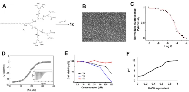
Figure 2 Synthesis, self-assembly, cytotoxicity and ionizable features of the amphiphilic
dendrimer 1c. (A) Synthesis of 1c through amidation of the ester-terminated dendrimer 1 using
N,N-dimethylethylenediamine. (B) A representative transmission electron microscopy (TEM)
image of the supramolecular dendrimer nanomicelles formed by 1c. (C) Critical micelle
concentration (CMC) of 1c estimated using a fluorescent spectroscopic assay with pyrene. (D)
Isothermal titration calorimetry (ITC) study on the demicellization of 1c in PBS buffer at pH
7.4; measured heat (Q) as a function of 1c concentration. In this panel, symbols represent
experimental values while the black line represents the Boltzmann data fit (R2 = 0.9983). Insert:
measured heat power vs. time elapsed during the titration. (E) Absence of cytotoxicity
following treatment of human embryonic kidney 293 (HEK293) cells with 1c, and comparison
of the cytotoxicity of dendrimers 1a and 1b. Cell viability was measured in triplicate using the
Remarkably, 1c is soluble in water, with solubility similar to that of the dendrimers 1a
and 1b. By virtue of its amphiphilic nature, 1c is able to self-assemble spontaneously in water
into supramolecular dendrimer micelles of 12–17 nm in size, as shown by transmission electron
microscopic (TEM) imaging (Fig. 2B). We then evaluated the critical micelle concentration
(CMC) of 1c using a fluorescent spectroscopic assay with pyrene (Fig. 2C). The CMC value
of 1c was around 17 µM, which is similar to that of 1a [19-20]. To further monitor the process of supramolecular micelle formation by 1c, we used isothermal titration calorimetry (ITC) (Fig.
2D and Fig. S4). The ITC results confirmed the ability of 1c to self-assemble into
supramolecular dendrimer nanomicelles at a CMC value of 19 µM at pH 7.4, in accordance with the results obtained from the fluorescent spectroscopic assay. Remarkably, 1c exhibited
much lower cytotoxicity than 1a and 1b, particularly at higher dendrimer concentrations, as
revealed by the toxicity assay on human kidney cells (HEK 293), mouse fibroblast cells (L929)
and Madin–Darby canine kidney cells (MDCK) (Fig. 2E and Fig. S5). The lower toxicity of
1c might be ascribed to the distinct tertiary amine terminal groups, which generated a relatively
lower surface charge for micelles formed by 1c (+23 mV) when compared with those formed
by 1a (+35 mV) and 1b (+40 mV) at pH 7.4 (Table S1). Collectively, these data confirm the
spontaneous self-assembly of 1c to form stable supramolecular dendrimer nanomicelles with a
favorable safety profile.
We next performed a potentiometric pH back titration of 1c, which showed continuous
buffering between pH 4.0 and 9.0, highlighting the ionizable nature of 1c (Fig. 2F). According
to this titration profile, 50% of the amine functionalities (8/16) of 1c were protonated at pH
7.4, likely corresponding to the eight tertiary amine terminals in 1c. At pH 5.0, corresponding
to the endosomal environment, 72% of the amine groups (12/16) were protonated, implying
that, in addition to the eight tertiary amine terminals, a further four tertiary amines located
repeatedly gave only small and constant endothermic peaks at pH 5.0 (Fig. S6 left), and the
corresponding heat flow curve could not be used to determine any CMC value of 1c under
these conditions. We reasoned that under acidic pH conditions 1c no longer forms stable
supramolecular nanomicelles because of the growing number of positive charges populating
the hydrophilic portions of the amphiphilic dendrimer. This causes stronger electrostatic
repulsions within a confined volume that eventually overcome the hydrophobic attractions
existing among the dendrimer hydrocarbon chains. Altogether, these results suggest that the
supramolecular dendrimer formed by 1c will bind to siRNA at physiological pH while allowing
endosomal release at an acidic pH.
Effectively, the supramolecular dendrimer micelles formed by 1c at pH 7.4 are
sufficiently positively charged to allow the formation of stable complexes with the highly
negatively charged siRNA molecules, thus protecting the siRNAs from degradation. We
studied this siRNA/dendrimer complex formation using a gel mobility shift assay. As Figure
3A illustrates, complete retardation of siRNA migration in the gel was observed in the presence
of 1c for N/P ratios above 2.5, suggesting formation of a stable siRNA/dendrimer complex.
TEM imaging further showed that the siRNA/dendrimer complexes at pH 7.4 were small and
spherical, i.e., ~40 nm in size (Fig. 3B), in line with the results obtained from dynamic light
scattering (DLS) analysis (Fig. 3C). In addition, we performed mesoscale molecular
simulations to investigate the interactions between siRNA and 1c, and representative
siRNA/dendrimer complexes formed are presented in Figure 3D. As clearly seen in Figure
3D, the siRNA molecules are uniformly distributed and reside on the surface of the
supramolecular dendrimers, highlighting the effective interaction between siRNA molecules
Figure 3 The supramolecular dendrimer issued from 1c formed small and stable complexes
with siRNA and protected the siRNA from degradation at physiological pH. (A) Gel retardation
of siRNA with 1c at varying N/P ratios indicates the formation of stable siRNA/dendrimer
complexes at N/P ratios ≥ 2.5. (B) TEM imaging and (C) DLS analysis of the siRNA/dendrimer
complex at pH 7.4. (D) A mesoscale molecular simulation of the siRNA/dendrimer complex
(color legend: siRNA, orange sticks; amphiphilic dendrimers, light blue sticks; micelle
hydrophobic cores, night blue spheres. Water and counterions are portrayed as a white field for
clarity). (E) ITC data for the formation of the siRNA/dendrimer complex. Integrated ITC data
(symbols) and relative fitting (solid line) using a sigmoidal model obtained from the titration
of 1c with siRNA in PBS at pH 7.4 (T = 25 °C). (F) The siRNA/ 1c complexes protect siRNA
against RNase degradation.
We also assessed the thermodynamics of binding between siRNA and the supramolecular
dendrimer formed by 1c using ITC (Fig. 3E). This revealed a negative enthalpic variation,
indicates that the driving force behind stable siRNA/dendrimer complex formation is
fundamentally electrostatic in nature. The related entropy variation upon formation of the
siRNA/dendrimer complex was also favorable (i.e., TDS = +2.92 kcal/mol), which originated mainly from the release of water molecules and counterions into bulk solvent upon
siRNA/dendrimer binding. Overall, the resulting negative Gibbs binding free energy (DG = – 7.91 kcal/mol) confirms that the formation of stable siRNA/dendrimer complexes is a
spontaneous and favorable thermodynamic process. Importantly, the siRNA/dendrimer
complex effectively protected siRNA from enzymatic degradation (Fig. 3F). Naked siRNA
was rapidly degraded within 5 min by RNase, whereas siRNA in complex with the dendrimer
was not degraded readily by RNase and remained intact even after 120 min. Taken together,
our results show that the supramolecular dendrimer formed by 1c is sufficiently protonated and
positively charged at physiological pH to enable interaction with siRNA to form small and
stable siRNA/dendrimer complexes within which the siRNA is shielded from enzymatic
degradation. These are important prerequisites for successful siRNA delivery.
Benefiting the favorable ionizable properties of the tertiary amine terminals, the siRNA
complexes formed with 1c exhibited a surface potential of +18 mV at pH 7.4, which is
substantially lower than that of siRNA complexes formed with 1a (+31 mV) and 1b (+35 mV)
(Table S1). At +18 mV, the surface potential is neither neutral nor too high to be
disadvantageous for siRNA delivery; complexes with too high surface charges may disrupt cell
membranes and induce eventual toxic effects, whereas neutral complexes have limited
colloidal stability and aggregate into large particles that precipitate. Importantly, this positive
surface potential is also sufficient and beneficial to promote cellular uptake via favorable
electrostatic interactions with the cell membrane, which is negatively charged. Notably, when
the pH decreased from 7.4 to 5.0, the zeta potential of the siRNA/1c complexes increased from
(+48 mV). This indicates that siRNA dissociates from the siRNA/dendrimer complex at pH
5.0, which is further supported by the results obtained from the ITC experiment examining the
interaction between 1c and siRNA at pH 5.0. As shown in the right panel of Figure S6, no
stable binding was detected between siRNA and 1c under acidic conditions, highlighting that
siRNA is released readily from the dendrimer complex at pH 5.0. This is in sharp contrast with
the data at pH 7.4, where siRNA and dendrimer 1c form a stable complex (Fig. 3). We further
evaluated the siRNA release from its dendrimer complex using the heparin-coupled ethidium
bromide assay (Fig. 4). As siRNA and dendrimer 1c form a stable complex at pH 7.4, no siRNA
release was detectable using heparin at the low dose of 2 unit/mL, and only 9 % siRNA release
was observed when using heparin even at the high dose of 20 unit/mL. In contrast, more than
25% siRNA was released at pH 5.0 even at the low heparin dose of 2.0 unit/mL, whereas almost
all free siRNA escaped from the siRNA/1c complex at the heparin dose of 20 unit/mL. There
results highlight that the siRNA/1c complex was not stable at pH 5.0 and disassembled upon
addition of heparin, hence releasing siRNA readily and easily. Collectively, these data support
our rationale that, by virtue of the ionizable nature of the tertiary amine terminals within 1c,
the siRNA/dendrimer complex forms easily at neutral pH but disassembles readily under acidic
Figure 4: Release profile of siRNA from the siRNA/1c complexes at N/P 10 as a function of
heparin concentration at pH 5.0 and pH 7.4 using heparin displacement - coupled ethidium
bromide fluorescent assay.
For effective gene silencing, the siRNA/dendrimer delivery system must be able to cross
cell membranes to enter cells, then escape from the acidic endosomes before releasing the
siRNA cargo to reach the RNAi machinery for gene silencing. We therefore studied the cellular
uptake and intracellular trafficking of the siRNA/dendrimer complexes in human pancreatic
cancer Panc-1 cells using confocal fluorescence microscopy. Specifically, siRNAs were
labeled with the red fluorescent tag Cy3, and the green fluorescent probe LysoTracker was
used to trace the acidic endosomes (Fig. 5). Fluorescence imaging revealed considerable red
fluorescent signals of Cy3 within Panc-1 cells following treatment with Cy3-siRNA/dendrimer
complexes after 1 h (Fig. 5A), and the uptake increased as the incubation time increased (Fig.
5E). In contrast, no obvious cellular uptake was observed when cells were treated with
demonstrate clearly the effective internalization of siRNA mediated by the dendrimer
nanovector.
Figure 5 Cellular uptake of the siRNA/dendrimer 1c complexes in human pancreatic cancer Panc-1 cells studied using confocal microscopic imaging and FACS flow with fluorescent dye
(Cy3)-labeled siRNA. Cellular uptake of the siRNA/dendrimer complexes in Panc-1 cells at 1
h (A, B, C, D) and 5 h (E, F, G, H) post-treatment, and (I) in the presence of inhibitors of
endocytosis pathways: cytochalasin D (inhibitor of micropinocytosis), chlorpromazine
endocytosis). (A, E) The red channel images highlight the Cy3-labeled siRNA/1c complexes;
(B, F) the green channel images show the endosomes labeled with LysoTracker® Green
DND-26; (C, G) the blue channel images show the nuclei of Panc-1 cells stained with the fluorescent
probe Hoechst 33342 staining solution; and (D, H) the merged images. (I) Cellular uptake of
the siRNA/1c complex is inhibited by cytochalasin D, genistein and chlorpromazine, which are
inhibitors of micropinocytosis, caveolae-mediated endocytosis and clathrin-mediated
endocytosis, respectively.
We next studied the uptake mechanism for the observed internalization of the siRNA/1c
complexes using specific inhibitors of endocytic pathways. Cytochalasin D is a
macropinocytosis inhibitor, whereas chlorpromazine and genistein are specific inhibitors of
clathrin-mediated endocytosis and caveolae-mediated endocytosis, respectively. Remarkably,
a significant reduction in the cell uptake of siRNA/1c complexes was observed when all these
inhibitors were applied accordingly (Fig. 5I). These results highlight that micropinocytosis,
clathrin-mediated endocytosis and caveolae-mediated endocytosis are all actively involved in
uptake of the siRNA/1c complex.
Subsequent intracellular trafficking of the siRNA/1c complex was studied using confocal
microscopic study. Co-localization of the majority of the red fluorescent signals of Cy3-labeled
siRNA and the green fluorescent signals of LysoTracker after 1 h of treatment (Fig. 5B–5D)
revealed trafficking of siRNA/1c complexes into endosomes. We further observed, after 5 h
treatment, considerable dispersion of the red Cy3 fluorescent signals of siRNA and separation
from the green fluorescent signals of LysoTracker (Fig. 5F–5H). These dispersed red
fluorescent signals represent the effective release of Cy3-labeled siRNAs from endosomes into
entered cells via endocytosis and then escaped from the endosomes before effectively releasing
siRNA.
Encouraged by these promising features of the siRNA/dendrimer complex, we then
assessed 1c-mediated siRNA delivery and gene silencing in different human cancer cells,
including pancreatic cancer Panc-1 cells, liver cancer HepG2 and Hep3B cells, and colorectal
cancer HT-29 cells (Fig. 6 and Fig. S7). Two different oncogenes, Myc and Akt2, were selected
as the target genes for the silencing study, because they both are promising targets for cancer
treatment. Akt2 is a threonine/serine kinase frequently activated in human cancers, with
essential roles in enhancing cancer cell proliferation and tumor progression [26, 27], whereas
Myc is an undruggable proto-oncogene that is associated with the outputs of major cellular
events such as cell cycle progression, cellular transformation and apoptosis [27, 28]. Treatment
with the corresponding siRNAs delivered by the supramolecular dendrimer formed with 1c
inhibited Myc and Akt expression effectively at both the mRNA (Fig. 6A and 6D) and protein
levels (Fig. 6B and 6E), leading to potent anticancer activity (Fig. 6C and 6F). Remarkably,
such RNAi efficacy was significantly higher than that obtained with the current gold standard
vector Lipo2000, and no gene silencing occurred when cells were treated with 1c alone, the
target siRNA alone, or the control scrambled siRNA complexed with 1c (Fig. 6 and Fig. S7).
In addition, neither the dendrimer 1c alone nor the scramble siRNA/dendrimer complex
showed any notable cytotoxicity (Fig. S8). These data highlight that dendrimer 1c is capable
of delivering siRNA efficiently and inducing effective gene silencing without adverse
Figure 6 Supramolecular dendrimer-mediated siRNA delivery and gene silencing of the oncogenes Akt2 and Myc as well as the subsequent antiproliferative effect on human liver
cancer HepG2 cells (A, B, C) and human pancreatic cancer Panc-1 cells (D, E, F) when
compared with no treatment (control), cells treated with dendrimer 1c alone, Akt2 or Myc
siRNA alone, or the scrambled siRNA/1c complex using 50 nM siRNA at an N/P ratio of 10.
The commercially available vector Lipo2000 was used as a reference control. Akt2 and Myc
expression at the mRNA (A, D) and protein (B, E) levels were determined using qRT-PCR and
western blotting, respectively. (C, F) Cell viability of HepG2 and Panc-1 cells upon treatment
with the siRNA/1c complex was assessed using the MTT test.
We further validated the efficacy of 1c-mediated siRNA delivery and consequent
anticancer activity in pancreatic cancer patient-derived organoids using siRNA targeting the
oncogene Myc (Fig. 7). Notably, patient-derived organoids are emerging as robust and
effective preclinical models that can be compared with traditional genetically engineered
animal models and patient-derived xenograft models (PDXs) [30]. More importantly, human
accurately predict responses to therapeutic treatments [31]. Myc is an undruggable oncogene
that promotes cancer cell proliferation and tumor progression [28, 29]. Therefore, using
Myc-targeting siRNA constitutes a promising strategy to combat pancreatic cancer [32], for which
there is no effective treatment. In this study, from the PaCaOmics cohort (clinical trial
registration number NCT01692873) [33, 34], we specifically selected two pancreatic cancer
patient-derived-organoid models, PDAC087T and PDAC115T, as both exhibit elevated
expression levels of Myc. Treatment of organoids PDAC087T and PDAC115T with
1c-delivered siRNA targeting Myc produced significant inhibition of Myc expression at both
mRNA and protein levels, with subsequent inhibition of cancer cell proliferation (Fig. 7).
Figure 7 Delivery of Myc siRNA in patient-derived cancer organoids by the supramolecular dendrimer formed with 1c, resulting in better gene silencing and antiproliferative effect than
Lipo2000. Pancreatic cancer patient-derived organoids PDAC087T and PDAC115T were
treated with the Myc siRNA delivered by 1c using 50 nM siRNA at an N/P ratio of 10, while
the commercial vector Lipo2000 was used for comparison. Myc mRNA (A, D) and protein (B,
E) levels were assessed using qRT-PCR and western blotting, respectively. (C, F) The viability
assay. Non-treated organoids and organoids treated with dendrimer 1c alone or Myc siRNA
alone were used as controls.
The gene silencing efficiency of the siRNA/dendrimer 1c complex in patient-derived
cancer organoids was lower than that observed for the experimental cancer cell lines. This
observation can be ascribed to the intrinsic three-dimensional (3D) organization of the cancer
cells within the organoids. Cell proliferation is often slower and lower in 3D organoids because
of space restrictions and limitations when compared with that of cancer cells seeded on plates
as two-dimensional (2D) organizations. Additionally, penetration and delivery of the
siRNA/vector complexes will be less favorable within the interior of the 3D organoids because
of the dense cellular organization in 3D when compared with that of cancer cells cultured on
2D plates. All these critical factors can reduce the efficiency of gene silencing in cancer
organoids when compared with that of cancer cells cultured on 2D plates. Nevertheless, our
supramolecular dendrimer formed by 1c showed significantly higher gene silencing and
anticancer activity when compared with the gold reference Lipo2000. These results
demonstrate that the supramolecular dendrimer formed by 1c is a promising system for
functional siRNA delivery. In addition, siRNA-based treatment in combination with human
cancer organoid models can also offer a reliable strategy for developing personalized treatment.
3 Conclusions
In summary, we report the ionizable supramolecular dendrimer nanovector formed by
self-assembly of the amphiphilic dendrimer 1c and its effective delivery of siRNA for RNAi-based
gene silencing in treating cancer. By virtue of its amphiphilic nature, 1c readily self-assembled
into a supramolecular dendrimer nanovector, which acted as an efficient and safe ionizable
positive surface charge of the resulting siRNA/dendrimer complex. Contextually, it is also able
to induce effective endosomal escape and subsequent cytoplasmic siRNA release at an acidic
pH, as supported by its pH titration profile and the experimental data for siRNA binding and
endosomal release. Remarkably, the siRNA/dendrimer complexes protected the siRNA from
degradation and delivered the siRNA effectively into cancer cells to target the corresponding
oncogenes for gene silencing. The siRNA/dendrimer 1c complex showed considerable
anticancer activity in different human cancer cell lines and in patient-derived-organoid cancer
models, outperforming the current gold standard Lipofectamine 2000. In contrast to our
previous dendrimers [19, 21], the 1c dendrimer nanosystem displayed almost no discernible
cytotoxicity when tested on human kidney cells (HEK 293), mouse fibroblast cells (L929) and
Madin–Darby canine kidney cells (MDCK). With effective and non-toxic delivery features,
combined with simple formulation, the supramolecular dendrimer nanosystem formed by 1c
holds great potential in siRNA delivery for both genomic and therapeutic applications, in
particular for primary immune cells, which are very sensitive to delivery materials and
therefore require especially effective and non-noxious delivery [35-37]. The present study
highlights that imparting ionizable properties via tertiary amine functionalities is an effective
and practical approach for siRNA delivery with a favorable safety profile. This study also offers
a new perspective for a supramolecular dendrimer in biomedical applications.
Acknowledgments
This work was supported by the Ligue Nationale Contre le Cancer (LP, ZL), China Scholarship
Council (WL, LD), Italian Association for Cancer Research (IG17413) (SP), the French
National Research Agency under the frame of the H2020 Era-Net EURONANOMED
European Research projects ‘Target4Cancer’, ‘NANOGLIO’, ‘TARBRAINFECT’,
from COST Action CA 17140 ‘Cancer Nanomedicine from the Bench to the Bedside’
supported by COST (European Cooperation in Science and Technology).
Author contributions
LP coordinated the project. DD and ZL synthesized the agents; DD, LD, AT and SG
characterized the agents; EL and DM performed ITC; ND and JLI provided the patient tumor
models; YJ carried out cell uptake experiments; LD performed the toxicity and gel shift studies;
WL performed all siRNA delivery experiments; DD, WL, LD, YJ, ZL, EL, AT, EL, NG, JLI,
SG, SP and LP analyzed data; and LP wrote the paper with contributions from DD, WL and
SP. All authors proofed the manuscript.
Associated content
Supporting information: Table S1, Scheme S1 and Figures S1-S8, as well as all detailed
experimental methods and protocols. This information is available free of charge via the
Internet.
References:
1. Setten, R. L.; Rossi, J. J.; Han, S. The Current State and Future Directions of
RNAi-based Therapeutics. Nat. Rev. Drug Dis. 2019, 18 (6), 421–446.
2. Delivering the Promise of RNA Therapeutics. Nat. Med. 2019, 25 (9), 1321.
3. Ledford H. Gene-silencing Technology Gets First Drug Approval After 20-year Wait.
Nature. 2018, 560 (7718), 291-292.
4. Mullard, A. RNAi agents score an approval and drive an acquisition. Nat. Rev. Drug
5. Kanasty, R.; Dorkin, J. R., Vegas, A., Anderson, D. Delivery Materials for siRNA
Therapeutics. Nat. Mater. 2013, 12 (11), 967-77.
6. Dong, Y.; Siegwart, D. J.; Anderson, D. G. Strategies, Design, and Chemistry in siRNA
Delivery Systems. Adv. Drug. Deliv. Rev. 2019, 144, 133-147.
7. Kim, B.; Park, J.; Sailor, M. J. Rekindling RNAi Therapy: Materials Design
Requirements for In Vivo siRNA Delivery. Adv. Mater. 2019, 31(49), 1903637.
8. Yin, H.; Kanasty, R.; Eltoukhy, A.; Vegas, A.; Dorkin, J.; Anderson, D. Non-viral
Vectors for Gene-Based Therapy. Nat. Rev. Genet. 2014, 15 (8), 541-555.
9. Ho, W.; Zhang, X.; Xu, X. Biomaterials in siRNA Delivery: A Comprehensive Review.
Adv. Healthc. Mater. 2016, 5 (21), 2715-2731.
10. Rietwyk, S. Peer, D.; Next-Generation Lipids in RNA Interference Therapeutics. ACS
Nano. 2017, 22 (8), 7572-7586.
11. Cooper, B. M.; Putnam, D. Polymers for siRNA Delivery: A Critical Assessment of
Current Technology Prospects for Clinical Application. ACS Biomater. Sci. Eng. 2016,
2 (11), 1837-1850.
12. Khandare, J.; Calderon, M.; Dagia, N.; Haag, R. Multifunctional Dendritic Polymers in
Nanomedicine: Opportunities and Challenges. Chem. Soc. Rev. 2012, 41(7),
2824-2848.
13. Liu, X.; Rocchi, P.; Peng, L. Dendrimers as Non-viral Vectors for siRNA Delivery.
New J. Chem. 2012, 36 (2), 256-263.
14. Cao, Y.; Liu, X.; Peng, L. Molecular Engineering of Dendrimer Nanovectors for siRNA
Delivery and Gene Silencing. Front. Chem. Sci. Eng. 2017, 11, 663–675.
15. Svenson, S., The dendrimer paradox – high medical expectations but poor clinical
16. Dong, Y.; Yu, T.; Ding, L.; Laurini, E.; Huang, Y.; Zhang, M.; Weng, Y.; Lin, S.; Chen,
P.; Marson, D.; Jiang, Y.; Giorgio, S.; Pricl, S.; Liu, X.; Rocchi, P.; Peng, L.; A Dual
Targeting Dendrimer-Mediated siRNA Delivery System for Effective Gene Silencing
in Cancer Therapy. J Am Chem Soc. 2018, 140 (47), 16264-16274.
17. Liu, X.; Wang, Y.; Chen, C.; Tintaru, A.; Cao, Y.; Liu, J.; Ziarelli, F.; Tang, J.; Guo,
H.; Rosas, R;. Giorgio, S.; Charle, L.; Rocchi, P.; Peng, L. A Fluorinated Bola‐
amphiphilic Dendrimer for On‐demand Delivery of siRNA, via Specific Response to
Reactive Oxygen Species. Adv. Funct. Mater. 2016, 26 (47), 8594-8603.
18. Liu, X.; Zhou, J.; Yu, T.; Chen, C.; Cheng, Q.; Sengupta, K.; Huang, Y.; Li, H.; Liu,
C.; Wang, Y.; Posocco, P.; Wang, M.; Cui, Q.; Giorgio, S.; Fermeglia, M.; Qu, F.; Pricl,
S.; Shi, Y.; Liang, Z.; Rocchi, P.; Rossi, J.; Peng, L. Adaptive Amphiphilic
Dendrimer-based Nanoassemblies as Robust and Versatile siRNA delivery Systems. Angew. Chem.
Int. Ed. 2014, 53 (44), 11822-11827.
19. Yu, T.; Liu, X.; Bolcato-Bellemin, A.; Wang, Y.; Liu, C.; Erbacher, P.; Qu, F.; Rocchi P.; Behr, J.; Peng, L. An Amphiphilic Dendrimer for Effective Delivery of Small Interfering RNA and Gene Silencing in Vitro and in Vivo. Angew. Chem. Int. Ed. 2012,
51 (34), 8478.
20. Chen, C.; Posocco, P.; Liu, X.; Cheng, Q.; Laurini, E.; Zhou, J.; Liu, C.; Wang, Y.;
Tang, J.; Col, V.; Yu, T.; Giorgio, S.; Fermeglia, M.; Qu, F.; Liang, Z.; Rossi, J.; Liu,
M.; Rocchi, P.; Pricl, S.; Peng, L. Mastering Dendrimer Self-Assembly for Efficient
siRNA Delivery: From Conceptual Design to In Vivo Efficient Gene Silencing, Small,
2016, 12 (27), 3667-3676.
21. Liu, X.; Liu, C.; Zhou, J.; Chen, C.; Qu, F.; Rossi, J.; Rocchi, P.; Peng, L. Promoting siRNA Delivery via Enhanced Cellular Uptake Using an Arginine-decorated
22. Kulkarni, J. A.; Cullis, P. R.; Meel, R. Lipid Nanoparticles Enabling Gene Therapies:
From Concepts to Clinical Utility. Nucleic Acid Ther. 2018, 28(3), 146-157.
23. Cullis, P. R., Hope, M. J., Lipid Nanoparticle Systems for Enabling Gene Therapies,
Mol. Ther. 2017, 25 (7), 1467-1475.
24. Evers, M. J. W.; Kulkarni, J. A.; Meel, R.; Cullis, P. R.; Vader, P.; Schiffelers, R. M.
State‐of‐the‐Art Design and Rapid‐Mixing Production Techniques of Lipid
Nanoparticles for Nucleic Acid Delivery. Small Methods, 2018, 2 (9), 1700375.
25. Wu, J.; Zhou, J.; Qu, F.; Bao, P.; Zhang, Y.; Peng, L. Polycationic Dendrimers Interact
With RNA Molecules: Polyamine Dendrimers Inhibit the Catalytic Activity of Candida
Ribozymes, Chem. Commun. 2005, 21 (3), 313-5.
26. Manning, B. D.; Toker, A. AKT/PKB Signaling: Navigating the Network, Cell, 2017,
20(3), 381-405.
27. Franke, T. F. PI3K/Akt: Getting it Right Matters, Oncogene, 2008, 27(50), 6473-88.
28. Dang, C. V. MYC on the path to cancer, Cell, 2012, 149 (1), 22-35.
29. Chen, H.; Liu, H.; Qing, G. Targeting Oncogenic Myc as a Strategy for Cancer
Treatment, Signal Transduct. Target Ther. 2018, 3, 5.
30. Tuveson, D.; Clevers, H. Cancer modelling meets human organoid technology. Science,
2019, 364 (6444), 952-955.
31. Vlachogiannis, G.; Hedayat, S.; Vatsiou, A.; Jamin, Y.; Fernández-Mateos, J.; Khan,
K.; Lampis, A.; Eason, K.; Huntingford, I.; Burke, R.; Rata, M.; Koh, D.; Tunariu, N.;
Collins, D.; Hulkki-Wilson, S.; Ragulan, C.; Spiteri, I.; Moorcraft, S.; Chau, I.; Rao,
D. Watkins, N. Fotiadis, M. Bali, M. Darvish-Damavandi, H. Lote, Z. Eltahir, E.
Smyth, Sa.; Begum, R.; Clarke, P.; Hahne, J.; Dowsett, M.; Bono, J.; Workman, P.;
Sadanandam, A.; Fassan, M.; Sansom, O.; Eccles, S.; Starling, N.; Braconi, C.;
Model Treatment Response of Metastatic Gastrointestinal Cancers, Science, 2018, 359,
920-926.
32. Wirth, M.; Mahboobi, S.; Krämer, O. H.; Schneider, G. Concepts to Target MYC in
Pancreatic Cancer, Mol Cancer Ther, 2016, 15 (8), 1792-1798.
33. Dusetti, N.; Iovanna, J. Organoids from Pancreatic Ductal Adenocarcinoma, Med Sci
(Paris), 2020, 36, 57-62.
34. Bian, B.; Juiz, N.; Gayet, O.; Bigonnet, M.; Brandone, N.; Roques, J.; Cros, J.; Wang,
N.; Dusetti, N.; Iovanna, J. Pancreatic Cancer Organoids for Determining Sensitivity to
Bromodomain and Extra-Terminal Inhibitors (BETi), Front Oncol. 2019, 9, 475.
35. Ellert-Miklaszewska, A.; Ochocka, N.; Maleszewska, M.; Ding, L.; Laurini, E.; Jiang,
Y.; Roura, A.-J.; Giorgio, S.; Gielniewski, B.; Pricl, S.; Peng, L.; Kaminska, B.
Efficient and Innocuous Delivery of siRNA to Microglia Using an Amphiphilic
Dendrimer Nanovector, Nanomedicine, 2019, 14, 2441–2458.
36. Garofalo, S.; Cocozza, G.; Porzia, A.; Inghilleri, M.; Raspa, M.; Scavizzi, F.; Aronica,
E.; Bernardini, G.; Peng, L.; Ransohoff, R. M.; Santoni, A.; Limatola, C. Natural Killer
Cells Modulate Motor Neurons-Immune Cells Cross Talk in Amyotrophic Lateral
Sclerosis, Nat. Commun. 2020, 11, 1773.
37. Chen, J.; Aleksandra, E.-M.; Garofalo, S.; Dey, A.; Tang, J.; Jiang, Y.; Clément, F.;
Marche, P.; Liu, X.; Kaminska, B.; Santoni, A.; Limatola, C.; Rossi, J.; Zhou, J.;
Peng, L., Synthesis and use of an amphiphilic dendrimer for siRNA delivery into