Archives • 2016 • vol.1 • 31-43
HIGH IN VITRO ANTIUROLITHIATIC EFFECT OF PITURANTHOS
SCOPARIUS ROOTS EXTRACTS
Benalia, H.1; Djeridane, A.1,2,*; Bensafieddine, F.1; Yousfi, M.1,2
1Laboratoire des sciences fondamentales, Université Amar Telidji. PB 37G, 03000, Laghouat- Algérie. 2laboratoire des sciences chimique et physiques appliquées, ENS de Laghouat, BP 4033, 03000, Laghouat- Algérie.
*amardjeridane@yahoo.fr; a.djeridane@mail.lagh-univ.dz
Abstract
The richness of the steppe zone by the medicinal plants and their diversity traditional uses in the region of Laghouat, allowed us to study the inhibitory effect of Herniaria fontanesii J.Gay, Pituranthos scoparius and Cynodon dactylon (L) extracts on the formation of calcium oxalate kidney stones. Firstly, we have carried out phytochemical screening and quantitative analysis of phenolic compounds of various extracts from the three plants. The obtained results proved that our extracts are rich in catechic tannins and C-, O-heterosides, with a total phenolic content ranged from 0.867 to 38.835 mg of gallic acid equivalent per 1 g of dry matter. The in vitro antiurolithiatic activity of different plants extracts has been carried out by two different models. In the turbidimetric assay, we have determined spectrophotometrically the effect of the extracts (1 g/L) on the oxalocalcic crystallization, induced by addition of oxalate in urines from healthy subject. Whereas, the gravimetric assay is based to measure the variation of calcium urate and uric acid renal calculi weight, after putting them in contact with 5 ml of the extracts (5g/L) during 15 days. In the two assays, the antiurolithiatic activity was compared with that of two antiurolithogenesis inhibitory standards: sodium citrate and Succinimide pharbiol. The achieved results measured by the two tests, show clearly that the hydromethanolic extracts of Pituranthos scoparius roots have provided very important antiurolithiasic power (>40% of inhibition) compared to the standard inhibitors. However, this result is a great step forward towards the search for an effective treatment for the urinary calculi formation. But, this work will have to be confirmed by in vivo experiments in order to validate these in vitro observations on the contributions of these three plants in the treatment of the oxalocalcic renal calculi.
Key Words Medicinal plants, Pituranthos scoparius roots, phytochemical screening, kidney stones, Calcium oxalate, antiurolithiasic.
_______________________________________ http://pharmacologyonline.silae.it
Introduction
During the past decades, the use of herbal medicines continues to expand rapidly across the world. Many people now take herbal medicines or herbal products for their health care in different national health-care settings [1]. However, the safety of herbal medicines has become a major concern to both national health authorities and the general public. The world health organization (WHO) requested to compile a list of medicinal plants and to establish international specifications for the most widely used medicinal plants and simple preparations. The WHO estimated that 80% of the populations of developing countries rely on traditional medicines, mostly plant drugs, for their primary health care needs. Therefore, the recent studies were focused on the scientific evaluation of traditional drugs of the vegetable which are rich in a wide variety of secondary metabolites, such as tannins, terpenoids, alkaloids, and flavonoids [2]. In Algeria, the alternative medicine is still largely requested by the population for several reasons which are being the same ones in other countries. Essentially, patients are not satisfied with the treatments which they receive and preferred herbal remedies which appear to be more natural without harmful side-effects are also inexpensive. For example, the conventional urinary calculi medication like diuretics and anti-inflammatory drugs are the most common prescription used to treat calcium renal calculi oxalocalcic, but they can cause serious side effects [3]. It effects about 2-15% of the world population mostly in the industrialized countries. It is the third most prevalent disorder in the urinary system with recurrence rate of 50% [4]. The highest average frequency is between 30 and 50 years of age, with an obvious male prevalence [5]. There are mainly four types of calculi. About 75% of stones are calcium containing, composed of calcium oxalate or calcium oxalate mixed with calcium phosphate and 15% of stones are the so-called triple phosphate or struvite stones, composed of magnesium ammonium phosphate. It is almost associated with infection. Uric acid stones from 6% and cystine stones 1-2% [6]. A large number of plant drugs have been used in Algeria since ancient times which claim efficient cure of urinary stones. Amongst the medicinal plants used in urolithiatic are Cynodon dactylon (L) and Herniaria fontanesii J.Gay. Pituranthos scoparius (Guezzah) family Asteraceae is an aphyllous perennial plant where its leaves are used by the local population as jaundice and antiurolithiatic natural remedies.
However, no scientific study has been reported regarding the anti-urolithiatic property of Pituranthos scoparius root extract. Therefore, the objective of the present study was to investigate and to validate the antiurolithiatic property of Pituranthos scoparius extracts in the inhibition/dissolution of calcium oxalate and uric acid stones.
Material and methods
Plants and reagents
Plants (Cynodon dactylon (L), Pituranthos scoparius and Herniaria fontanesii J.Gay.) were collected from a local herbalist in December 2014 flowering times between March and May in the area of the Algerian Saharian Atlas at 40 km (at 40 km at the north of Laghouat). The samples were identified at the Agronomic National Institute of Alger, and the voucher specimens were deposited at the laboratory of Fundamental Sciences, University of Laghouat. The investigated plants were dried and stored in dark. All chemicals were of the highest quality available and were purchased from Sigma-Aldrich (France).
Preparation of the extracts
The extracts have been prepared by maceration and decoction. The decoction of dried plant material was prepared by boiling 2 g of powder in 100 ml of distilled water for 5 min. While, the maceration of 2 g of powder was soaked in 100 ml of 4 different mixture solvents: acetone (100%), methanol (100%), acetone-water (50%-50%) and methanol-water (50%- 50%) at room temperature for 24 hours with occasional shaking. Then, each extract was filtered through a single layer of muslin cloth, and then final filtrate was collected by passing it through a Whatman grade 1 filter paper in a Büchner funnel under vacuum. The filtrates were evaporated to dryness on a Rotary Evaporator under reduced pressure. Dried extracts were dissolved, placed in a bottle, stopped and then stored at -4 0C until used. We obtained then, 5 acetonic extracts, 5 methanolic extracts, 5 aqueous extracts, 5 hydromethanolic extracts and 5 hydroacetonic extracts.
Phytochemical analysis of the extracts
The phytochemical screening on 25 prepared extracts was carried out to determine the presence of alkaloids, flavonoids, tannins, saponins, starch, glycosides, anthocyances, coumarins, sterols/triterpenes, anthraquinones and reducing compounds by using standard qualitative procedures as described by Trease and Evans [7], Sofowora [8] and Harborne [9].
_______________________________________ http://pharmacologyonline.silae.it
Determination of phenolic content
The concentration of total phenolics in different plant extracts was estimated by the Folin-Ciocalteu procedure [10], which is considered as the best method for total phenolics (included tannins) determination. The total phenolic content of the samples was expressed as gallic acid equivalents (GAE), which reflected the phenolic content as the amount of gallic acid (mg) in 1 gram of dry material.
Thin layer chromatography (TLC) Analysis
The presences of different active constituents in hydromethanolic extract of Pituranthos scoparius roots were analyzed through two-dimensional TLC analysis. The one-dimensional TLC analysis chromatogram was developed by using commercially available aluminum sheets of Silica gel 60 F254 (Merck) with ethyl acetate /dichloromethane/cyclohexane in the volume ratio 4.7/3.5/1.76 as eluent. The mobile phase for two-dimensional TLC analysis was: dichloromethane / methanol / water in volume ratio 5/4.16/0.84. Spots were observed under UV light at 366 nm and 254 nm before and after spraying with Dragendorff reagent, 10% ethanolic aluminum chloride, Liebermann-Burchard reagent and vanillin sulphuric acid reagent.
In vitro antilithiatic activity Turbidimetric method
The in vitro antilithiatic activity of the extract was tested in terms of inhibition of calcium oxalate formation by the extracts in the presence and absence of inhibitors (standard drugs and extract) [11]. The Precipitation of calcium oxalate at 37°C and pH 6.8 has been studied by the measurement of turbidity at 620nm. A spectrophotometer UV/Visible (Shimadzu 1800) was employed to measure the turbidity caused due to formation of calcium oxalate in treatments.
Twenty four hours urine sample was collected from a healthy subject in a propylene bottle containing sodium azide as an antibacterial agent. 100 μl of inhibitor solution at 1 mg/ml were added to an aliquot of 1 ml of urine and allowed to warm up to 37°C. Finally, 1 ml of 25 mM sodium oxalate solution was added and tubes were incubated at 37°C for 300 sec. Each set was replicated five times. At the end of the experiment, tubes were read at 620 nm. Tubes with no extract added were used as control. The antilithiatic activity obtained for each plant extract was compared with that of Succinimide Pharbiol and sodium citrate as positive controls. The rate of nucleation was estimated by
comparing the induction time in the presence of the extract with that of control. Data was represented in percentage inhibition and calculated using the following mathematical formula:
Inhibition % = {1-[Ai/ Ac]} x 100
Where; Ai: Absorbance in the presence of inhibitor (drugs/extracts), Ac: Absorbance without Inhibitor (Control).
Gravimetric method
The dissolution percentage of renal calculus (C1: uric acid; C2: sodium urate) was evaluated by the ratio of these weight losses using analytical balance. Pure renal stones were incubated with 5 ml of plant extracts or standard drugs at 5 g/l during 2 weeks under magnetic stirring. Only, the extracts that have showed inhibition percent higher than 40% in the turbidimetric model, have been tested in this assay. Each experiment was performed in triplicate. The mass loss of the stones was measured after the two weeks period. The amount of remaining undissolved stones is subtracted from the total quantity used in the experiment in the beginning to know the total quantity of dissolved. The antiurolithiasic activity was expressed by the percentage of dissolution of the renal calculus (D %).
D % = ((m0 – mf)/ m0) x 100
Where; D%: Rate of dissolution, m0: Initial stone weight, mf: Undissolved stone weight.
Statistical analysis
All the treatments were performed in triplicates and each data point in the results is the mean of three replicates. All experiments were repeated at least once. The statistical significance of the treatment effect was expressed as mean ± SEM.
Result and discussion
Determination of total phenol content
The obtained crude extracts present thick viscous appearance with different colors. The percentage yields of the extracts were ranged between 2.40% and 38.83% (Table 1). Herniaria fontanesii J. Gay aerial parts exhibited the highest crude extract content, while the lowest percent yield is revealed in Pituranthos scoparius roots extract. Moreover, the average of percent yields of acetonic extracts is at least lower than that of aqueous or methanolic extracts. Recent studies have shown that phenolic plant extracts contribute significantly to the
_______________________________________ http://pharmacologyonline.silae.it
antilithiatic activity [12, 13]. Therefore, one of the purposes of this study were to investigate the phenolic content of different parts of plants and to identify the best extraction solvent to further characterize the antilithiatic properties of these extracts and their effect on health.
The average of total phenol contents of twenty five plant extracts measured by Folin-Ciocalteu assay is presented in Table 1. The average total phenols content of ethanolic extracts was significantly different (p < 0.05) from aqueous, acetonic or methanolic extracts. The amount of total phenolics changes in different plants varies from 0.867et 5 .160 mg GAE/g of dry material. The highest total phenolic levels have been detected in the methanolic and hydromethanolic fractions, and the lowest in acetonic extracts. Also, results show that among tested samples, aerial plant extracts have significantly higher (p <0.005) total phenols content than those found in roots for the same plant. According to recent studies [14-17], the difference in polarities of extracting solvents might influence the solubility of chemical constituents in a sample and its extraction yield. Therefore, the selection of an appropriate solvent system is one of the most relevant steps in optimizing the recovery of total phenolics and other bioactive compounds from extracts.
Phytochemical analysis of extracts
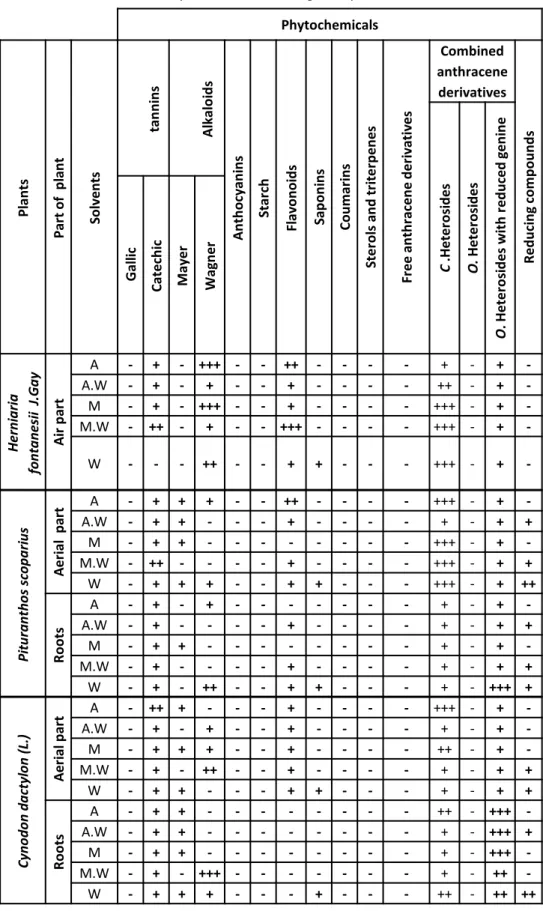
Medicinal plants contain some organic compounds which provide definite physiological action on the human body and these bioactive substances include tannins, alkaloids, carbohydrates terpenoids, steroids and flavonoids. Thus, qualitative phytochemical studies were performed on extracts using suitable chemicals and reagents to confirm the presence of alkaloids, flavonoids, tannins, saponins, starch, glycosides, anthocyanins, coumarins, sterols/triterpenes, anthraquinones and reducing compounds. The phytochemical characteristics of the twenty five extracts of three medicinal plants tested were summarized in the Table 2. Phytochemical analysis showed the presence and absence of certain chemical constituents. Moreover, the results revealed the presence of medically active compounds in the different studied extracts. The results of qualitative phytochemical studies indicate that the maximum numbers of chemical constituents were present in the aqueous extract of Pituranthos scoparius aerial part when compared to the other extracts. As well, it could be seen that tannins and heterosides were present in all the plant extracts.
Saponins were present only in the aqueous extracts of all plant parts. Moreover, alkaloids are present in all Herniaria fontanesii J. Gay extracts with Wagner reaction but they are absent with Mayer reagent. The appearance of the reducing compounds is also verifiable in almost extracts of Pituranthos scoparius and Cynodon dactylon (L.) in particular those which contain water. The flavonoids are also present in almost all plants extracts except the Cynodon dactylon (L.) roots extracts. They are presented in variable quantities but with higher contents in the Herniaria fontanesii J. Gay aerial part extracts. Therefore, starch, glycosides, anthocyanins, coumarins, sterols/triterpenes and anthraquinones were absent on all plants extracts.
However, the absence of some phytochemicals constituents in all extracts like coumarins, anthocyanins, sterols and terpenes does not justify their total absence in these plants but, the extraction methods used in our work are not satisfactory to extract desired metabolites. For example, phytochemical analyses which were carried out by some researchers [18-20] on the two parts of Pituranthos scoparius extracts revealed the presence of coumarins in certain extracts. Moreover, terpenoids are present also in almost all the studied extracts. Furthermore, some researchers [21, 22] reported that Cynodon dactylon (L.) contains sterols and terpenoids, which does not corroborate with our results.
Evaluation of antiurolithiatic activity
Urinary calculi are the third prevalent disorder in the urinary system. Urinary calculi may cause obstruction, hydronephrosis, infection and hemorrhage in the urinary tract system [23]. Surgical operation, lithotripsy and local calculus disruption using high-power laser are widely used to remove the calculi. However; these treatments are relatively costly, painful and require expert hands with availability of appropriate equipments [24]. Currently, phytotherapy or traditional medicine was employed for the the evaluation of antiurolithiatic activity, and certain medicinal herbs has been used in traditional medicine to treat urinary stones. Hence, caryophyllaceae, fabaceae and apiaceae are the most widely plant families used for the treatment of kidney stones [25].
The objective of the present study was to investigate and to validate the antiurolithiatic property of Herniaria fontanesii J. Gay, Pituranthos scoparius and Cynodon dactylon (L.) extracts in experimentally-induced urolithiatic in vitro. The choice of our investigated plants is based on two criteria: first, _______________________________________
in this domain there are few studies in Algeria that deals with these plants, and the second is that these plants have ethnopharmacological data indicating their traditional utilization against urinary stones. The formation of such urinary calculus involves several physicochemical events, e.g. nucleation, growth and aggregation, but the mechanism of these processes remains incompletely understood. The inhibiting potential of normal urine is attributed to the organic substances present in all urines. Most of the natural inhibitors are not present in sufficient concentrations [26]. Therefore, to prevent this physiological malfunction, the precipitation of calcium oxalate was carried out in the absence and the presence of twenty five extracts which compared to two standard inhibitors (sodium citrate and Succinimide pharbiol).
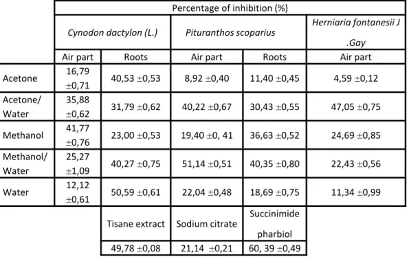
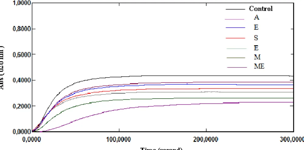
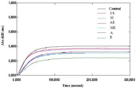
In our study, the incubation of healthy subject man urine with sodium oxalate at 37°C and pH 6.8 results the calcium oxalate precipitation. Measurements of the turbidity with the presence and without of inhibitor at 620 nm during 300 seconds provide the following curves (Fig. 1-7). Then the percentage of inhibition was calculated at the same concentration of all inhibitors (1 g/l). All the extracts showed inhibition of calcium oxalate crystallization at this concentration (Table 3) when compared with sodium citrate. But, the hydromethanolic extract of Pituranthos scoparius aerial part and the aqueous extract of Cynodon dactylon (L.) roots showed significant inhibition on calcium oxalate crystallization (51,14 and 50,59% respectively) when compared with Sussinimide pharbiol. Moreover, best inhibition in stone nucleus formation (turbidity) was seen by the hydromethanolic and hydroacetonic extracts as compared to the control which is significantly different from other extracts. Furthermore, there is no significant linear correlation observed between the antiurolithiatic activity and total phenolic content According to the phytochemical analysis, extracts which found to contain flavonoids showed good anticristallo-oxalocalcic activities. This observation is agreeing with that of literature investigation which indicates that flavonoids can reduce oxalocalcic crystallization [27]. Thus, the hydroacetonic extract of the aerial part of Herniaria fontanesii J.Gay which is traditionally used for the treatment of kidney stones 4 have provided high percent inhibition (47.05%). This is probably due to the presence of polyphenols which can form a complex with the calcium cations [26]. Also, the hydromethanolic extracts of the aerial part and roots of Pituranthos scoparius prevent 51.14% and
40, 35% of calcium oxalate formation respectively. This result can be interpreted that extracts have oxalocalcic inhibitors such as tannins. These compounds which contain acid functional group may directly imply complexation of calcium ions, and consequently the reduction of oxalocalcic crystallization [28, 29]. As well, hydromethanolic and hydroacetonic extracts of Cynodon dactylon (L.) aerial part inhibit oxalocalcic crystallization with percentages exceeding 35%. That can be related to the presence of alkaloids. Moreover, the acetonic and aqueous roots extracts of the same plant exhibit good inhibitions properties (higher than 40.50%), which can be due to their high content in saponins [30]. Thus, we noted that extracts exhibiting oxalocalcic inhibition percentage upper than 47%, contain catechic tannins, C-Heterosides and O-Heterosides. Subsequently, it is thought that the anticristallo-oxalocalcic activity of these extracts could be due to the existence of these three phytochemical families.
After the screening of anticristallo-oxalocalcic effect of our extracts by using the turbidimetric model, another in vitro assay (gravimetric model) was carried out to select the most effective extracts providing kidney stones dissolution. For this reason, extracts which having anticristallo-oxalocalcic inhibition percentages higher than 40% have been testing for their dissolution power against two types of kidney stones (uric acid: CAS number is 54495-64-6; formula C5H4N4O and calcium urate: CAS number is 36-1619; formula C10H6CaN8O6). Kidney stones were exposed for two week, in contact with extracts and the percentage of dissolution was calculated by comparing the residual weight of calculus to their initial weight [31]. The obtained results of this assay are shown in Table 4. According to the results exposed in Table 4, we can see that plants extracts were appeared to be more effective than the sodium citrate solution and less effective than Succinimide pharbiol used as standards. Therefore, the dissolution rates of sodium citrate solution for acid uric and sodium urate stones were 11.72% and 6.88% respectively. While, Succinimide pharbiol drug exhibited higher dissolving power than plant extracts, with dissolution rates of 42.47% and 75.95% for acid uric and sodium urate calculus respectively. However, the hydromethanolic extract of Pituranthos scoparius roots had the most efficient dissolving power for acid uric calculi with 46.39% dissolution rate. Whereas, the hydroacetonic extract of Pituranthos scoparius aerial part and the methanolic extract of Cynodon dactylon (L.) roots revealed a dissolution rates of 30.33% and 28.6% respectively, _______________________________________
for sodium urate calculi. Thus, methanolic and acetonic extracts of the two parts of Cynodon dactylon (L.) exhibited a weak dissolving activity with percentage of dissolution ranging between 1.37 and 9.11%. Moreover, uric acid calculi were better dissolved by some extracts than sodium urate and vice versa. Hence, the good dissolving power of hydromethanolic extract of Pituranthos scoparius roots can be resulted from the interaction between uric acid and molecules present in this extract during the dissolution process. According to the phytochemical analysis of this extract, we suppose that its higher activity could be due to the presence of tannins, flavonoids or anthracene derivative. This observation is supported by certain research studies [32, 33].
Moreover, phytochemical screening by one and two-dimensional thin-layer chromatography of this extract allowed us to highlight the presence of flavonoids (AlCl3Reagent, yellow fluorescence spot under UV light) (Fig. 7-10). It is also possible to deduce that the polar compounds, which appear as brown spot using vanillin-sulphuric reagent, were glycosides (Fig. 11). Furthermore, TLC plates which showed blue-violet coloured spots with vanillin-sulphuric acid reagent (Fig. 11) and blue fluorescent spots with Liebermann-Burchard reagent (Fig. 12) indicates the presence of terpenoids. These observations were also made by some researchers [34]. In conclusion, these in vitro results should be confirmed in vivo in order to develop a potent antilithiatic agent from this plant, as this property of the extract is advantageous in preventing urinary stone formation by inducing the excretion of small particles from the kidney and reducing the chance of their retention in the urinary tract. The mechanism by which the plant exerts its effects remains unknown and could be the objective of study in future. The plant extract may contain phytochemicals that inhibit the growth of calcium oxalate monohydrate crystals, thus phytochemicals responsible for this activity could be analyzed in future studies.
Conclusion
Upto date, the scientific documentation regarding in vitro antiurolithiatic activity of Cynodon dactylon (L.) extracts has been reported, although it has not reported for Herniaria fontanesii J. Gay and
Pituranthos scoparius extracts up till
now. Subsequently, we have explored in our study the antiurolithiatic activities of twenty five extracts of three local medicinal plants by utilizing two different in vitro models. The present investigation
provides useful information on antiurolithiatic activity of these plant extracts, and the hydromethanolic extract of Pituranthos scoparius roots was showed maximum antiurolithiatic power of both in vitro assays in comparison to the other all extracts and standards.
From the present study it is concluded that urinary stones could be dissolved with hydromethanolic extract of Pituranthos scoparius roots and without the aid of surgical intervention. Consequently, the present study provides scientific proof for traditional claim of Pituranthos scoparius as antiurolithiatic. So, further in vivo studies are required to support the ethnomedicinal claim. Therefore, the present investigation will be supportive to the scientific documentation related in vitro studies. Correlation between in vitro and in vivo studies may be helpful to understand the molecular mechanism of litholysis process and to reveal phytochemicals of the extract responsible for dissolving or disintegrating renal calculi. Further studies need to isolation and purification of active phytoconstituents with potent antiurolithiatic activity.
References
1. Lartigau-Roussin, C., Une approche de la Médecine traditionnelle à Mayotte: Des plantes en question. Bull bat-Hist & Géo Mayotte 2002;6:38-43.
2. (OMS) Organisation mondiale de la santé Aide-mémoire 2003;134.
3. Juyal, D., Bisht, G., Singh, A., Antilithiatic Effect of Ethanolic Extract of Stevia Rebaudiana Bert. Pharmacologyonline 2010;2:517–523.
4. Atmani, F., Khan, S., Effects of an extract from Herniaria
hirsuta on calcium oxalate crystallization in vitro. BJU Int
2000;85:621–625.
5. Guyton, A., Hall, J., Text book of Medical Physiology. Philadelphia Saunders 2006; 11:307-325.
6. García Alvarez, J.L., Torrejón Martínez, M.J. , Arroyo Fernández, M. Development of a method for the quantitative analysis of urinary stones, formed by a mixture of two components, using infrared spectroscopy. Clinical Biochem 2012; 45: 582-587.
7. Trease, G.E., Evans, W.C.W.B., Scandars Company Ltd. London, Pharmacognosy 1989; 14: 269-300.
8. Sofowora, A., Medicinal Plants and Traditional Medicine in Africa. 2nd Edn.Spectrum Books Limited, Ibadan, Nigeria 1993;1-153.
9. Harborne, J.B., Phytochemical methods guide to modern technique of plant analysis. 2nd ed. Chapman and Hall; London 1984;4-16.
10. Singleton, V.L., Ross, J.A., Colorimetry of total phenolics with phosphomolybdic-phosphotungstic acid reagent. Amer J Enol Vitic 1956; 16, 144-158.
11. Atmani, F., Slimani, Y., Mbark, A.N., et al., In Vitro and in
Vivo Antilithiasic Effect of Saponin Rich Fraction Isolated
from Herniaria hirsuta. J Bras Nefrol 2006; 28:199-203. 12. Novaes Ada, S. , Da Silva Mota, J. , Barison A. , Veber,
C.L. , Negrão, F.J. , Kassuya, C.A. , De Barros, M.E. Phytomedicine : Inter J Phytother Phytopharma 2014; 21: 523-528.
_______________________________________ http://pharmacologyonline.silae.it
13. Rakesh, K., Bijarnia, K., Puri, S., et al., The most potent antilithiatic agent ameliorating renal dysfunction and oxidative stress from Bergenia ligulata rhizome. J Ethnopharmacology 2014; 158: 85-932.
14. Koffi, E., Sea, T., Dodehe, Y., et al., Effect of solvent type on extraction of polyphenols from twenty three Ivorian plants. J Anim Plant Sci 2010;(3):550- 558.
15. Djeridane, A., Hamdi, A., Bensania, W., et al., The in vitro evaluation of antioxidative activity, glucosidase and α-amylase enzyme inhibitory of natural phenolic extracts. Diab Metab Synd: Clin Res Rev 2015;(9):324-331.
16. Boussoussa, H., Hamia, C., Djeridane, A., et al., Effect of different Solvent Polarity on Extraction of Phenolic Compounds from Algerian Rhanterium adpressum Flowers and their Antimicrobial and Antioxidant Activities. Cur Chem Biol 2014; 8: 43-50.
17. Sulaiman, S.F., Sajak, A.A., Ooi, L.K., et al., Effect of solvents in extracting polyphenols and antioxidants of selected raw vegetables. J Food Comp Anal 2011;24:506-515.
18. Adida, H., Benariba, N., Bechiri, A., et al., Étude phytochimique et évaluation du pouvoir antiradicalaire des extraits de Pithuranthos scoparius. Phytothérapie 2015; 1-6.
19. Dahia, M., Siracusa, L., Laouer, H., et al., Constituents of the Polar Extracts from Algerian Pituranthos scoparius. Nat Prod Comm 2009; 4: 1691-1692.
20. Houria, A., Esma, F., Rabah, D., et al., In vitro antibacterial activity of Pituranthos scoparius from Algeria. Int J Biol Chem Sci 2014; 8: 2095-2108.
21. Arumugam, N., Boobalan, T., Raja, P., et al., Antimicrobial activity and phytochemical screening of Cynodon dactylon and Carica papaya. Res Biotech 2014; 5: 21-31.
22. Suresh, K., Deepa, P., Harisaranra, J.R., et al., Antimicrobial and Phytochemical Investigation of the Leaves of Carica papaya L., Cynodon dactylon (L.) Pers., Euphorbia hirsuta L., Melia azedarach L. and Psidium guajava L. Ethnobot Leaflets 2008; 12:1184-1191
22. Patel, R.K., Patel, S.B., Shah, J.G., Anti-Urolithiatic Activity of Ethanolic Extract of Seeds of Benincasa Hispida (Thumb). Pharmacologyonline 2011; 3: 586-591.
23. Rieu, P., Lithiases d'infection. Annal Urol 2005; 39: 16-29. 24. Amar, A., Harrache, D., Atmani, F., et al., Effect of Parietaria
officinalis on the crystallization of calcium oxalate in urine. Phytothérapie 2010; 8:342-347.
25. Doddola, S., Pasuplati, H., Konganti, B., et al., Evaluation of Sesbania grandiflora for antiurolithiasic and antioxidant properties. J Nat Med 2008; 62:300-307.
26. Grases, F., Ramis, M., Costa-Bauzá, C., et al., Effect of Herniaria hirsuta and Agropyron repens on calcium oxalate urolithiasis risk in rats. J Ethnopharmacol 1995; 45:211-214. 27. Das, I., Gupta, S., Pandey, V.N., et al., Inhibition and
dissolution of calcium oxalate crystal by Benrbris Vulgaris -Q and other metabolites. J Crystal Growth 2004; 267: 654-661. 28. Van Staveren, C.J., Van Eerden, J., Van Veggel, F.C.J.M., et al., Complexation of neutral guests and electrophilic metal cations in synthetic macrocyclic hots. J Am Chem Soc 1998; 110:4994-5008.
29. Soundararajan, P., Mahesh, R., Rmesh, T., et al., Effect of Arva lanata on calcium oxalate urolithiasis in rats. Indian J Exp Bio 2006; 44:981-986.
30. Saso, L., Valentini, G., Leone, M.G., et al., Development of an in vitro assay for the screening of substances capable of dissolving calcium oxalate crystals. Urol Int 1998; 61:210-214.
31. Oyewo, E.B., Akanji, M.A., Adekunle, A.S., Immunomodulation Capabilities of Aqueous Leaf Extract of Phyllanthus amarus in male Wistar Rats . Report Opin 2012; 4:22-37.
32. Tanzeer, K., In Vitro and in Vivo Studies on Antilithiatic Properties of Trachyspermum Ammi. Doctoral thesis, University of Information Technology, Solan 2009; 56. 33. Akhanovna, M., Boua, B., Kouadio, K.C., et al., Sur l’analyse
qualitative et pharmacologique de 2 plantes antihypertensives utilisées à N’gramanssabo en Côte d’Ivoire. Revue « Nature Technologie ». B- Sci Agr Biol 2013; 8:02-12.
_______________________________________ http://pharmacologyonline.silae.it
_______________________________________ http://pharmacologyonline.silae.it
Cynodon dactylon (L.) Pituranthos scoparius Herniaria fontanesii J.Gay
Aerial part Roots Aerial part Roots Aerial part
R (%) TP (mg/g) R (%) TP (mg/g) R (%) TP (mg/g) R (%) TP (mg/g) R (%) TP (mg/g) A 30,46 1,377±0,025 7,57 0,867±0,021 5,05 2,143±0,016 2,40 0,984±0 ,016 7,23 4,971±0,036 A.W 32,00 2,710±0,118 12,73 2,051±0,017 22,02 4,839±0,140 15,67 1,542±0,053 34,31 2,755±0,026 M 20,57 1,880±0,0276 13 ,91 1,333±0,017 12,72 3,58±0,132 10,49 6,230±0,091 37,27 2,489±0,013 M.W 16,57 5,160±0,197 12,90 0,822±0,015 10,44 2,133±0,069 16,21 4,745±0,249 38,83 4,926±0,030 W 20,13 2,759±0,026 13,42 1,369±0,051 19,43 4,607±0,075 18,01 2,754±0,022 25,65 0,968±0,042
Table 1. Extraction yield and total phenolic content in different plants extracts
(R (%): Extraction yield, TP: Total phenolic content (mg/g), A: Acetone (100%), A.W: Acetone/water (50%/50%), M: Methanol (100%), M.W: Methanol/water (50%/50%), W: water (100%)).
_______________________________________ http://pharmacologyonline.silae.it
Table 2. Phytochemical screening of all plants extracts Phytochemicals Pl an ts Par t of p lan t So lv e n ts tan n in s A lkal o id s A n th o cy an in s Star ch Fl av o n o id s Sap o n in s Co u m ar in s Ste ro ls an d tr ite rp e n e s Fr e e an th rac e n e d e ri vativ e s Combined anthracene derivatives R e d u ci n g co m p o u n d s C .H e te ro si d e s O. H e te ro si d e s O . H e te ro si d e s wi th r e d u ce d g e n in e Gal lic Cat e ch ic M ay e r Wag n e r H ern iari a fon ta n es ii J.Gay A ir p ar t A - + - +++ - - ++ - - - - + - + -A.W - + - + - - + - - - - ++ - + -M - + - +++ - - + - - - - +++ - + -M.W - ++ - + - - +++ - - - - +++ - + -W - - - ++ - - + + - - - +++ - + -Pi tu rant h o s s copa ri u s A e ri al p ar t A - + + + - - ++ - - - - +++ - + -A.W - + + - - - + - - - - + - + + M - + + - - - - - - - - +++ - + -M.W - ++ - - - - + - - - - +++ - + + W - + + + - - + + - - - +++ - + ++ Roots A - + - + - - - - - - - + - + -A.W - + - - - - + - - - - + - + + M - + + - - - - - - - - + - + -M.W - + - - - - + - - - - + - + + W - + - ++ - - + + - - - + - +++ + Cyno do n da ctyl o n ( L. ) A e ri al p ar t A - ++ + - - - + - - - - +++ - + -A.W - + - + - - + - - - - + - + -M - + + + - - + - - - - ++ - + -M.W - + - ++ - - + - - - - + - + + W - + + - - - + + - - - + - + + Roots A - + + - - - - - - - - ++ - +++ -A.W - + + - - - - - - - - + - +++ + M - + + - - - - - - - - + - +++ -M.W - + - +++ - - - - - - - + - ++ -W - + + + - - - + - - - ++ - ++ ++
Average of 3 readings was taken: (+++) Very strongly positive, (++) Strongly positive, (+) Positive test, (-) Negative test.
_______________________________________ http://pharmacologyonline.silae.it
Table 3. Shows reduction of Calcium oxalate nucleus formation by plants extracts compared
with control drugs.
(C1: Uric acid; C2: Sodium urate) Percentage of inhibition (%)
Cynodon dactylon (L.) Pituranthos scoparius Herniaria fontanesii J .Gay
Air part Roots Air part Roots Air part Acetone 16,79 ±0,71 40,53 ±0,53 8,92 ±0,40 11,40 ±0,45 4,59 ±0,12 Acetone/ Water 35,88 ±0,62 31,79 ±0,62 40,22 ±0,67 30,43 ±0,55 47,05 ±0,75 Methanol 41,77 ±0,76 23,00 ±0,53 19,40 ±0, 41 36,63 ±0,52 24,69 ±0,85 Methanol/ Water 25,27 ±1,09 40,27 ±0,75 51,14 ±0,51 40,35 ±0,80 22,43 ±0,56 Water 12,12 ±0,61 50,59 ±0,61 22,04 ±0,48 18,69 ±0,75 11,34 ±0,99
Tisane extract Sodium citrate Succinimide pharbiol 49,78 ±0,08 21,14 ±0,21 60, 39 ±0,49
Percentage of dissolution (D%)
Cynodon dactylon (L.) Pituranthos scoparius Herniaria fontanesii J.Gay
Air part Roots Air part Roots Air part
C1 C2 C1 C2 C1 C2 C1 C2 C1 C2 Acetone - - 9,11 1,35 - - - -Acetone/ Water - - - - 10,26 30,33 16,49 17,89 Methanol 8,96 3,07 - - - -Methanol/ Water - - 6,30 28,6 13,44 9,68 46,39 14,72 - -Water - - 14,68 5,45 - - -
-Tisane extract Sodium citrate Succinimide pharbiol
C1 C2 C1 C2 C1 C2
17,33 24,56 11,72 6,88 42,47 75,95
_______________________________________ http://pharmacologyonline.silae.it
Figure 1. Change in turbidity without and with extracts from the aerial
part of Herniaria fontanesii J.Gay
Figure 2. Change in turbidity without and with extracts from the aerial
part of Pituranthos scoparius
Figure 3. Change in turbidity without and with extracts from
_______________________________________ http://pharmacologyonline.silae.it
Figure 4. Change in turbidity without and with extracts from the aerial
part of Cynodon dactylon (L.)
Figure 5. Change in turbidity without and with extracts from Cynodon dactylon (L.) roots
_______________________________________ http://pharmacologyonline.silae.it
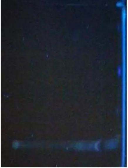
Figure 7. One-dimensional TLC fingerprinting
profile of hydromethanolic extract of
Pituranthos scoparius roots under UV light
(366nm).
Figure 9. One-dimensional TLC fingerprinting
profile of hydromethanolic extract of
Pituranthos scoparius roots under UV light
(254nm).
Figure 8. Two-dimensional TLC fingerprinting
profile of hydromethanolic extract of Pituranthos scoparius roots under UV light (366nm).
Figure 10. Two-dimensional TLC fingerprinting
profile of hydromethanolic extract of
Pituranthos scoparius roots under UV light
(366nm) derivatived with AlCl3(1 %)
Figure 11. One-dimensional TLC fingerprinting
profile of hydromethanolic extract of
Pituranthos scoparius roots derivatived with
vanillin-sulfuric acid reagent
Figure 12. Two-dimensional TLC fingerprinting
profile of hydromethanolic extract of Pituranthos
scoparius roots under UV light (366nm) derivatived