32 Imane et al.
RESEARCH PAPER
OPEN ACCESS
Antimicrobial and antioxidant activity of
Ammi visnaga
(L)
phenolic extracts and their effects on planktonic and biofilm
growth of food spoilage
Bacillus cereus
Belkacem Imane
*, Rebai Ouafa, Djibaoui Rachid
Laboratory of Microbiology and vegetal Biology, Faculty of Natural Sciences and Life,
University of Mostaganem, Algeria
Key words:Ammi visnaga, Bacillus cereus, Biofilm, Polyphenols, Flavonoids, Antioxidant
http://dx.doi.org/10.12692/ijb/9.4.32-47
Article published on October 11, 2016
Abstract
Ammi visnaga (L) is a species from Apiaceae family (Umbelliferae), it is widely used in Algeria. It is supposed to be an interesting source of phenolic compounds which can be used against biofilm growth of bacteria. Bacillus cereus, a crucial pathogenic bacterium that causes food poisoning, is known as a producer of gastrointestinal diseases. In the present work we used water, acetone, ethanol and methanol to extract phenolic compound from the plant Ammi visnaga (L). The extracts were evaluated for their antioxidant activity and their effects on planktonic cells, swarming motility and biofilm growth of Bacillus cereus isolates. The results indicate that 70% methanolic extract represent the highest amount of total phenols (176mg GAE/g), and the lowest amount was obtained with acetone extract (18, 66mg GAE/g). Flavonoids extractability was found to be highest with ethanolic extract (22mg QE/g). Among all the extracts of A. visnaga (L), methanolic extract 70% showed the most potent radical scavenging ability (IC 50: 1, 46mg/ml) and the highest reducing power values from 1,129 to 1,974 at 700nm. DPPH assay of plant extracts was well correlated with FRAP assay (R2=0, 7018) and a good
correlation was found between antioxidant activity (IC 50) and polyphenols content of different extracts (R2=0,
8153). No correlation was found between total polyphenol and flavonoids contents (R2=0, 4267). The obtained
results show that A. visnaga (L) extracts might possess high antimicrobial activities and methanolic extract at 10mg/ml was more effective to swarming motility and biofilm formation in Bacillus cereus strains.
*
Corresponding Author: Belkacem imane imane.belkacem@univ-mosta.dz
International Journal of Biosciences | IJB |
ISSN: 2220-6655 (Print), 2222-5234 (Online) http://www.innspub.net Vol. 9, No. 4, p. 32-47, 201633 Imane et al.
Introduction
The ability of many pathogenic bacteria to adhere to surfaces and to form biofilms has major implications in a variety of industries including the food industry, where biofilms create a persistent source of contamination causing food spoilage. Microbial adhesion to surfaces and consequent biofilm formation is a survival strategy that has been studied and documented in recent decades (Watnick and Kolter, 2000). Plant secondary metabolites (phytochemicals) have demonstrated promising antimicrobial properties when applied against planktonic cells and biofilms. Natural antimicrobial products can be attractive to the food industry in that they control natural spoilage microorganisms (Tajkarimi et al., 2010).
Therefore, new antimicrobial products need to be identified and their antimicrobial action against bacterial biofilms must be assessed. Previous studies (Cushnie and Lamb, 2005; Vaquero et al., 2007; Simões et al., 2009a) have demonstrated that phenolic substances, including simple phenols and phenolic acids, are a major class of phytochemicals that have already demonstrated significant antimicrobial properties. Phenolics of higher plants are ubiquitous low molecular compounds. They are the most widespread molecules among secondary plants metabolites, and are of high significance in plant development (Curir et al., 1990).
A great number of medicinal plants containing flavonoids have been reported their antibacterial activity. The plant Ammi visnaga (L) Lam. khown as Noukha (in Algeria) or Khella (in some parts of North Africa) is classified under the Apiaceae (Umbelliferae) family. It is found growing widely in North Africa, Asia and Europe (Hegnauer, 1973), were it is used in traditional medicines to treat gastrointestinal cramps. In Algeria Ammi visnaga (L) is largely used in traditional treatment of digestive diseases and culinary but not very well studied scientifically. The major components of the plant Ammi visnaga (L) are furanochromones and coumarins. Khellin and visnagin are the most biologically active of them. (Benigni et al., 1962; Hegnauer, 1973).
Furthermore, Bacillus cereus is a spore former bacteria, responsible for the spoilage of different food products. It is widespread in nature and in many raw and processed foods. It can survive during the cooking process, resist to pasteurization and produce emetic toxins. (Granum and Lund, 1997; Finlay et al., 2002).
The cooked food products when stored at room temperature, the spores can germinate, proliferate, and produce emetic and diarrhoeal toxins leading to poisoning. The adhesion of B. cereus to surfaces is mainly due to its high hydrophobicity, to spores surface charges, and to the long appendages covering its surfaces. (Andersson et al., 1995). B. cereus cells can attach on stainless steel surfaces and form a biofilm which cause problems in several processes of food industry (Peng et al., 2002). The bacterial biofilms are generally formed by cells clusters gathered with an extra-cellular material to colonize surfaces.
The plant Ammi visnaga (L) is widely used in Algerian culinary, but it has been rarely used for improving their antioxidant and antimicrobial effects. The main purpose of this study was to investigate the antioxidant activity and to evaluate the phenolic and flavonoids content of A. visnaga phenolic extracts, as well as their effects on planktonic cells and biofilms of Bacillus cereus isolates causing foodborne spoilage. Materials and methods
Isolation of Bacillus cereus strains from different Food Sources
Several Samples from local commercial supermarket and home-made foods were collected. The used foods are mainly: fresh and raw ground meats, poultry, fish, dairy products and some cooked dishes. All food samples were transported in sterile plastic boxes. The isolation of Bacillus species was performed according to the conventional procedure by serial dilution in sterile phosphate buffered saline. 10g of food sample was added in 90 mL of phosphate buffered saline; the different solutions were heated at 85 C° for 10 min.
34 Imane et al.
100µl from the appropriate dilutions were surface plated on LB agar, and on Mannitol egg-yolk polymyxin B (MYP) agar plates (Vanderzant and Splittstoesser, 1992). Incubation was carried at 30 °C for 48h. The characterization of the isolates was performed by studying colonies morphology, Gram stain, cell forms and biochemical tests, using API 20 E system and Bergey’s Manual of Systematic Bacteriology. (Cappuccino and Sherman, 2004). Screening of Bacillus cereus toxicity
Lecithinase activity: plates were prepared by adding egg yolk emulsion up to 5% (v/v) to nutritive agar. Each isolate was spotted on the medium and the plates were incubated at 30°C. Opaque zones around the colonies, caused by hydrolysis of lecithin indicated lecithinase production. (Guttmann and Ellar, 2000).
Hemolytic activity: The hemolytic activity was determined at 33°C on 5% sheep blood agar plates by surface inoculation as described by Pruss et al. (1999). The isolates were spotted on blooded nutritive agar medium. (Collins et al., 2001). The strains were classified as α (partial), β (total), or non-hemolytic. Amylase activity: The ability to hydrolyze starch was tested by inoculating on the starch agar medium; the zones were detected by adding lugol to the plate’s surfaces. (Collins et al., 2001).
Caseinase activity: Caseinase was identified according to the method of Gudmudsdo (1996) on milk agar medium. The isolated bacteria were streaked on the appropriate medium for 24 h at 37°C. A transparent zone around the colonies indicated caseinase activity.
Antibiogram pattern of the isolated strains
To select Bacillus cereus showing resistance to antibiotics, all isolates were tested for their sensitivity to antibiotics using following antibiotics: Penicillin G, Doxycycline (30µg), Erythromycin (15µg), Norfloxacine (10µg), Amoxicillin (30µg), Ampicillin (10µg), Cephalothin (30µg), Carbenicillin (100µg), Oxacillin (1µg), Piperacillin (30µg),
Trimethoprim-Sulfamethoxazole (25µg), Tobramycin (30µg), Rifampin (5µg) and polymyxin B (30µg). The antibiogram was realized on Mueller– Hinton agar, using disc diffusion method as described by Bauer et al., (1966). The Inocula were set to 0.5 McFarland or (OD= 0.08 to 0.1) at 620 nm, which corresponds to 108 CFU/ mL. The plates were incubated at 37°C for 24h. The inhibition zones around the disc was measured and interpreted as sensitive, moderate or resistant.
Plant material
The plant Ammi visnaga (L) was studied following its large use in Algerian culinary and its potential medicinal uses. The plant was collected during spring season from the region of Sidilakhdar (Mostaganem, Algeria), and identified by Microbiology and vegetal biology Laboratory at Mostaganem University. The plant was washed with distilled water and dried at room temperature under shade. The dried aerial parts were powdered by a blender and stored away from light for further studies.
Determination of moisture content
10 g of powdered aerial parts of Ammi visnaga (L) was weighed and put in an oven (70°C) until dryness. Moisture lost was determined by the difference between initial fresh weight and constant weight after drying. All samples were analyzed in triplicate and the result was expressed by percentage of moisture of the sample studied.
Extraction procedure
An absolute methanol, water and three 70% (v/v) organic solvents (Methanol, Acetone and ethanol) were used to extract phenolic compounds. 10% w/v of powdered plant material was extracted by 100ml of each solvent. The extraction was performed during 30 min. Centrifugation was done at 3000 rpm/ 20min, and vacuum filtration was used to separate the liquid extract. The filtrates were collected and the solvent was evaporated at 40°C using a rotary vacuum evaporator. The aqueous extract was obtained by soaked 10 g of powdered plant in 100 ml sterile water for 5 mi, the mixtures were centrifuged at 3000 rpm for 20 min. The supernatant was used for the determination of total phenolic content and antioxidant activity. (Oboh et al., 2009).
35 Imane et al.
Total phenolic content
Ammi visnaga (L) extracts phenolic content was determined by adding 5 ml of Folin-ciocalteau reagent (1:10) and 4 ml of 7.5% aqueous sodium carbonate to 0.5 ml of different extracts. The mixtures were kept in obscurity at room temperature for 15 min. The optical density was then measured at 765 nm using UV spectrophotometer. (Singleton and Rossi, 1965). The results were reported as Gallic acid equivalent (GAE) per gram of dry weight. Gallic acid concentrations were prepared as standard in methanol.
Flavonoid content
To determine the flavonoid content (TFC), the aluminum chloride colorimetric assay was used as described by Liu et al. (2008). A volume of 2 ml of diluted extract was allowed to 200µl of 0. 5% sodium nitrite and incubated for 5 min. Then, 200µl of aluminum chloride 10% was added to the mixture. 2 ml of sodium hydroxide 1M was added to the mixture after 6 min, and the absorbance wasreaded at 510 nm. Quercetin was used for the calibration curve. The results of flavonoids content were expressed as mg quercetin equivalents (QE) per g. All the samples were analyzed in tryplicate.
Antioxidant activity of Ammi visnaga (L) extracts Free radical scavenging assay (DPPH): to determine the ability of scavenging of phenolic extracts on 1,1-diphenyl-2-picrylhydrazyl (DPPH) free-radicals the method of Shimada et al., (1992) was used. 2ml of each plant extracts at different concentrations was added to 0.5 ml of 1 mM DPPH prepared in methanol. After shaking, the mixture was left at room temperature to stand for 30 min in the dark. The absorbance was measured at 517 nm against an aliquot blank. Methanol was used as a control. Scavenging ability (%) = [A0–A1/A0] ×100. The higher values explain greater antioxidant activity of the tested sample, but higher IC 50 value indicates a weaker capacity to scavenge DPPH radicals.
Ferric reducing power (FRAP): The reducing power was determined by adding 1 mL of varying concentration of sample extracts to 2.5 mL phosphate buffer (0.2 M, pH 6.6), then the solution was mixed with 2.5 mL of potassium ferricyanide 1%.
After incubation at 50°C for 20 min, 2.5 mL of 10% acid trichloroacetic (w/v) was added to the mixture. After centrifugation at 3000 rpm for 10 min, the upper layer (2.5 mL) was mixed with 2.5 mL of deionized water. Finaly, 0.5 mL of 0.1% ferric chloride was added. The absorbance was measured at 700 nm. Disilled water was used as negative control. The assay was done in triplicate. (Oyaizu, 1986). Antimicrobial activity of Ammi visnaga (L) extracts Agar-well diffusion assay: The antibacterial activity of the different plant extracts was evaluated against five isolates of food spoilage Bacillus cereus and a reference strain Bacillus cereus ATCC 14579. The turbidity of the bacterial suspensions was adjusted to an equivalent to 0.5 McFarland. The agar-well diffusion assay was performed according to the recommended method of Valgas et al. (2007). A standardized bacterial inoculum was uniformly surface spread on a Mueller-Hinton agar. Next, 80 μL of each plant extract dissolved in distilled water (10mg /ml) was added into the wells of 6 mm in diameter, the plates were incubated at 30°C for 24h and the measure of the diameter of inhibition zones was done. The Ampicillin (30 μg) and well containing the solvent only were used as controls. All tests were done in triplicate.
Swarming behaviour assay: The effect of methanolic extract on swarming migration of Bacillus cereus food spoilage isolates was realized according to the method cited by Liaw et al., (2000), bacterial suspensions were grown in Brain-Heart Infusion (BHI) broth and incubated overnight at 37 °C. After incubation, the cultures were adjusted to an OD =1 ± 0.05 at 620 nm. 5 μl of an overnight bacterial culture was spotted centrally onto the surface of dry PPGAS (phosphate-limited peptone–glucose– ammonium salt) agar plates (KCl 20 mM, NH4Cl 20 mM, MgSO4 1.6 mM, Tris–HCl (pH 7.2) 120 mM, peptone 1.0%, glucose 0.5%) without or with 0, 5; 1, 5 or 10 mg/ ml of phenolic compounds, then incubated at 37ºC for 24h. For monitoring swarming motility, glutamate 0.05% was used instead of NH4Cl.
36 Imane et al.
Crystal violet biofilm assay: Cell suspensions were prepared by inoculating 3 ml of TSB (tryptic soy broth) with bacterial Bacillus cereus strains and were cultured overnight at 37°C and then diluted in fresh media to an OD =0,06 at 620nm. The biofilm growth inhibition performed adopting method of biofilm inhibition spectrophotometric assay in 96 well microplates as described by Regev-Shoshani et al., (2010). 100 μl of each prepared bacterial suspension of the tested isolates was added into 96 well microplates and 100µl of different concentration of methanolic extract of Ammi visnaga (L)was added and incubated at 37º C for 48h, 200 μl of 1% w/v aqueous solution of crystal violet was added after removing the liquid suspension. After 30 minutes, the wells were washed thoroughly after removing the dye and 200µl of ethanol at 95% was added and incubated for 15 minutes. The reaction solution was read in an ELISA reader at 595 nm. The TSB was used as a blank. After incubation, the MIC was defined as the lowest concentration of A.visnaga (L) extract that exhibit an inhibition of visible growth.
Reduction of biofilm biomass was calculated as following: % inhibition =OD in control-OD in treatment/OD in control* 100.
Statistical analysis
All the experiments were carried out in triplicate. The data were analyzed by ANOVA. The IC50 values were calculated from linear regression analysis. Correlations between variables were established by excel.
Results
Isolation and identification of the isolated Bacillus cereus strains
46 isolates were obtained by isolation from different food samples, 24 isolates were selected after screening of their ability of resistance to penicillin G and other antibiotics (Fig. 1). The Colonies of isolated bacteria were big waxy white or gray, surrounded by an opacity zone. 19 of them were unable to catabolize mannitol when cultured on MYP agar.
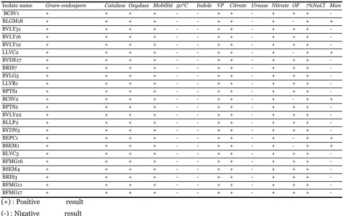
Table 1. Identification of isolates of food spoilage Bacillus cereus.
Isolate name Gram-endospore Catalase Oxydase Mobilité 50°C Indole VP Citrate Urease Nitrate OF 7%NaCl Man
BCSV1 + + + + - - + + - + + + - BLGM18 + + + + - - + + - + - + + BVLY31 + + + + - - + + - + + + - BVLY16 + + + + - - + + - + + + - BVLY12 + + + + - - + + - + + + - LLVC2 + + + + - - + + - + - + + BVDE17 + + + + - - + + - + + + - BRIS7 + + + + - - + + - + + + - BYLG5 + + + + - - + + - + + + - LLVB1 + + + + - - + + - + + + - BPTS1 + + + + - - + + - + + + - BCSV2 + + + + - - + + - + - + + BPTS2 + + + + - - + + - + + + - BVLY22 + + + + - - + + - + + + - BLLP2 + + + + - - + + - + + + - BYDN3 + + + + - - + + - + + + - BEPC1 + + + + - - + + - + - + + BSEM1 + + + + - - + + - + - + + BLVC3 + + + + - - + + - + + + - BFMG16 + + + + - - + + - + + + - BSEM4 + + + + - - + + - + + + - BRIS3 + + + + - - + + - + + + - BFMG11 + + + + - - + + - + + + - BFMG17 + + + + - - + + - + + + - (+) : Positive result (-) : Nigative result
37 Imane et al.
The isolates were gram positive rods. Motile, and grow positively in 7% NaCl but could not grow at 50°C. Following results shown in table (1), 19 isolates were belonged to Bacillus cereus and 5 to Bacillus subtilis. The identification of the above species was
confirmed by API 20E system as recommended by bergy’s manual. All Bacillus cereus stains were beta-haemolytic producing phospholipase C, amylase and caseinase. The obtained results indicate that the studied Bacillus cereus strains are currying a level of toxicity and virulence.
Table 2. Antimicrobial activity of Ammi visnaga (L) extracts against isolates of Bacillus cereus.
Plant extracts (solvent) BVDE17 BPTS3 BCSV1 BRIS3 BSEM4 ATCC 14579 Acetone 70% 6+0,81 9+0,91 8+0,03 7+0,02 9+0,05 8,33+0,057 AQ (water) 8,33+0,85 9,33+1,07 11,33+0,037 9,33+0,025 9,33+0,057 9,33+0,057 MeOH 70% 21+1,05 15,33+1,15 15,66+0,05 15,33+0,057 15,33+1 12,66+0,057 MeOH 100% 14+1 12,66+1,09 13+0,047 12,66+0,047 12,66+0,047 11,33+0,057 EtOH 70% 11,33+0,87 12,66+1,09 12,33+0,057 11+0,037 11,66+0,87 10,33+0,057 Antibiotic sensitivity
The profile of antibiotic resistance of the tested strains to different antibiotics was performed. All Bacillus cereus strains showed high resistance towards Penicilline G (100%), but found variably resistant to the other antibiotics tested (Fig. 1). All the strains showed high sensitivity towards Norfloxacine (89, 13%), Doxycycline (84, 78%), Erythromycin (78, 26%), but were found less sensitive to Amoxicillin (21, 73%) Oxacillin (8, 69%) and Ampicillin (4, 34%). Among the sample studied, the aerial part of the plant Ammi visnaga (L) showed high moisture content which showed a value of 90.46 ± 0.09%.
Effect of concentration and solvent type on total phenols yield
The results in figure (2) show the extraction yield of total phenols and flavonoids using different solvents. The phenolic content of all extracts ranged from 18, 66 to 172, 66 mg/g. The highest values was found in the aqueous methanol extract (172, 66 mg/g) which was significantly higher than ethanol extract 38 mg/g and acetone extract which represent the lowest yield with 18, 66 mg/g. The total flavonoid contents in Ammi visnaga (L) ranged from 3, 3 to 22 mg EQ/g. Ethanolic extract showed the highest value, while the lowest value was obtained by the pure (100%) methanol extract. (Fig.2). In general, considering all the solvents used in this study,
aqueous methanol at 70% was found to be the most effective solvent to extract total phenols from plants. To improve the effect of the concentration of extraction solvent, we have used absolute, aqueous methanol and water extract. We also found that the yield of extraction of total phenols obtained by aqueous methanol was higher (172, 66 mg/g) than absolute methanol and water extract with 96 mg/g and 21, 66 mg/g respectively.
DPPH radical scavenging activity assay
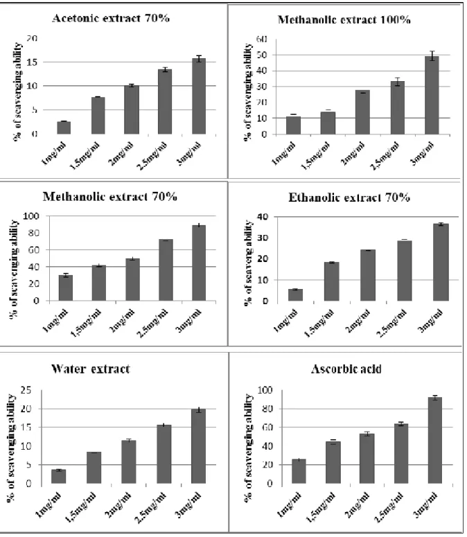
In terms of antioxidant activities, and in comparison with ascorbic acid (AA) as positive control, it was found that methanolic extracts represented the highest DPPH scavenging activity (89, 21%) at concentration of 3mg/ ml, this value was significantly different from ethanol and acetone extracts at the same concentration. (Fig. 3). These values were lower than those obtained by ascorbic acid (positive control) which represent 91, 79%. Regardless the type of solvent, extracts with concentrations 70% of solvent used presented higher DPPH radical scavenging capability than those with their respective absolute methanol and water extracts. This result was similar to thus observed by total polyphenol content. The Figure 4 represents the IC 50 values obtained for the investigated Ammi visnaga (L) extracts. Methanolic extract was found to be 1, 46 mg/ml while the acetonic extract was 3,1. The results indicate that the scavenging ability of methanolic extract of Ammi visnaga (L) on DPPH radical was strong.
38 Imane et al.
Figure 5 represent the results of correlation between antioxidant activities (DPPH and FRAP methods) and correlation between total polyphenol and flavonoid content.
Free Reducing power
The results of the reducing power of Ammi visnaga (L) extracts at different concentration used showed a range of absorbance values from 0.4 to 2.57 at 700 nm (Fig. 6). It was also found that high concentration of each extract increase the reducing power. Table 3. Minimum Inhibitory Concentration (MIC) and % biofilm inhibition by Ammi visnaga (L) methanolic extract for Bacillus cereus isolates.
MIC mg/ml
BRIS3 BVDE17 BPTS2 BSEM4 BCSV1
OD595nm %Inhibition OD595nm %Inhibition OD595nm %Inhibition OD595nm %Inhibition OD595nm %Inhibition
C 0,18+0,01B 0 0,19+0,01A 0 0,11+0,02J 0 0,15+0,01E 0 0,17+0,01C 0
0,5 0,15+0,02D 15,78 0,112+0,01J 41,05 0,091+0,01N 17,27 0,139+0,01F 7,33 0,123+0,01H 27,64
1 0,11+0,01 I 38,88 0,097+0,02L 48,94 0,086+0,01O 21,81 0,126+0,01G 16 0,104+0,01K 38,82
5 0,096+0,01L 46,66 0,058+0,01S 69,47 0,063+0,01R 42,72 0,095+0,01M 36,66 0,076+0,01P 55,29
10 0,066+0,01Q 63,33 0,04+ 0,01U 78,94 0,028+0,01W 74,54 0,055+0,01T 63,33 0,032+0,01V 81,17
Absolute and aqueous methanolic extracts exhibited a high reducing power in comparison with the other plant extracts. Methanolic extract at 70% represent the highest value of 1.974±0.14 at 3mg/ml that might attribute to the collective antioxidant effects of phenolics and flavonoids.
Antimicrobial activities of Ammi visnaga (L) extracts Agar-well diffusion assay: The results of antibacterial activity against strains of Bacillus cereus are illustrated in table 2.
The inhibition zones of bacterial strains obtained by the methanolic extract were in the range of 12, 66 ± 0.5 to 21 ± 1 mm, which was significantly different from those obtained by ethanolic extract and acetonic extract with 10,33+0,05 to 12,66+0,05 and 7+0,05 to 9+0,05, respectively. It can be observed that the extracts of A. visnaga (L) possessed an inhibitory effect on all tested strains, depending on solvent of extraction and amounts of phenolic compounds.
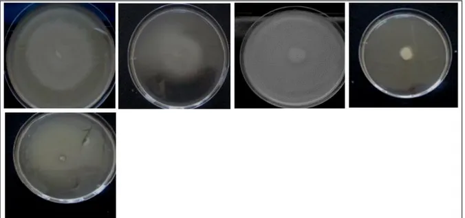
Fig. 1. Bacillus cereus isolates sensitivity towards antibiotics studied. Swarming motility assay: The obtained results of
motility revealed that methanolic extract possess a significant effect on swarming motility of strains tested.
The diameter of swarming zones was highly correlated to the increasing of phenolic concentrations and the highest zone diameter was obtained by the concentration of 10 mg/ml. (Fig. 7).
39 Imane et al.
Biofilm inhibition assay: In biofilm quantification assay, a significant decrease was observed when bacterial strains grown in the presence of the methanolic extract. The apppropriate concentrations of methanolic extract (0, 5; 1; 5 and 10 mg/ml) provides a decrease in biofilm growth. The reduction in biofilm biomass of the isolates tested range from 63, 33 to 81, 17%. (Table 3).
The biomass of biofilm formed by the different strains of Bacillus cereus decrease with the increase of the concentration of the methanolic extract and this reduction differ from stain to another. These results supposed that the methanolic extract of A. visnaga (L) has a strong effect against biofilm of Bacillus cereus due to their total phenolic content.
Fig. 2. Polyphenol and flavonoids content of Ammi visnaga (L) extracts. Discussion
Although, substantial amount of data are available about the incidence of B. cereus in various types of food and food poisoning cases worldwide. (Stenfors et al., 2008). Currently, the selective MYP plating medium is recommended by food authorities as standards for the detection of B. cereus.
The main identification feature of this medium is the lecithinase activity, resulting in opaque precipitation zones around suspect colonies. (Szabo et al., 1984). The opaque zone might be due to the fact that the egg yolk tellurite emulsion used is a lipoprotein basically composed of lecithin, and lecithinase breaks down lecithin to produce an insoluble precipitate, resulting in opaque zones around the colonies. (Hussain et al., 2011).
The obtained results designate that the studied Bacillus cereus strains are currying a level of toxicity and virulence, as cited by Oh (2006), Kashid and
Ghosh (2010) which reported that the major features of B. cereus on plating media, produced caseinase, lecithinase with hemolytic activity. In this regard, Awny et al. (2010) reported that haemolysin, lecithinase and caseinase of B. cereus and Staphylococcus aureus from food samples are considered as virulence and toxicity factors. Our results are supported by the broad picture of antibiotic susceptibility patterns of B. cereus as shown by Fenselau et al. (2008).
Precedent works showed that antimicrobial susceptibility of Bacillus cereus was highly susceptible to Erythromycin, Ciprofloxacin, Streptomycin, Chloramphenicol, and less sensitive to Ampicillin, Cloxacillin, Ampiclox, and Cotrimazole, (Umar et al., 2006). Our finding can prove that actually antibiotics are widely used in foods, consequently they can be an important factor to transfer resistant antibiotic foodborne pathogens which can be very harmful to human health. (Khan et al., 2000).
40 Imane et al.
Fig. 3. Scavenging ability of different extracts of Ammi visnaga (L). The difference in the yield of total phenols obtained
from different plant extracts was mainly due to the difference in the nature of phenolic compounds obtained by each solvent used. (Ignat et al., 2011). In addition, Horax et al. (2005) highlighted the importance of solvents effect that significantly influenced the quantity of phenolic compounds extracted. Following Ross et al. (2009), a great number of phenolic compounds can be obtained by methanol extraction
such as phenolic acids, flavanons, flavanols, anthocyanins, catechins, and procyanidins. These results highlight that the polarity of the extraction solvent used can influence the yield of extracted phenolic compounds. This result is in line with those obtained by Zhao et al. (2006), the solubility and the extraction yield of chemical constituents of a sample can be affected by the difference in polarity of the solvent used for extraction.
41 Imane et al.
The quantification of the total phenolic content in the extracts showed that the aqueous mixtures at 70% solvent and in comparison with pure solvents, were more effective to extract the phenolic compounds. (Rødtjer et al. 2006) Lapornik et al. (2005) suggest that ethanol and methanol extracts (70%) represent higher values of total polyphenols than water extracts.
The results of free radical scavenging ability (DPPH assay) agreed with the results reported by Zhang et al. (2013). DPPH scavenging is significantly influenced by solvents used for extraction of polyphenols. Phenolic compounds have been investigated to be a strong hydrogen donators to the DPPH radical. (Von Gadow, Joubert, &Hansmann, 1997).
Fig. 4. IC 50 of different extracts of Ammi visnaga (L). The antioxidant activity of the majority of plants is mostly due to the presence of phenolic compounds. It is known as free radical scavengers. (Skerget et al., 2005). The antioxidant activity estimation and the ability of solvent to dissolve a selected group of antioxidant compounds can be influenced by the change in solvent polarity. (Zhou & Yu, 2004). The correlation between antioxidant activity and total phenol contents has been largely studied in different food products. (Kiselova et al., 2006).
Our results of antioxidant activity obtained by the reducing power assay showed that the methanolic extract had the highest antioxidant activity values at 700 nm, this result was similar to those obtained from the yield of total phenolic compounds. Previous researches and reports in the literature agree with our results, phenolic compounds are the major phytochemicals responsible for the antioxidant capacity of natural extracts (Cevallos-Casals et al., 2006), maybe due to their redox properties, which permit them to act as reducing agents, hydrogen donors, and singlet oxygen quenchers. (Chang et al., 2001).
The antibacterial effect can be explained by the relationship between the number of hydroxyl groups and their position on the phenol group and their relative toxicity to microorganisms, the increase in hydroxylation induce an increase of antimicrobial activity. (Marjorie, 1999). The Ammi visnaga (L) or Noukha has been shown to be effective against microorganisms. (Abroush et al., 2001). Jaradat et al. (2015) found that the plant extracts of Ammi visnaga (L) obtained by using organic solvents have shown a strong antimicrobial activity than the aqueous extract and variations among species were obvious. Baydar et al., (2004) have suggest that the antibacterial activity of plant extracts is proportional to the amount of phenolic compounds in investigated plant.
Some researchers suggested that tannic acid and tannin are the main phenolic acids which can stopped P. aeruginosa swarming motility without reducing their growth capacity (Omay and Tufenkji, 2011). Proteins can have a strong effect on the mechanism of inhibition of swarming motility that is propably due to binding and precipitation of phenolic compounds to them. (Pratt and Kloter, 1998).
42 Imane et al.
Fig. 5. Correlation between polyphenol and flavonoid content (a) and between antioxidant activity (IC 50) and polyphenol content of extracts of Ammi visnaga (L) (b). (c) Represent correlation between IC 50 and flavonoid content, and (d) correlation between antioxidant activities DPPH and FRAP methods.
In clinical and foodborne pathogenesis biofilm associated infection is known as a trigger to chronic diseases, food spoilage. Even dairy and refrigerated food spoilage was also created by the bacterial biofilm. (Teh et al., 2014; Mizan et al., 2015). Some previous works revealed that the enzymatic activity of glucosyltransferase which permit the colonization of
bacteria and their adherence can be affected by the phenolic compounds. (Yanagida et al., 2000, Gregoire et al., 2007). Biofilm formation can be inhibited by the action of phenolics due to lack of iron, some phenolics have intermediate property of iron chelating. (Devosset al., 1999).
43 Imane et al.
The peptide’s capability to cover either the surface of biomaterial or the bacterium itself, reduce the adherence of microorganisms on surface and decrease the biofilm growth. (Segev-Zarko et al., 2015) Developed studies are demonstrating that there is a biological rationale between quorum sensing and
biofilm which work on a coordinate manner leading to spoilage. (Bai and Vittal, 2014).) Bacterial AHLs can be imitate by substances secreted by several plants and affect the regulated behaviors of quorum-sensing plant-associated bacteria, respectively. (Teplitski et al., 2000).
Fig. 7. Effect of Ammi visnaga (L) methanolic extract on swarming motility of Bacillus cereus. Conclusion
In conclusion, the extracts of Ammi visnaga (L) obtained by using solvents with less polarity were strong radical scavengers than those obtained with high polarity solvents. The selection of the appropriate solvent is a important step to optimize the extraction of total polyphenol, flavonoid and antioxidant activities of the investigated plant extracts. Methanol was recommended as the best solvent for the production of antioxidant compounds from the aerial parts of the plant of A. visnaga (L). It also possess a strong antibacterial and antibiofilm activity against Bacillus cereus strains responsible for food spoilage. For this, it can be used in food industries as a natural preservative. However, further studies are needed to explore the individual or major polyphenolic groups and other bioactive compounds in the extracts of A. visnaga (L) and their contribution to health care and biotherapy. In addition, in vivo assay of antioxidant are recommended to confirm the strong potentiel of Ammi visnaga (L) to treat diseases caused by food spoilage.
Acknowledgements
This research was funded by the laboratory of Microiology and vegetal biology at Abdelhamid Ibn badis University in Algeria.
References
Abroush Z, Majd A, Rezaee MB. 2001. Evaluation of antimicrobial effect of Tooth pick plant, Master of Science thesis. Biology department. Azad University.
Andersson A, Rönner U, Granum PE. 1995. What problems does the food industry have with the spore forming pathogens Bacillus cereus and Clostridium perfringens? International Journal of Food Microbiology 28, 145-155.
Awny NM, AbouZeid AM, Abdo MA. 2010. Prevalence of toxigenic bacteria in: some Egyptian food, in: Proceeding of Fifth Scientific Environmental Conference, Alexandria Egypt. 107–124.
44 Imane et al.
Bai AJ, Vittal RR. 2014. Quorum sensing inhibitory and anti-biofilm activity of essential oils and their in vivo efficacy in food systems. Food Biotechnology 28, 269–292.
http://dx.doi.org/10.1080/08905436.2014.932287. Bauer AW, Kirby WMM, Sherris, JC Turck M. 1966. Antibiotic susceptibility testing by a standardized single disk method. American Journal of Clinical Pathology 36, 493-496
Baydar NG, Ozkan G, Sagdiç O. 2004. Total phenolic contents and antibacterial activities of grape (Vitisvinifera L.) extracts. Food Control 15, 335–339. http://dx.doi.org/10.1016/S0956-7135(03)00083-5. Benigni R, Capra C, Cattorini PE. 1962. Piantemedicinali - Chimicafarmacologia e terapia. Vol. 1, Inverni& Della Beffa, Milano. 60-82.
Cappucino JG, Sherman N. 2004. Microbiology A Laboratory Manuel Pearson Education (Singapore) Indian Branch. New Delhi.
Cevallos-Casals BA, Byrne D, Okie WR, Cisneros-Zevallos L. 2006. Selecting new peach and plum genotypes rich in phenolic compounds and enhanced functional properties. Food Chemistry 96, 273–280.
http://dx.doi.org/10.1016/j.foodchem.2005.02.032 Chang ST, Wu JH, Wang SY, Kang PL, Yang NS, Shyur LF. 2001. Antioxidant activity of extracts from acacia confuse bark and heartwood. Journal of agriculture and food chemistry 49, 3420-3424 Collins CH, Lyne PM, Grange JM. 2001. Collins and Lyne’s microbiological methods. 7th ed. London: Arnold.
Cushnie TPT, Lamb AJ. 2005. Antimicrobial activity of flavonoids. International Journal of Antimicrobial Agents 26, 343-356.
http://dx.doi.org/10.1016/j.ijantimicag.2005.09.002.
Curir PV, Sumere CF, Termini A, Barthe P, Marchesini A, Dolci M. 1990. Flavonoid accumulation is correlated with adventitious roots formation in Eucalyptus gunnii Hook micropropagated through axillary bud stimulation. Journal of Bacteriology 92, 1148–1153.
Devoss JJ, Rutter K, Schroeoder BG, Barry CE. 1999. Iron acquisition and metabolism by mycobacteria. Journal of Bacteriology 181, 4443-4451.
Fenselau C, Havey C, Teerakulkittipong N, Swatkoski S, Laine O, Edwards N. 2008. Identification of b-lactamase in antibiotic-resistant Bacillus cereus spores. Applied and Environmental Microbiology 74, 904-6.
http://dx.doi.org/10.1128/AEM.00788.
Finlay WJJ, Logan NA, Sutherland AD. 2002. Bacillus cereus emetic toxin production in cooked rice. Food Microbiology 19, 431-439.
http://dx.doi.org/10.1006/fmic.2002.0505.
Granum PE, Lund T. 1997. Bacillus cereus and its food poisoning toxins. FEMS Microbiology Letters 157, 23-228.
http://dx.doi.org/10.1111/j.1574-6968.1997.tb12776. Gregoire S, Singn AP, Vorsa N, Koo H. 2007. Influence of Cranberry phenolics on glucan synthesis by glucosyltransterase and Streptococcus mutansacidigenicity. Journal of Applied Microbiology 103, 1960-1968.
http://dx.doi.org/10.1111/j.1365-2672.2007.03441.x. Gudmundsdo BK. 1996. Comparison of extracellular proteases produced by Aeromonas salmonicida strains isolated from variousfish species. Applied Bacterioly 80, 105–113.
http://dx.doi.org/10.1111/j.1365-2672.tb03196.x Guttman DM, Ellar DJ. 2000. Phenotypic and genotypic comparisons of 23 strains from the Bacillus cereus complex for a selection of khown and putative B. thuringesis virulence factors. FFMS MicrobialLeH. Jul 1, 118(1), 7-13.
45 Imane et al.
Hegnauer R. 1973. ChemotaxonomiederPflanzen, Band 8, 418-433.
Horax R, Hettiarachchy N, Islam S. 2005. Total phenolic contents and phenolic acid constituents in 4 varieties of bitter melons (Momordicacharantia) and antioxidant activities of their extracts. Journal of Food Science 70, C275-80.
http://dx.doi.org/10.1111/j.13652621.2005.tb07173.x. Hussain MAA, Sanousi SME. 2011. Prevalence of Clostridium prefringens and Clostridium prefringens-like organisms in faecalsamples of domestic animals. Journal of Veterinary Medicine and Animal Health 2, 89–101.
Ignat I, Volf I, Popa VI. 2011. A critical review of methods for characterization of polyphenolic compounds in fruits and vegetables. Food Chemistry 126, 1821–1835.
http://dx.doi.org/10.1016/j.foodchem.12.026. Jaradat N. 2015. Phytochemical Screening and In-vitro Evaluation of Antioxidant and Antimicrobial Activities of the Entire Khella Plant (Ammi visnaga.L.) A member of Palestinian Flora. Palestine International Journal of Pharmacognosy and Phytochemical Research 7(1), 137-143.
Kashid SG, Ghosh JS. 2010. Production, isolation and characterization of exotoxin produced by Bacillus cereus 2156 and Bacillus licheniformis NCIM-5343, Journal of Pharmacological and Toxicological Methods1, 50–55.
Khan SA, Nawaz MS, Khan AA, Cerniglia CE. 2000. Transfer of erythromycin resistance from poultry to human clinical strains of Staphylococcus aureus. Journal of Clinical Microbiology 38, 1832– 1838.
Kiselova Y, Ivanova D, Chervenkov T, Gerova D, Galunska B, Yankova T. 2006. Correlation between the in vitro antioxidant activity and polyphenol content of aqueous extracts from bulgarian herbs. Phytotherapy Research 20(11), 961- 965.
http://dx.doi.org/10.1002/ptr.1985.
Lapornik B, Prošek M, Wondra AG. 2005. Comparison of extracts prepared from plant by-products using different solvents and extraction time. Journal of Food Engineering 71, 214-222.
http://dx.doi.org/10.1016/j.jfoodeng.2004.10.036. Liaw SJ, Lai HC, Ho SW, Luh KT, Wang WB. 2000. Inhibition of virulence factor expression and swarming differentiation in Proteus mirabilis by p-nitrophenylglycerol. Journal of Medical Microbiology 49, 725-731.
http://dx.doi.org/10.1099/0022-1317-49-8-725. Liu X, Zhao M, Wang J, Yang B Jiang Y. 2008. Antioxidant activity of methanolic extract of emblica fruit (Phyllanthusemblica L.) from six regions in China. Journal of Food Composition and Analysis 21, 219–228.
http://dx.doi.org/10.1016/j.jfca.2007.10.001.
Marjorie CM. 1999. Plant products as antimicrobial agents. Clinical Microbiology Reviews 12(4), 564– 582.
http://dx.doi.org/10.1021/jf0100907.
Mizan MFR, Jahid IK, Ha SD. 2015. Microbial biofilms in seafood: a food-hygiene challenge. Food Microbiology 49, 41–55.
http://dx.doi.org/10.1016/j.fm.2015.01.009.
Oboh G, Alimiluyi AO, Akidahansi AA. 2009. Changes in polyphenols and antioxidant activity during fermentation of some under-utilized legumes. Food science and Technology.Interdium Vol 15 Issue 1, 41-46.
http://dx.doi.org/10.1177/1082013208101022. Oh MH. 2006. Ecology of Toxigenic Bacillus Species in Rice Products, University of New South Wales School of Chemical Engineering and Industrial Chemistry. International Journal of Food Science & Technology. Sydney.
Omay C, Tufenkji N. 2011. The swarming motility of Pseudomonas aeruginosa is blocked by cranberry proanthoeyanidins and other tannin-containing materials. Applied and environmental microbiology 77(a), 3061- 3067.
46 Imane et al.
Oyaizu M. 1986. Studies on products of browing reaction: antioxidative activity of products browing reaction prepared from glucosamine. The Japanese Journal of Nutrition 44, 228-234 ().
Peng JS, Tsai WC, Chou CC. 2002. Inactivation and removal of Bacillus cereus by sanitizer and detergent. International Journal of Food Microbiology 77, 11-18.
http://dx.doi.org/10.1016/S0168-1605(02)00060-0. Pratt LA, Kotter R. 1998. Genetic analysis of Escherichia coli biofilm formation roles of flagella, motility and chemotaxis. Molecular Microbiology 30, 285-293.
http://dx.doi.org/10.1046/j.1365-2958.1998.01061.x. Pruss BM, Dietrich R, Nibler B, Martlbauer E, Scherer S. 1999. The hemolytic enterotoxin HBL is broadly distributed among species of the Bacillus cereus group. Applied and Environmental Microbiology 65, 5436–5442.
Regev-shoshani G, Ko M, Miller C, Av-Gay Y. 2010. Slow release of nitric oxide from charged catheters and its effect on biofilm formation by Escherichia coli. Antimicrobial Agents and Chemotherapy 54, 273-279.
http://dx.doi.org/10.1128/AAC.00511-09.
Rødtjer A, Skibsted LH, Andersen ML. 2006. Antioxidative and prooxidative effect of extracts made from cherry liqueur pomace. Food Chemistry 99, 6-14.
http://dx.doi.org/10.1016/j. foodchem. 2005.07.011. Ross KA, Beta T, Arntfield SD. 2009. A comparative study on the phenolic acid identified and quantified in dry beans using HPLC as affected by different extraction and hydrolysis methods. Food Chemistry 113, 336–344.
http://dx.doi.org/10.1016/j.foodchem.2008.07.064. Segev-Zarko L, Saar-Dover R, Brumfeld V, Mangoni ML, Shai Y. 2015. Mechanisms of biofilm inhibition and degradation by antimicrobial peptides. Biochemical Journal 468, 259-270.
http://dx.doi.org/10.1042/BJ20141251.
Shimada K, Fujikawa K, Yahara K, Nakamura T. 1992. Antioxidative properties of xanthan on the autoxidation of soybean oil in cyclodextrin emulsion. Journal of Agricultural and Food Chemistry 40, 945-948.
http://dx.doi.org/10.1021/jf00018a005.
Singleton VL, Rossi JA. 1965. Colorimetry of total phenolics with phosphomolybdic–phosphotungstic acid reagents. American Journal of Enology and Viticulture 16, 144–158.
Simões M, Bennett RN, Rosa EA. 2009a. Understanding antimicrobial activities of phytochemicals against multidrug resistant bacteria and biofilms. Natural Product Reports 26, 746-757. http://dx.doi.org/10.1039/b821648g.
Skerget M, Kotnik P, Hadolin M, Hras AR, Simonic M, Knez Z. 2005. Phenols, proanthocyanidins, flavones and flavonols in some plant materials and their antioxidant activities. Food Chemistry 89, 191–198.
http://dx.doi.org/10.1016/j.foodchem.2004.02.025. Stenfors ALP, Fagerlund A, Granum PE. 2008. from soil to gut: Bacillus cereus and its food poisoning toxins. FEMS Microbiology Reviews 32, 579-606.
http://dx.doi.org/10.1111/j.1574-6976.2008.00112.x. Szabo RA, Todd ECD, Rayman MK. 1984. Twenty-four hour isolation and confirmation of Bacillus cereus in foods. Journal of Food Protection 47, 856–860.
Tajkarimi MM, Ibrahim SA, Cliver DO. 2010. Antimicrobial herb and spice compounds in food. Food Control 21, 1199-1218.
http://dx.doi.org/10.1016/j.foodcont.2010.02.003. Teh KH, Flint S, Palmer J, Andrewes P, Bremer P, Lindsay D. 2014. Biofilm an unrecognised source of spoilage enzymes in dairy products? International Dairy Journal 34, 32–40. http://dx.doi.org/10.1016/j.idairyj.2013.07.002.
47 Imane et al.
Teplitski M, Robinson JB, Bauer WD. 2000. Plants secrete substances that mimic bacterial N-acyl homoserine lactone signal activities and affect population density-dependent behaviors in associated bacteria. Molecular Plant-Microbe Interactions Journal 13, 637-648.
http://dx.doi.org/10.1094/MPMI.13.6.637.
Umar AS, Yerima MB, Uzal U. 2006. Antimicrobial sensitivities of Bacillus cereus isolated from food samples sold in Bauchi metropolis to selected antibiotics. Nigerian Journal of Microbiology 20(1), 655-661.
Valgas C, Souza SMD, Smânia EFA, SmâniaJr A. 2007. Screening methods to determine antibacterial activity of natural products. Brazilian Journal of Microbiology 38, 369–380.
http://dx.doi.org/10.1590/S15178382200700020003 4.
Vanderzant C, Splittstoesser DF. 1992. Compendium of Methods for the Microbiological Examiantion of Foods, 3rd edn. Washington DC, American Public Health Association.
Vaquero MJR, Alberto MR, de Nadra MCM. 2007. Antibacterial effect of phenolic compounds from different wines. Food Control 18, 93-101. http://dx.doi.org/10.1016/j. foodcont.2005.08.010. Von Gadow A, Joubert E, Hansmann CF. 1997. Comparison of the antioxidant activity of rooibos tea (Aspalathuslinearis) with green, oolong and black tea. Food Chemistry 60, 73–77.
http://dx.doi.org/10.1016/S0308-8146(96)00312-3.
Watnick P, Kolter R. 2000. Biofilm, city of microbes. Journal of Bacteriology 182, 2675-2679. http://dx.doi.org/10.1128/JB.182.10.26752679.2000. Yanagida A, Kanda T, Tanabe M, Matsudaira F, Oliveira CJG. 2000. Inhibitory effects of apple polyphenols and related compounds on cariogenic factors of mutans streptococci. Journal of Agricultural and Food Chemistry 48 (11), 5666-5671.
http://dx.doi.org/10.1021/jf000363i.
Zhang G, Hu M, He L, Fu P, Wang L, Zhou J. 2013. Optimisation of microwave assited enzymatic extraction of polyphenols from waste peanuts shells and evaluation of its antioxidant and antibacterial activities in vitro. Food and bioproducts processing 91, 158-168.
http://dx.doi.org/10.1016/j.fbp.2012.09.003.
Zhao M, Yang B, Wang J, Li B, Jiang Y. 2006. Identification of the major flavonoids from pericarp tissues of lychee fruit in relation to their antioxidative activities. Food. Chemistry 98, 539.
http://dx.doi.org/10.1016/j.foodchem.2005.06.028. Zhou K, Yu L. 2004. Effects of extraction solvent on wheat bran antioxidant activity estimation. Lebennsmittel-Wissenschaftund-Technologie 37, 717–721.