HAL Id: cea-01830786
https://hal-cea.archives-ouvertes.fr/cea-01830786
Submitted on 14 Jan 2019HAL is a multi-disciplinary open access archive for the deposit and dissemination of sci-entific research documents, whether they are pub-lished or not. The documents may come from
L’archive ouverte pluridisciplinaire HAL, est destinée au dépôt et à la diffusion de documents scientifiques de niveau recherche, publiés ou non, émanant des établissements d’enseignement et de
Single crystal CVD diamond membranes as Position
Sensitive X-ray Detector
K. Desjardins, C. Menneglier, M. Pomorski
To cite this version:
K. Desjardins, C. Menneglier, M. Pomorski. Single crystal CVD diamond membranes as Posi-tion Sensitive X-ray Detector. Journal of InstrumentaPosi-tion, IOP Publishing, 2017, 12, pp.C12046. �10.1088/1748-0221/12/12/C12046�. �cea-01830786�
Single crystal CVD diamond membranes as Position
Sensitive X-ray Detector
K. Desjardins,a,1C. Menneglieraand M. Pomorskib
aSynchrotron SOLEIL,
L’Orme des Merisiers, Saint-Aubin BP 48, Gif-sur-Yvette, 91190, France bCEA-LIST, Diamond Sensors Laboratory,
91191, Gif-sur-Yvette, France
E-mail: [email protected]
Abstract: Transparent X-ray Beam Position Monitor (XBPM) has been specifically developed for low energy X-ray beamlines (1.4 keV < E < 5 keV) allowing to transmit more than 80% of 2 keV energy beam. The detector is based on a free-standing single crystal CVD diamond membrane of 4 µm thickness with position-sensitive DLC (Diamond-Like Carbon) resistive electrodes in duo-lateral configuration. The measured X-ray beam induced current (XBIC) due to the interaction of X-rays with diamond membrane allows precise monitoring of the absolute beam flux and the beam position (by the reconstruction of its center-of-gravity) at beam transmissions reaching 95%. This detector has been installed at SOLEIL synchrotron on the SIRIUS beamline monochromator output and it has shown charge collection efficiency (CCE) reaching 100% with no lag-effects and excellent beam intensity sensitivity monitoring. X-ray beam mapping of the detector showed an XBIC response inhomogeneity of less than 10% across the membrane, corresponding mainly to the measured variation of the diamond plate thickness. The measured beam position resolution is at sub-micron level depending on the beam flux and the readout electronics bandwidth.
Keywords: Beam-line instrumentation (beam position and profile monitors; beam-intensity mon-itors; bunch length monitors); Instrumentation for synchrotron radiation accelerators; Detector design and construction technologies and materials
Contents
1 Introduction 1
2 Experimental setup 2
3 Experimental results 4
3.1 XBIC and Charge Collection Efficiency (CCE) 4
3.2 The homogeneity of the XBIC response 6
3.3 X-ray Beam Position reconstruction and linearity 6
3.4 X-ray Beam Position resolution capability 6
4 Conclusion 8
1 Introduction
Many beamlines at modern light-source facilities are working with X-ray beams focused down to a few tenths of a micron, and record X-ray data within a millisecond speed. Such beamlines need fast diagnostics to precisely regulate the position of X-ray beam, where the beam stability is crucial for the success of experiments. In order to improve the performance of current beam diagnostics systems, Detector group of SOLEIL synchrotron and Diamond Sensors Laboratory (LCD) of CEA-LIST started in 2012 development of XBPMs based on single crystal chemical vapor deposited (scCVD) diamond [1]. A standardized production of 40-micrometer thick resistive electrode duo-lateral diamond Position-Sensitive Detectors (PSD) has been elaborated recently. This is a unique solution on the market allows in-beam detectors with only carbon material in the direct beam. More than ten sensors are already installed and operational at SOLEIL’s beam lines. A routine performance of these compact transparent in-beam scCVD diamond PSDs includes submicron position resolution and absolute beam intensity monitoring, with a minimum beam distortion and a transmittance reaching > 95%, which is state-of-the-art. Following the success of 40-micron thick scCVD diamond PSDs, our effort focused at fabrication of diamond XBPMs based on < 5 micrometers scCVD diamond membranes [2] in order to allow precise beam monitoring for lower energy X-ray beamlines (E < 5 keV). An argon/oxygen plasma dry deep-etching technique developed at Diamond Sensors Laboratory of CEA-LIST was used to produce free-standing, large-area 3-micron thick scCVD diamond membranes. Those were used for XBPMs fabrication and later installed at Soleil’s SIRIUS beamline for characterization purposes [3]. These detectors demonstrated: a precise measurement of beam intensity, a relatively good homogeneity and submicron position resolution capability depending on the beam flux and acquisition bandwidth. In the following, we present the performances of this super-thin diamond PSD.
2 Experimental setup
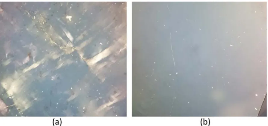
A super thin diamond XBPM has been made from a “standard grade” element six ltd. scCVD dia-mond slab with lateral dimensions of 4.5 mm× 4.5 mm and 300 µm thickness [www.e6.com]. This sample was laser sliced and polished to 40 µm thick plate by ALMAX easyLab [http://www.almax-easylab.com]. According to producer, the so called “standard grade” diamond has a substitutional nitrogen content of < 1 ppm, which reduces significantly life time of the charge carriers making this material useless for thick (∼ 40 microns). XBPMs use it, however the cost is significantly reduced (×10) compared to so called “electronic grade” diamond with nitrogen content of < 1 ppb. The struc-tural quality of diamond plate supplied has been checked by crossed-polarizer optical microscopy. The excellent structural quality is evidenced for a standard grade samples with almost perfect large area with no strain visible (figure1.b) which has been compared to the highly birefringent electronic grade diamond plate with high concentration of structural defect (dislocations, inclusions) density giving rise to strained bulk regions, and clearly visible as bright traces in figure1.a. The sam-ple thickness homogeneity and quality of surfaces polishing have been characterized by an IR-FT technique at the SMIS beamline optical characterization laboratory of SOLEIL.
Figure 1. Crossed-polarizer optical microscopy images obtained for two ElementSix sc-CVD diamond
plates: (a) electronic grade, (b) low-birefringence standard grade.
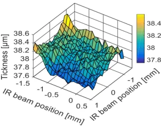
The thickness of 38.1 µm in average has been measured (figure 2) with variation less than 0.7 µm (peak-to-peak) and microscopic roughness of 130 nm rms over the 4 mm2surface. Similar characteristics were measured for several diamond samples and currently these specifications are guaranteed by the ALMAX easyLab for each fabricated plate.
A method of membrane fabrication was described elsewhere [4]. In short: using an ar-gon/oxygen plasma deep-etching technique, a free-standing auto supported membrane is produced in the central area of the diamond slab surrounded by a thicker frame. Thickness typically ranges from 1 micron to several microns, with macroscopic lateral dimensions comparable to diamond sample size typically∼ 3 × 3 mm. The structure and dimensions of the scCVD diamond membrane used in this work is depicted in figure3(left).
After hot acid cleaning, a∼ 100 kOhm/square resistive layer of Diamond Like Carbon (DLC) (thickness of∼ 200 nm) has been deposited on both diamond surfaces by a PVD sputtering apparatus using simple shadow masks. Finally, two pairs of lateral strip electrodes (Ti/Au) at the extremities
Figure 2. A thickness cartography obtained by an FT-IR microscope on laser sliced and re-polished 40 micron
thick scCVD diamond plate before deep plasma etching to the membrane.
Figure 3. A cross-sectional schematic view of free-standing scCVD diamond membrane PSD with its
dimensions (in blue — diamond membrane, in black — DLC resistive layers, in green — four lateral electrodes made of Ti/Au. A photograph of fabricated scCVD diamond PSD ready-to-mount on the dedicated readout pcb.
of DLC electrodes have been deposited with PVD and shadow masks. A complete scCVD diamond PSD ready for the mounting on the readout printed circuit board (pcb) is presented in figure3right. Such prepared diamond membrane PSD is mounted and wire-bonded onto a specially designed ceramic and installed in a vacuum chamber on SIRIUS beamline at SOLEIL for characterization purposes.
The XBIC signals from four Ti/Au lateral electrodes are connected to a four-channel current-to-voltage amplifier (LOCuM-4, by ENZ www.enz-de.de) and the output of LOCuM-4 voltages are digitized by an ADC (Adlink 2010). The detector bias voltage was supplied directly from the LOCUM as depicted in figure4.
The position of the X-ray beam is reconstructed in the two dimensions by a simple center-of-gravity algorithm: X = Kx× Ix1− Ix2 Ix1+ Ix2 , Y = Ky× Iy1− Iy2 Iy1+ Iy2 , (2.1)
where X and Y are coordinates of the position of X-ray beam center, Kx & Ky are the scale factor equal to L/2 with L the distance between collecting electrodes and I (x1,2 or y1,2) is the signal collected, respectively, from horizontal (X) and vertical (Y ) electrode pairs.
Figure 4. Diagram of the scCVD diamond membrane duo lateral PSD and signal generation principles.
The intensity of X-ray beam absorbed in diamond membrane is given by the sum of the signals collected from one of the electrode pairs (i.e. It =|Ix1+ Ix2| = |Iy1+ Iy2|).
3 Experimental results
3.1 XBIC and Charge Collection Efficiency (CCE)
The PSD diamond was irradiated with synchrotron X-rays on the SIRIUS beamline (E = 4 keV, Flux ∼ 1013ph/s), and the sum of XBICs of each pair of lateral electrodes was measured as a function of the detector bias voltage. Results are shown in figure5. Despite the rather high concentration of the nitrogen impurity, giving rise to a deep 1.7 eV (below conduction band) trapping center, present in the standard grade ElementSix diamond, a full CCE was evidenced for only 2 V/µm for the detector active area consisting of 4 microns thick membrane. A quick saturation of XBIC signal is reached due to the extremely short < 40 ps charge carriers transit time in such a thin layer, which anyhow is significantly shorter than the nitrogen altered lifetime of charge carriers.
The measured total induced XBIC of It = 63 µA is in excellent accordance with the expected current and it is given as:
It = q× Eph εp × A
Eph × Φ, (3.1)
where q is the value of the electron charge, Ephis the incident-beam energy and εp = 13.25±0.5 eV is the average electron-hole pair creation energy for X-ray absorption in diamond, A is the absorption factor depending on the energy Ephand diamond thickness, Φ is the X-ray beam flux. Thus, the complete CCE of the sensor allows absolute monitoring of X-ray beam flux during the experiment with a high precision (figure6). Additionally, we do not observe any persisting currents or lag-effects frequently reported for defective pcCVD XBPMs.
The figure 7shows the perfect time response of the scCVD membrane PSD XBIC generated during the beamline shutter opening.
Figure 5. XBICs vs. detector bias for the 4 µm thick scCVD diamond membrane PSD.
Figure 6. Total current time scan. Signal to noise ratio better than 0.1% (ADC sampling 100 Hz)
Figure 7. OFF-ON response of the scCVD diamond membrane PSD XBIC vs. time for shutter beam opening
3.2 The homogeneity of the XBIC response
The XBIC spatial homogeneity of the PSD was determined by a 2D scan of an X-ray beam spot of 200 µm × 200 µm size over the detector active area. The 2D map of XBIC image in figure8shows the total intensity inducted for all scanning positions of the beam on the PSD. For the 1 mm2center of the active area, we measured a variation of only 20 nA rms corresponding to 1.2% a fluctuation.
Figure 8. Full-area 2D homogeneity map of total XBIC (left) and in 2 × 2 mm of the center of active area
(right)
The XBIC spatial fluctuation correlates with the variation in the thickness of the diamond plate observed before the etching process (figure2), thus this variation is mainly related to X-ray beam absorption. Spatial inhomogeneity does not alter significantly the position monitoring if only small beam spots are used, however for better performance or precise beam intensity monitoring of moving beams the XBIC must be corrected for the sample thickness variation (by calibration) or thickness homogeneity must be improved for future devices.
3.3 X-ray Beam Position reconstruction and linearity
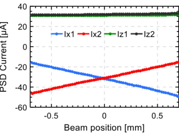
The scale factor in horizontal and vertical directions (Kx& Kyin equation (2.1)) have been checked by recording the PSD currents versus beam position (figure9) and by fitting the difference over the sum versus beam position with a linear function. The scale factor is in accordance with the inter-electrode dimensions (Lx = 2.7 mm & Ly = 3.5 mm) with Kx = 1.3 mm and Ky = 1.9 mm.
Figure 10illustrates the very good agreement of X-ray beam reconstructed position with real position of thin PSD after the Kx, Kyfactors calibration.
3.4 X-ray Beam Position resolution capability
The position resolution depends on the incident beam flux (intensity of XBIC), and the bandwidth of the readout electronics. A complete study of this dependence, based on experimental measurements compared to the theoretical model, is under study. For these particular experimental conditions the position resolution capability of the diamond PSD detector is illustrated in the figure 11. The detector was displaced by 10 microns using stepper motors (curve in blue), the X-ray beam reconstructed position for an ADC integration time of 100 ms and a X-ray beam flux of 1013ph/s
Figure 9. Cross-over response of the diamond membrane PSD while scanning the beam through the detector
in horizontal direction (Ix1, Ix2).
Figure 10. Reconstructed beam position vs. beam position coordinates obtained from X step motor with the
scale factor determined.
Figure 11. Time scan of the X-ray beam position with a 10 microns displacement of the scCVD diamond
membrane PSD for an ADC integration time of 100 ms. In blue — PSD motor displacement, in red — PSD reconstructed position.
(XBIC of 63 µA) is displayed in red. As presented, the relative X-ray beam movement is perfectly reconstructed by the PSD with a standard deviation of about 130 nm rms for these conditions.
4 Conclusion
We have fabricated and demonstrated the performances of a scCVD diamond membrane PSD intended for in-beam monitoring of the tender X-ray beamlines. The excellent charge collection efficiency, the relatively good homogeneity for the XBIC response and submicron beam position resolution were demonstrated at less than 4% absorption of the incident X-ray beam at 4 keV. Two ready-to-use detectors have been prepared for SIRIUS beamline. More sensors are planned for SIRIUS and METROLOGIE beamlines but the thinning process remains a key issue in the production of these membranes. Due to the presence of structural defects within the diamond bulk and despite the screening procedure the yield of pinholes free membranes fabrication is rather far from 100%.
Acknowledgments
We would like to thanks the SIRIUS beamline teams for their support and beam-time necessary for the characterization of the very thin PSD XBPM.
References
[1] K. Desjardins et al., Characterisation of CVD Diamond devices as XBPMs at SOLEIL, J. Phys. Conf.
Ser. 425(2013) 212004.
[2] K. Desjardins et al., Ultra-thin optical grade scCVD Diamond as X-ray Beam Position Monitor, J.
Synchrotron Rad. 21(2014) 1217.
[3] G. Ciatto et al., SIRIUS: A new beamline for in situ X-ray diffraction and spectroscopy studies of
advanced materials and nanostructures at the SOLEIL Synchrotron, Thin Solid Films 617 (2016) 48. [4] M. Pomorski et al., Super-thin single crystal diamond membrane radiation detectors, Appl. Phys. Lett.
103 (2013) 112106.
[5] A.I. Harris, Spectroscopy with multichannel correlation radiometers,Rev. Sci. Instrum. 76(2005) 054503[astro-ph/0504449].
[6] G.F. Knoll, Radiation detection and measurements, John Willey and Sons, Inc., New York (2000). [7] V. Dangendorf, Time-resolved fast-neutron imaging with a pulse-counting image intensifier, in
proceedings of International workshop on fast neutron detectors and applications, University of Cape Town, South Africa, 3–6 April 2006,PoS(FNDA2006)008.