HAL Id: tel-01127126
https://tel.archives-ouvertes.fr/tel-01127126
Submitted on 7 Mar 2015HAL is a multi-disciplinary open access archive for the deposit and dissemination of sci-entific research documents, whether they are pub-lished or not. The documents may come from teaching and research institutions in France or abroad, or from public or private research centers.
L’archive ouverte pluridisciplinaire HAL, est destinée au dépôt et à la diffusion de documents scientifiques de niveau recherche, publiés ou non, émanant des établissements d’enseignement et de recherche français ou étrangers, des laboratoires publics ou privés.
microenvironments : single cell behaviors for collective
functions
Caterina Tomba
To cite this version:
Caterina Tomba. Primary brain cells in in vitro controlled microenvironments : single cell behav-iors for collective functions. Physics [physics]. Université de Grenoble, 2014. English. �NNT : 2014GRENY039�. �tel-01127126�
THÈSE
Pour obtenir le grade de
DOCTEUR DE L’UNIVERSITÉ DE GRENOBLE
Spécialité : Physique pour les sciences du vivant
Arrêté ministériel : 7 août 2006
Présentée par
Caterina TOMBA
Thèse dirigée parCatherine VILLARD et codirigée parAlice NICOLAS
préparée au sein du l’ Institut Néel et du Laboratoire des Technologies de la Microélectronique
dans l’Ecole Doctorale de Physique de Grenoble
Primary brain cells in
in vitro controlled microenvironments
single cell behaviors for collective functions
Thèse soutenue publiquement le05/12/2014, devant le jury composé de :
Christine METIN
Institut du Fer à Moulin, Rapporteur
Vincent STUDER
Institut interdisciplinaire de Neurosciences, Rapporteur
Yves COUDER
Laboratoire Matière et Systèmes Complexes, Examinateur
Frédéric SAUDOU
Grenoble Institut des Neurosciences, Examinateur
Catherine VILLARD
Institut Néel, Directeur de thèse
Alice NICOLAS
Cellules primaires du cerveau en microenvironnements contrôlés in vitro Résumé :Du fait de sa complexité, le fonctionnement du cerveau est exploré par des méthodes très diverses, telles que la neurophysiologie et les neurosciences cognitives, et à des échelles variées, allant de l’observation de l’organe dans son ensemble jusqu’aux molécules impliquées dans les processus biologiques. Ici, nous proposons une étude à l’échelle cellulaire qui s’intéresse à deux briques élémentaires du cerveau : les neurones et les cellules gliales. L’approche choisie est la biophysique, de part les outils utilisés et les questions abordées sous l’angle de la physique. L’originalité de ce travail est d’utiliser des cellules primaires du cerveau dans un souci de proximité avec l’in vivo, au sein de systèmes in vitro dont la structure chimique et physique est contrôlée à l’échelle micrométrique. Utilisant les outils de la microélectronique pour un contrôle robuste des paramètres physico-chimiques de l’environnement cellulaire, ce travail s’intéresse à deux aspects de la biologie du cerveau : la polarisation neuronale, et la sensibilité des cellules gliales aux propriétés mécaniques de leur environnement. A noter que ces deux questions sont étroitement imbriquées lors de la réparation d’une lésion. La première est cruciale pour la di-rectionalité de la transmission de signaux électriques et chimiques et se traduit par une rupture de symétrie dans la morphologie du neurone. La seconde intervient dans les mécanismes de re-colonisation des lésions, dont les propriétés mécaniques sont altérées. Les études quantitatives menées au cours de cette thèse portent essentiellement sur la phénoménologie de la croissance de ces deux types de cellules et leur réponse à des contraintes géométriques ou mécaniques. L’objectif in fine est d’élucider quelques mécanismes moléculaires associés aux modifications de la structure cellulaire et donc du cytosquelette.
Un des résultats significatifs de ce travail est le contrôle de la polarisation neuronale par le simple contrôle de la morphologie cellulaire. Ce résultat ouvre la possibilité de développer des architectures neuronales contrôlées in vitro à l’échelle de la cellule individuelle.
Mots clés : Cellules primaires du cerveau, neurones d’hippocampe, polarisation neuronale, cellules gliales du cortex cérébral, motifs d’adhésion et de rigidité, mécanique cellulaire, méca-nosensibilité, cytosquelette, biophysique
Abstract: The complex structure of the brain is explored by various methods, such as neurophysiology and cognitive neuroscience. This exploration occurs at different scales, from the observation of this organ as a whole entity to molecules involved in biological processes. Here, we propose a study at the cellular scale that focuses on two building elements of brain: neurons and glial cells. Our approach reaches biophysics field for two main reasons: tools that are used and the physical approach to the issues.
The originality of our work is to keep close to the in vivo by using primary brain cells in in vitro systems, where chemical and physical environments are controlled at micrometric scale. Microelectronic tools are employed to provide a reliable control of the physical and chemical cellular environment. This work focuses on two aspects of brain cell biology: neuronal polarization and glial cell sensitivity to mechanical properties of their environment. As an example, these two issues are involved in injured brains. The first is crucial for the directionality of the transmission of electrical and chemical signals and is associated to a break of symmetry in neuron morphology. The second occurs in recolonization mechanisms of lesions, whose mechanical properties are impaired. During this thesis, quantitative studies are performed on these two cell types, focusing on their growth and their response to geometrical and mechanical constraints. The final aim is to elucidate some molecular mechanisms underlying changes of the cellular structure, and therefore of the cytoskeleton.
A significant outcome of this work is the control of the neuronal polarization by a simple control of cell morphology. This result opens the possibility to develop controlled neural ar-chitectures in vitro with a single cell precision.
Keywords: Primary brain cells, hippocampal neurons, neuronal polarization, cortical glial cells, chemical and mechanical pattering, cellular mechanics, mechanosensitivity, cytoskeleton, biophysics
Acknowledgments, remerciements
First of all, I would like to thank you, the reader, for your interest in this PhD work. For these few pages, I prefer to switch to my daily language, the French, in order to say "un grand merci" to all the people I had the chance to meet during my PhD and that contributed to realize this experience.
Ces trois ans m’ont donné l’occasion de nourrir ma curiosité pour le monde qui nous en-toure et de plonger dans le monde de la biophysique. La richesse de cette aventure a pour protagonistes la vitalité, la passion et la curiosité des différentes personnes qui ont participé de façon active à la réalisation de ce projet de thèse ou que j’ai connu grâce aux collaborations et les conférences auxquelles j’ai pu participer. Je ne pourrai pas les citer toutes mais j’en suis très reconnaissante.
Je tiens à remercier en quelques lignes ici mes directrices de thèse, Catherine Villard et Alice Nicolas, grâce à qui j’ai pu découvrir la beauté de ce domaine de recherche et son approche. Merci pour m’avoir toujours soutenue et encouragée par votre optimisme et confiance. Je vous remercie pour votre dynamisme qui m’a permis d’explorer différents laboratoires et de m’appro-cher de leurs compétences de biologistes. Merci pour ces trois ans de travail intense et amusant. Je tiens également à remercier tous les membres du jury, Christine Métin, Vincent Studer, Yves Couder et Frédéric Saudou, d’avoir accepté d’évaluer mon travail, pour leur commentaires et leur regard intéressé et critique très enrichissant.
Je souhaite remercier mes équipes, l’équipe Termodynamique des Petits Systèmes à l’Ins-titut Néel dirigée par Olivier Bourgeois, et l’équipe Micronanotechnologies pour la santé au LTM dirigée par David Peyrade. Je tiens à remercier Alain Schuhl, Pierre-Etienne Wolf et Benjamin Canals à l’Institut Néel et Olivier Joubert et Jumana Boussey au LTM pour leur soutient comme aussi André Sulpice et tout le personnel du CRETA pour leur accueil toujours chaleureux.
Un merci spécial à Antoine Triller de l’IBENS pour m’avoir accueilli dans son laboratoire jusqu’à que je puisse être autonome dès les premières étapes nécessaires pour mes manips et merci à toute l’équipe, en particulier à Sabrina Colasse pour sa patience et délicatesse utilisée pour m’amener à apprendre les techniques de dissection.
Je tiens à rappeler la générosité et l’hospitalité également de :
• Isabelle Marty, Julien Fauré et en particulier Julie Brocard du GIN à Grenoble pour l’initiation et le rodage à la culture cellulaire,
• Catherine Picart et Claire Monge du LMGP à Grenoble pour leur échanges sur les cellules musculaires,
• Giorgio Scita et Andrea Disanza, Amanda Oldani, Dario Parazzoli et Massimiliano Garrè iii
première vagues d’actine en fluorescence,
• Casper Hoogenraad, Ines Ferreira et Kah Wai Yau de l’Université de Utrecht pour leur sympathie et persévérance,
• Laurent Malaquin et toute l’équipe à l’Institut Curie de Paris pour leur investissement dans l’aventure des cellules primaires aimantées dans des systèmes microfluidiques, • Patricia Bassereau et Thomas Bornschlögl de l’Institut Curie de Paris pour la courte
mais enthousiasmante expérience avec les pinces optiques,
• Giovanni Longo et Simone Ruggeri à l’EPFL à Lausanne pour leur maitrise avec l’AFM qui a battu l’élasticité imparable des neurones,
• Christine Métin à l’institut du Fer à Moulin à Paris qui a fait tout le possible pour réussir les manips,
• Lionel Bureau du LiPhy à Grenoble pour sa disponibilité à partager la fabrication des brosses de PNIPAM.
Merci beaucoup aussi à :
• Nir Gov du Weizmann Institute à Rehovot pour les échanges enrichissants et son intérêt insatiable pour notre système,
• Nora Dempsey, Frédéric Dumas-Bouchiat, Luiz Fernando Zanini et Damien Le Roy pour leur enthousiasme et sourire malgré les contraintes de manipuler des objets vivants, • toute la formidable et impeccable équipe de Nanofab,
• Pierre Brosse pour ses questions sur les neurones parmi ses conseils patients et utiles pour apprendre à me servir du tour,
• Laetitia Marty, Vincent Bouchiat et Graziella Kavaklian pour leur aide à Nanochimie, • Ghislain Bugnicourt qui a rendu son manuscrit à mon premier jour de thèse et qui n’a
jamais manqué de me transmettre son enthousiasme et des bons conseils pour démarrer au mieux cette aventure,
• Céline Braïni qui a pris la suite du passage de témoin, avant lequel j’ai pu profiter de sa bonne compagnie et soutient, que ce soit en salle blanche ou devant une tasse de thé et muffins,
• les stagiaires Beilun Wu, Soha Souk et Yohan Lecomte pour leurs questions et les bons moments gourmands passés ensemble
... de l’Institut Néel et à...
• Michel Moussus pour m’avoir toujours soutenue et motivée, pour son sang froid, ses conseils en béton et le meilleur café français,
• David Fuard qui m’a initiée à la salle blanche et a su toujours intervenir avec la bonne solution dans les moments cruciaux,
• Abbas Mgharbel pour ses explications et conseils sur les gels et pour avoir renoncé à sa yaourtière pour assurer un confort essentiel des neurones entre Paris et Grenoble, • Nicolas Bouchonville d’avoir pris à coeur la suite des mesures AFM,
• Olivier Lecarme, Julien Cordeiro, Thibault Honegger, Clarisse Benoit, Kevin Berton, Maxime Darnon, Camille Petit-Etienne... pour leur vitalité contagieuse et motrice et pour la bonne ambiance au labo et à l’extérieure
... du LTM,
... sans oublier Danielle Gulino, qui a relu mon manuscrit avec beaucoup d’intérêt en apportant des remarques constructives aussi bien avant qu’après la rédaction, Camille Migdal, pour un travail d’équipe hyper efficace et plaisant, Marie Courçon, Alexandre Morel, Mélanie Char-bit, Helena Polena et Isabelle Vilgrain du C3 au CEA à qui je dois un merci particulier pour leur bonne humeur toujours présente, comme aussi pour les petits délices qui soulageaient les longues journées au labo.
Je tiens également à remercier Stéphanie Lacour et Cédric Paulou de l’EPFL à Lausanne, Cédric Allier et Srikanth Vinjimore Kesavan du CEA à Grenoble pour les courtes mais très enrichissantes manips ensemble, le Prof. Balestra de l’Université de Turin pour ses conseils. Une grande partie des résultats de ce travail n’aurait pas pu être observée sans l’accès aux mi-croscopes mis à disposition par Odile Fihol-Cochet et Didier Grunwald du CEA, Jean-Philippe Kleman de l’IBS et Michaël Betton du LiPhy à Grenoble, que je souhaite tous remercier pour leurs conseils et présence importante.
Au GIN de Grenoble, je voudrais remercier aussi Antoine Depaulis, pour sa bonne humeur, toujours disponible pour des bons conseils, et Mireille Albrieux, de nous avoir accueilli dans son labo pour nos premières aventures de dissection à Grenoble.
Merci beaucoup à Christine Martinelli, Patricia Poirier, Carméline Meli, Mathilde Mauro, Louise Infuso de l’Institut Néel, Sylvaine Cetra et Marielle Clot du LTM, Sandrine Ferrari de l’école doctorale de Physique et toutes les gestionnaires qui ont toujours fait leur mieux pour simplifier les démarches parfois emmêlées du système administratif, à Cécile Nemiche de la repro pour ses conseils et compréhension.
Un souvenir spécial à Franck Omnès qui m’a guidée à la découverte de l’Institut Néel la toute première fois que je rentrais au labo et qui m’a transmis son enthousiasme pour la re-cherche à l’interface entre physique et biologie.
Mille mercis à ...
Stéphanie Monfront pour la riche expérience avec la Fondation Nanosciences, l’équipe d’Innov-Doc pour la belle synergie, Sandrine, Julien, Jonathan et Jonathan pour leur accueil chaleu-reux pendant mes visites à Paris, ma troupe de Percevalière pour son énergie puissante et les moments ensemble inoubliables, les autres covoitureurs franco-italiens pour les conversations dépaysantes et les questions motivantes sur mon sujet de recherche.
Je vais terminer par remercier de tout coeur toutes les personnes chaleureuses dont la présence a marqué le cheminement de ma thèse et qui ont alimenté mon enthousiasme au cours de la rédaction, pour leur curiosité assidue pour mon travail et leur esprit critique, leur soutient jour par jour et les renforts culinaires, les pique-niques régénérants en montagne : ma famille qui a fait tout le possible pour assister à la soutenance et qui a assuré la réussite du pot, Ben, Sania, Rachel, Alex, Raph, Antho, Virginie, Yann, Daniela, Véro, Cyril, Sofia, Luca, Layal, Antoine, Cec, Jerem, Six, Marta, Vale... Grazie!
Contents
Introduction . . . 1
I Introduction to brain cells 9 I.1 Functions and morphology . . . 14
I.2 Brain cells structure . . . 17
I.2.1 Microtubules . . . 17
I.2.2 Actin filaments . . . 19
I.2.3 Intermediate filaments . . . 21
I.2.4 Axon and dendrites . . . 21
I.2.5 Growth cone . . . 23
I.3 Neuronal cell growth and polarization . . . 24
I.3.1 Mechanisms of neuronal polarization process . . . 25
I.3.2 Molecular neuronal growth and guidance . . . 25
I.3.3 Waves . . . 26
I.4 Brain cell adhesion in vitro . . . 27
I.4.1 Extracellular matrix receptors and adhesion proteins . . . 28
I.5 Brain cell response in controlled microenvironments. . . 31
I.5.1 Chemical, mechanical and topographical stimuli . . . 31
I.5.2 Mechanical properties of neuronal cells . . . 36
I.6 Control from single cell to population. . . 38
I.6.1 Active cell entrapment and long term soma positioning . . . 39
I.6.2 Neuronal architecture and polarity . . . 40
I.6.3 Neuronal polarity at population levels . . . 42
I.6.4 Coming research approaches and applications . . . 43
I.7 Context and objective of this work . . . 44
Bibliography . . . 46
II Materials and Methods 57 II.1 Patterned substrates . . . 62
II.1.1 Chemical patterns . . . 62
II.1.2 Physical patterns . . . 75
II.2 Primary cell cultures . . . 84
II.2.1 Neuronal and mixed neuronal–glial cell cultures . . . 84
II.2.2 Pure glial cell cultures . . . 87 vii
II.2.3 Fixation . . . 88
II.2.4 Immunofluorescence . . . 89
II.3 Microscopy observations . . . 91
II.3.1 Time-lapse experiments . . . 91
II.3.2 Fixed cells . . . 91
II.4 Analysis methods . . . 92
II.4.1 Neurite length of isolated cells. . . 92
II.4.2 Statistical tests . . . 92
II.4.3 Neurite volume: atomic force and digital holographic microscopes . . . . 94
II.4.4 Waves detection . . . 95
II.4.5 Optical tweezers . . . 96
II.5 Cell positioning by magnetic traps . . . 96
II.5.1 Micropatterned hard magnetic particles in PDMS . . . 97
II.5.2 Micropatterned soft magnetic particles in glass . . . 99
II.6 Summary . . . 101
Bibliography . . . 102
III Neuronal growth under chemical adhesive constraints 105 III.1 How neurite width controls neuronal growth . . . 112
III.1.1 Neurite growth . . . 112
III.1.2 Neuronal polarization . . . 114
III.1.3 Neurite volume . . . 119
III.1.4 Summary about the influence of neurite width in neuronal growth . . . 123
III.2 Behind the role of the neurite width in the axonal specification . . . 124
III.2.1 Changing the adhesive width along a same neurite . . . 124
III.2.2 About axonal and dendritic markers . . . 129
III.2.3 Summary about our biophysical approach of neuronal polarization . . . 132
III.3 Growth cone like waves along neurites . . . 133
III.3.1 Neurite elongation and neuronal polarization: which mechanisms? . . . 133
III.3.2 Neurite width influences wave characteristics . . . 133
III.3.3 Waves as force generating structures . . . 140
III.3.4 Summary about wave dynamics . . . 158
III.4 Molecular high resolution investigation . . . 160
III.4.1 Actin structure and effectors . . . 161
III.4.2 Microtubules organization and associated proteins . . . 162
III.4.3 Summary about waves and molecular aspects . . . 165
III.5 Toward controlled neuronal networks . . . 166
III.6 Conclusion. . . 169
CONTENTS ix
IV Glial cells under physical constraints 175
IV.1 Glial cells sensitivity to the substrate stiffness . . . 180
IV.1.1 Methodology . . . 181
IV.1.2 Mixed neuronal–glial cell cultures . . . 182
IV.1.3 Pure glial cell cultures . . . 191
IV.1.4 Discussion about glial cell mechanosensitivity in mixed and pure cultures 198 IV.1.5 Mechanical constraints at the cell level . . . 201
IV.2 Glial cells sensitivity to the substrate topography . . . 204
IV.3 Discussion . . . 206
Bibliography . . . 209
Conclusions and perspectives 211
Introduction
Since the pioneering observations of Ramón y Cajal more than one century ago, the very fast development of imaging technologies has given the possibility to investigate living systems, and in particularly the brain, from submicrometric scale to the level of the whole organ. Therefore, it does not surprise the growing charm around the understanding of the brain organization and working. Although some knowledge about nervous system are now well established and largely diffused, this complex system is far from being understood and still attract the attention of scientists and people in general.
The complexity of the nervous system is partially due to its large panel of functions and properties of mechanical, electrical and chemical origins, that distinguishes it from most other tissues or organs. Therefore, the study of this biological system is approached from different points of view. Nervous system can be considered for its morphological properties, as well as for its capabilities to respond to a stimulus, to propagate and transfer nerve impulses, or for its role in interaction with other organs. Nevertheless, it is crucial to consider nervous system as relatively autonomous but fundamentally integrated in a larger biological system. In the same way, single neurons are entities with independent functions that have to be taken into account as a component of a larger computational entity that is the nervous system.
The surge of innovative technologies giving access to the cell and sub-cell scale environment has allowed significant advances in the understanding of nervous system. The variety of tools coming from genetics, physics, chemistry and optics opens new approaches and new questions that motivate scientists. This context gives the great advantage to promote the encounter of diverse disciplines that, each of them, provides its portion of knowledge and a combinatory of approaches. The interest for the brain is that way addressed from both a computational and a cellular point of view. These two angles of study enrich one another and are associated to complex questions like the information processing in neuronal networks and the mechanisms underlying the origin and the development of neurodegenerative diseases and brain cancers.
This PhD project has mainly taken place in a laboratory of physics. The point of view of a physician is the guideline of this work, supported by enriching interactions with biolo-gists. Indeed, several collaborations and exchanges with biologists in France and Europe have been undertaken along this PhD to maintain a constructive dialog between these two domains of research. The particular context of Grenoble has played a fundamental role thanks to a large proximity of several laboratories of research and to a technological environment highly developed in the microelectronic context.
In this PhD thesis, we focus on the issue of brain cell growth from a morphological point of view at the scale of single cells. In particular, our interest is oriented towards the interplay between cell morphology and its functions. From this point of view, a neuron is characterized by a rather spherical cell body, named the soma, and arboreal extensions, called neurites, that in polarized functional neurons differentiate into two types: dendrites and axons. Dendrites are organized into a tree structure that collects the electrical activity produced by pre–synaptic neurons, whereas the axon represents the long extension that conveys the output signal toward post–synaptic neurons.
During this PhD project, experiments have been performed using primary brain cells. These kind of cells differentiate from cell lines, that are quasi immortal cells with a nearly unlimited capability of division. Cell lines have thus characteristics similar to cancer cells and are populations that became stable after several steps in vitro, far from their original environment in vivo. Contrarily, primary cells are more sensitive to their environment and better reflect the properties of the organ of their origin.
Primary brain cells have been obtained from rodent embryos. Indeed, at least for studies on cell brain behavior, rodents are considered as the best experimental compromise between avail-ability and pertinence of brain tissues and ethical aspects. In particular, we have studied the in vitro behavior of hippocampal neurons and cortical glial cells. Neurons from hippocampus are a basic reference of primary brain cells but they are still poorly employed in microstructured in vitro environments. Glial cells are another fundamental cell type in brain. They are known for their metabolic and regulatory role of neuron activity but they are also at the origin of most of brain cancers and of the mechanical changes in injured brains. This work is thus constituted of two main topics. Firstly, the establishment of the neuronal polarization, or differentiation, as a function of morphological changes associated to this crucial phenomenon in the neuron development. Secondly, the sensitivity of glial cells to the physical and mechanical properties of cell environment.
Our study highlights the importance of the interactions of cells with the physical and chemical properties of their environment. These interactions are at the basis of the development of a single cell and of its specific functions. In that sense, our approach is humbly inspired by the work on the living system geometrical organization of the Scottish D’Arcy Thompson (On Growth and Form, 1917) until the Chilean H. Maturana (Autopoiesis and Cognition: The Realization of the Living, with F. Varela, 1980).
The methodology employed in this work is based on the fabrication of controlled environ-ments in order to investigate the neurons and glial cells response to topographical, mechanical and chemical cues. Indeed, although the nervous system is a complex network of interconnected cells, one way to understand its complexity is to dissect it into simpler elements. For that, it is necessary to study the properties of its cellular components, that are mainly neuronal and glial cells. This is currently possible thanks to the contribution of microelectronic technologies and of the development of instruments of microscopic observation that give access to a fine control of the physical and chemical properties of the cell environment, as well as to a deep analysis at the single cell level.
INTRODUCTION 3
In the first chapter of this manuscript, we will introduce the general context of our work in order to describe the basics of cell neurobiology, the specific vocabulary associated to brain cells (highlighted in bold) and the main steps of neuronal development in vitro. This section will give some motivations underlying the studies on the cellular microenvironment and an overview of the principal examples reported by the literature. We will mainly focus on studies with primary brain cells, for the same reason we have made the choice to work with this kind of cells, that is to keep a close relationship with the in vivo real environment.
The subject of the second chapter will be the methods employed as well as some additional methods we have developed and tested, but not used for the results exposed in this manuscript. The different techniques explored reflect the growing impact of microelectronics technologies into life science. We will conclude this chapter with the presentation of some techniques to trap cells in controlled microstructured substrates whose principles are based on magnetic interactions.
In the two last chapters, we will report experimental results about neuronal and glial cell responses to chemical and physical contraints of their environment. In the third chapter, we will analyze the influence of the adhesion geometry on the neuronal growth. We will especially focus on the neurite elongation and the neuronal polarization. More precisely, we will report observations on neurons whose branches differ in width or number. Possible biophysical mechanisms underlying the control of the neurite growth and polarization will be proposed and discussed, eventually based on theoretical models.
In the fourth chapter, we will discuss about the mechanosensitivity of glial cells on patterns of rigidity at different scales. This work represents a new exploratory study of the role played by the chemistry of the glial cell environment in their sensitivity to the mechanical properties of their substrates. A short section about the glial cell response to topography will conclude this chapter.
At the end of each of these two last chapters we will provide a summary of our results. Finally, more significant results will be recapitulated in a general conclusion discussing the various perspectives that have come to light from this PhD work.
INTRODUCTION 5
Introduction
Depuis les observations pionnières de Ramón y Cajal il y a plus d’un siècle, le développement très rapide des technologies d’imagerie a ouvert la possibilité d’étudier les systèmes vivants, et en particulier le cerveau, de l’échelle submicrométrique à celle de l’organe vu dans son ensemble. Ces techniques d’imagerie toujours plus résolues ont engendré un véritable engouement vers la compréhension de l’organisation du cerveau et de son fonctionnement. Bien que certaines connaissances sur le système nerveux soient maintenant bien établies et largement diffusées, ce système complexe est loin d’être compris et attire encore fortement l’attention à la fois des scientifiques et du grand public.
La complexité du système nerveux est en partie due à sa variété de fonctions, associées à un environnement mécanique, électrique et chimique, qui le distingue de la plupart des autres tissus ou organes. De ce fait, l’étude de ce système biologique est abordé sous différents points de vue. Le système nerveux peut aussi être observé pour ses propriétés morphologiques, pour sa capacité à répondre à un stimulus et à propager et transférer l’influx nerveux, ou pour son rôle dans l’interaction avec d’autres organes. Il est crucial de considérer le système nerveux comme relativement autonome mais fondamentalement intégré dans un système biologique plus grand. De la même manière, les neurones sont des entités ayant des fonctions indépendantes qui doivent être pris en compte en tant que composants d’une entité plus grande qui est le système nerveux. L’émergence de technologies innovantes donnant accès à l’environnement à l’échelle cellu-laire et sub-cellucellu-laire a permis des avancées significatives dans la compréhension du système nerveux. La variété des outils issus de la génétique, de la physique, de la chimie et de l’op-tique ouvre de nouvelles approches et de nouvelles questions qui motivent les scientifiques. Ce contexte donne le grand avantage de favoriser la rencontre de disciplines différentes qui, chacune d’entre elles, apportant sa part de connaissances et d’approches possibles. Une spéci-ficité du cerveau est l’importance d’approcher son fonctionnement à la fois avec un point de vue cellulaire et un point de vue in silico. Ces deux angles d’étude s’enrichissent l’un l’autre et sont associées à des questions complexes comme le traitement de l’information dans des réseaux neuronaux, les mécanismes sous-jacents à l’origine et au développement de maladies neurodégénératives ou l’invasion de cancers du cerveau.
d’un physicien est la ligne directrice de ce travail, soutenu par l’enrichissement des interactions avec les biologistes. En effet, plusieurs collaborations et échanges avec des biologistes en France et en Europe ont été entreprises le long de cette thèse afin d’assurer un dialogue constructif entre ces deux domaines de recherche. Le contexte particulier de la ville de Grenoble a joué un rôle fondamental grâce à une grande proximité de plusieurs laboratoires de recherche et à un environnement technologique très développé dans le contexte de la microélectronique.
Dans cette thèse, nous nous concentrons sur la question de la croissance des cellules du cerveau à partir d’une analyse morphologique à l’échelle de cellules individuelles. En particulier, notre intérêt est orienté vers l’interaction entre la morphologie des cellules et leurs fonctions. De ce point de vue, un neurone est caractérisé par un corps cellulaire plutôt sphérique, appelé soma, et des ramifications, nommées neurites, qui dans les neurones fonctionnels une fois polarisés se différencient en deux types : les dendrites et les axones. Les dendrites sont organisées en une structure arborescente qui recueille l’activité électrique produite par les neurones pré– synaptiques, alors que l’axone représente une longue extension qui transmet le signal de sortie vers les neurones post–synaptiques.
Au cours de ce projet de thèse, des expériences ont été effectuées en utilisant des cellules primaires du cerveau. Ce type de cellules se différencie des lignées cellulaires, devenues stables après plusieurs étapes in vitro, loin de leur milieu d’origine in vivo. Bien que plus contraignantes à manipuler, les cellules primaires sont plus sensibles à leur environnement et reflètent mieux les propriétés de l’organe d’origine.
Les cellules primaires du cerveau ont été extraites d’embryons de rongeurs. En effet, au moins pour les études sur le comportement des cellules du cerveau, les rongeurs sont considé-rés comme le meilleur compromis expérimental entre la disponibilité et la pertinence des tissus du cerveau et les aspects éthiques. Nous avons étudié le comportement in vitro des neurones issus d’une région cérébrale appelée hippocampe et des cellules gliales corticales. Les neurones de l’hippocampe sont une référence fondamentale des cellules primaires du cerveau, mais ils sont encore peu utilisés dans des environnements microstructurés in vitro. Les cellules gliales constituent l’autre grand type de cellules du cerveau. Elles sont connues pour leur rôle méta-bolique et régulateur et de l’activité neuronale. Elles sont également à l’origine des cancers du cerveau et du remodelage de l’environnement cellulaire dans les cerveaux blessés. Ce travail est donc constitué de deux thèmes principaux : la mise en place de la polarisation neuronale (ou différenciation axonale), en fonction de changements morphologiques associés à cette étape clé de développement, puis la sensibilité des cellules gliales aux propriétés physiques et mécaniques du micro-environnement cellulaire.
INTRODUCTION 7
physiques et chimiques de leur environnement. Ces interactions sont déterminantes pour la croissance d’une cellule isolée et ses fonctions spécifiques. Dans ce contexte, notre approche est modestement inspiré par le travail sur l’organisation géométrique des systèmes vivants de l’écossais D’Arcy Thompson (On Growth and Form, 1917) jusqu’au chilien H. Maturana (Autopoiesis and Cognition : The Realization of the Living, avec F. Varela, 1980).
La méthodologie utilisée dans ce travail est basée sur la fabrication d’environnements contrôlés afin d’étudier la réponse des neurones et des cellules gliales à des signaux topo-graphiques, mécaniques et chimiques. En effet, bien que le système nerveux soit un réseau complexe de cellules interconnectées, une façon de comprendre sa complexité est de le sub-diviser en éléments plus simples. Pour cela, il est nécessaire d’étudier les propriétés de ses composants cellulaires, qui sont principalement des cellules neuronales et gliales. Ceci est ac-tuellement possible grâce à la contribution des technologies de la microélectronique qui donnent accès à un contrôle subcellulaire des propriétés physiques et chimiques de l’environnement de la cellule, et à l’élaboration d’instruments d’observation microscopique qui permettent une analyse approfondie des mécanismes cellulaires.
Dans le premier chapitre de ce manuscrit, nous allons présenter le contexte général de notre travail afin de décrire les bases de neurobiologie cellulaire, le vocabulaire spécifique as-socié aux cellules du cerveau (indiqués en gras) et les principales étapes du développement neuronal in vitro. Cette section donnera les motivations sous-jacentes des études effectuées dans des micro-environnements artificiels contrôlés et un aperçu des principaux exemples rap-portés par la littérature. Nous nous concentrerons principalement sur l’état de l’art portant sur des cellules primaires du cerveau, pour la même raison que celles pour lesquelles nous avons fait le choix de travailler avec ce type de cellules.
Le sujet du deuxième chapitre sera dédié aux méthodes employées ainsi qu’à d’autres mé-thodes que nous avons développées et testées, mais qui ne sont pas utilisés pour les résultats exposés dans ce manuscrit. Les différentes techniques explorées reflètent l’impact croissant des technologies de la microélectronique dans les sciences de la vie. Nous terminerons ce chapitre par la présentation de quelques techniques basées sur des interactions magnétiques permettant de capturer des cellules sur des sites prédéterminés lors de l’étape d ?ensemencement.
Les deux derniers chapitres suivants sont dédiés spécifiquement aux réponses des cellules neu-ronales et gliales à des contraintes chimiques et physiques de leur environnement. Dans le troisième chapitre, nous analyserons l’influence de la géométrie d’adhésion sur la croissance neuronale. Nous mettrons l’accent sur l’élongation neuritique et la polarisation neuronale. Plus précisément, nous rapporterons nos observations sur des neurones dont les branches varient en largeur ou en nombre. De possibles mécanismes biophysiques sous-jacents contrôlant la crois-sance des neurites et la polarisation seront proposés et discutés, éventuellement basés sur des
modèles théoriques.
Dans le quatrième chapitre, nous discuterons de la sensibilité des cellules gliales aux pro-priétés mécaniques de leur environnement sur des motifs de rigidité à différentes échelles. Ce travail présente une étude exploratoire du rôle joué par la chimie de l’environnement des cellules gliales dans leur réponse aux propriétés mécaniques de ce même environnement. Une courte section sur la réponse des cellules gliales à la topographie terminera ce chapitre.
À la fin de chacun de ces deux derniers chapitres, nous fournirons un résumé de nos résultats. Enfin, les résultats les plus significatifs seront récapitulés dans une conclusion générale qui discutera les différentes perspectives résultant de ce travail de thèse.
Chapter I
Introduction to brain cells
Contents
I.1 Functions and morphology . . . 14 I.2 Brain cells structure. . . 17 I.2.1 Microtubules . . . 17 I.2.2 Actin filaments . . . 19 I.2.3 Intermediate filaments . . . 21 I.2.4 Axon and dendrites . . . 21 I.2.5 Growth cone . . . 23 I.3 Neuronal cell growth and polarization . . . 24 I.3.1 Mechanisms of neuronal polarization process . . . 25 I.3.2 Molecular neuronal growth and guidance . . . 25 I.3.3 Waves . . . 26 I.4 Brain cell adhesion in vitro . . . 27 I.4.1 Extracellular matrix receptors and adhesion proteins . . . 28 I.5 Brain cell response in controlled microenvironments . . . 31 I.5.1 Chemical, mechanical and topographical stimuli . . . 31 I.5.2 Mechanical properties of neuronal cells. . . 36 I.6 Control from single cell to population . . . 38 I.6.1 Active cell entrapment and long term soma positioning. . . 39 I.6.2 Neuronal architecture and polarity . . . 40 I.6.3 Neuronal polarity at population levels . . . 42 I.6.4 Coming research approaches and applications . . . 43 I.7 Context and objective of this work . . . 44 Bibliography. . . 46
11
Chapitre I : résumé
Dans ce chapitre d’introduction nous présentons quelques propriétés fondamentales de l’organisation cellulaire et subcellulaire des cellules du cerveau, les processus impliqués dans leur développement et leur différentiation. Cela permet de poser le contexte biophysique et d’introduire les éléments nécessaires à la compréhension de ce travail de thèse. Une étude bibliographique portant sur l’interaction des cellules primaires du cerveau avec des microen-vironnements contrôlés décrit et compare les travaux récents. Ce chapitre se termine par une présentation succinte de la finalité et la validité des méthodes et des approches expérimentales choisies.
13
List of abbreviations and nomenclatures
AIS . . . Axonal Initial Segment AnkG . . . AnkirinG
CAMs . . . Cell Adhesion Molecules
CAMSAPs . . . Calmodulin–regulated spectrin–associated proteins CNS . . . Central Nervous System
DIV . . . Days In Vitro
DRG . . . Dorsal-Root Ganglia neurons EB . . . End–Binding proteins
ECM . . . Extracellular matrix FAs . . . Focal Adhesions FAK . . . Focal Adhesion Kinase GFAP . . . Glial Fibrillary Acid Protein MAPs . . . Microtubule–Associated Proteins PDMS . . . PolyDiMethylSiloxane
PLL, PDL . . . Poly-L-lysine, Poly-D-lysine PLL–PEG . . . Poly-L-lysine PolyEthyleneGlycol PLO . . . Poly-ornithine
PNS . . . Peripheral Nervous System µCP . . . Microcontact printing 3D, 2D, 1D . . . 3, 2 and 1 dimensions
I.1
Functions and morphology
The nervous system assures the survival of living organisms by its capacity to orchestrate their interaction with the external environment, which at the most basic level means to co-ordinate the movements produced by the body and to process the signals from the various organs, including sensory organs. The centralized control role played by the nervous system is characterized by its complex and very quick capabilities to integrate information. During the course of the Evolution, the nervous system has gained in complexity and efficiency, reaching its higher level in primates with the emergence of consciousness. A high brain plasticity and development throughout life is what distinguishes humans from the others primates. The level of complexity of the nervous system in the organisms is often associated to a corresponding ca-pacity to acquire new learning and to adjust behaviors in reaction to the external environment. All responses to the various internal or external stimuli determines the organism behavior. In that respect, the brain function is strongly associated to the foundation of biological individu-ality, to such an extent that the existing legislation in most of the world equates the cessation of all brain activity to death. Most questions about identity, consciousness and intelligence are still open, gathering the joined efforts of neuroscientists, psychologists and philosophers.
In vertebrate organisms, the nervous system is composed of the central (CNS) and of the peripheral (PNS) nervous system. The CNS includes the brain and the spinal cord and results in a centralized control of the information coming both from the organism and its environment. The CNS and the PNS are surrounded by three sheets of connective tissues forming the meninges. The PNS is constituted of nerves leading from and to the CNS and of a set of ganglia that are groups of nerve cells providing intermediary connections with the CNS. Cells from the CNS will be the focus of this work. The CNS is mainly composed of neurons and of more numerous glial cells.
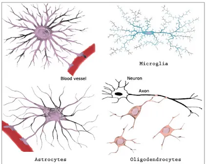
Glial cells name takes origin from the Greek word "glue", thanks to their first known role of support and protection of the nervous tissue. Glial cells are grouped in three principal types (Figure I.1): astrocytes, microglia and oligodendrocytes.
Astrocytes, taking their name from their star-like shape, ensure several functions in the nervous system: they supply glucose to neurons, they contribute to regulate the composition of the interstitial fluid in the nervous tissue and to the formation of the blood-brain barrier, they modulate synaptic activity, releasing and uptaking for example glutamate [1]. In the fourth chapter we will discuss their interaction with neurons on mechanosensitive aspects. Microglia are macrophage cells with an immune defense role. The third kind of glial cells has a similar role than Schwann cells in the PNS, i.e. to wrap a myelin sheath around neuronal extensions.
I.1. Functions and morphology 15
Figure I.1: Morphologies of the three principal types of glial cells: astrocytes, microglia and oligodendrocytes. Adapted from Ganong W.F., Review of Medical Physiology, 22nd Edition, 2005.
There is a growing interest for glial cells in general and for their active involvement in various brain functions, including the regulation of the strength of neuronal connections [2,3]. Glial cells, unlike mature neurons where the mitotic cell cycle is arrested [4], may retain the potential to divide and this capacity is one of the reasons of the glial origin of most of brain cancers. They are also associated to the mechanical changes observed in the injured brain. Neurons are excitable cells characterized by a resting intra-cellular potential (∼ -70 mV ) induced by the difference of ions concentrations, mainly choloride (Cl−), sodium (Na+) and potassium (K+), on opposite sides of the cellular membrane. The membrane potential can locally switch to positive values in neuronal processes conveying electrical signals. The neuron morphology reflects its function to collect, process and transmit electrical signals through chemical junction named synapses. From a mature neuron cell body, or soma, emerge one axon (sometimes two in specific neuronal types) that conveys the output signal toward post-synaptic neurons, and multiple dendrites organized into a tree structure that collects the electrical activity produced by pre-synaptic neurons. Soma and dendrites mainly compose the grey matter of the CNS whereas the white matter is composed of bundles of myelinated axons. In mammals, the typical size of a soma is on the order of 10 µm, the axon and dendrites diameter of less than 1 µm [5]. The different steps of neuronal maturation in vitro will be described in Section I.3.
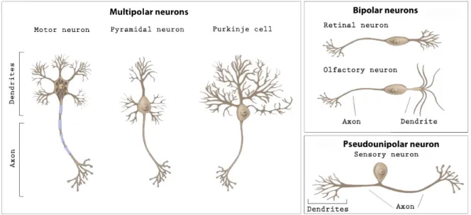
There are over 200 different varieties of neuronal cells. The geometry of axons and dendrites and synapses localization change strongly with the cell type, depending on their role in the neural circuit (Figure I.2). For this reason, a better understanding of the interplay between morphologies and associated functions is crucial.
Figure I.2: Basic neuron types of different brain areas, classed by their morphology. Adapted from wiseGEEK.com.
Brain can be divided into functional areas, which dimensions are species-dependent. Their local architecture is nevertheless quite conserved, in relation to their biological function. For example, the olfactory bulb is more developed in mice than in human beings, where the cerebral cortex is the most developed region (Figure I.3).
Figure I.3: Longitudinal (A) and transverse (B) sections of human and rat (A) or mouse (B) brains, showing functional similarities although in different proportions. Adapted from [6].
Interestingly, there are no qualitative differences between neurons of the human brain and those of other mammals or other vertebrates. However, numerous variables differentiate the brains of several species like their size, their number of neurons and glial cells, the number of their sub-types, the molecular nature of synapses [7] and the architecture of the connectivity between neurons.
I.2. Brain cells structure 17
hippocampus or the cortex of rodent embryos at eighteen days of gestation (E18 embryos). The main reason is that the dissociated hippocampal tissue provides a relatively homogeneous cell population. Indeed, pyramidal neurons, so-called for their pyramidal-shaped soma, are the predominant neuron type present in the hippocampus at this stage of development. These cells are excitatory glutamatergic neurons, whereas the majority of the remaining neurons are interneurons, GABAergic cells making inhibitory synaptic terminals [8]. Moreover, the hippocampus development has been well-characterized in vitro. On a functional point of view, the hippocampus plays an important role in spatial memory and in learning processes, it is essential for the consolidation of long-term memories, acquired from experiences, and it may be necessary in the encoding of novel associative information in short-term memory. Cerebral cortex encompasses about two-thirds of the brain mass and it is responsible, amongst other functions, of consciousness, memory, language and thinking.
I.2
Brain cells structure
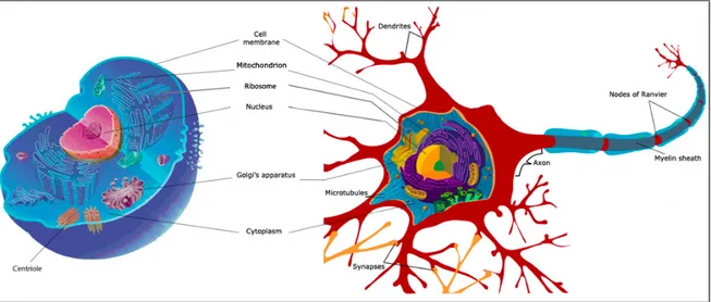
Neurons, as all eukaryotic cells including glial cells (Figure I.4), have a common envelope named the plasma membrane. This membrane, that contains many transmembrane proteins and macromolecules, is at the interface between the extracellular matrix (ECM) and the internal cytoplasm. The cytoplasm is composed of a medium called the cytosol where are immersed the nucleus containing genes, various organelles (e.g. the mitochondria which pro-vides energy, the Golgi’s apparatus and the ribosomes that are able to synthesize proteins) and three types of filaments. These filaments are microtubules, actin and intermediate filamentsthat, organized in a network, form the cytoskeleton of the cell. These three impor-tant structures will be detailed in the following subsections (I.2.1,I.2.2,I.2.3). The cytoskeleton plays a critical role in the dynamical properties of the cell, e.g. migration, polarization, forces generation and external signals transduction. In neurons, the cytoskeleton adopts specific or-ganizations that will be described in the subsections entitled: axon, dendrites and growth cone(I.2.4,I.2.5).
I.2.1 Microtubules
Microtubules are nucleated from the centrosome, an organelle composed of two centri-oles, and they form the mitotic spindles that guide the chromosomes during cell division. Recent studies have shown that most of microtubules dispersed in the different regions of neu-rons are not only nucleated and released from the centrosome [9] but they can originate from a acentrosomal assembly [10]. This phenomenon already known from plants and yeasts [11], could be important for axonal specification, thanks for example to the presence in the axon of some microtubule severing proteins like katanin [4].
Figure I.4: Common components between a typical eukaryotic animal cell (left), like glial cells, and a neuron (right). Adapted from Wikimedia Commons.
Microtubules are highly dynamic structures, with an external and internal diameter typically of 22 nm and 12 nm and a length varying between fraction to tens of microns [12]. Their tubular architecture makes them the stiffest filaments in the cytoskeleton, giving structural support to cell shapes. The tubular structure of these protofilaments is polarized with a plus (+) end (polymerization) and a minus (−) end (depolymerization) and is made up of α and β-tubulin dimers (FigureI.5).
Figure I.5: Schematic representation of microtubule structure. A) Tubulin heterodimer (α and β subunits) and a polarized protofilament. B) Protofilament network composed of parallel protofil-aments with the same orientation. C) Electronic image of a microtubule segment showing a ring of 13 protofilaments [13].
I.2. Brain cells structure 19
Of note, the growth of the microtubule + end is coordinated by the end–binding (EB) proteins family, composed of three members (EB1 – 3) [14]. In neurons, EB1 is implicated in axonal transport [15], whereas EB3 plays the role of a molecular link between microtubules and the actin cytoskeleton [16]. The polarization of microtubules determines the direction of motion of vesicles and organelles along the filaments. Kinesin and dynein are the molecular motors associated to microtubules, moving respectively toward their + and - ends. A more recent family of microtubule minus–end binding proteins called calmodulin–regulated spectrin– associated proteins (CAMSAPs) have been described for their role of regulation of microtubules dynamics. It has been shown that mammalian CAMSAP family members bind specifically to microtubule minus–ends and protect them against kinesin-13–induced depolymerization [17]. In particular, CAMSAP2 specifically localizes to noncentrosomal microtubule minus–ends. In neurons, it stabilizes the free microtubule minus–ends in order to control neuronal polarity and development, playing a key role for proper microtubule organization [18].
In general, microtubule–associated proteins (MAPs) localized along microtubules promote their stabilization and organization.
I.2.2 Actin filaments
Actin filaments, or F-actin, are organized in a double helix of 7 – 9 nm of diameter and composed of monomers of globular actin, or G-actin. They are polarized and dynamic like microtubules and their thin structure makes them very flexible (FigureI.6.A). Actin polymer-ization is stimulated by nucleating factors, e.g. formins and Arp2/3 complex.
Actin filaments self assemble in 3D, 2D or 1D structures such as cortex, lamellipodium and filopodium (Figure I.6.B). Lamellipodia are large projections of the leading edge of the cell used to explore its environment and to move. Thin filopodia are filled with oriented, bundled actin filaments that usually spread beyond the front of the lamellipodium with the + end to-ward the protrusion direction. Actin-binding proteins, such as Eps8, are enriched in the growth cones, in particular in the focal adhesions and in the filopodium. These regulatory proteins are involved in the axonal filopodia formation and, more in general, in the control of the actin dynamics in developing neurons [19]. Actin self assembly and bundling are the result of the cooperation of numerous proteins, e.g. IRSp53, Ena/VASP, WASp/Scar. Antiparallel associa-tions of actin filaments are found in stress fibers, making possible the production of mechanical forces inside the cell by shifting actin filaments respectivly to each others. The actin filaments relative motion involved in cell contractility is allowed by myosin-II, an ATP–dependent motor protein (Figure I.6.C).
Figure I.6: A) Actin polymerization stages: from nucleation of G-actin monomers (red), to intermediary stable actin complexes (violet) to a gradual elongation of actin filaments (F-actin). In the steady state, actin filaments are organized in a polarized double helix [20]. B) Example of actin structures in glial cells at 2 DIV. The grey rectangle corresponds to the zoom in the inset: lamellipodium (green arrow), filopodia (yellow arrow) and stress fibers (blue arrow). Red, phalloidin. Blue, nucleus. Scale bar: 20 µm. C) Myosin II molecules (in blue and in white) are associated in an antiparallel fashion to move (see arrows) towards the + ends of antiparallel actin filaments leading to acto-myosin contractility [21].
I.2. Brain cells structure 21
I.2.3 Intermediate filaments
Intermediate filaments are responsible for structural support. They are less dynamic than the two others polymers (microtubules and actin filaments) and they are not polarized. Their molecular structure depends on the cell type but they generally adopt an helical shape com-posed of dimer proteins organized in protofilaments, with a typical diameter of 10 nm, i.e. an intermediate size between the diameters of microtubules and of actin filaments.
In neurons, they are called neurofilaments and play a major role in the maturation and the maintenance of axonal integrity as well as in the establishment of axonal diameter [13]. Their defective trafficking and mutations have also been implicated in mechanisms involved in several neurological disorders, e.g. Parkinson’s disease and amyotrophic lateral sclerosis, and in neuronal death [22].
Astrocytes in the CNS express a specific type of intermediate filament proteins: the glial fib-rillary acid protein (GFAP). In embryonic glial cells, the vimentin intermediate filament, a protein characteristic of mesenchymal cells (e.g. fibroblasts), is also present.
I.2.4 Axon and dendrites
Axons and dendrites are neuronal protrusions grouped under the generic term of neurites. These cylindrical membrane protrusions are essentially composed of a bundle of axial micro-tubules, a small proportion of neurofilaments and a peripheral actin cortex. Tau and MAP2 are distinctive microtubule-associated proteins of axons and dendrites respectively. Both play a role in promoting microtubule stabilization.
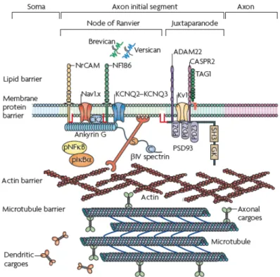
Axon, the transmitter pole of neurons, morphologically differs from dendrites by its more homogeneous aspect and its thinner and constant diameter. Its length varies depending on the neuron type from hundreds of microns to a few meters in large animals. Microtubules are all oriented with their + end towards the tip of the axon [23]. The proximal region of the axon near the soma is called axonal initial segment (AIS). In this area, a high density of sodium (Na+) channels fulfills the necessary conditions to initiate the action potential [24]. A diffusion barrier is established at the AIS level, associated to the segregation of specific axonal proteins like ankyrinG (ankG) [25]. The interplay between ankG and AIS microtubules that support the maintenance of neuronal polarity is coordinated by microtubule plus–end–binding proteins EB1 and EB3 [26].
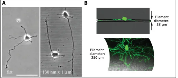
The characteristic length of the AIS is typically 30 – 40 µm. A similar characteristic length has been revealed in axotomy experiments where the remaining stump retains a memory of its axonal nature only when longer than 35 µm [27] (Figure I.7).
Figure I.7: A) GFP-positive hippocampal neuron at 10 DIV. Axon is identified by the arrow. B) Axon is cut at 33 µm from the cell body (red dashed line). C) Responses of axotomized neurons: above a threshold of ∼ 35 µm neurons mostly regrow their original axon. From [27]. In the myelinated axons of vertebrates, clustered Na+ channels are then activated at the nodes of Ranvier to rapidly propagate action potentials along the axon [29]. Nodes of Ran-vier (see FigureI.4) represent myelin sheath gaps of approximately 1 µm in length, leading to uninsulated areas in axons that are necessary for their electrical processes.
The terminal region of the axon generally includes several branches, ending with a specialized area called the synaptic button. This is the presynaptic part of the synapse. It contains an actin network that mainly represents the site where neurotransmitter vesicles and the en-ergy suppliers mitochondria are localized. Kinesins allow directional transfer of molecules from soma to axon terminals, i.e. using vesicles to lead along microtubules materials required for renewal of the membrane.
Unlike axons, dendrites, the receptors poles of neurons, possess microtubules with reverse polarities, i.e. with their + or - ends directed toward the same side of the cell.
Some observations have shown that microtubules are involved in mRNA localization. More-over, ribosomes, whose function is to decode mRNA information, have been observed closely associated to microtubules. A specific distribution of mRNA triggers a locally controlled syn-thesis of proteins, e.g. mRNA coding for MAP2 that is localized to dendrites [30]. For instance, cytoskeletal proteins involved in the formation of dendritic spines, small membrane protru-sions that are the main sites of excitatory synaptic inputs [31], could be synthesized in the dendrites. Of note, in mammal PNS axons the ability to synthesize proteins is higher than in the CNS, that might be directly correlated to the high capacity of axon spontaneous regener-ation observed in the peripheral nervous system [32].
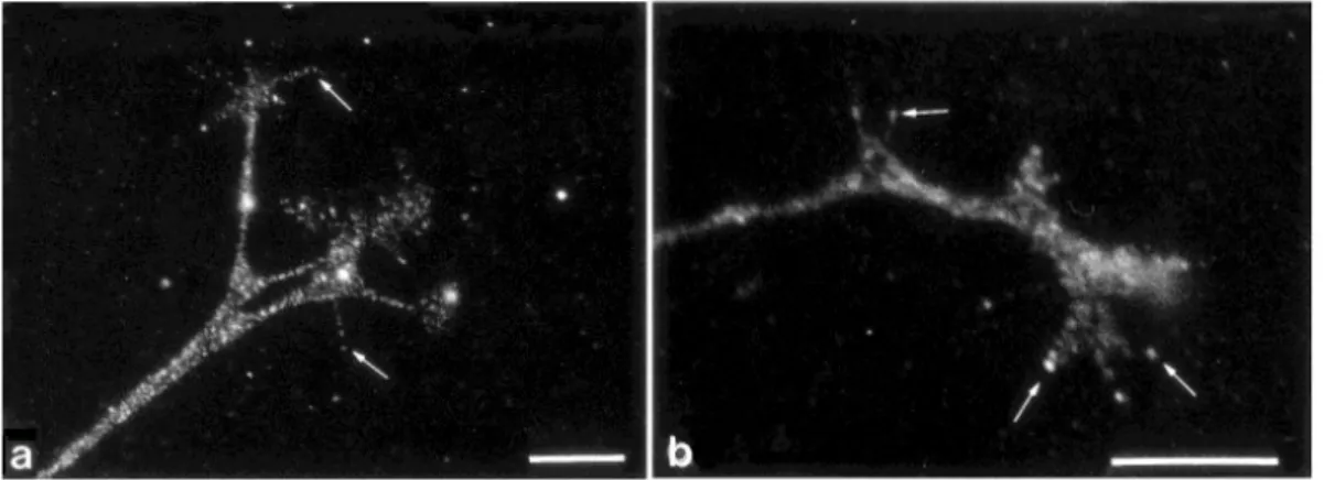
Recently, the development of high-resolution techniques in vitro gives the possibility to analyse the structural organization of axon and dendrites. Stochastic optical reconstruction microscopy (STORM) has revealed periodical ringlike structures around the axon circumference at a sub-micrometric scale [33]. These rings are composed of actin and adducin, an actin-capping protein, with a periodicity of ∼ 180 to 190 nm and alternated with spectrin rings, a cytoskele-tal protein. Interestingly, these periodic rings have not been observed in dendrites (FigureI.8).
I.2. Brain cells structure 23
Figure I.8: STORM imaging of cytoskeletal organization of axons and dendrites in hip-pocampal neurons al 7 DIV [33]. A) Three-dimensional image of actin in a dendrite (top) and an axon (bottom). Color-scale shows from violet to red the z positions closest to farthest from the substratum, respectively. B) Actin, adducing and βII-Spectrin immunostaining in an axon.
The consequence of these actin structures in the regulation of shape and adhesive properties of axons and dendrites remains an open issue. The state of the art of our knowledge on the molecular features of neuronal adhesions will be exposed in a dedicated section (I.4).
I.2.5 Growth cone
The growth cone is another important specific structure of neurons. The growth cone is formed at the neurite tip during development to ensure neurite guidance. Its high dynamics and sensitivity toward guidance molecules play a key role in neuronal growth and polarization, as much as in neuronal adhesion, as will be discuss in next sections (I.3,I.4). Beside, it has been shown that a growth cone produce mechanical forces.
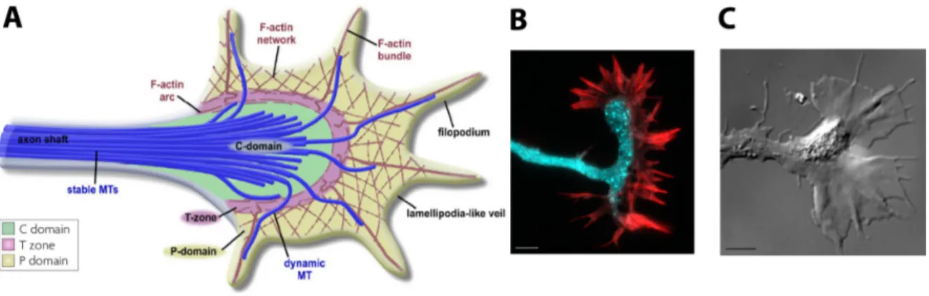
Structurally, a growth cone is composed of three areas that determine its shape and motion: a central zone filled with microtubules, an intermediary zone and a peripheral one, composed of lamellipodia and filopodia (Figure I.9). The intermediary area is made of an actin arc enclos-ing the microtubules comenclos-ing from the shaft and receivenclos-ing the retrograde actin flow from the peripheral zone. Myosin II, a main component of the actin arc, seems to actively constrain mi-crotubules of the central domain through acto–myosin contractility, leading to the stabilization of microtubules [34].
Figure I.9: Growth cone. A) Schematic representation of the three areas of a growth cone: the central (C) domain with stables microtubules, the transitional (T) zone with the actin arc enriched of myosin II and the peripheral domain (P), composed of lamellipodia and filopodia. Adapted from [34]. B) Example of growth cone in a mouse hippocampal neuron at 2 DIV. Cyan: synapsin. Red: phalloidin. Scale bar: 10 µm. C) Differential Interference Contrast image of a growth cone of a Xenopus spinal neuron [35]. Scale bar: 5 µm.
I.3
Neuronal cell growth and polarization
Embryonic cortical and hippocampal neurons development in vitro has been described by several stages identified by the morphological changes that occur during maturation [36,37,
38] (FigureI.10). Soon after plating, lamellipodia and filopodia are observed at the periphery of the soma. After several hours, these structures become immature neurites [39]. The navigation of neurites in this phase is mainly guided by the presence of the growth cone. Then, one neurite starts to elongate faster than the other. This longest neurite will progressively acquire the molecular specificities of the axon and the other neurites will later fully differentiate into dendrites. Finally, the dendritic spines that characterize a mature functional network will appear after about two weeks of culture.
Figure I.10: The generic stages of development of cortical/hippocampal neurons. Time refers to mouse hippocampal neurons, expressed in Days In Vitro (DIV). Adapted from [38].
I.3. Neuronal cell growth and polarization 25
I.3.1 Mechanisms of neuronal polarization process
Neuronal polarization is the event that leads to the axonal specification. The common denominator of neuronal polarization both in vivo and in vitro is the symmetry breaking expressed by the distinct specific molecular and morphological characteristics of axons and dendrites [38] (FigureI.10).
Contrarily to the non–uniform chemical and topographical in vivo environment where nu-merous signals may orient the cell asymmetry associated with the polarization process, the emergence of a structural asymmetry in vitro results from a stochastic process leading to a random choice of the localization of axonal specification [40].
The mechanisms at the base of the breaking of symmetry associated to axonal specification during neuronal polarization are more and more investigated. Recent studies have focused on the interplay between extracellular signals and cytoskeletal organization, i.e. actin stability and microtubule protrusion [40]. Local instability of the actin network restricted to a single growth cone is a physiological signal that triggers neuronal polarization [41]. Axon specifica-tion is also associated with an increased microtubule stabilizaspecifica-tion in one of the neurites, but it is actually unclear how this stability is achieved.
Recently, it has been shown that, in cultured hippocampal neurons and in cortical slices, laminin promotes neuronal polarization, i.e. axon specification and growth, through adhesive contacts and a resultant regulation of directional microtubule assembly [42]. Both in vertebrate and invertebrate species, growth cones interact with their environment, i.e. with other cells, the physical substrate or diffusive molecular gradients. These aspects will be the subject of the next subsection.
I.3.2 Molecular neuronal growth and guidance
Over the past decade, a large effort has been made in developmental neurobiology to iden-tify the repulsive and attractive molecules that guide axons.
Proteins that promote axonal outgrowth have been arranged in three distinct classes: dif-fusible molecules, such as trophic factors, components of the ECM and cell adhesion molecules (CAMs) [43]. Trophic factors regulate neuron growth and survival, as it was demonstrated by V. Hamburger and R. Levi Montalcini [44], who received the Nobel Prize in 1986 for the purification of the first trophic factor in the 1950s: the nerve growth factor (NGF). ECM constituentswill be more detailed in a dedicate section (I.4). Let us mention here that proteoglycan proteins specifically regulate the structural organisation of the ECM, modu-late growth factor activities and cellular adhesive and motility events, such as cell migration and axon outgrowth [45]. CAMs include calcium independent (immunoglobulin superfamily and integrins) and dependent (cadherins and selectins) proteins.
N-CAM and L1 belong to the immunoglobulin superfamily. L1 is mainly observed on fasci-culating axons (i.e. axons growing on top of each others) whereas N-CAM is predominantly involved in the stabilization of the cell contacts and in the interactions between glial cells and neurons [46]. They also contribute to more complex processes like axonal pathfinding, target recognition, synapse formation and synaptic plasticity.
The main proteins that contribute to axonal navigation are the semaphorins. They act as axonal guidance during brain development of vertebrates by the capacity of growth cones to sense their signals.
Semaphorin-3A (Sema3A), the first discovered semaphorin, in 1993 [47], is repulsive for the axon and attractive for the dendrites of cortical neurons [48]. More generally, semaphorins are able to drive nerve fiber fasciculation as shown for example by the capacity of Sema3A to inhibit the branching of cortical axons growing on two-dimensional substrates [49].
Amongst others guidance molecules, we report the most commonly known netrins, slits and ephrins families.
Growth and guidance of CNS axons are also crucially influenced by the interaction with astroglial cells. Neurite outgrowth and growth cone motility are mainly mediated by two receptors: N–cadherin and β1–class integrin. The first one is a Ca2+–dependent cell adhesion molecule, the second one is an ECM receptor. Chick ciliary ganglion neurons grown on cultured astrocytes had shown to be strongly influenced by these receptors at E8, whereas the influence of β1–integrin vanished at later stages [50]. The reduction of the integrin function during development, i.e. the interactions with several ECM proteins, including laminin, could be associated to the limited ability of adult CNS neurons to regenerate, in coherence with the regenerative potential played by laminin [51].
N-cadherin may have a primary importance in neuritic growth and it could be also implicated in adhesion between nerve and muscle in vivo [52], as well as in synaptogenesis [53].
I.3.3 Waves
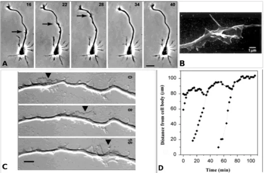
To conclude this short introduction to neuronal growth and polarization, this section gives the basics of growth cone–like structures named "waves" that have been first observed in in vitro rat hippocampus cultures, mainly during the first stages (i.e. 2 and 3, FigureI.10) [54]. Waves are created at the soma level and travel to the neurite tip at a speed of approximately 3 µm/min [54] (Figure I.11.A-C). They occasionally move in a retrograde direction (less than 5% of waves). These structures are similar to growth cones in their dynamics and composition [55]. Some previous works have suggested that these dynamical membrane deformations may recruit many actin–binding proteins, like GAP–43, Ezrin, Cortactin and the axon–promoting Shootin–1 protein [54, 56]. However, a global understanding of their molecular structure is
I.4. Brain cell adhesion in vitro 27
still missing.
The wave arrival at the neurite tip is systematically correlated with an initial retraction followed by an elongation of the neurite, leading to a net elongation, as well as an increase in the growth cone dynamics [57] (Figure I.11.D).
Figure I.11: A) Wave propagation along an axon (see arrows): phase contrast image of a rat hippocampal neuron after approximately 30 h in culture. Times are shown in minutes and correspond to the respective times in the graph in D). Scale bar: 20 µm. B) Scanning electron image showing 3D wave morphology of the rat hippocampal neuron. Scale bar: 1 µm. C) Wave propagation (see arrows) toward the growth cone of a rat hippocampal neuron (out-field), recorded after approximately 24 h in culture. Times are shown in minutes. Scale bar: 5 µm. D) Diagram of axon tip (circles) and wave (diamonds) positions in function of time during the wave propagation. Wave arrival is associated to an axon retraction and a subsequent elongation. Adapted from [57,54].
Waves also contribute to the axon growth and to the creation of new branches [55]. The frequency of these waves is higher along the nascent axon (approximately 2 times more than in other neurites). Moreover, they were observed in hippocampal slices, confirming that they are not in vitro artifacts [55]. Several fundamental open issues related to waves remain, like their mechanisms of propagation and their possible role in the neuronal polarization process as well as in the axonal transport mechanism [55,56].
I.4
Brain cell adhesion in vitro
Cell adhesion mechanisms are involved at all levels, i.e. from the maintenance of the cohesion of the neural tissue to the regulation of synaptic contacts in the mature nervous system. Cell adhesion molecules directly or indirectly interact with cytoplasmic proteins and cytoskeletal structures and therefore actively participate to various processes like cell spreading,