HAL Id: hal-01226720
https://hal.archives-ouvertes.fr/hal-01226720
Submitted on 20 Nov 2015HAL is a multi-disciplinary open access archive for the deposit and dissemination of sci-entific research documents, whether they are pub-lished or not. The documents may come from teaching and research institutions in France or abroad, or from public or private research centers.
L’archive ouverte pluridisciplinaire HAL, est destinée au dépôt et à la diffusion de documents scientifiques de niveau recherche, publiés ou non, émanant des établissements d’enseignement et de recherche français ou étrangers, des laboratoires publics ou privés.
molecular spin-state photo-switching: a
molecule-to-lattice energy transfer
Andrea Marino, Marco Cammarata, Samir F. Matar, Jean-François Létard,
Guillaume Chastanet, Matthieu Chollet, James Michael Glownia, Henrik T.
Lemke, Eric Collet
To cite this version:
Andrea Marino, Marco Cammarata, Samir F. Matar, Jean-François Létard, Guillaume Chastanet, et al.. Activation of coherent lattice phonon following ultrafast molecular spin-state photo-switching: a molecule-to-lattice energy transfer. Structural Dynamics, AIP, 2016, 3 (2), 023605 (15 p.). �10.1063/1.4936290�. �hal-01226720�
Activation of coherent lattice phonon following ultrafast molecular
spin-state photo-switching: a molecule-to-lattice energy transfer
A. Marino,1 M. Cammarata,1 S. F. Matar,2 J.-F. Létard,2 G. Chastanet,2M. Chollet,3J. M. Glownia,3
H. T. Lemke,3 and E. Collet.1,a)
1
Institute de Physique de Rennes, UMR 6251 University Rennes1-CNRS 35042 Rennes, France
2
CNRS, Université de Bordeaux, ICMCB, 87 avenue du Dr A. Schweitzer, Pessac, F'33608, France
3
LCLS, SLAC National Laboratory, Menlo Park, 94025, CA, USA.
a)
Author to whom correspondence should be addressed. Electronic mail: eric.collet@univ-rennes1.fr
We combine ultrafast optical spectroscopy with femtosecond X-ray absorption to study the photo-switching dynamics of the [Fe(PM-AzA)2(NCS)2] spin-crossover molecular solid. The
light-induced excited spin-state trapping (LIESST) process switches the molecules from low spin (LS) to high spin (HS) states on the sub-picosecond timescale. The change of the electronic state (<50 fs) induces a structural reorganization of the molecule within 160 fs. This transformation is accompanied by coherent molecular vibrations in the HS potential and especially a rapidly damped Fe-ligand breathing mode. The time-resolved studies evidence a delayed activation of coherent optical phonons of the lattice surrounding the photoexcited molecules.
I. INTRODUCTION
Many chemical and biological systems can switch between different electronic and structural states within less than one picosecond under light excitation. Such transformations, as well as their recovery towards the stable state, involve not only the photoactive center with a molecular transformation, but also its environment (Schoenlein et al., 1991; Elsaesser et al., 1991; Barbara et
al., 1992; Rosspeintner et al., 2013; Jun et al., 2015 and Levantino et al., 2015). For example, the energy transfer between a "hot" photo-excited molecule and its surrounding solvent is at the origin of molecular vibrational cooling and solvent heating. The light-control of molecular transformations expands nowadays towards materials science, offering new opportunities to impact the macroscopic state of materials with a light pulse (Koshihara et al., 2009). Molecular crystals are promising systems composed of many interacting elements which can give rise to collective transformations as ferroelectricity (Collet et al., 2003; Okamoto et al., 2004 and Uemura et al., 2010) or metal-insulator transitions for instance (Chollet et al., 2005; Kawakami 2009; Gao et al., 2013 and Servol
et al., 2015). In such systems, the charge redistributions induced by light at the intra- and inter-molecular levels activate (through the couplings between charge and structural degrees of freedom) coherent optical phonons in the early stages of the transformation, which results from the energy redistribution from the absorbing centers to the molecular and lattice degrees of freedom.
Spin-crossover (SCO) materials belong to a family of prototypical systems showing photo-chromic and photo-magnetic responses between two competitive states of different spin multiplicities (Gütlich and Goodwin 2004, Halcrow 2013). In the present case of study, FeII-based SCO complexes, of 3d6 electronic configuration can be switched from a diamagnetic low-spin state (LS S=0, t2g6eg0) to a paramagnetic high-spin state (HS S = 2, t2g4eg2). Weak continuous wave (cw)
laser irradiation at low temperature is known as an efficient way for controlling SCO materials by light. By choosing the appropriate excitation wavelength, it is possible to selectively populate the HS state (LIESST) or LS state (reverse-LIESST) (Hauser 1986) which are long-lived in solids below a characteristic TLIESST temperature (Létard et al., 1999) and transient above (Lorenc et al.,
2009, Lorenc et al., 2012). In solution the photoinduced HS state is transient at ambient temperature and pump-probe studies were used to unveil the ultrafast molecular switching and explore the intersystem crossing (ISC) dynamics. The use of complementary probes, sensitive to different degrees of freedom, strongly help investigating both ultrafast electronic and structural dynamics (Gawelda et al., 2007; Consani et al., 2009; Bressler et al., 2009; Cannizzo et al., 2010; Zhang et
al., 2014; Marino et al., 2014 and Auböck et al., 2015). LIESST in molecular solids shows very similar features, and it is well established that the strong electron-structural coupling plays a central role in the photo-induced trapping of the HS state (van Veenendaal et al., 2010; Cammarata et al., 2014; Bertoni et al., 2015a and Bertoni et al., 2015b). However, the strong interest of performing ultrafast LIESST in the solid state resides in the active nature of the crystalline solid: the constituting molecules can switch during the complex out-of-equilibrium pathway under the effect of lattice pressure or heating (Lorenc et al., 2009, Lorenc et al., 2012, Kaszub et al., 2013, Marino
et al., 2015 and Bertoni et al., 2015c). However, the associated energy transfer process from the absorber molecule to its crystalline environment was not investigated thus far. Here we study the elementary electronic and structural processes involved during LIESST in the SCO material [Fe(PM-AzA)2(NCS)2]. For understanding the molecule-to-lattice energy transfer, we compare the
photoresponse in [Fe(PM-AzA)2(NCS)2] crystals with the dynamics of solid solution crystals where
these SCO molecules are diluted in a passive [Zn(PM-AzA)2(NCS)2] matrix. II. EXPERIMENT
A. Sample description and steady state conversions.
The molecular structure of the SCO compound Fe(PM-AzA)2(NCS)2
(cis-bis(thiocyanato)bis[(N-2'-pyridylmethylene)-4-(phenylazo)aniline]), as obtained by X-ray diffraction (Guionneau et al., 1999), is represented in Fig. 1. This FeII system of d6 electronic configuration undergoes a spin-crossover from a diamagnetic LS (S=0) to a paramagnetic HS (S = 2) state (Létard et al., 1999 and Guionneau et al., 1999). It is monitored in Fig. 1a with magnetic susceptibility (χM) measurements.
The χMT product gives direct information on the spin state and shows a smooth spin crossover
centered at T(1/2) = 184 K, typical of non cooperative systems, (Ksenofontov et al., 1998 and
Guionneau et al., 1999). Several techniques sensitive to different degrees of freedom can probe the spin state switching (Halcrow et al., 2013). It is well established with ligand field theory that the change of spin state is associated with an important reorganization of the ligand, bounded here by N atoms to the Fe.
In this family of SCO compounds, the main structural deformation of the FeN6 octahedron is the
change of the average Fe−N distance, from ⟨Fe−N⟩LS ≈ 1.97 Å to ⟨Fe−N⟩HS ≈ 2.16 Å revealed by
X-ray diffraction (Marchivie et al., 2003 and Guionneau et al., 2012). Therefore, X-X-ray absorption near edge spectroscopy (XANES) at the FeII K-edge can be used to monitor the structural reorganization accompanying the SCO (Briois et al., 1995), and the maximum change is observed around 7125 eV (Bressler et al., 2009; Cammarata et al., 2014). Fig. 1b shows that the thermal equilibrium spin-crossover can be monitored by the change of X-ray absorption (XAS @ 7215 eV). In addition, the change of electronic distribution is associated with a color change of the material and consequently the spin state conversion can also be monitored with optical measurements (Marino et al., 2013, Goujon et al., 2008). Fig. 1c reports the overall reflectivity change with temperature and it is clear that these three measurements, probing a similar thermal SCO, give complementary fingerprints of the SCO phenomenon. They also prove the occurrence of the photo-switching due to 830 nm light excitation (Fig. 1a & 1c) below TLIESST = 44 Kin this compound
(Létard et al., 1999). We note a smaller change of magnetic susceptibility after photoexcitation at low temperature, compared to the thermal change (Fig. 1a), which is due to the rather thick powder sample used compared to the laser penetration depth, giving an overall limited fraction of photo-switched molecules (20 %). Diffuse optical reflectivity (OR) data (Fig. 1c) probing mostly the surface show a greater fraction of molecules in the photoinduced HS state. These conventional techniques are too slow for investigating the photoswitching dynamics on the time scales of elementary processes, which typically fall in the sub-picosecond range. Here we use two complementary femtosecond pump-probe techniques: optical spectroscopy and X-ray absorption. The[Fe(PM-AzA)2(NCS)2] crystals appear fully dark and optical transmission measurements can't
be performed in the visible range (VIS) (Marino et al., 2013) but only in the near-infrared region (NIR) where 50 m thick crystals become more transparent. Fig. 2a shows that because of this high absorption, the diffuse optical reflectivity (OR) of the pure HS [Fe(PM-AzA)2(NCS)2] is weak
below 1200 nm. The changes in the VIS part of the optical reflectivity (OR) spectrum measured on single crystals (Marino et al., 2013) constitute a nice fingerprint of the SCO (Fig. 2b), as observed
at thermal equilibrium in the spectral region of interest (630 nm - 750 nm). An isosbestic point is evidenced around690 nm. The OR increases during the LS → HS switching below 690 nm, whereas it decreases above. The absorption decreases when the [Fe(PM-AzA)2(NCS)2] molecules
are diluted in a [Zn(PM-AzA)2(NCS)2] crystalline matrix. Usually such dilution of SCO molecules
into isostructural lattice, which show no spin transitions, is used to modulate the coupling of the SCO molecule to the lattice and shifting phase transition to higher or lower temperature through the chemical pressure effect (Hauser, 2004). Here we take advantage of the optical properties of the matrix to study LIESST, because the Zn-based molecules of 3d10 electronic configuration are optically silent in the 550-1050 nm, as confirmed by the OR diffuse spectra of the pure Zn compound. These Zn-based molecules adopt the same crystal structure as Fe-based ones, but do not present any spin crossover (Guionneau et al., 2012). Consequently, the optical changes observed in the [Zn0.9Fe0.1(PM-AzA)2(NCS)2] materials are only attributed to its constituting
[Fe(PM-AzA)2(NCS)2] SCO molecules.
All these specific optical properties can be explained by an accurate description of excited states and the time dependent DFT (TD-DFT) approach is an excellent tool (Martin et al., 2003). The natural transition orbitals (NTO) account for hole–particle pairs and can be used to interpret the excited states corresponding to the absorption of light. TD-DFT as implemented in the Gaussian 09 package (Gaussian; 09) was applied for obtaining the NTO of the LS and HS [Fe(PM-AzA)2(NCS)2] as well as [Zn(PM-AzA)2(NCS)2] starting from geometry optimized molecule with
b3lyp/lanl2dz hybrid functional – basis set. Fig. 3a shows the calculated oscillator strength change between LS and HS [Fe(PM-AzA)2(NCS)2]. The main features are the appearance of an absorption
band above 1500 nm in the HS state, which is attributed to d-d transition from t2g-like to eg-like
orbitals as shown by the NTO in Fig. 3b. The LS d-d band is around 1200 nm in the LS state, where the ligand field is higher. The d-d transition in an Fe-N6 Oh symmetry are strictly forbidden, but the
[Fe(PM-AzA)2(NCS)2] molecule in the crystal lies in general position (its point symmetry is C1)
and the FeN6 core is not exactly octahedral, with 6 different Fe-N bonds (in the 1.94 to 1.98 Å
range) and N-Fe-N angles different from 90° (in the 80-93° range). In the LS state, the calculated oscillator strength of the d-d transitions are weak. In addition, because of the low symmetry, the t2g
-like orbitals are no more degenerate (this is also true for the 2 eg-like orbitals) as schematically
shown in the energy diagram (Fig. 3b). Consequently, different t2g-like -> 2 eg-like transitions are
calculated and the ones with strongest oscillator strength are shown at 1166 nm, 1236 nm and 1250 nm (bleu bars in Fig. 3a and bleu arrows in Fig. 3b). In average, the d-d splitting is 10Dq 1.02 eV. We used such d-d transition in another SCO solid to induced the LS-to-HS photoswitching (Marino
et al., 2014). In the HS state the deviation from Oh symmetry of the FeN6 core is larger (Guionneau
et al., 1999; Buron et al., 2012) for the Fe-N bonds (in the 2.06 to 2.27 A range) and the N-Fe-N angles from 90° (in the 74-99° range). Consequently the t2g-like -> 2 eg-like transitions are stronger
and shift to lower energy because of the 0.2 Å Fe-N elongation. The strongest t2g-like -> 2 eg-like
transitions are calculated at 1085 nm, 1450 nm and 1650 nm (Fig. 3a, Fig. 3b) and in average the d-d splitting is around-d 0.92 eV. Therefore the average d-decrease of the ligand-d field-d from the LS to the HS states is D10Dq = 0.1 eV.
The calculated absorption change from LS to HS states (decreasing above 600 nm and increasing below) are also in agreement with the reflectivity change reported in Fig. 2a, even though the isosbestic point is shifted by 90 nm with respect to our observations. The optical transitions around
the isosbestic point (690 nm) correspond to MLCT transition, both for LS and HS states, as illustrated by the NTO shown in Fig. 3b. The optical absorption of [Zn(PM-AzA)2(NCS)2] is
calculated to be very low in the 500-800 nm range, in agreement with the high reflectivity in Fig. 2a, confirming that this compound is optically silent in this range. These optical results at thermal equilibrium are of importance for the time-resolved analysis performed hereafter.
B. Experimental Conditions for femtosecond studies
The optical pump-probe experiments were configured in NIR-transmission and VIS-reflection geometry with a quasi-collinear configuration of pump and probe beams. The temporal evolution of Optical Density (OD) and Optical Reflectivity (OR) were respectively obtained from the relative change of transmitted and reflected probe signals. The LS-to-HS photoswitching dynamics was firstly investigated on pure [Fe(PM-AzA)2(NCS)2] single crystals (with typical dimensions of (500
± 50) × (70 ± 5) × (50 ± 5) μm3
), and then on [Zn0.9Fe0.1(PM-AzA)2(NCS)2] crystals with 10 % of
Fe molecules diluted in a passive zinc matrix. The sample temperature was controlled with a standard liquid nitrogen cryostream (Oxford Instruments Cryojet) and set for all experiments at 130 K to allow the HS-to-LS back relaxation within less than 1 ms (probe repetition rate of 1 kHz for the stroboscopic acquisition). For the optical experiments, two monochromatic ultrashort laser pulses of the duration of 40 fs generated with two optical parametric amplifiers (OPA) were used as pump and probe leading to an overall instrumental response function (IRF) on the order of 80 fs. The pump wavelength was set to 800 nm on a MLCT band (where it efficiently induces LS-to-HS conversion as shown in Fig. 1) with an excitation density of 0.4 mJ/mm2, typically switching 1-2 % of the molecules in the crystal (Lorenc et al., 1-2011-2; Collet et al., 1-2011-2). The photo-response of such SCO materials is linear on the ps timescale with the excitation energy per pump pulse (Bertoni
et al., 2015 and Marino et al., 2015) and the data presented here were collected well below the sample damage (found around 1.0-1.5 mJ/mm2). Data treatment involved iterative fitting of single/double exponential test functions convoluted with a Gaussian IRF of 80 fs. The residual oscillations observed in the OR curves were treated with a time dependent FFT analysis, in order to highlight the activation time of inter- and intra- molecular modes. For time scans up to 8 ps we used a smoothing procedure above 1 ps, with a 200 fs time window (10 experimental time steps) for increasing the signal to noise ratio and showing better the 1.25 ps modes.
Time-resolved X-ray absorption measurements (XAS) were performed at the XPP end station of the LCLS X-FEL (X-ray Free Electron Laser) in Stanford, USA (Chollet et al., 2015). For this experiment 50 fs pump laser pulses, also centered at 800 nm were used with an excitation density of 0.5 mJ/mm2. The change in XAS was recorded with 30 fs X-ray probe pulses centered at 7125 eV. This energy is sensitive to the changes accompanying the SCO , as observed during the thermal crossover (Fig. 1c), or in ultrafast studies in other SCO compounds (Bressler et al., 2009 and Cammarata 2014). The IRF of 110 (10) fs at FWHM was obtained with a X-ray/optical cross correlator designed to synchronize the optical and the X-ray laser pulses (Harmand et al., 2013).
III. RESULTS
A. Ultrafast structural trapping of the HS state
The OR dynamical time (dt) traces, recorded at selected probe wavelengths and reported in Fig. 4a, enable to track in real time the ultrafast LIESST dynamics. The single color pump-probe measurements obtained with the probe set at 640 nm, 690 nm and 720 nm clearly reproduce the optical fingerprints characteristic of the LS → HS switching (Fig. 2b), with an ultrafast OR increase at 640 nm and decrease at 720 nm as the oscillator strength changes from LS to HS states in this spectral region. On the other hand, time-resolved analysis around the isosbestic point (690 nm), where LS and HS states contribute equally, allows an isolated observation of the dynamics of the intermediate state(s) involved during the spin-state photo-switching. Such intermediate states (INT), as the initially photoexcited 1MLCT state (t2g5eg0L1), are responsible for the transient reflectivity
peak around dt=0. An exponential fit, taking into account our 80 fs IRF indicates that the INT state(s) decays toward the HS state within less than 50 fs. This peak is found of Gaussian shape and its width is limited by our experimental time resolution. This 50 fs decay from the 1MLCT (possibly together through other possible INT states) was also reported in other SCO compounds (Marino et
al., 2013; Cammarata et al., 2014 and Auböck 2015).
Whereas optical measurements give mostly access to the change of electronic state, time-resolved X-ray absorption spectroscopy at the FeII K-edge enables to track in real time the structural reorganization around the FeN6 core and especially to the change in 〈Fe-N〉 distance. Figure 4b
reports the time course of the XAS measured at 7125 eV. The XAS increase at this energy is a clear characteristic fingerprint of the formation of the HS structure, as observed at thermal equilibrium (Fig. 1c) when 〈Fe-N〉 elongates from the equilibrium value of the LS state (⟨Fe−N⟩LS ≈ 1.97 Å) to
the one of the HS state (⟨Fe−N⟩HS ≈ 2.16 Å) measured by X-ray diffraction (Guionneau et al., 1999
and Marchivie et al., 2003). An exponential fit convolved with our Gaussian IRF of 110 (10) fs allowed an accurate determination of the structural reorganization around Fe, with an elongation time constant τ〈Fe-N〉 = 160 (20) fs, similar to the one reported for other SCO molecules in crystal
and solution (Bressler et al, 2009; Lemke et al, 2013 and Cammarata et al, 2014). The signal remains constant for longer delays measured up to 6 ps (inset of Fig. 4b).
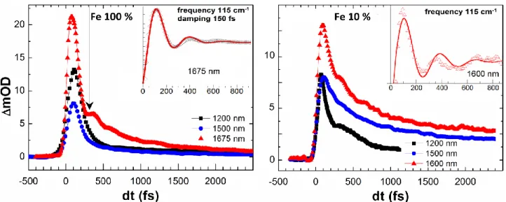
The optical transmission measurements in the spectral region probing the d-d transition of the HS state around 1200-1600 nm (Fig. 3) are also sensitive to the amplitude of the ligand field, which is modulated by the Fe-N distance (Bertoni et al, 2015a). Fig. 4c & Fig. 5 report the time evolution of the optical density (OD), recorded in transmission geometry for different probe wavelengths in the 1200-1675 nm spectral range. The increase of absorption after photo-excitation of the LS state corresponds to the appearance of the HS d-d transitions. The OD increase of the HS state in these low energy bands results therefore from the d-d gap narrowing. Here again an intermediate OD peak is observed around dt=0 as the MLCT state also absorbs. The exponential fit of the time trace indicates that the decay towards the HS state occurs within 150 (20) fs. This timescale differs from the 50 fs decay of the MLCT state and may be associated with a contribution of an intermediate triplet state to this transient absorption. It is difficult to identify here because of its weak optical fingerprint, but it was directly observed on the pathway from MCLT to HS by x-ray absorption techniques (Zhang et al., 2014). This absorption change is governed by the t2g- eg gap narrowing as
fs measured by XAS. A slower 750 fs decay is also observed and associated with vibrational cooling already observed in preliminary measurements (Marino et al., 2013). The apparent decay in Fig. 4c & Fig. 5 is therefore not associated with a decay of the HS state, as confirmed by constant XAS after 100s fs.
On top of this OD exponential decay, an oscillation also shows up at this probe wavelength. The insets of Fig. 5 show the oscillating component obtained by subtracting the exponential decay to the data. Since the MLCT peak decays within 150 fs, this 115 cm-1 oscillation (290 fs period), observed up to 1 ps, is therefore attributed to the HS state. It falls in the frequency range of the 〈Fe-N〉 breathing molecular mode reported in a similar SCO compounds (Baranovic et al., 2004; Ronayne
et al., 2006; Sousa et al., 2013; Cammarata et al., 2014 and Bertoni et al., 2015a). As the ligand field (Fe-N distance) modifies the amplitude of the d-d absorption at 1675 nm, this mode is therefore very likely associated with the breathing of the Fe-N6 core shell. Since the symmetry of
this molecule is very low (the 6 Fe-N bonds are not symmetry equivalent) there is no proper breathing mode with the totally symmetric Fe-N stretching. However the observed mode, probed around d-d transition, has Feligand breathing character. The oscillation of the signal is rapidly damped, within 150 (30) fs. This effect may come from a damping of the amplitude of the molecular oscillations, but a dephasing of the response of the photo-switched molecules, resulting from energy transfer to other modes or to collision, is also very likely. It is interesting to underline that such a breathing mode cannot be attributed to impulsive Raman process in the ground LS state because on the one hand the LS breathing frequency is significantly higher (150 cm-1) and on the other hand the LS state is silent at 1675 nm. This mode is also observed in our IR data in the diluted compound (Fig. 5) but the signal is more noisy due to the weak concentration of SCO molecules. In addition to this mode, another vibration (67 cm-1, 500 fs period) is observed for probing wavelengths around the isosbestic point (Fig. 6). Contrary to the 115 cm-1 mode, the 67 cm-1 mode it is not rapidly damped as few oscillations are observed during the first 2 ps. Since the absorption at this wavelength involves MLCT states (Fig. 3), this low frequency mode is very likely a molecular ligand torsion. This is also in agreement with calculations founding many ligand torsion modes in this frequency range (Baranovic et al., 2004; Ronayne et al., 2006; Sousa et al., 2013; Cammarata et al., 2014; Bertoni et al., 2015a and Bertoni et al., 2015b).
All these results are in good agreement with the recent literature reporting on the ultrafast LIESST for several FeII compounds, both in solution and solids (Cammarata et al., 2014; Bertoni et al., 2015a and Auböck et al., 2015). Since this LIESST dynamics is mostly the same for an isolated molecule in solution and for a molecule embedded in a single crystal, our results underline also that the ultrafast LIESST in SCO solids is strictly confined to a molecular response on the sub-ps timescale. Its MLCT state is short-lived (<50 fs) and the HS state is rapidly reached. Once in the HS potential the molecular structure changes and the Fe-N bonds expand towards the equilibrium value. The breathing mode is then activated and because of the excess energy the system oscillates in the HS potential where other modes, like ligand torsion, are activated. Our results are also consistent with the theoretical description of the LS → HS photo-switching process as resulting from the coherent activation and fast damping of the breathing mode, bringing and keeping the system in the HS potential (van Veenendaal et al., 2010 and Cammarata et al., 2014). We observe indeed that the 290 fs period breathing mode is highly damped within 150 fs. However, in Fig. 6 we can observe
another oscillation with 1.25 ps period, activated later and persisting up to 8 ps. Such features were not observed in solution and their nature is investigated in detail in the next section.
B. Activation of coherent optical lattice phonon
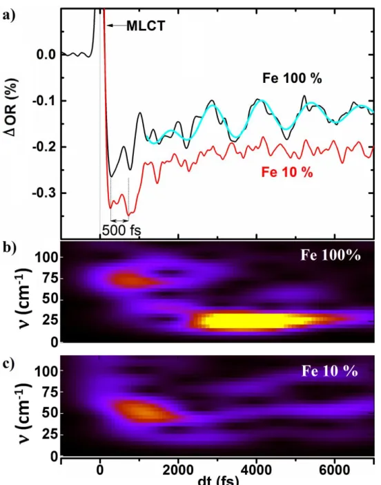
From a first look at Fig. 6 it is possible to identify in the OR time traces the short MLCT peak, followed by two main oscillating components. During the first two ps the weak 67 cm-1 oscillation (500 fs period) discussed above appears. Then the OR starts to oscillate with much higher amplitude and these slower oscillations vanish around 8 ps. The fit of this mode, taking into account an exponential increase and decay of its spectral weight, is shown by the cyan thick lines (using the same parameters for the different time traces), and gives a 1.25 ps period (28 cm-1). The oscillating component of Fig. 7 was extracted from the average bi-exponential fit describing decays of MLCT to HS state and the vibrational cooling (Marino et al., 2013) and we plot in Fig. 7 its time dependent fast Fourier transform (t-FFT) analysis. This analysis confirms the presence of the two different modes at around 67 cm-1 and 28 cm-1. The 67 cm-1 mode is activated as the HS potential is reached and is observed in the 0-2 ps range and corresponds to molecular coherent oscillations in the photoinduced HS state. It is important to notice how the amplitude of the 28 cm-1 mode is maximum at 4 ps: it gradually increases around 2 ps and decreases until vanishing around 8 ps. We observe in Fig. 7 a spectral weight transfer from the 67 cm-1 to the 28 cm-1 modes. Such low frequency modes in the 30 cm-1 range are usually associated to large amplitude ligand torsion (Ronayne et al., 2006). In molecular solids, this frequency range also corresponds to inter-molecular optical phonons, mixing inter-molecular displacement and torsion (Uemura and Okamoto 2010 and Servol et al., 2015). It seems therefore difficult to discriminate from these results the nature of such a mode. Is it associated with the local oscillation of the photo-excited molecules or is it associated with an optical phonons?
For answering to this question, the same experiments were performed on solid solution crystals, where photoactive [Fe(PM-AzA)2(NCS)2] molecules are diluted in a passive and optically silent
crystalline matrix of [Zn(PM-AzA)2(NCS)2] molecules. We used the solid solution [Zn(1-x)Fex
(PM-AzA)2(NCS)2] with x = 0.1. By performing such time resolved analysis on diluted crystals, we hope
to access only to the photo-response of the isolated photo-active Fe-based molecules and to learn more about its interaction with the crystalline environment. We should underline that a dilution of 10 % means that one Fe-based molecule is in average isolated in a box surrounded by 2×2×2 Zn-based molecules.
Figure 7 compares the dynamical time traces of optical reflectivity recorded for λprobe = 700 nm and
optical density in the IR for the pure and diluted crystal. The same experimental conditions were used with λpump = 800 nm, with a pump fluence of 0.4 mJ/mm2. The photo-response of the diluted
sample is similar to the one of the pure Fe compound. It starts with an ultrashort (<50 fs) MLCT peak partially shown in Fig. 6, zooming on the zone of interest, followed by the 500 fs (67 cm-1) oscillations evident in the pure compound, and less pronounced in the diluted one. The lower oscillation amplitude in the diluted crystal is due to a smaller density of Fe-based molecules and the higher noise for this record forbids an accurate extrapolation of the high frequency modes. The amplitude of the 28 cm-1 mode in the pure crystal is of the same order as the optical change between the first 500 fs and the signal around 2 ps. In the diluted crystal we observe a similar optical change between 500 fs and 2 ps and consequently the signal/noise ratio looks good enough to observe the
lattice phonon mode. Since it is not the case, we conclude that the 28 cm-1 oscillations are absent in the optical data, as confirmed by the time-dependent FFT (Figure 7). At that stage, we should keep in mind that such low frequency modes usually have lattice optical phonon character involving molecular bending, translation and/or orientation (Ronayne et al., 2006). In addition, the pure LS compound made of [Fe(PM-AzA)2(NCS)2] molecules, is absorbing at the 700 nm probe wavelength
and this absorption creating MLCT state can be modulated by molecular vibration. This is not the case for the [Zn(PM-AzA)2(NCS)2] lattice matrix of the diluted compound, which is optically silent
at 700 nm (Fig. 2). We therefore come to the conclusion that the 28 cm-1 vibration observed only in the pure compound is a lattice optical phonon mode.
IV. Discussion
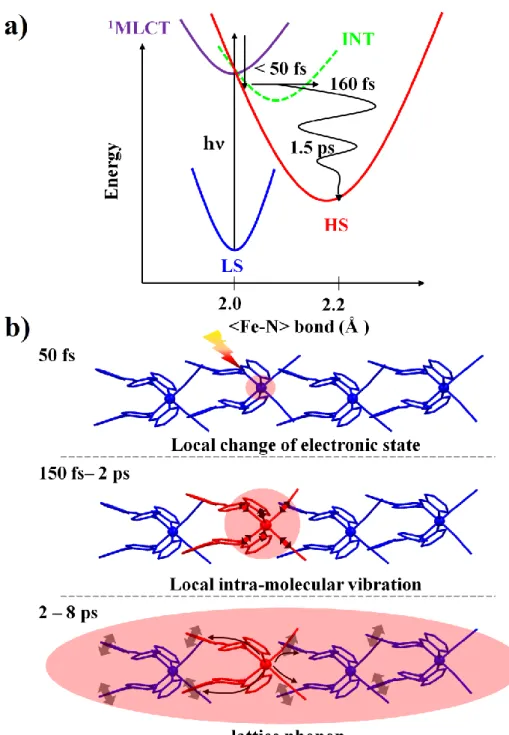
The results reported here give essential insight on the LIESST process in crystals and are complementary to ultrafast studies of LIESST at the molecular level performed for molecules in solution. Regarding the LIESST phenomenon itself, the use of complementary probes (Optics and X-ray) allowed to determine the temporal evolution of the electronic and structural degrees of freedom at the molecular scale. The results underline that during LIESST the electronic and structural degrees of freedom are strongly coupled and evolve in sequential steps. A global picture schematically describing the photophysics mechanism of LIESST is represented in Fig. 8a, with different potential energy curves representing the main electronic states of the system. After photoexcitation of the LS molecular state, the system reaches the 1MLCT state, which decays within less than 50 fs, as observed with optical reflectivity around the HS/LS isosbestic point sensitive to this intermediate state. As the less bonding HS potential is rapidly reached, a new equilibrium 〈Fe-N〉 bond length is defined and its elongation occurs, as observed with time resolved XAS (fig. 3b) and IR optical spectroscopy (sensitive to the HS t2geg gap). This average bond elongation occurs
within τ〈Fe-N〉 ∼ 160 fs, and activates the intramolecular Fe-ligand stretching mode as the molecule
oscillates in its new HS potential. Since this HS state is reached on a timescale corresponding typically to a half vibrational period of the Fe-N breathing mode, any intermediate state cannot be structurally relaxed and therefore can only serve as dynamically mixed mediators. These results reported here in the [Fe(PM-AzA)2(NCS)2] SCO solid find a common agreement with recent works
reporting the fast population of the HS state (Gawelda et al., 2007; Consani et al., 2009; Bressler et
al., 2009; Marino et al., 2013; Zhang et al., 2014 and Marino et al., 2014) and the consecutive coherent activation of different intra-molecular molecular modes in the HS potential in solution (Consani et al., 2009 and Auböck et al., 2015) and in the solid state (Cammarata et al., 2014; Bertoni et al., 2015a and Bertoni et al., 2015b). Once in the HS potential, a non radiative vibrational cooling occurs (Cannizzo et al., 2010 and Bertoni et al., 2012) and the energy is transferred to the surrounding medium. In their high time resolution measurements Auböck et al. observed a 30-50 fs phase shift of the Fe-N stretching mode and interpreted this as the characteristic the arrival time in the HS potential. Our time resolution (80 fs) is not good enough to give such accurate numbers but our results agree with such observation. What is new here with respect to previous ultrafast studies of LIESST is the evidence of the energy transfer from the absorber SCO molecule to its surrounding lattice, activating so an optical lattice phonon. It is this energy transfer which is responsible for lattice heating and drives the consecutive lattice expansion observed on ns timescale and the thermal population of the HS state observed on µs timescale (Lorenc et al., 2012; Collet et al., 2012; Bertoni et al., 2015 and Marino et al., 2015). Such lattice modes are not activated
immediately after photo-excitation: the phase shift of this mode optical lattice phonon mode (250 (40) fs) may be interpreted as an activation of the mode due to molecular swelling, which occurs on similar timescale. However, since the intensity of this mode increases during the 4 first ps, the process responsible for its activation is probably a coupling of the lattice modes with the molecular modes. Indeed, the intra-molecular vibration and the optical lattice phonon look sequentially activated, with a spectral weight transfer from the 67 cm-1 to the 28 cm-1 modes. In the Zn matrix, the 28 cm-1 lattice vibration is not observed when energy is transferred from the Fe SCO molecule to the passive Zn-based lattice. This is due to the fact that the Zn-based lattice is optically silent, prohibiting so the observation of this mode. This result confirms that such vibrations are located on the lattice surrounding the absorber molecule and corresponds to optical lattice phonons. Considering the great deal of modes in the few tens of cm-1 in molecular crystals, it might seem surprising that only a particular one is present. We attribute this observation to the selectivity of the probe rather than a selective excitation process. Indeed as discussed above light at 690 nm should involve a transition to the ligand orbitals thus being sensitive to the lattice vibrations coupled to the ligand torsion. Supporting this idea is the observation (in similar FeIII compounds) of different lattice modes at different probe wavelengths (Bertoni et al., 2015). One should also note that lattice modes at higher frequencies are probably excited as well. However, the energy transfer from the molecule to the lattice is longer than the oscillation periods of these modes, which are not activated coherently and consequently do not modulate the time-resolved signal.
Figure 8b is a schematic representation of this process following the molecular LIESST described above. One more time, we underline that the pump pulse locally photoswitches within a fragment of time the absorbing Fe-based molecules in the crystal. As these undergo LIESST, the HS potential is reached with an excess of energy in the order of 1-2 eV. The population of the antibonding eg
orbitals leads to a reorganization of the FeN6 octahedron, occurring beyond a doubt within 160 fs,
which in turns triggers the coherent activation of intra-molecular vibrations during the first 2 ps, also observed in the IR for in the diluted compound (Fig. 5). As the molecule couples to its environment via intermolecular contacts, the vibrational relaxation of the HS state occurs and a coherent optical phonons of the lattice surrounding the photoexcited molecules is activated.
These findings evidence that the photo-switched SCO molecule in the crystal rapidly equilibrates with the environment, transferring within 2 ps the energy (released on the molecule by the laser pulse) to the lattice. Since the lattice modes are coherently activated, the mechanism involved in the process is very likely a coupling between the intra-molecular modes and the lattice modes. Indeed, phononphonon coupling is known as an efficient way to transfer energy between different vibration modes (Uemura and Okamoto 2010; Fröst et al., 2011). This process is of high importance for SCO solids as it is established that lattice expansion, induced here by its resulting heating (Collet et al., 2012), can drive low spin to high spin transformation through the elastic coupling of the SCO molecules mediated by the lattice (Spiering et al., 1982; Lorenc et al., 2009 and Enachescu et al., 2009).
Acknowledgements:
This work was supported by the Institut Universitaire de France, Rennes Métropole, ANR (ANR-13-BS04-0002), Centre National de la Recherche Scientifique (CNRS) (PEPS SASLELX), and Fonds Européen de Développement Régional (FEDER). A.M. thanks CNRS and Région Bretagne for PhD funding (COMMAND). Portions of this research were carried out at the Linac Coherent Light Source (LCLS) at SLAC National Accelerator Laboratory. LCLS is an Office of Science User Facility operated for the U.S. Department of Energy Office of Science by Stanford University.
References:
Aubock G. and Chergui M. "Sub-50-fs photoinduced spin crossover in [Fe(bpy)3]2+" Nature Chemistry 7, 629–633 (2015)
Baranovic G., Babic D. "Vibrational study of the Fe(phen)2(NCS)2 spin-crossover complex by density-functional calculations." Spectrochem. Acta A 60, 1013, (2004)
Barbara P.F., Walker G.C. and Smith, T.P. "Vibrational modes and the dynamic solvent effect in electron and proton transfer." Science 256, 975 (1992)
Bertoni R., Lorenc M., Tissot A., Servol M., Boillot M.-L. and Collet E. "Femtosecond spin-state photo-switching of molecular nanocrystals evidenced by optical spectroscopy" Angew. Chem. Int. Ed. 51, 7485–7489 (2012)
Bertoni R., ammarata ., Lorenc ., atar S. F., L tard .-F., Lemke H. T., and Collet E. "Ultrafast Light-Induced State Trapping Photophysics Investigated in Fe(phen)2(NCS)2 Spin-Crossover Crystal" Acc. hem. Res. 48, 774−781 (2015)a
Bertoni R., Lorenc M., Laisney J., Tissot A., Moreac A., Matar S. F., Boillot M.-L. and Collet E. "Femtosecond spin-state photo-switching dynamics in an FeIII spin crossover solid accompanied by coherent structural vibrations" J. Mater. Chem. C (2015)b
Bertoni R., Lorenc M., Tissot A., Boillot M.-L. and Collet "Femtosecond photoswitching dynamics and microsecond thermal conversion driven by laser heating in FeIII spin-crossover solids " Coordination Chemistry Reviews 282–283, 66–76 (2015)c
Bressler C., Milne C., Pham V-T., El Nahhas A., van der Veen R.M., Gawelda W., Johnson S.L., Grolimund D., Kaiser D., Borca C.N., Ingold G., Abela R., Chergui M., «Femtosecond XANES Study of the Light-Induced Spin Crossover Dynamics in an Iron(II) Complex», Science 323, (5913), p489-492, (2009).
Briois V., Moulin C., Sainctavit P., Brouder C. and Flank A.-M. "Full Multiple Scattering and Crystal Field Multiplet Calculations Performed on the Spin Transition FeII(phen)2(NCS)2 Complex at the Iron K and L2,3 X-ray Absorption Edges" J. Am. Chem. Soc. 117, 1019-1026 (1995)
Buron-Le Cointe M., Hébert J., Baldé C., Moisan N., Toupet L., Guionneau P., Létard J. F., Freysz E., Cailleau H., and Collet E. " Intermolecular control of thermoswitching and photoswitching phenomena in two spin-crossover polymorphs" Phys. Rev. B 85, 064114 (2012)
Cammarata M., Bertoni R., Lorenc M., Cailleau H., Di Matteo S., Mauriac C., Matar S. F., Lemke H., Chollet M., Ravy S., Laulhé C., Létard J.-F. and Collet E. "Sequential Activation of Molecular Breathing and Bending during Spin-Crossover Photoswitching Revealed by Femtosecond Optical and X-ray Absorption Spectroscopy" Phys. Rev. Lett. 113, 227402 (2014)
Cannizzo A., Milne C., Consani C., Gawelda W., Bressler C., van Mourik F., Chergui M. "Light-induced spin crossover in Fe(II)-based complexes: The full photocycle unraveled by ultrafast optical and X-ray spectroscopies" Coord. Chem. Rev. 254, (21-22), 2677-2686, (2010)
Chollet M., Guerin L., Unchida N., Fukaya S., Shimoda H., Ishikawa T., Matsuda K., Hasegawa T., Ota A., Yamochi H., Saito G., Tazaki R., Adachi S-I., Koshihara S. "Gigantic Photoresponse in 1/4-Filled-Band Organic Salt (EDO-TTF)2PF6" Science 307, 86-89, (2005)
Chollet, M., Alonso-Mori, R., Cammarata, M., Damiani, D., Defever, J., Delor, J. T., Feng, Y., Glownia, J. M., Langton, J. B., Nelson, S., Ramsey, K., Robert, A., Sikorski, M., Song, S., Stefanescu, D., Srinivasan, V., Zhu, D., Lemke, H. T. & Fritz, D. M. (2015). J. Synchrotron Rad. 22, 503-507.
Collet E., Lemée-Cailleau M.H., Buron-Le Cointe M., Cailleau H., Wulff M., Luty T., Koshihara S., Meyer M., Toupet L., Rabiller P., Techerts S. "Laser-induced ferroelectric structural order in an organic charge-transfer crystal" Science 300, 612-615, (2003)
Collet E., Lorenc M., Cammarata M., Guerin L., Servol M., Tissot A., Boillot M.L., Cailleau H., Buron-Le Cointe M. "100 Picosecond Diffraction Catches Structural Transients of Laser-Pulse Triggered Switching in a Spin-Crossover Crystal" Chem. Eur. J. 18, 2051-2055, (2012)
Collet E., Moisan N., Baldé C., Bertoni R., Trzop E., Laulhé C., Lorenc M., Servol M., Cailleau H., Tissot A., Boillot M.L., Graber T., Henning R., Coppens P., Buron-Le Cointe M. "Ultrafast spin-state photoswitching in a crystal and slower consecutive processes investigated by femtosecond optical spectroscopy and picosecond X-ray diffraction" Phys. Chem. Chem. Phys. 14, 6192 (2012). Consani C., Premont-Schwarz M., Cannizzo A., El Nahhas A., van Mourik F., Bressler C., Chergui M. "Vibrational coherences and relaxation in the high-spin state of aqueous [Fell(bpy)3]2" Angew. Chem. Int. Ed. 48, 39, (2009)
Elsaesser T. and Kaiser, W. "Vibrational and vibronic relaxation of large polyatomic molecules in liquids." Annu. Rev. Phys. Chem. 42, 83 (1991)
Enachescu C., Stoleriu L., Stancu A., Hauser A. "Model for Elastic Relaxation Phenomena in Finite 2D Hexagonal Molecular Lattices". Phys. Rev. Lett. 102, 257204 (2009)
Fröst M., Manzoni C., Kaiser S., Tomioka Y., Tokura Y., Merlin R., Cavalleri A. " Nonlinear phononics as an ultrafast route to lattice control". Nat. Phys. 7, 854 (2011).
Gao M., Lu C., Hubert J-R., Liul C.L., Marx A., Onda K., Koshihara S-Y., Nakano Y., Shao X., Hiramatsu T., Saito G., Yamochi H., Cooney R.R., Moriena G., Sciani G., Miller D.R.J. "Mapping molecular motions leading to charge delocalization with ultrabright electrons" Nature 496, 343-346 (2013)
Gaussian 09, Revision D.01, Frisch, M. J.; Trucks, G. W.; Schlegel, H. B.; Scuseria, G. E.; Robb, M. A.; Cheeseman, J. R.; Scalmani, G.; Barone, V.; Mennucci, B.; Petersson, G. A.; Nakatsuji, H.; Caricato, M.; Li, X.; Hratchian, H. P.; Izmaylov, A. F.; Bloino, J.; Zheng, G.; Sonnenberg, J. L.; Hada, M.; Ehara, M.; Toyota, K.; Fukuda, R.; Hasegawa, J.; Ishida, M.; Nakajima, T.; Honda, Y.; Kitao, O.; Nakai, H.; Vreven, T.; Montgomery, J. A., Jr.; Peralta, J. E.; Ogliaro, F.; Bearpark, M.; Heyd, J. J.; Brothers, E.; Kudin, K. N.; Staroverov, V. N.; Kobayashi, R.; Normand, J.; Raghavachari, K.; Rendell, A.; Burant, J. C.; Iyengar, S. S.; Tomasi, J.; Cossi, M.; Rega, N.; Millam, M. J.; Klene, M.; Knox, J. E.; Cross, J. B.; Bakken, V.; Adamo, C.; Jaramillo, J.; Gomperts, R.; Stratmann, R. E.; Yazyev, O.; Austin, A. J.; Cammi, R.; Pomelli, C.; Ochterski, J. W.; Martin, R. L.; Morokuma, K.; Zakrzewski, V. G.; Voth, G. A.; Salvador, P.; Dannenberg, J. J.; Dapprich, S.; Daniels, A. D.; Farkas, Ö.; Foresman, J. B.; Ortiz, J. V.; Cioslowski, J.; Fox, D. J. Gaussian, Inc., Wallingford CT, 2009.
Gawelda W., Cannizzo A., Pham V-T., van Mourik F., Bressler C., Chergui M. "Ultrafast Nonadiabatic Dynamics of [FeII(bpy)3]2+ in Solution" J. Am. Chem. Soc. 129, (26), 8199-8206 (2007)
Goujon A., Varret F., Boukheddaden K., Chong C., Jeftic J., Garcia Y., Naik A. D., Ameline J.C. and Collet E. "An optical microscope study of photoswitching and relaxation in single crystals of the spin transition solid [Fe(ptz)6](BF4)2, with image processing" Inorg. Chim. Acta 361 4055–4064
(2008)
Guionneau P., Létard J.-F., Yufit D. S., Chasseau D., Bravic G., Goeta A. E., Howard J. A. K. and Kahn O. " Structural approach of the features of the spin crossover transition in iron(II) compounds" J. Mater. Chem. 9, 985–994 (1999)
Guionneau P., Lakhloufi S., Lemée-Cailleau M.-H., Chastanet G., Rosa P., Mauriac C., Létard J.-F. "Mosaicity and structural fatigability of a gradual spin-crossover single crystal." Chem. Phys. Lett. 542, 52–55, (2012)
Gütlich P., Goodwin H.A, "Topics in Current Chemistry, Spin crossover in Transition Metal Compounds, Vol I, II and III», Springer Berlin, 234, (2004)
Halcrow M. A. "Spin Crossover Materials: Properties and Applications" (Wiley, West Sussex) ISBN 9781119998679 (2013)
Harmand M., Coffee R., Bionta M.R., Chollet M., Fritz D.M., Lemke H.M., Medved N., Ziaja B., Toleikis S., Cammarata M., "Achieving few-femtosecond time-sorting at Hard X-ray Free Electron Lasers" Nature Photonics 7, (2013)
Hauser A. “Reversibility of light-induced excited spin state trapping in the Fe(ptz)6(BF4)2 and the Zn1-xFex(ptz)6(BF4)2 spin-crossover systems” hem. Phys. Lett. 124, 6, (1986)
Hauser A. "Light-induced spin-crossover and the high-spin -> low-spin relaxation". Topics in Current Chemistry, Spincrossover in Transition Metal Compounds, Vol II" (eds P. Gütlich, H. A. Goodwin), Springer, Berlin, 234, 155-198 (2004)
Jun S., Yang C., Kim T. W., Isaji M., Tamiaki H., Ihee H., Kim J., "Role of thermal excitation in ultrafast energy transfer in chlorosomes revealed by two-dimensional electronic spectroscopy." Phys. Chem. Chem. Phys., 17, 17872-17879 (2015)
Kaszub W., Buron-Le Cointe M.Lorenc M., Boillot M.L., Servol M., Tissot A., Guerin L., Cailleau H., Collet E. "Spin-State Photoswitching Dynamics of the [(TPA)FeIII(TCC)]SbF6 Complex" Eur. J. Inorg. Chem. 5-6,992-1000 (2013)
Kawakami Y., Iwai S., Fukatsu T., iura ., Yoneyama N., Sasaki T., and Kobayashi N. “Optical Modulation of Effective On-Site Coulomb Energy for the Mott Transition in an Organic Dimer Insulator” Phys. Rev. Lett. 103, 066403, (2009)
Koshihara S-Y. Book "The LXIII Yamada Conference on Photo-Induced Phase Transition and Cooperative Phenomena (PIPT3)" Journal of Physics: Conference Series, 148, (2009)
Ksenofontov V., Levchenko G., Spiering H., Gütlich P., Létard J.-F., Bouhedja Y. and Kahn O. “Spin rossover behaviour under pressure of Fe(PM-L)2(NCS)2 ompounds with Substituted
2’-pyridylmethylene 4-anilino ligands” hem. Phys. Lett. 294, 545-553 (1998)
Lemke H. T., Bressler C., Chen L. X., Fritz, D. M., Gaffney K. J., Galler A., Gawelda W., Haldrup K., Hartsock R. W., Ihee H., Kim J., Kim K. H., Lee J. H., Nielsen M. M., Stickrath A. B., Zhang W., Zhu D., Cammarata M. "Femtosecond X‑ray Absorption Spectroscopy at a Hard X‑ray Free Electron Laser: Application to Spin Crossover Dynamics." J. Phys. Chem. A. 117, 735 (2013) Létard J.-F., Capes L., Chastanet G., Moliner N., Létard S., Real . A., and Kahn O. “Critical Temperature of the LIESST Effect in Iron(II) Spin Crossover Compounds” Chem. Phys. Lett., 313, 115-120, (1999)
Levantino M., Schiró G., Lemke H. T., Cottone G., Glownia J. M., Zhu D., Chollet M., Ihee H., upane A. and ammarata . “Ultrafast myoglobin structural dynamics observed with an X-ray free-electron laser” Nature ommunications 6, 6772, (2015)
Levantino M., Lemke H. T., Schiró G., Glownia ., upane A., and ammarata . “Observing heme doming in myoglobin with femtosecond X-ray absorption spectroscopy” Struct. Dyn. 2, 041713, (2015)
Lorenc M., Hebert J., Moisan N., Trzop E., Servol M., Buron-Le Cointe M., Cailleau H., Boillot M., Pontecorvo E., Wulf M., Koshihara S., Collet E. "Successive Dynamical Steps of Photoinduced Switching of a Molecular Fe(III) Spin-Crossover Material by Time-Resolved X-ray Diffraction" Phys. Rev. Lett. 103, 028301, (2009)
Lorenc M., Balde C.H., Kaszub W., Tissot A., Moisan N., Servol M., Buron-Le Cointe M., Cailleau H., Chasle P., Czarnecki P., Boillot M., Collet E. "Cascading photoinduced, elastic, and thermal switching of spin states triggered by a femtosecond laser pulse in an Fe(III) molecular crystal», Physical Review B 85, 054302, (2012)
Marchivie M., Guionneau P., Létard J.-F., Chasseau D.. "Towards direct correlations between spin-crossover and structural features in iron(II) complexes". Acta Cryst. B59, 479-486 (2003).
Marino A., Servol M., Bertoni R., Lorenc M., Mauriac C., Létard J.F., Collet E. "Femtosecond optical pump–probe reflectivity studies of spin-state photo-switching in the spin-crossover molecular crystals [Fe(PM-AzA)2](NCS)2" Polyhedron 66, 123–128, (2013)
Marino A., Chakraborty P., Servol M., Lorenc M., Collet E., and Hauser A. "The Role of Ligand-Field States in the Ultrafast Photophysical Cycle of the Prototypical Iron(II) Spin-Crossover Compound [Fe(ptz)6](BF4)2" Angew. Chem. Int. Ed. 53, 3863 –3867 (2014)
Marino A., M. Buron-Le Cointe, M. Lorenc, R. Henning, A. D. DiChiara, K. Moffat, N. Bréfuel, E. Collet. "Out-of-equilibrium dynamics of photoexcited spin-state concentration waves " Faraday Discussion 177, (2015)
Martin R. "Natural transition orbitals." J. Chem. Phys. 118, 4775, (2003)
Okamoto H., Ishige Y., Tanaka S., Kishida H., Iwai S., and Tokura Y. “Photoinduced phase transition in tetrathiafulvalene-p-chloranil observed in femtosecond reflection spectroscopy” Physical Review B 70, 165202 (2004)
Ronayne K. L., Paulsen H., Höfer A., Dennis A. C., Wolny J. A., Chumakov A. I., Schünemann V., Winkler H., Spiering H., Bousseksou A., Gütlich P., Trautwein A. X., McGarvey J. J. "Vibrational spectrum of the spin crossover complex [Fe(phen)2(NCS)2] studied by IR and Raman spectroscopy,
nuclear inelastic scattering and DFT calculations". Phys. Chem. Chem. Phys. 8, 4685 (2006).
Rosspeintner A., Lang B. and Vauthey E. "Ultrafast photochemistry in liquids." Annu. Rev. Phys. Chem. 64, 247 (2013)
Schoenlein R. W., Peteanu L. A., Mathies R. A., Shank C. V. "The First Step in Vision: Femtosecond Isomerization of Rhodopsin" Science 254, 412-415 (1991)
Servol M., Moisan N., Collet E., Cailleau H., Kaszub W., Toupet L., Boschetto D., Ishikawa T., Moréac A., Koshihara S, Maesato M., Uruichi M., Shao X., Nakano Y., Yamochi H., Saito G., Lorenc M. "Local response to light excitation in the charge-ordered phase of (EDO−TTF)2SbF6"
Phys. Rev. B 92 024304 (2015)
Sousa C., de Graaf C., Rudavskyi A., Broer R., Tatchen J., Etinski M., Marian C.M." Ultrafast deactivation mechanism of the excited singlet in the light-induced spin crossover of [Fe(2,2’-bipyridine)3]2+". Chem. Eur. J. 19, 17541 – 175 (2013)
Spiering H., Meissner E., Koppen H., Muller E.W. and Gutlich P. "The effect of the lattice expansion on High Spin Low Spin transitions" Chemical Physics 68, 65-71 (1982)
Uemura H., Okamoto H., "Direct Detection of the Ultrafast Response of Charges and Molecules in the Photoinduced Neutral-to-Ionic Transition of the Organic Tetrathiafulvalene-p-Chloranil Solid" Phys. Rev. Lett. 105, 258302 (2010)
van Veenendaal M., Chang J., Fedro A.J. "Model of Ultrafast Intersystem Crossing in Photoexcited Transition-Metal Organic Compounds" Physical Review Letters 104 (25), 061401, (2010)
Zhang W., Alonso-Mori R., Bergmann U., Bressler C., Chollet M., Galler A., Gawelda W., Hadt R.G., Hartsock R.W., Kroll T., Kjær K.S., Kubiček K., Lemke H.T., Liang H.W., Meyer D.A., Nielsen M.M., Purser C., Robinson J.S., Solomon E.I., Sun Z., Sokaras D., van Driel T.B., Vankó G., Weng T.C., Zhu D., Gaffney K.J. " Tracking excited-state charge and spin dynamics in iron coordination complexes." Nature 509 (7500), 345-348 (2014).
Figure Captions:
FIG. 1. Thermal spin crossover of the [Fe(PM-AzA)2(NCS)2] compound (top) monitored with the
evolution of a) χM.T product, b) X-ray absorption at 7125 eV, c) total optical reflectivity. The
increase of T and OR under cw light excitation at 830 nm in a) and c) is related to the long-lived LIESST phenomenon at low temperature.
FIG. 2. a) diffuse reflectivity spectra collected on powders of [Fe(PM-AzA)2(NCS)2] (▬),
[Zn0.9Fe0.1(PM-AzA)2(NCS)2] (▬) and [Zn(PM-AzA)2(NCS)2] (▬) crystals. b) optical reflectivity
spectra of a [Fe(PM-AzA)2(NCS)2] single crystal acquired at different temperatures during the SCO
from pure LS (100 K) and HS (270 K) states.
FIG. 3. a) The TD-DFT calculated absorbance spectra for LS, HS and Zn compounds. b) at 690 nm
NTO of hole and particle correspond to transitions from t2g-like to L-like orbitals as shown here for
FIG. 4. Ultrafast dynamics in the 0-2500 fs range for the [Fe(PM-AzA) 2(NCS)2] single crystal
upon excitation in the MLCT bands at 800 nm (0.4-0.5 mJ/mm2) observed by a) two-color pump-probe reflectivity measurements with dynamical time traces for 640 nm, 690 nm and 720 nm pump-probe,
b) Time-resolved change of XAS at 7125 eV after laser irradiation at 800 nm for the
[Fe(PM-AzA)2(NCS)2], c) two-color pump-probe transmission measurements at 1675 nm. The fits (solid red
lines) take into account the IRF of 80 fs for a) and 110 fs for b) and c). The inset of b) covers the 0-6 ps time window, whereas the inset of c) covers the first ps.
FIG. 5. Ultrafast dynamics in the 0-2500 fs range probed in the infra-red part on the HS d-d bands
for the pure (100% Fe) e crystal (left) and for the 10 % diluted [ n e crystal (right) after photoexcitation at 800 nm (0.4 mJ/mm2), the
insets show the oscillating part of the signal. For the Fe 100% compound, the fit at 1675 nm (red curve) gives a 115 cm-1 frequency and a 150 fs damping. For the fit at 1600 nm in the Fe 10% compound the frequency was fixed to 115 cm-1 because of the noise limit and a 150 (50) fs damping was found.
FIG. 6. Dynamical OR time traces recorded at 680 nm 690nm and 700 nm after photoexcitation at
800 nm (0.4 mJ/mm2) and showing in phase oscillations of the reflectivity signal Thick cyan curves represents the fit to the data with a sine function multiplied with an increasing and decreasing exponential terms
FIG. 7. a) Comparison of the oscillating dynamics accompanying the ultrafast LIESST (
and ) for the the pure crystal (black) and for the 10 % diluted [ crystal (red). b) time dependent FFT analysis
of the oscillating component of the OR at 700 nm showing the sequential activation of two different vibrations at 67 cm-1 and 28 cm-1 for the pure crystal (b) and the diluted compound (c). Thick cyan curves represents the fit to the data with a sine function multiplied with an increasing and decreasing exponential terms
FIG. 8. a) Schematic representation of the energy transfer from the absorber Fe-ion to the crystal
lattice. A first laser excitation release an excess of energy of 1.5 eV on the Fe-ion. Due to the electron-phonon coupling, the ultrafast bond elongation triggers coherent structural
intra-molecular vibrations. A strong phonon-phonon coupling transfer the excess of energy from the molecule to the crystal lattice via phonon-phonon coupling. b) Schematic summary of the ultrafast LIESST mechanism activated via LS 1
MLCT laser excitation. Decay of the excited 1MLCT state in less than 50 fs where possible intermediate (INT) states are involved in the intersystem crossing to the HS state. The HS electronic configuration implies a shift of the reaction coordinates to higher
〈Fe-N〉 bond lengths. This elongation is measured to occur in 160 fs. The HS potential is reached
in a vibrational excited state where the non radiative vibrational cooling inside the HS potential to its bottom occurs in 1.5 - 2 ps. The pink ellipsoids schematically represent the redistribution of the energy initially deposited on the molecule.

![FIG. 2. a) diffuse reflectivity spectra collected on powders of [Fe(PM-AzA) 2 (NCS) 2 ] (▬), [Zn 0.9 Fe 0.1 (PM-AzA) 2 (NCS) 2 ] (▬) and [Zn(PM-AzA) 2 (NCS) 2 ] (▬) crystals](https://thumb-eu.123doks.com/thumbv2/123doknet/14456694.519763/19.892.99.808.109.370/fig-diffuse-reflectivity-spectra-collected-powders-aza-crystals.webp)
![FIG. 4. Ultrafast dynamics in the 0-2500 fs range for the [Fe(PM-AzA) 2 (NCS) 2 ] single crystal upon excitation in the MLCT bands at 800 nm (0.4-0.5 mJ/mm 2 ) observed by a) two-color pump-probe reflectivity measurements with dynamical t](https://thumb-eu.123doks.com/thumbv2/123doknet/14456694.519763/20.892.221.673.100.963/ultrafast-dynamics-crystal-excitation-observed-reflectivity-measurements-dynamical.webp)


