HAL Id: tel-01079444
https://tel.archives-ouvertes.fr/tel-01079444
Submitted on 2 Nov 2014
HAL is a multi-disciplinary open access archive for the deposit and dissemination of sci-entific research documents, whether they are pub-lished or not. The documents may come from teaching and research institutions in France or abroad, or from public or private research centers.
L’archive ouverte pluridisciplinaire HAL, est destinée au dépôt et à la diffusion de documents scientifiques de niveau recherche, publiés ou non, émanant des établissements d’enseignement et de recherche français ou étrangers, des laboratoires publics ou privés.
Lavinia Vija
To cite this version:
Lavinia Vija. Androgen Signaling in Sertoli Cells. Human health and pathology. Université Paris Sud - Paris XI, 2014. English. �NNT : 2014PA11T031�. �tel-01079444�
UNIVERSITE PARIS-SUD
ÉCOLE DOCTORALE :
Signalisation et Réseaux Intégratifs en
Biologie
Laboratoire Récepteurs Stéroïdiens, Physiopathologie Endocrinienne et Métabolique
Reproduction et Développement
THÈSE DE DOCTORAT
Soutenue le 09/07/2014 par
Lavinia Magdalena VIJA
SIGNALISATION ANDROGÉNIQUE DANS LES
CELLULES DE SERTOLI
Directeur de thèse : Jacques YOUNG Professeur (Université Paris Sud)
Composition du jury :
Président du jury : Michael SCHUMACHER DR1 (Université Paris Sud)
Rapporteurs : Serge LUMBROSO Professeur (Université Montpellier I) Mohamed BENAHMED DR1 (INSERM U1065, Université Nice))
Examinateurs : Nathalie CHABBERT-BUFFET Professeur (Université Pierre et Marie Curie) Gabriel LIVERA Professeur (Université Paris VII)
PARIS SUD UNIVERSITY
École Doctorale :
Signalisation et Réseaux Intégratifs en
Biologie
Steroid Receptors, Metabolic and Endocrine Physiopathology Laboratory
Reproduction and Development
PhD THESIS
Public dissertation on the 09/07/2014 By
Lavinia Magdalena VIJA
ANDROGEN SIGNALING IN SERTOLI CELLS
Thesis Coordinator : Jacques YOUNG Professor (Université Paris Sud)
Thesis Commitee :
President of the jury: Michael SCHUMACHER DR1 (Université Paris Sud)
Referees: Serge LUMBROSO Professor (Université Montpellier I)
Mohamed BENAHMED DR1 (INSERM U1065, Université Nice)
Examinators: Nathalie CHABBERT-BUFFET Professor (Université Pierre et Marie Curie)
Gabriel LIVERA Professor (Université Paris VII)
Motto:
“I am not searching for the answers,
I am just trying to understand the questions”
ACKNOWLEDGEMENTS
It was a long journey finally ending with the dissertation. I would like to gratefully remember those people who supported me during this important period of my life.
Dear Professor Jacques YOUNG, allow me to respectfully thank you for your support. From the moment you proposed to coordinate this project, you continued to generously share your professional experience and to teach me how to develop a scientific reasoning and how to discover the amazing universe of translational research. Thank you for having shared with me not only your clinical experience but also the seriosity and careful art of performing a bibliographic search and especially forhaving taught me the wisdom and the witty spirit and many other secrets necessary for writing a good scientific paper.
Dear Marc LOMBES, words are not enough to express my gratitude for having so kindly adopted me in your research unit and for all the support you offered me during the last four years. Your permanent disponibility, your suggestions, always correct and pertinent to all my scientific or personal problems, were always remarkable. Thank you for all your help. I sincerely wish that this was just the beginning of a long journey in the universe of scientific research in molecular endocrinology, and that we shall continue new projects and new French-Romanian collaborations.
Dear Professor Serge LUMBROSO and Doctor Mohamed BENAHMED allow me to respectfully thank you for all your support and understanding and for the enthusiasm you accepted to review this work. Words are not enough to express my respect and my gratitude for you.
I would also like to sincerely thank Professor CHABBERT-BUFFET for all your help, support and time you shared so kindly with me during these last years; it is a pleasure and an honor for me that you accepted to be my thesis examinator. I would also like to thank Professor Gabriel LIVERA, for having so kindly accepted to be one of the examinators. I would also like to sincerely thank to all the members of the INSERM Unit 693, from Seniors (Professor AnneGUIOCHON-MANTEL, Hugues LOOSFELT, Nadine BINART, Jérôme B, Séverine, Jérôme F, Larbi and Damien, with a special thanks forSay- who always came up with a solution and support, even in those moments when experiments were not always associated with remarkable results, and who always showed a sign of friendship for everyone) to my collegues. Dear Adela, Bruno, Julien, Emmanuelle, Catherine, Junaid, Nathalie, Audrey, Anne-Lise, Ségolène, Jérôme N, and all students and collegues, with which I shared great moments, week-ends and long days of work, sometimes ending up at midnight, I am really happy I met you. Friendship is the most beautiful homeland as a song used to say and you have completely proven that this was true.Thank you, Geri MEDURI for having shared with me the secrets of the immunohistochemistry, for the magic of those great Italian dishes you prepare with so much love and for the pleasure of listening and supporting me.And last but not least, I would like to thank to all my friends and collegues from the “C.I. Parhon” National Institute of Endocrinology, as well as to all my colleagues and Seniors who allowed me to discover and to get competences in Nuclear Medicine (Professor Philippe Chaumet-Riffaud, Professor Alain Prigent, Frederique Archambaud and all the doctors from Bicêtre, StLouis and Avicenne Hospitals), to my family who understood my choices,as well as to all my friends from my both countries.
TABLE OF CONTENTS
Foreword 1
Introduction
1. Testicular differentiation and development 5
Factors involved in male sex differentiation
A. Undifferentiated state 5
B. Gonadal differentiation 8
C. Hormonal control of the masculinization process 15
C.1.Introduction 15
C.2.Fetal testis development
C.3.Fetal testicular hormonal milieu responsible for masculinization
16 C.4.Endocrine regulation of the fetal testis major cell types
a) Leydig cells and testosterone production in human fetal testis 17
b) Fetal Sertoli cells 19
2. Human testicular physiology: seminiferous tubules and spermatogenesis
2.1. Testicular development from birth to adulthood 21
2.1.1.Postnatal Sertoli cells 21
2.1.2.Postnatal Leydig cells 24
2.1.3.Leydig cells and steroidogenesis 25
2.1.4.Postnatal germ cells and spermatogenesis 26
2.2 Hormonal regulation of spermatogenesis 28
2.2.1.Roles of FSH in spermatogenesis 29
2.2.2.Roles of androgens/LH in spermatogenesis 30
2.2.3.Roles of estrogens in spermatogenesis 33
2.2.4.Roles of thyroid hormones in spermatogenesis 34
2.2.5.Roles of insulin in spermatogenesis 35
3. Nuclear receptors(NRs): general structure and classification
3.1.Nuclear receptors: classification 36
3.2.Nuclear receptors: general structure 39
4 Androgens and the Androgen receptor(AR)
4.1Androgen action 40
4.1.1.Testosterone:biosynthesis and metabolism 41 4.1.2.Dihydrotestosterone:biosynthesis and metabolism 43
4.1.3.5-α-reductases 43
4.2.The Androgen Receptor (AR) 44
4.2.1. AR genomic signaling and effects 44
4.2.1.1. AR functional domains
a) The NH2-terminal domain (NTD) b) The DNA-binding domain (DBD) c) The hinge region
d) The ligand binding domain (LBD) e) AR domains interactions
45
4.2.1.2.Transcriptional regulation of AR regulated target genes in testis 50 4.2.1.3.Posttranscriptional and posttranslational modifications of AR 50 1. AR Phosphorylation
3. Acetylation 4. Methylation 5. SUMOylation 6. Ubiquitination
Crosstalk between posttranslational modifications of AR
4.2.2.The Androgen Receptor:non genomic signaling and effects 55 4.3. The Androgen Receptor and the molecular biology of androgen
insensitivity
55 4.3.1Androgen insensitivity syndrome in humans 56 4.3.2. Androgen insensitivity syndrome in spontaneous and generated
AR-knockout models
59
5 Androgen receptor coregulators 60
5.1.Nuclear receptor(NR) coregulators
5.1.1.History of coregulators 60
5.1.2.General factors involved in coregulators functionality 61
5.1.3.Coactivators 65
5.1.4.Corepressors 66
5.1.5.Mechanism of interaction between steroid receptors(SR) and coregulators
67 5.1.6.Coregulators involvement in biological processes 69
5.1.7.Coregulators and human disease 70
5.2.Androgen receptors (AR) coregulators
5.2.1.Introduction 71
5.2.2.Classification of AR coregulators 71
5.2.3.Modulation of AR transcriptional activation by androgen receptor coactivators
72 5.2.4. Modulation of transcriptional repression by androgen receptor
corepressors
78 5.2.5.Animal knockouts and the biological roles of AR coregulators 80 5.2.6. Androgen receptor coregulators in human physiology and
pathology
81 5.2.6.1.Androgen receptor coregulators in androgen insensitivity
syndromes (AIS) and male infertility
81 5.2.6.2.Androgen receptor coregulators in prostate cancer 85 5.2.6.3.Androgen receptor coregulators in other pathologies 87 5.2.6.4. Androgen receptor coregulators: future directions 87 89 Objectives
Results
97
First part:Androgenic regulation in Sertoli cells 99
Paper 1.Androgen-dependent stabilization of Androgen Receptor
in the novel murine Sertoli cell line, ST38c
Lavinia Vija, Kahina Boukari, Hugues Loosfelt, Geri Meduri, Say
Viengchareun, Nadine Binart, Marc Lombès and Jacques YoungMol
Cell Endocrinol. 2014 Mar 25;384(1-2):32-42
2.Objectives 103
3.Discussion 105
4.Limits, further perspectives and conclusions
4.1. AR induced-AMH repression: hypothesis, results and perspectives 4.1.1.Introduction 4.1.2.Objectives 4.1.3.Results 4.1.4. Discussion 111 111 111 112 113 113 5.Conclusion 117
Second part: Androgen receptor coregulators (SRC-2 and HBO1): expression and characterization during testicular development
Paper2. Expression and characterization of androgen receptor
coregulators, SRC-2 and HBO1, during human testis ontogenesis and in androgen signaling deficient patients.
Lavinia Vija, Geri Meduri, Eva Comperat, Viorel Vasiliu, Vincent
Izard, Sophie Ferlicot, Kahina Boukari, Philippe Camparo, Say Viengchareun, Elisabeth Constancis, Constantin Dumitrache, Marc Lombès, Jacques Young. Mol Cell Endocrinol. 2013 Aug
15;375(1-2):140-8.
1. Introduction 123
1.1. Rationale for SRC-2 selection 125
1.2. Rationale for SRC-2 selection 126
2. Murine and human testicular samples and patient profiles 130
2.1. Murine testicular samples 130
2.2. Human testicular samples 130
2.3. Data related to the genetic profile of the studied patients 131
3. Additional results 136
3.1. SRC-2 and HBO1 mRNA expression during murine testicular postnatal development
136 3.2. SRC-2 and HBO1 cellular localization in adult murine
seminiferous tubules
136 3.3. SRC-2 and HBO1 expression during human postnatal testicular
development
137 3.4. Cellular localization of SRC-2 and HBO1 in human adult
seminiferous tubules
137 3.5. SRC-2 and HBO1 expression in human testicular pathologies with impaired androgen signaling
141 3.6. Androgens induce HBO1 gene expression in SMAT1 Sterol cells
in the presence of AR
146
4. Discussion, limits and perspectives 147
Third part: Androgen regulation in Sertoli cells Additional papers
in a post-pubertal patient with 5α-Reductase type 2 Deficiency. Review of the literature in a perspective of evaluation of potential fertility of these patients.
Lavinia Vija, Sophie Ferlicot, Diana Paun, Hélène Bry-Gauillard,
Gabriela Berdan,Issam Abd-Alsamad, Marc Lombès and Jacques Youngaccepted in BioMedCentral Endocrine Disorders May 2014
1.Introduction and Objectives 152
2.Discussion and perspectives 154
Paper 4. Healthy birth after testicular extraction of sperm and
ICSI from an azoospermic man with mild androgen insensitivity syndrome caused by an androgen receptor partial loss-of-function mutation.
Massin N, Bry H, Vija L, Maione L, Constancis E, Haddad B, Morel Y, Claessens F, Young J.
Clin Endocrinol (Oxf). 2012 Oct;77(4):593-8.
163
List of figures
Number Page
Figure 1 Migration of primordial germ cells 6
Figure 2 Main steps in gonadal differentiation during embryonic development
13 Figure 3 Stages and genetic regulation in normal male sexual
differentiation
14 Figure 4 Regulation of testosterone production in the human male fetus 19
Figure 5 Human testicular sections 22
Figure 6 Sertoli cell proliferation in humans follows two periods 24
Figure 7 Human testicular steroidogenesis 26
Figure 8 The 6 stages of spermatogenesis defined by Clermont 28 Figure 9 Mechanism for nuclear receptor action of class I nuclear
receptors
37 Figure 10 Mechanism for nuclear receptor action of class II nuclear
receptors
38 Figure 11 Structure of testosterone and dihydrotestosterone 40
Figure 12 Model of androgen action 42
Figure 13 Molecular structure of the androgen receptor 47 Figure 14 Functional domains of the androgen receptor 49 Figure 15 Coregulators are involved in chromatin remodeling 62 Figure 16 Representative scheme of in vivo putative substrates for HAT
and possible effects of their enzymatic activities
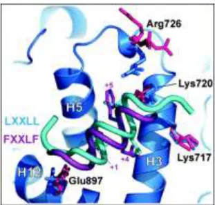
64 Figure 17 General structure of the SRC/p160 family of coactivators 66 Figure 18 Model of coregulator recruitment by steroid receptors 68 Figure 19 Schematic presentation of the AR transcription modulation by
coactivators
76 Figure 20 AR FxxLF and LXXLL binding modes to the AR LBD 77 Figure 21 Androgen stimulated ST38c cells express low levels of Rhox5
mRNA with no androgenic regulation.
110 Figure 22 In the ST38c cells, Amh mRNA expression is not upregulated
upon mRNA and protein AR silencing, using a siRNA approach
115
Figure 23 AR protein expression in ST38c SC after 48 and 72 h of transfection with siRNA AR.
116 Figure 24 Relative Src-2 (A), Hbo1 (B) and Ar (C) mRNA expression in
murine postnatal testis at various ages
138 Figure 25 Relative Ar (A) Src-2 (B), Hbo1 (C) and mRNA expression in
murine adult primary cultures of spermatogonia and Sertoli cells
139
Figure 26 Cellular localization of SRC-2 and HBO1 in human adult testis by double immunostaining
140
Figure 28 HBO1 and AR immunostaining in 7 complete androgen insensitivity (CAIS) patients, presenting various AR mutations
144 Figure 29 SRC-2 and HBO1 immunostaining in seminiferous tubules with
various degrees of spermatogenesis impairment.
145 Figure S1 ST38c cells express the androgen receptor with high affinity
for ligand
104 Figure S2 Expression of transfected AR in the presence of SRC-2,
HBO1 or their empy plasmid vectors.
128 Figure S3 SRC2 and HBO1 nuclear immunostaining quantification in
testicular samples obtained fromnormal adults and patients with androgen insensitivity syndromes.
129
List of tables
Number Page
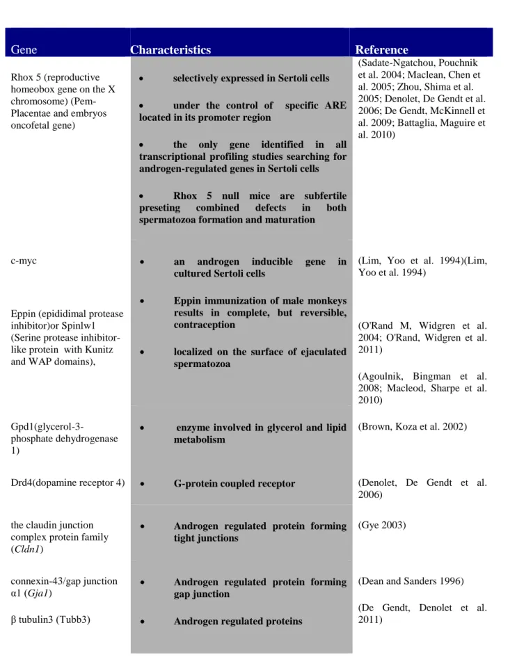
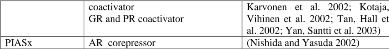
Table 1 Selection of androgen receptor regulated genes in Sertoli cells 54
Table 2 Clinical classification of AIS 58
Table 3 Selected recent reports on several most known receptor coregulators 63 Table 4 List of some coregulators that modulate AR transcriptional activity
and that are identified in normal human tissues and certain human pathologies
73
Table 5 Table resuming androgen receptor coregulator animal knockout models
83 Table 6 Main characteristics of previously reported Sertoli cell lines 108 Table 7 Main clinical features of patients included in the study 133 Table 8 Presentation of the genetic profile (mutations in AR, TACR3 and
SRD5A2 genes) for the patients whose testicular samples were included in the present study.
134
Table 9 Immunohistochemical detection of anti-Mullerian hormone (AMH) and androgen receptor (AR) within Sertoli cells
161 Supplem
ental Table 1
Mus musculus primer sequences for real time PCR 118
Supplem ental Table 2
Oligonucleotide 5’-3’ primer sequences of murine ARsiRNA 119
Supplem ental Table 3
Oligonucleotide primer sequences of human and murine primers for real-time quantitative reverse transcriptase- PCR.
List of aminoacids
A
Alanin
I
Isoleucine
R
Arginine
C
Cystein
K
Lysine
S
Serine
D
Aspartic acid
L
Leucine
T
Threonine
E
Glutamic acid
M
Methionine
V
Valine
F
Phenylalanine
N
Asparagine
W
Tryptophan
G
Glycin
P
Proline
Y
Tyrosine
ABBREVIATIONS
ABP Androgen binding protein AMH Anti Mullerian hormone AR
5ARD
Androgen receptor
5α-reductase type 2 deficiency ARE Androgen responsive elements DBD DNA-binding domain
DHH Desert hedgehog gene ER Estrogen receptor
FSH Follicle stimulating hormone FSHR FSH receptor
GnRH Gonadotropin releasing hormone HAT Histone acetyl transferase
HBO1 Histone acetyltransferase binding to origin recognition complex hCG Human chorionic gonadotropin
INSL3 Insulin-like 3
LBD Ligand binding domain LH Luteinizing hormone LHR LH receptor
LNCaP Lymph node carcinoma of the prostate NCoR Nuclear hormone receptor corepressor N-ter Amino terminal
Pem Placental embryonic oncofetal gene PTM Peritubular myoid cells
SF1 Steroidogenic factor 1 SGP-2 Sulphated-glycoprotein -2
SMRT Silencing mediator of retinoid and thyroid hormone receptor SRC-2 Steroid receptor coactivator-2
SRY Sex determining region of Y SOX9 Sry-like HMG box9
TAU-1,5 Transcription activation unit 1,5 TDF Testis determining factor
mRNA Messenger RNA RNApolII RNA polymerase II
3βHSD 3β-hydroxiosteroid deshydrogenase
WAGR Wilms’ tumor, Aniridia, Genitourinary abnormalities and mental retardation
Foreword
1 The androgen receptor (AR) is essential for the development and maintenance of the masculine phenotype and for spermatogenesis. Whether androgens are produced as early as around 7 gestational weeks, in accordance with the expansion of Sertoli cells number during the latter half of the first trimester and the beginning of the second trimester of gestation, Sertoli cells do not express AR during fetal development in both rodent and human. Moreover, AR expression in Sertoli cells exhibits a specific pattern during postnatal testicular development. During fetal life and in the neonate, during the “mini-puberty“period, the transient coexistence of high concentrations of androgens and AMH are related to physiological androgen insensitivity, explained by the absence of functional AR expression in SC during these developmental periods. At puberty, androgenic stimulation of a functional AR in Sertoli cells is associated with the entry in meiosis and spermatogenesis. Moreover, AMH expression significantly decreases in adulthood, in association with the androgenic stimulation of a functional AR present on Sertoli cells, as well as with the meiotic entry of spermatocytes.
The main objective of the thesis was the better comprehension of the physiological molecular mechanisms of AR regulation in Sertoli cells during murine and human ontogenesis.
The first part of this manuscript, represented by the Introduction, presents the scientific background, related to the expression and regulation of the androgen receptor (AR) and its molecular partners, during testicular developmental stages. The introduction is organized in five chapters. The first chapter presents the main factors contributing to prenatal testicular differentiation and development. The second chapter is meant to be a short overview about testicular development from birth to adulthood, presenting data related to the contribution of testicular cellular components to the main testicular functions: steroidogenesis and spermatogenesis. In the last part of this chapter I insisted on the hormonal regulation of spermatogenesis, in order to introduce the main contributions of the androgenic regulation. The third chapter shortly introduces general data related to nuclear receptors structure and classification, whereas the forth chapter is related to the molecular aspects of androgens and the androgen receptor. The last chapter is dedicated to AR molecular partners, notably to the roles and specific expression of the AR coregulators in physiology, in specific animal models, as well as their known implications in human pathologies related to androgen regulation.
Foreword
2 The introductory part is followed by a general presentation of the objectives of the thesis project. The objectives are organized as a list of questions.
The results are organized in three parts and focus several aspects related to the androgenic regulation of Sertoli cells. Each part includes either the published manuscript related to the question announced in the Objectives area, or the manuscript submitted. Each paper is preceded by an introductory part including the objectives of the work and followed by additional data covering discussions about the limits and the potential perspectives related to the presented work.
The first paper describes the characteristics of a novel, immortalized murine Sertoli cell line, which was generated in the INSERM U693, the research unit where I developed my thesis project. This mature Sertoli cell line exhibits important endogenous AR expression, as well as androgenic transcriptional regulation and posttranslational stabilization. Moreover, we used this novel cellular model to test the hypothesis that in mature, postpubertal Sertoli cells, AMH extinction is directly regulated by the AR.
The second paper describes the expression and characterization of two AR coregulators (steroid receptor coactivator-2, SRC-2 and histone acetyltransferase binding to origin recognition complex, HBO1) during human testicular developmental stages and in patients with impaired androgen signaling, due to mutations in the AR (androgen insensitivity syndromes). We show for the first time the cellular distribution of these coregulators in human testis and we provide arguments related to their roles in the regulation of the AR transcriptional activity. In addition to the publication, the expression of these coregulators studied during murine ontogenesis and also in several other human pathologies related to impaired androgen production or infertility are also presented in the last part of this chapter.
The third part presents two additional papers that I sign as the first or the third author, in which different aspects of androgen regulation in Sertoli related to the contribution of testosterone or its reduced metabolite, dihydrotestosterone, in the regulation of spermatogenesis, or the contribution of both androgens and spermatogenesis to AMH extinction in mature seminiferous tubules, are presented, starting each one, from a case report. The first paper describes the histological and immunohistochemical characteristics of a patient
Foreword
3 with 5-α reductase type 2 deficiency, while the second paper describes the first healthy birth after testicular extraction of sperm and intracytoplasmic sperm injection from an azoospermic man with mild androgen insensitivity syndrome.
The last chapter, named General conclusions, summarizes the most important and original contibutions of this work for the better understanding of human testicular physiology.The manuscript is finalized with a presentation of the future perspectives and research themes that might contribute to better understanding, diagnosis and treatment of male infertility.
Foreword
5 Chapter 1
Testicular differentiation and development
The process of human sex differentiation is complex and comports three sequential stages, as well as the combined action of hormones and cell signaling, allowing the formation of a male or female urogenital apparatus.
Distinct from the fact that genetic sex is determined during the fecundation of an X-chromosome bearing oocyte by an X or Y X-chromosome bearing spermatozoid, the gonads remain undifferentiated until the 7th week of gestation. It has been generally accepted that the presence of the Sry gene on the Y chromosome determines the gonads to become testis, while testicular hormones are important for the differentiation of internal and external male genital organs and for the development of the secondary sexual characteristics.
Factors involved in male sex determination
A.Undifferentiatied state (bipotential gonad)
The bipotential gonadal ridge, develops at 4 gestational weeks in the human embryo as a thickening of the mesodermic mesonephros covered by the coelomic epithelium, after the division of the urogenital ridges (precursors of the urinary and adreno-gonadal ridge), followed by the division of the adreno gonadal ridges (common precursor of the adrenal and gonadal glands). Gonads are subsequently colonized by the primordial germ cells (of ectodermal origin) that have migrated from the dorsal part of the yolk sac wall near the base of the allantois, after having crossed the dorsal mesentery of the hindgut (Fig.1.). The mesonephros and the coelomic epithelium also give rise to Wolffian ducts (primordial for epididymes, vasa deferentia amd seminal vesicles in the male fetus) and to Mullerian ducts (that give rise to Fallopian tubes, uterus and upper vagina in the female).
Biochemical, genetic and molecular experimental studies in mice and clinical observations in patients with disorders of sexual differentiation (DSD), allowed the identification in the XY embryo, of several factors that play essential roles in the differentiation of the gonads into a testis.
The bipotential gonad expresses genes such as WT1, SF1, DMRT1. Subsequently, GATA4/Fog2, WT1 and SF1 promote the expression of SRY in male.
6 Several factors such as PAX2, EMX2, LHX9 have been identified in the urogenital system of mouse embryos; however, no genitourinary defects or sex development defects have been identified in humans (Biason-Lauber 2010).
Fig.1. Migration of primordial germ cells (Rey, R, Josso N, 2009; Endotext)
WT1
The WT1(Wilms tumour1) gene encodes a zinc finger DNA-binding protein that plays important roles in development as well as tumour suppression. WT1 functions upstream of two orphan nuclear receptors: SF1(steroidogenic factor1) and DAX1(dosage-sensitive sex reversal, adrenal hypoplasia congenital, X chromosome). WT1 and SF1 have a simultaneous expression in the undifferentiated gonad and synergize to enhance transcription of AMH (Anti Mullerian Hormone) while DAX 1 represses this interaction (Nachtigal, Hirokawa et al. 1998; Park and Jameson 2005).
WT1 is expressed in the gonadal ridge and its expression is maintained throughout development to adulthood. Once the gonad has differentiated, WT1 is expressed in Sertoli cells.
Its invalidation in mice is lethal leading to absence of the gonads, kidneys and adrenal glands (Kreidberg, Sariola et al. 1993).
7 Mutations in the WT1 gene have been identified in patients with renal tumours (Wilms tumour), or isolated diffuse mesangial sclerosis, and complex tumours such as Wilms tumour, aniridia, genitor-urinary anomalies and mental retardation syndrome (WAGR syndrome), Frasier syndrome, Denys-Drash syndrome (DDS) or Meacham syndrome (Jeanpierre, Denamur et al. 1998; Royer-Pokora, Beier et al. 2004; Biason-Lauber 2010).
SF1
Human SF1 (NR5A1) (steroidogenic factor 1) is a protein belonging to the nuclear receptor superfamily, expressed in the developing adrenal gland and in the bipotential gonad (Ramayya, Zhou et al. 1997; Hanley, Ball et al. 1999). Its expression is maintained in the somatic cells of the differentiated testis where, together with SRY supports SOX9 expression (Sekido and Lovell-Badge 2008). In Sertoli cells, SF1 activates AMH expression at around 7-8 weeks of gestation, leading to the regression of the Mullerian ducts in the male fetus (Fig.1.2. and 1.3.). Starting from 8 weeks of gestation, SF1 activates the steroidogenic cascade in Leydig cells, leading to the androgenization of male external genitalia(Biason-Lauber 2010).
Deletion of NR5A1 gene, encoding for SF1 in XY mice results in impaired adrenal development, testicular dysgenesis with persistence of Mullerian structures and female external genitalia (Lin and Achermann 2008).
SF1 dysfunctions related to heterozygous loss of function mutations in NR5A1 or single nucleotide polymorphisms are associated with a wide range of reproductive phenotypes in humans ranging from complete testicular dysgenesis with Mullerian structures, through individuals with mild clitoromegaly/micropenis or genital ambiguity or cryptorchidism, to severe penoscrotal hypospadias or even anorchia(Lin and Achermann 2008).
DMRT1
Another human gene specifically expressed in the testis is DMRT1 (Doublesex and mab-3 related transcription factor 1). DMRT1 presents sequence similarity to genes that regulate the sexual development of nematodes and insects (Raymond, Shamu et al. 1998; Matson, Murphy et al. 2011).
8 Matson et al (Matson, Murphy et al. 2011)found that combined hemizygosity of DMRT1 and DMRT2, genes mapping on the 9p, is responsible for gonadal dysgenesis, with a pleiotropic spectrum, varying from streak gonad to hypoplastic testes.
DMRT1, expressed in murine Sertoli and germ cells, contributes to male determination by repressing multiple female-promoting genes (such as FOXL2) and activating male-promoting genes. Matson et al. demonstrated that deletion of DMRT1 during fetal development induces postnatal feminization of the testis, causing male-to-female primary sex reversal. Moreover, deletion of DMRT1 in adults can re-program differentiated Sertoli cells into apparent granulosa cells. DMRT1 mutant mice are born male, but present withanomalies in Sertoli cells and lack germ cells, suggesting that this gene is not essential for the gonadal differentiation (Raymond, Murphy et al. 2000). In humans, deletions of the long arm of chromosome 9, removing DMRT1, are associated with XY male-to-female sex reversal.
GATA4
GATA4, a member of the GATA family of transcription factors, is abundantly expressed in Sertoli cells throughout embryonic development (Biason-Lauber 2010).
GATA4 enhances AMH gene transcription through a direct interaction with the nuclear receptor SF-1(Tremblay and Viger 1999). In 46, XY humans, GATA4 mutations were related to atrial septal heart defects and genital ambiguity (Biason-Lauber 2010).
B.Gonadal differentiation
Starting from the 7th week of gestation, in the XY embryo, a specific panel of factors regulates the differentiation process of the undifferentiated gonad into a testis. Genetic approaches on human sex-reversal and sexual dysgenesis syndromes as well as mouse gene knockout studies allowed the identification of sex-determining and gonad-formation genes, such as those encoding for the transcription factors SRY, SOX9, SF1, DAX1, DMRT1 and cell-signaling molecules AMH, WNT4andFGF9.
9 SRY
In 1940, Alfred Jost performed elegant surgical experiments on rabbit embryos revealing that the ablation of the undifferentiated gonad leads to female phenotype (Jost 1947). On the other hand, sex determination was shown to be chromosomally controlled. On the basis of these two principles it was hypothesized that the Y chromosome must encode for a so-called testis-determining factor (TDF), comprising one or several genes on the Y chromosome. Moreover, molecular analysis related to the presence of the Y chromosome in several human pathologies, such as Klinefelter syndrome, which is caused by a sexual aneuploidy, demonstrated that Y chromosome was associated exclusively to a male phenotype(Nordqvist 1995). Initially named Testis Determining Factor (TDF), this factor then became SRY (Sex-Determining Region of Y), as Goodfellow’s team revealed in 1989 that 46, XX males expressed a fragment of the Y chromosome(Palmer, Sinclair et al. 1989). Furthermore, Jager et al presented a case of 46, XY female with a frameshift mutation of the SRY gene (Jager, Anvret et al. 1990). SRY is the site of the majority mutations responsible for XY gonadal dysgenesis.
However, despite more than 20 years of research, little is known about SRY function and mechanism of action. As it is located on the Y chromosome, SRY is minimally conserved and vulnerable to degradation. It belongs to the HMG (high mobility group)-box containing family of nuclear transcription factors and contains a HMG DNA-binding motif, representing the only region conserved between species (Biason-Lauber 2010).
Expressed only in the XY gonadal ridge, SRY is enhanced by WT1 and SF1, whereas an overexpression of WNT4 and DAX1 inhibits SRY action (Rey and Grinspon 2011).
Human and mouse SRY are expressed before the differentiation of the bipotential gonad into a testis. Moreover, SRY has been detected in both nucleus and cytoplasm of Sertoli cells in human fetal and neonatal testis as well as in spermatogenic cells from adult human testis (Modi, Shah et al. 2005).
SOX9
SOX9 (Sry-like HMG box9) is another candidate gene and a transcription factor, critical for testicular differentiation and chondrogenesis. Human SOX9 contains a HMG domain, capable of directing nuclear import and DNA bending and shares 70% amino acid homology to the
10 HMG box of SRY. In addition, the SOX9 protein contains additional protein domains, including two transcriptional activation domains, downstream of the HMG box. Unlike SRY, it is very highly conserved.
SOX9 is up regulated in the male gonad under the influence of SRY, driving Sertoli cell differentiation. If SRY is absent or inactive, SOX9 is silenced, leading to the development of the follicle cells and the ovary.
SOX9 has a cytoplasmic expression in both murine and human genital ridges, while its expression increases soon after SRY expression and becomes exclusively confined to the nucleus of Sertoli cells in the differentiated testis(Morais da Silva, Hacker et al. 1996; de Santa Barbara, Moniot et al. 2000). In female mice, SOX9 expression is absent in the differentiated ovary, while transcripts and weak cytoplasmic expression is still detected in human female after the gonadal differentiation(Morais da Silva, Hacker et al. 1996; de Santa Barbara, Moniot et al. 2000; Harley, Clarkson et al. 2003).
When sexually undifferent female gonads were cultured in the presence of Leptomycin B (an inhibitor of nuclear export) a sex reversal phenotype was observed, due to a potential role for NES (a SOX9 nuclear export sequence) driven nuclear export in the regulation of the sex-specific gene expression (Gasca, Canizares et al. 2002).
Ectopic expression of SOX9 is sufficient to induce testis differentiation in XX mice gonads, suggesting that SOX9 is required downstream of SRY to initiate the male pathway(Vidal, Chaboissier et al. 2001).
Moreover, transgenic mice with an insertional mutation, odsex (ods), lacking a gonad-specific regulatory element upstream to the SOX9 promoter, that mediates repression of SOX9, develop as sterile XX males lacking SRY and showing no skeletal defects(Bishop, Whitworth et al. 2000).
46 XY, SOX9 null mice develop gonads into ovaries(Kobayashi, Chang et al. 2005). Conditional SOX9 null mutants in Sertoli cells show normal embryonic testis development and are initially fertile, but become sterile with age from dysfunctional spermatogenesis. Moreover, SOX9 and SOX8 double nullizygous mice do not differentiate testis cords into seminiferous testis tubules and present upregulation of ovary-specific markers and downregulation of Sertoli intercellular junctions, highlighting that concerted SOX9 and SOX8 function in Sertoli cells is essential for the maintenance of testicular function(Barrionuevo, Georg et al. 2009).
11 In humans, SOX9 mutations are responsible for campomelic dysplasia characterized by male to female sex reversal in about 75% of cases, and by a skeletal malformation syndrome characterized by shortness and bowing of the long bones and typical facial appearance that includes hypertelorism, depressed nasal bridge, flat face, low-set ears, micrognathia and macrocephaly(Foster, Dominguez-Steglich et al. 1994; Moog, Jansen et al. 2001).
DAX1
DAX1 (dosage-sensitive sex reversal, adrenal hypoplasia critical region, on chromosome X, gene 1; also known as NROB1, nuclear receptor subfamily 0, group B, member 1) encodes for a member of the orphan nuclear receptor family of transcriptional regulators that is expressed in steroidogenic tissues (gonads, adrenals), the ventromedial hypothalamus (VMH), and pituitary (Jadhav, Harris et al. 2011). DAX1 interacts with a series of corepressors, of which some are expressed in human testis (Biason-Lauber 2010). DAX1 represses SF1 transcriptional activation of steroidogenic genes; it interacts with and represses estrogen-receptor target reporter genes. Several DAX1 mutations cause not only adrenal insufficiency but also hypogonadotropic hypogonadism (Jadhav, Harris et al. 2011), consistent with DAX1 expression in both hypothalamus and pituitary, suggesting a common function for DAX1 as a repressor of SF1 in both adrenal and gonadal tissues.
Transgenic XY mice with DAX1 overexpression show delayed testicular development, while duplications of the X chromosome containing DAX1 cause dosage sensitive sex-reversal (Bardoni, Zanaria et al. 1994; Muscatelli, Strom et al. 1994).
AMH
Also known as Müllerian inhibiting substance (MIS), the Anti Müllerian Hormone (AMH) is a member of the transforming growth factor family. AMH, produced by fetal Sertoli cells, is responsible for regression of the Müllerian duct, staring from the 8th week of pregnancy (Fig.2.). SOX9, SF1, WT1 and GATA4 are transcription factors which contribute to the anti-Müllerian hormone production by Sertoli cells during fetal life (Fig.3.). Mutations of the
12 Fallopian tubes in male, syndrome called PMDS (persistent Müllerian duct syndrome)(de Santa Barbara, Moniot et al. 2000).
FGF9 and WNT4
FGF9 and WNT4 act in an antagonistic way to regulate sexual determination (Kim, Kobayashi et al. 2006). FGF9 is a fibroblast growth factor, expressed in seminiferous ducts, involved in testicular development, as XY mice lacking FGF9 develop as phenotypic females showing defects in testis cord formation and organization as a consequence of reduced and abnormal Sertoli cells and/or reduced migration and proliferation of interstitial cells (Colvin, Green et al. 2001).
WNT4 is a member of the WNT family of glycoproteins involved in developmental changes (Vainio, Heikkila et al. 1999). WNT4 is expressed in mesenchymal tissue surrounding Müllerian ducts. The bipotential gonad expresses both FGF9/WNT4. In the XY gonad, SRY stimulates SOX9 expression which favors FGF9 expression, while in the XX gonad, in the absence of the SRY, SOX9 is not anymore induced, and WNT4 participates in the ovary formation. WNT4 knock out female mice are masculinised, with few oocytes in their ovaries, suggesting a role of WNT4 in the fate of female germ cells.
Homozyguous WNT4 mutations lead to female to male sex reversal in 46, XX foetuses with ambiguous genitalia, gonadal dysgenesis or to a normal testis, adrenal hypoplasia and pulmonary and cardiac anomalies, called SERKAL syndrome (Mandel, Shemer et al. 2008; Biason-Lauber 2010).
Epigenetic mechanisms involved in sexual differentiation
DNA methylation is a crucial element in the epigenetic regulation. Guo et al. (Guo, Zhu et al. 2014) revaled, in a recent publication in Nature, in July 2014, that in huma embryos the major wave of genome-wide demethylation is complete at the 2-cell stage, and that, the demethylation of the paternal genome is faster than that of the maternal genome, so that, at the end of the zygotic stage the genome-wide methylation level in male pronuclei is lower than that in female pronuclei.These data suggest that testicular and Sterol cell development as well as the disorders of sexual differentiation are not only explained by the detection of
13 genetic alterations on DNA sequencing, but also related to multiple complex alterations in gene methylation or demethylation, that cannot be easily depicted.
Events in male sexual differentiation Gestational Weeks (GW)
Events in female sexual differentiation
Development of Wolffian ducts 4-5
Migration of germ cells 5 Migration of germ cells
6 Development of Mullerian ducts
Differentiation of seminiferous tubules 7
Regression of Mullerian ducts Appearance of Leydig cells
8
9 First meiotic prophase in oogonia
Prostate and external genitalia begin Complete Mullerian ducts regression
10 Wolffian ducts regression
Fetal testes in the internal inguinal rings Seminal vesicles formation
Penile urethra completed
12-14 First primordial follicles
16 First primary ovarian follicles
Maximum of Leydig cells. Peak of testosterone
17
Leydig cell regression 20
22-24 First multilayer (secondary) ovarian follicles
Canalisation of the vagina
Inguinoscrotal phase of testicular descent 27-36 Cessation of oogonia multimplication
Fig.2.Main steps in gonadal differentiation during embryonic development. Data derived and adapted from (Sizonenko 1993; Biason-Lauber 2010). Green- phase of undifferentiated gonad;Blue-phases of differentiation of the male gonad; Red- phases of differentiation of the female gonad
14 Fig.3.Stages and genetic regulation in normal male sexual differentiation.
Adapted from (Sekido and Lovell-Badge 2008; Biason-Lauber 2010; Rey and Grinspon 2011)
SF1, WT1 and DMRT1/2 are involved in gonadal ridge differentiation from the mesonephric mesoderm and the coelomic epithelium; SRY, positively regulated by WT1 and SF1, and antagonized by DAX1, directs gonadal differentiation to the male pathway, upregulating SOX9. Sertoli cells secrete AMH, responsible for regression of Mullerian ducts. AMH expression is triggered by SOX9 and enhanced by SF1 and WT1. Testosterone binds to the androgen receptor and promotes differentiation of Wollfian ducts into epididymides, vasa deferentia and seminal vesicles, while dihydrotestosterone drives differentiation of the urogenital sinus and external genitalia.
15
C.Hormonal control of the masculinization process
C.1.Introduction
Masculinization refers to the transformation of the indifferent fetus with a testis into a phenotypic male with internal and external male genitalia. The fetal testes are the most important endocrine regulators for male development and masculinization. Interestingly, in humans and most mammalian species, the endocrine role of the testis in the critical period of fetal masculinization is independent of, and occurs before the regulation by the hypothalamic-pituitary axis(O'Shaughnessy and Fowler 2011).
C.2.Fetal testis development
To summarize, under genetic control, as above mentioned (SRY, SOX9, FGF9), testes differentiate from the genital ridge at about 6 weeks of gestation in human (Fig.2.), and about 12 days post coitum (dpc) in mouse.
Primordial germ cells migrate along the dorsal mesentery and reach the gonadal ridge by 32-35 days, the majority reaching the seminiferous cords. Sertoli cells differentiate from precursors within the coelomic epithelium and migrate at the end of the sixth week, into the developing gonad, where they gather with the primordial germ cells. Sertoli cells begin to express AMH starting from the 8th week, which determines Mullerian duct regression, process completed by the end of the 9th week of gestation (Rey, Lukas-Croisier et al. 2003).
At 7-8 gestational weeks in human (and at 12.5 dpc in mouse), the seminiferous cords arise from the endothelial cells that migrate from the mesonephros towards the developing gonad and enclose Sertoli and germ cells (O'Shaughnessy and Fowler 2011).
At 6 weeks in human and 12.5dpc in mice, fetal Leydig cells start to differentiate from mesenchymal-like stem cells within the interstitial space between the cords (O'Shaughnessy, Baker et al. 2006), under the regulation of factors such as: desert hedgehog gene (DHH) (secreted by the Sertoli cells(Pierucci-Alves, Clark et al. 2001)), the PDGFR-α (Brennan, Tilmann et al. 2003) and the X-linked aristaless-related homeobox gene (Arx)(expressed in the peritubular myoid cells(Kitamura, Yanazawa et al. 2002)).
Peritubular myoid cells (PTM) start to develop at around 8 weeks of gestation in humans and 13 dpc in mice (O'Shaughnessy and Fowler 2011).
16 Finally, at the end of the 8th week of gestation, the human testis is organized in testicular cords (comprising somatic Sertoli cells and germ cells, surrounded by PTM) and interstitial tissue, containing somatic Leydig cells.
C.3.Fetal testicular hormonal milieu responsible for masculinization
The action of three testicular hormones (AMH, androgens, INSL3) secreted by the fetal testis is determinant for male internal and external genital differentiation.
Sertoli cells secrete AMH since the 8.5th week of gestation, stimulated by direct action of SOX9 and SF1 that acts via type II AMH receptors in the mesenchyme surrounding the Mullerian ducts epithelium, inducing their regression. AMH also might play a role in the maturation of the steroidogenic functions of the Leydig cell population in MIS-KO mice (Wu, Arumugam et al. 2005).
Androgens and INSL3 are predominantly produced by the fetal Leydig cells.
Main testicular androgens are testosterone (T) and dihydrotestosterone (DHT). 5α-reductase type 2 converts testosterone to dihydrotestosterone in the prostate and external genitalia; moreover, DHT harbors the capacity for activating androgen receptor (AR) regulated genes of almost ten-times of that of T.
Testosterone can be detected in human testis at around 7 gestational weeks, fetal plasma concentration increasing in the early second trimester (11-13 wks), followed by a decline in the late second trimester (17-19 wks). Testosterone expression in fetal testis is in accordance with the expansion of Sertoli cells number during the latter half of the first trimester and the beginning of the second trimester of gestation (O'Shaughnessy, Baker et al. 2007).
Testosterone and dihydrotestosterone are also maintaining the Wolffian ducts and act on the external genitalia to induce formation of a penis and a scrotum.
INSL3 is, along with testosterone, a major product of human prenatal, neonatal and adult testicular Leydig cells.
INSL3 is responsible for the development of the gubernaculum testis, necessary for the testicular descent into the scrotum, process mandatory for normal spermatogenesis and fertility in adulthood. INSL3 knockout mice present with disruption in the abdominal phase of testicular descent (Zimmermann, Steding et al. 1999). In addition to its crucial function in testicular descent, INSL3 is suggested to play a paracrine role in germ cell survival and an endocrine role in bone metabolism. Like testosterone, INSL3 protein and transcript are
17 present in the human male fetus during the mid and late second trimester (Anand-Ivell, Ivell et al. 2008; Bay, Cohen et al. 2008), followed by an early postnatal peak and increasing secretion during puberty, resulting in high adult serum levels. INSL3 production is entirely dependent on the state of Leydig cell differentiation, and is stimulated by luteinizing hormone (LH). According to animal studies, fetal INSL3 production is, directly or indirectly, sensitive to estrogenic or androgenic compounds, as the gubernaculums testis presents both ER and AR; while androgens regulate growth of the gubernaculum and the inguinal phase of testicular descent, high estrogen exposure in utero can affect the gubernaculum development and suppress INSL3 production by the fetal cells (Emmen, McLuskey et al. 2000; Bay and Andersson 2011)
C.4.Endocrine regulation of the fetal testis major cell types
While in the post pubertal male, normal Sertoli and Leydig cell functions and spermatogenesis are critically dependent on pituitary gonadotropins (FSH and LH), in the fetus, the role of pituitary hormones is less clear and limited to the latter part of gestation(O'Shaughnessy and Fowler 2011). Placental gonadotropin is critical for normal fetal testicular development in both non-human primates and in human (O'Shaughnessy, Baker et al. 2006; O'Shaughnessy, Baker et al. 2007; Scott, Mason et al. 2009).
a) Leydig cells and testosterone production in human fetal testis
Androgens produced by fetal Leydig cells are essential for the fetal masculinization during a period, called “masculinization programming window”, located between 15 to 17 dpc in the rat and between 8 to 12 gestational weeks in the human(Scott, Mason et al. 2009), period when the hypothalamic-pituitary-gonadal axis is not yet functional, suggesting that gonadotropins, such as LH, are not necessary for fetal Leydig cell functions.
This hypothesis was confirmed by mouse models, as the hpg mouse (hypogonadal mouse, which harbors a deletion truncating the GnRH gene (Mason, Hayflick et al. 1986)), but exhibits normal circulating androgen levels at the end of gestation, despite a near-total deficiency of LH values (O'Shaughnessy, Baker et al. 1998), as well as by transgenic mouse models lacking the LH receptor or the LHβ subunit, which present with a normal testicular
18 phenotype at birth despite failure of post-natal Leydig cell development(Zhang, Pakarainen et al. 2004).
The murine testicular steroidogenesis independency from choriogonadotrophins and LH during the period of masculinization of the reproductive tract is contrasted by the crucial dependence on still poorly understood paracrine regulators (such as PDGFA, IGF 1, NAP, and members of the TGFβ family (O'Shaughnessy and Fowler 2011)).
In humans, the fetal Leydig cells also go through an early and short phase of independence from hormonal stimulation (6-7 wks of gestation), rapidly followed by the hormonal regulation of chorionic gonadotropin (hCG), which acts through the LH receptor (LHR).
Testosterone production is also independent of GnRH secretion, as this commences in the second trimester of pregnancy, after the “masculinization programming window”, explaining why GnRH mutations do not lead to severe masculinization impairment, but only to hypogonadotrophic hypogonadism with mild signs at birth, such as small penis or cryptorchidism (Young 2012).
The hCG/LH-independent testosterone production at early fetal ages (7 to 10 weeks) is still poorly understood. Rouiller-Fabre’s team shown that 7 gestational wk human fetal testis explants do not require hCG/LH stimulation in vitro to maintain testosterone production and that retinoid acid increases testosterone secretion only at this age and not afterwards, suggesting that paracrine factors, such as retinoid acid, might regulate fetal steroidogenesis at this developmental period(Lambrot, Coffigny et al. 2006).
In humans, hCG production peaks between 8 and 12 weeks of gestation paralleling fetal androgen levels, consistent with a primary Leydig cell dependence on hCG. At 12 weeks of gestation, as hCG levels decline, fetal LH begins to rise and susequently becomes important in maintaining Leydig cell function(Scott, Mason et al. 2009).
Evidence for both the hormone-independent and hCG-dependent phases comes from human individuals carrying inactivating mutations in genes encoding the LH receptor or LHβ subunit. In rare inactivating mutations of the LHR gene, XY individuals have an external female phenotype, while maintaining epididymis and ductus deferens, confirming a short period of testosterone production independent of LH stimulation, followed by loss of
19 testicular androgen production during the critical period of sexual differentiation(Wu and Chan 1999). In contrast, in patients with mutations in the LHβ gene (at present there are 6
LHβ gene mutations identified in both men and women(Scott, Mason et al. 2009; Basciani,
Watanabe et al. 2012)), fetal masculinization appears normal (presumably maintained by hCG) while post-natal masculine development fails in men (Fig.4.).
Fig.4. Regulation of testosterone production in the human male fetus. Impact of
inactivating mutations in genes encoding main actors involved in the hypothalamic-pituitary-testicular pathway of regulation (reproduced from (Scott, Mason et al. 2009)).
b) Fetal Sertoli cells
Fetal Sertoli cells are essential for the initial differentiation and development of the human testis, inducing differentiation of the fetal Leydig cell population and maintaining the gonocytes and the early spermatogonia populations through expression of different factors, such as Steel factor (kit ligand), glial-derived neurotrophic factor (GDNF), etc(Payne, Gallagher et al. 2010). Sertoli cells also secrete AMH, which acts to prevent Müllerian duct development in the developing male(Rey, Lukas-Croisier et al. 2003).
20 Sertoli cell proliferation occurs throughout fetal and prepubertal development, and germ cell number and fertility in the adult animal are dependent upon the final numbers of adult Sertoli cells(Sharpe, McKinnell et al. 2003).
While in the adult, Sertoli activity is modulated by FSH and androgens, fetal Sertoli cell development and proliferation are regulated mainly by androgens; altogether, the role of FSH or other hormones such as thyroid hormones are still controversial. FSHR knockout (FSHRKO) mice have normal numbers of Sertoli cells at birth, expressing most markers of Sertoli cell activity (Dhh, Pdgfa, Amh). Androgens appear to play a significant role in the establishment of Sertoli cell number during fetal development, because androgen-insensitive testicular feminized (Tfm) mice(Goldstein and Wilson 1972) and androgen receptor-knockout (ARKO) mice (Tan, De Gendt et al. 2005) have a significant reduction in Sertoli cell number at birth (Johnston, Baker et al. 2004).
However, Sertoli cells do not express AR during fetal development in both rodent and human (Shapiro, Huang et al. 2005; Chemes, Rey et al. 2008; Boukari, Meduri et al. 2009), and Sertoli cell numbers are normal at birth in transgenic mice that selectively lack AR in the Sertoli cells (SCARKO mouse(De Gendt, Swinnen et al. 2004)), suggesting that androgens might exert their function on fetal Sertoli cells via another testicular cell type. The intermediate might be the peritubular myoid cells (PTM) as immunohistochemical data suggest that PTM express AR in the fetal testis. This hypothesis was also confirmed by the PTM-specific AR-knockout (PTM-ARKO) mice (Welsh, Saunders et al. 2009), which have impaired Sertoli cell function and develop a 86% reduction in germ cells in adulthood (Welsh, Saunders et al. 2009).
Follicle-stimulating hormone (FSH) is a regulator of Sertoli cell proliferation, as confirmed by FSH-R knockout mice, which have a diminished postnatal Sertoli cell population (Migrenne, Moreau et al. 2012).However, the cessation of neonatal Sertoli cell proliferation occurs despite continual FSH exposure. The role of FSH in fetal Sertoli cell development in human and other mammalian species is not certain. Fetal human anencephaly is associated with reduced testis size, related to reduction in both Sertoli cells but also with severe reduction of Leydig cells (Baker and Scrimgeour 1980).
Thyroid hormones seem to be important in regulating Sertoli cell proliferation, although at present data are not very consistent. Neonatal hypothyroidism in both rodents and human is
21 associated with increased testicular size and Sertoli cell population, suggesting that hypothyroidism prolongs Sertoli cell proliferation. The precise mechanisms responsible for these effects are still unknown although some mechanistic hypotheses have been proposed(Holsberger and Cooke 2005).
Chapter 2
Human testicular physiology: seminiferous tubules and
spermatogenesis
2.1. Testicular development from birth to adulthood
Testicular postnatal structural development is related to its two main functions: steroidogenesis and spermatogenesis. In this respect, testicular structures could be divided in two main compartments: the interstitial tissue, containing Leydig cells, which are responsible for the testicular production of androgens and the seminiferous tubules, containing Sertoli and germ cells, which are peripherally delineated by peritubular myoid cells (Fig.5.A). Spermatogenesis is a complex cellular event consisting of all processes leading from germ cell populations development and proliferation during the pre- and postnatal periods to the formation of spermatids and spermatozoids in the postpubertal male (Fig.5.B).
This chapter aims at presenting the postnatal development of Sertoli, Leydig and germ cells, their involvement in spermatogenesis and steroidogenesis, the hormonal regulation of spermatogenesis, with a focus on Sertoli cells development and their main implications in human spermatogenesis.
2.1.1. Postnatal Sertoli cells
Sertoli cells play major roles in the support, nourishment and organization of germ cells, orchestrating the spermatogenesis.
Sertoli cell proliferation pattern is organized during two periods of life in all species; the first one occurs during fetal and neonatal life and the second during the peripubertal and pubertal period.
22 In rodents, both number and function of Sertoli cells are essential for the adult testis sperm production; murine Sertoli cells are mitotically active during the first two postnatal weeks with no significant change in Sertoli cell numbers occurring thereafter (Vergouwen, Jacobs et al. 1991).
Fig.5.Human testicular sections.A.Newborn testis seminiferous tubules measure 60-65µm,
have no lumina and contain germ cells (black arrows) and abundant Sertoli cells, with about 26 Sertoli cells per cross-section; B.Adult testis seminiferous tubules are larger (180-200 µm in diameter) and contain Sertoli cells (yellow markers) (which can be recognized by their large central nucleoli), spermatogonia (basal cells with pale cytoplasm, round nuclei and eccentric nucleoli), spermatocytes and spermatids. Hematoxylin immunostaining. Scale bars- 50µm.
23 In humans, Sertoli cell proliferation was quantified by stereological methods by Cortes et al, who established two distinct periods of Sertoli cell proliferation. Sertoli cells proliferate during fetal life, process that continues postnatally and during infancy, while the second proliferation period starts at puberty, in association with the initiation of spermatogenesis(Cortes, Muller et al. 1987) (Fig.6.).
Sertoli cell proliferation is gonadotrophin dependent, being most rapid during the first year of postnatal life, when a sixfold increase in testicular volume is observed. Establishment of an adequate number of Sertoli cells during childhood is crucial, since each Sertoli cell can only sustain a definite number of germ cells, Sertoli cell number determinating the efficiency of spermatogenesis.
During neonatal and prepubertal periods, Sertoli cells exhibit immunohistochemical markers of immaturity, such as AMH immunoreactivity(Franke, Pauls et al. 2004); the androgen receptor (AR) is absent at birth, its nuclear expression being evidentiated starting from around 4 years of life, increasing throughout adulthood(Chemes, Rey et al. 2008).
At puberty, FSH controls Sertoli cell proliferation, thereby strongly influencing the final number of Sertoli cells in adulthood. Both rodent and human Sertoli cells exhibit a plethora of morphological and functional features as they reach their mature, non-proliferative phenotype. This includes progressive reduction in anti-Müllerian hormone expression, emergence of androgen responsiveness, formation of the blood-testis barrier and polarized secretion, contributing to the formation of a seminiferous tubule with a lumen(Sharpe, McKinnell et al. 2003).
Mature Sertoli cells express AR and have a diminished expression of AMH(Boukari, Meduri et al. 2009); moreover, androgen stimulation of spermatogenesis requires direct androgen action on Sertoli cells(O'Shaughnessy, Verhoeven et al. 2010).
In addition, adult/mature Sertoli cells develop morphological changes (such as distinction of prominent nucleoli) and express adhesion molecules, such as connexin-43, as well as inter-Sertoli tight junction complexes, which represent the morphologic basis for the blood-testis barrier. The blood-testis barrier creates a specific microenvironment necessary for the ongoing spermatogenesis. A recent study, published in Science (Smith and Braun 2012),showed the organization of mature Sertoli cells tight junctions, such as zonula occludens 1, claudin 3 and claudin 11 and their roles in the constitution of the blood-testis-barrier and in spermatogenesis. Mature Sertoli cells synthesize proteins such as androgen-binding protein (ABP), sulfated glycoproteins (SGP), activin and inhibin (inhibin being an
24 important marker of spermatogenesis), important for the regulation of spermatogenesis, as well as several other proteins involved in the migration of differentiating germ cells (spermatocytes) towards the tubular lumen (such as metalloproteins,α2-macroglobulin). The number of Sertoli cells decreases with age, being positively correlated with daily sperm production(Nistal 2008).
Fig.6. Sertoli cell proliferation in humans follows two periods: the first one starts during fetal life comprising the neonatal and infantile periods, while the second period of proliferation begins at puberty (reproduced from (Sharpe, McKinnell et al. 2003)).
2.1.2. Postnatal Leydig cells
In humans, Leydig cell development is triphasic, as fetal, neonatal and adult Leydig cells are distinguished, all of them representing the main source of testicular testosterone(Prince 2001). Fetal Leydig cells are still present at birth, but disappear by the age of 6 months, explaining the significant levels of serum and testicular testosterone present in neonate boys. A second proliferation period, reaching a peak at 2-3 months postnatally, comprises the neonatal Leydig cells, which contribute to testosterone synthesis under LH stimulation(Codesal, Regadera et al. 1990). At puberty, neonatal population would progressively be replaced by the adult Leydig cell population or the mature Leydig cells. During adulthood (between 20 and 60 years), Leydig cells maintain a steady expression, covering approximately 4% of the mature testicular volume (Shalet 2009).
25 Postnatal Leydig cell proliferation is regulated by LH/hCG, as suggested by the parallel profiles of Leydig cells proliferation and LH secretion. Conversely, in cases of precocious puberty related to mutations which constitutively activate LHreceptor, Leydig cells become hyperplastic and produce high levels of testosterone in the context of prepubertal levels of luteinizinghormone (LH)(Shenker, Laue et al. 1993).
Leydig cell activity is also regulated by IGF1 as adult mouse models invalidated for IGF1, present diminished numbers of Leydig cells and lower circulating testosterone levels (Wang and Hardy 2004).
AMH modulates the number and steroidogenic function of Leydig cells by regulating the expression of steroidogenic enzymes (Racine, Rey et al. 1998).
In conclusion, Leydig cell development and postnatal activity is strongly regulated by LH, different paracrine factors having also their contribution.
2.1.3. Leydig cells and steroidogenesis
The major circulating androgen is testosterone, which is synthesized from cholesterol in Leydig cells. The conversion of cholesterol to testosterone involves the transfer of cholesterol to the inner mitochondrial membrane by the steroidogenic acute regulatory (StAR) protein and the side chain cleavage of cholesterol by the enzyme P450scc. For final conversion to testosterone, subsequent reactions require enzymes such as 3β-hydroxysteroid dehydrogenase (HSD17B6), 17α-hydroxylase and 17β-hydroxysteroid dehydrogenase type 3 (HSD17B3) (Scott, Mason et al. 2009).
The process of androgen biosynthesis requires the hormonal regulation by LH, which is recognized by its G-protein coupled membrane receptor on Leydig cells. Cholesterol is transferred to the inner layer of the mitochondrial membrane by the Steroidogenic Acute Regulatory Protein (StAR) (CYP11A). Leydig cells are the only cells in the testis that contain cytochrome P450 cholesterol side chain cleavage enzyme (P450scc) and 3β-hydroxysteroid dehydrogenase (3β-HSD), enzymes essential for the first two steps in testicular steroidogenesis, converting cholesterol to pregnenolone and pregnenolone to progesterone. Indeed, pregenenolone is produced in mitochondria, and then transferred to the endoplasmic reticulum, where it can be converted into 17-hydroxy-pregnenolone by the P450 17α-hydroxylase on the Δ5 Pathway, or takes the Δ4 Pathway in which 3β-hydroxysteroid dehydrogenase converts pregnenolone to progesterone (Fig.7.).
26 In humans, the Δ5 Pathway prevails the Δ4 Pathway; therefore, with the catalytic activities of P450hydroxylase/17, 20 lyase and 17β HSD type3, steroidogenesis proceeds to 17α-hydroxy-pregneneolone and DHEA (dehydroepiandrosterone) and then to androstendiol and under the enzymatic action of 3βHSD type III, to the ultimate product, testosterone.
Testosterone might be metabolized into 5α-dihydrotestosterone (by the 5α-reductase) (which would be detailed in the forthcoming chapters) or into estradiol (by the P450 aromatase).