Compressive Stress Enhances Coordinated Migration
of Mammary Carcinoma Cells
by Janet M. Tse
B.S. Chemical Engineering
University of California, Berkeley (2002) M.S. Chemical Engineering Practice Massachusetts Institute of Technology (2006)
SUBMITTED TO THE DEPARTMENT OF CHEMICAL ENGINEERING IN PARTIAL FULFILLMENT OF THE REQUIREMENTS FOR THE DEGREE OF
DOCTOR OF SCIENCE IN CHEMICAL ENGINEERING AT THE
MASSACHUSETTS INSTITUTE OF TECHNOLOGY February 2010
2010 Janet M. Tse. All rights reserved.
The author hereby grants to MIT permission to reproduce and to distribute publicly paper and electronic copies of this thesis document in whole or in part in any medium now
known or hereafter created.
Signature of Author:_______________________________________________________ Department of Chemical Engineering
January 19, 2010 Certified by:_____________________________________________________________
Rakesh K. Jain Andrew Werk Cook Professor of Tumor Biology, Harvard Medical School Thesis Supervisor Certified by:_____________________________________________________________ Robert S. Langer Institute Professor Thesis Supervisor Accepted by:_____________________________________________________________ William M. Deen Professor of Chemical Engineering
Compressive Stress Enhances Coordinated Migration
of Mammary Carcinoma Cells
by
Janet M. Tse
Submitted to the Department of Chemical Engineering on January 6, 2010 in Partial Fulfillment of the Requirements for the Degree of
Doctor of Science in Chemical Engineering ABSTRACT
Cancer research has traditionally focused on genetic and biochemical changes during tumor progression. Uncontrolled cell proliferation of a solid tumor in a confined space not only creates well-studied oxidative stress (hypoxia), but also generates growth-induced mechanical stress (compression). However, the importance of such compressive stress in tumor biology has been largely ignored. Our lab has previously shown that compressive stress influences tumor spheroid growth and stimulates production of extracellular matrix molecules. Others have also
demonstrated the importance of matrix rigidity in tumor development and enhanced tumor cell adhesion by hydrostatic pressure. Yet whether growth-induced compressive stress can enhance caner cell migration and invasion remains unclear.
The focus of this thesis is to evaluate the effect of anisotropic compressive stress on cancer cell motility. To mimic growth-induced compressive stress experienced by cancer cells in vivo, we developed an in vitro compression device for compressing a monolayer of cancer cells with precisely-defined normal forces. Here we show, for the first time, that externally-applied compressive stress resulted in faster migration of some mammary carcinoma cell lines.
Independent of multi-cellular micro-organization, compression induced migration of mammary carcinoma cells in a coordinated sheet, initiated by “leader cells” – single cells at the leading edge of the sheet, extending long filopodia. Accompanied by redistribution of fibronectin deposition, compression enhanced cell-matrix adhesion and stabilized cell distension, thereby promoting coordinated cell migration. Using a stochastic model to simulate 2-D collective cell migration, cell distension and uniform cell migration were found to be crucial factors for effective collective migration. Our finding on compression-induced coordinated migration of mammary carcinoma cells has significant implications for in vivo situations where epithelial cancer cells form a “coordinated” invading mass guided by “leader” cells. Our work suggests that compressive stress generated by proliferating cancer cells can distort their shape, enhance cell-substrate adhesion and stimulate formation of leader cells responsible for collective cell migration. This discovery could open the door to characterization of novel pathways driven by mechanical-stress and improved strategies for cancer treatment.
Thesis Supervisor: Rakesh K. Jain
Title: Andrew Werk Cook Professor of Tumor Biology, Harvard Medical School Thesis Supervisor: Robert S. Langer
Biographical Sketch
EDUCATION2006 Massachusetts Institute of Technology, M.S., Chemical Engineering Practice 2002 University of California at Berkeley,
B.S (High Honors) in Chemical Engineering PROFESSIONAL EXPERIENCE
06/00-12/00 Undergraduate Researcher, Department of Chemical Engineering UC Berkeley, Berkeley, CA
Advisor: Dr. Douglas S. Clark
01/01-12/02 Undergraduate Researcher, Department of Chemical Engineering UC Berkeley, Berkeley, CA
Advisor: Dr. Jay D. Keasling 06/01-08/01 Research Intern
Roche Bioscience, Palo Alto, CA
06/02-08/02 Research Intern, Drug Eluting Stent Unit Guidant Corporation, Santa Clara, CA 01/03-08/03 Chemical Engineer, Drug Eluting Stent Unit
Guidant Corporation, Santa Clara, CA
02/04-present Research Fellow, Edwin L. Steele Laboratory Massachusetts General Hospital, Charlestown, MA Advisors: Drs. Rakesh K. Jain and Robert S. Langer 02/05-03/05 Practice School, MIT Chemical Engineering
Novartis Pharmaceuticals, Suffern, NY 04/05-05/05 Practice School, MIT Chemical Engineering
General Mills, Golden Valley, MN
09/07-01/08 Teaching Assistant: Chemical/Biological Engineering Laboratory Department of Chemical Engineering, MIT, Cambridge, MA HONORS AND AWARDS
Xerox Technical Minority Scholarship (2000-2001) Elected to Tau Beta Pi (2001)
Robert T. Haslam Chemical Engineering Fellowship (2003)
Honorable Mention, National Science Foundation Graduate Research Fellowship (2003) PUBLICATIONS
Cheng, G., Tse, J., Jain, R.K., Munn, L.L. Micro-environmental mechanical stress controls tumor spheroid size and morphology by suppressing proliferation and inducing apoptosis in cancer cells. PLoS One, 4(2): e4632, 2009.
J.M. Tse, G. Cheng, J.A. Tyrrell, S.A. Wilcox-Adelman, Y. Boucher, R.K. Jain, L.L. Munn, “Compression-induced cell distension and adhesion stimulate coordinated migration of mammary carcinoma cells.” Submitted.
CONFERENCE PRSENTATIONS
“Mechanical stress-induced invasion of cancer cells: regulation of cell-cell contact and cell-substrate adhesion.” Annual Meeting of the Biomedical Engineering Society, Pittsburgh, PA, October 2009.
“Mechanical stress induces cancer cell alignment and migration.” Annual Meeting of the Biomedical Engineering Society, St. Louis, MO, October 2008.
Acknowledgements
I would like to start by thanking my thesis advisor Dr. Rakesh K. Jain for his support. His belief in my abilities has been palpable. He encourages me and pushes me to reach my full potential. He has provided me with countless opportunities to work on an exciting project and train myself to be a scientist and an engineer. His support for me has been boundless and I am deeply grateful. I would also like to specially thank one of my committee members Dr. Lance L. Munn for guiding me through my thesis project. His guidance, support and energy are inspirational. I am grateful to him for being unfailingly generous with his time. I would also like to thank Dr. Yves Boucher for his guidance during my first year in the lab.
I would also like to thank my co-advisor Dr. Robert S. Langer, my thesis committee members Drs. William W. Deen, Roger D. Kamm and Subra Suresh. They have provided me with invaluable advice and encouragement. I would also like to thank the MIT
Chemical Engineering Department for opening many educational avenues and building solid and rigorous foundation for my career.
My friends in the Steele Lab have always been generous with their time and talents, and have been patient with me as I learned many new things. They have truly been a source of encouragement and I thank them for this. In particular, I would like to thank Sung-Suk Chae for his generous time when I consulted him for guidance and advice in molecular biology experiments. I would also like to thank Wilson Mok, Patrick Au, Josh Tam, Jonathan Song, Temitope Sodunke, Gang Cheng and my fellow graduate students: Benjamin Diop and Vikash Chauhan for all their supports. When I was frustrated about my experiments, they would cheer me up and encourage me to be positive and move forward. They have all been generous with their time, and have been my good listeners. Thank you for making the lab a really enjoyable place and keeping me company on many a late night. I don’t know if I would be able to survive the frustration without fun and entertainment provided by them.
Finally, I would like to thank my family. Although I cannot recall a single conversation in which I discussed my research with any of them, they instilled in me the principles of hard work, persistence, patience and striving for excellence. These were essential traits for me to complete this thesis. I have never appropriately expressed to my parents the appreciation I feel for the sacrifices they have made for me. Especially, they gave up everything they had established in Hong Kong and moved to the United States simply for providing my brother and me with a better education. I hope that by dedicating this thesis to them I can begin to make up for that.
Table of Contents
Chapter 1: Introduction and background ………..13
Chapter 2: Migration potential of cancer cells in response to applied compressive stress ………..……. 44
Chapter 3: Role of compressive stress in leader-cell formation and migration …………87
Chapter 4: Role of compressive stress in cell adhesion and migration ………...126
Chapter 5: A stochastic model of coordinated cell migration ……….179
Chapter 6: Conclusions and future directions ……….……211
Appendix A………..226
Appendix B………..233
Appendix C………..236
List of Figures
Figure 1.1. Schematic representations of different experimental techniques used to apply mechanical stimulation to living cells ……….…. 27 Figure 1.2. Schematic diagram of how forces applied via ECM (A) or directly to the cell surface (B) transmit across integrins and focal adhesions to induce a biochemical
response, respectively ……….. 29 Figure 1.3. Compressive stress generated by cancer cells collapses blood vessels
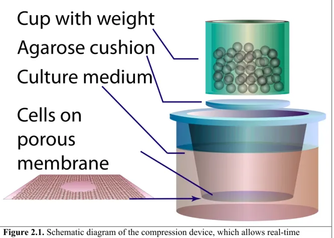
………... 31 Figure 1.4. Growth-induced mechanical stress distribution controls tumor spheroid shape ………33 Figure 2.1. Schematic diagram of the compression device, which allows real-time
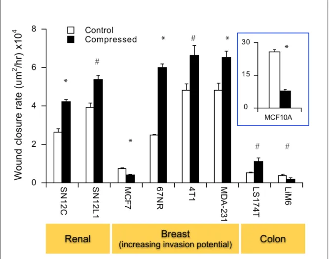
monitoring of cell migration exposed to a constant loading force .……….…. 49 Figure 2.2. Forced cell extrusion by compression occurs on the 8um-porous membranes, but not on the 0.4um ones ……….55 Figure 2.3. Compressive stress induces faster cell migration in multiple cancer cell lines, particularly more aggressive breast cancer cell lines ………... 58 Figure 2.4. Compressive stress causes no change or decreased cell proliferation. …..… 59 Figure 2.5. Compressive stress differentially influences cell migration behavior …..…. 61 Figure 2.6. Compressive stress induces cytoskeletal actin rearrangement ……….. 63 Figure 2.7. Compressive stress alters microtubule organization in cancer cells ………. 66 Figure 2.8. Compressive induces directional migration of 67NR cells in a coordinated manner ……….…. 68 Figure 2.9. Moderate stress enhances 67NR cell motility without a significant decrease in cell viability ………..71 Figure 2.10. Continuous compressive stress is required to maintain enhanced cell
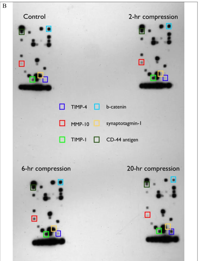
Figure 2.11. Compressive stress unlikely influence the genes associated with tumor metastasis or extracellular matrix and adhesion molecules arrays ……….. 76 Figure 3.1. Externally-applied stress enhances leader-cell formation ………..98 Figure 3.2. Both control and compressed 67NR cells at the leading edge of the “wound” are polarized but the compressed ones have larger projected cell-substrate contact area ………..100 Figure 3.3. Compressive stress induces 67NR cell extension and filopodial protrusions ………..101 Figure 3.4. Compression-induced leader cells and enhanced cell migration were
reproduced in circular patterns created by cell micro-contact printing ………..103 Figure 3.5. Compressive stress induces leader-cell formation independent of geometry-driven polarization ………..105 Figure 3.6. Free-cell perimeter determines leader-cell formation: geometry-driven in uncompressed cultures vs. cell distension-induced by compressed cultures …………..107 Figure 3.7. Rac activity is not required for compression-induced protrusions …..….…110 Figure 3.8. Compressive stress tends to reduce Cdc42 activity despite enhanced
protrusions ………...114 Figure 3.9. Perturbation of Cdc42 activity reduces compression-induced cell movement but not formation of leader cells with filopodial protrusions ……….115 Figure 4.1. E-cadherin-mediated cell-cell adhesion in MCF10A cells does not contribute to reduced migration under compression ………....140 Figure 4.2. E-cadherin-mediated cell-cell adhesion in MCF7 cells does not contribute to reduced migration under compression ……….…...142 Figure 4.3. Ectopic expression of E-cadherin in 67NR mammary carcinoma cells has no effect on compression-induced migration behavior ………144 Figure 4.4. Blocking N-cadherin function does not abolish compression-induced 67NR cell migration ………..146 Figure 4.5. Compression-induced cell-substrate adhesion appears to be related to
Figure 4.6. Compression increases fibronectin deposit at the cell-substrate interface independent of fibronectin synthesis ………..…150 Figure 4.7. Compression promotes FAK-mediated initial adhesions, which could in turn mature into vinculin-containing focal adhesions ………153 Figure 4.8. Integrin β1 is responsible for fibronectin-associated migration of 67NR cells ………...156 Figure 4.9. Integrin β1 is involved in compression-induced migration …..…….……...158 Figure 4.10. Compression reduces the total paxillin expression level, but increases the fraction of phosphorylated paxillin associated with cell-substrate interaction ………...161 Figure 4.11. Actomyosin contractility reduces compression-induced directional cell migration……….……….164 Figure 5.1. Assignment of cells to the defined square cell pattern on the grid surface ………..182 Figure 5.2. The cell protrudes at its leading edge without rear retraction until it doubles in size………...185 Figure 5.3. The cell protrudes at its leading edge and then retracts at the rear…………186 Figure 5.4. Force-body diagram of the cell being modeled……….190 Figure 5.5. Simulation of coordinated migration of cells initially arranged in a square pattern under no stress condition……….194 Figure 5.6. Simulation of the base condition (uncompressed case in Fig. 5.5) with the maximum frontal length allowed for each cell being doubled………....196 Figure 5.7. Simulation of the base condition (uncompressed case in Fig. 5.5) with the protrusion/migration rate being kept constant, independent of free cell perimeter
(FCP)………197
Figure 5.8. Simulation of the base condition (uncompressed case in Fig. 5.5) with the FCP-dependent protrusion/migration rate being doubled………197
Figure 5.9. Simulation of the base condition (uncompressed case in Fig. 5.5) with the same number of initial protrusions assigned for all cells around the periphery of the
Figure 5.10. Comparison of the shape change index values between the experimental and different model conditions………...199
Figure 5.11. Compression could increase cell frontal extension and facilitate constant cell protrusion/translocation rate, resulting in enhanced coordinated cell migration……….202
Figure 5.12. Simulation of collective cell migration in a rosette pattern under stress-free
condition………..205
Figure 5.13. Simulation of collective cell migration in a rosette pattern under compression: increased cell frontal extension and constant, FCP-independent cell
protrusion/translocation rate………206
Figure 5.14. Simulation of collective cell migration in a circle pattern under stress-free
condition………..207
Figure 5.15. Simulation of collective cell migration in a circular pattern under compression: increased cell frontal extension and constant, FCP-independent cell
protrusion/translocation rate………....208
Figure 6.1. Conceptual model of compression-modulated coordinated cell
migration………..…218 Appendix Figure A1: Compression-induced 67NR migration is observed on both porous and nonporous surfaces….………..……….227 Appendix Figure A2: Gene tables for SABiosciences microarrays ………....228 Appendix Figure A3: Compressive stress does not induce nuclear localization of beta-catenin ……….230 Appendix Figure A4: Compressive stress has no significant effect on fibroblast
migration………..231 Appendix Figure B1: Compression induces lamellipodial protrusions in LS174T colon carcinoma cells ………..………...233 Appendix Figure B2: Fibronectin-coated patterns created with PDMS stamps ……….234 Appendix Figure C1: Loss of E-cadherin-mediated cell adhesion has no effect on LS174T or LiM6 colon carcinoma cell migration ………237
Appendix Figure C2: Blocking fibronectin-integrin interaction with RGD peptides or an antibody to fibronectin does not necessarily reduce fibronectin-induced migration …..240 Appendix Figure C3: Compression appears to reduce the phosphorylated level of focal adhesion kinase (FAK) ………...242 Appendix Figure D1: Simulation of collective migration under stress-free condition with various action probability ………...………....244 Appendix Figure D2: Histogram showing migration rates for simulations from Appendix Figure D1 ……….………...247
List of Tables
Table 1.1: Studies of mechanical stress in tumor biology………...…35
Table 4.1: Antibodies used for immunofluorescence microscopy ………….………….132
Table 4.2: Various integrin blocking antibodies………..135
Table 5.1: Key parameters for the cell state ………...183
Table 5.2: What model parameter could be affected by compression? ………..…195
Chapter 1: Introduction and background
Portions of the chapter have been taken from:
G. Cheng, J. Tse, R.K. Jain, L.L. Munn, “Micro-environment mechanical stress controls tumor spheroid size and morphology by suppressing proliferation and inducing apoptosis in cancer cells.” PLoS One. 2009; 4(2): e4632.
Introduction
Cancer is the second leading cause of death in the United States, accounting for nearly 25% of total deaths [1]. The American Cancer Society estimates that about 565,650 Americans will die of cancer and 1.44 million new cancer cases will be diagnosed [1].
Cancer is a disease characterized by uncontrolled growth of abnormal cells, which have undergone DNA mutation. The most commonly mutated gene in human tumors is p53, which is a tumor suppressor gene promoting arrest in G1 and G2 checkpoints of the cell cycle, apoptosis, and DNA repair in response to damaged DNA [2]. The accumulation of DNA mutations promotes the development of cancers, not all of which form solid
tumors. For instance, leukemias are cancers of white blood cells, which do not form solid tumors, but circulate in the blood vessels. However, over 85% of human cancers form solid tumors, such as carcinomas, sarcomas and adenocarcinomas [1].
These solid tumors transform over time from a benign cell mass into an invasive phenotype. Eventually, those malignant tumor cells spread to other parts of the body (metastasis) and become fatal. Cancer research has historically focused primarily on studying the role of genetic and biochemical changes in tumor progression. For instance, it is well established that intratumoral hypoxia (cancer cells starved of oxygen) activates certain genes, which promotes cancer cell motility and invasion. However, in growing solid tumors, cancer cells also experience compressive stress generated by uncontrolled cell proliferation in a confined space[3,4]. While it has been long known that mechanical
stimuli are essential for normal physiological processes such as tissue remodeling and maintenance [5-7], endothelial cell biology[8,9], and morphogenesis [10,11], the role of such compressive mechanical force (transmitted through pericellular matrix and cells) in cancer progression has not been widely investigated.
The importance of mechanical stress in tumor biology is being recognized increasingly. Our lab has previously shown that compressive stress influences tumor spheroid growth [3,4] and stimulates production of extracellular matrix molecules [12]. Others have also demonstrated the importance of matrix rigidity in tumor development [13-15] and enhanced tumor cell adhesion by hydrostatic pressures[16-18]. Yet whether growth-induced compressive stress can impose selection pressure for cancer cells with enhanced migratory and invasive potentials remains unclear.
The focus of this thesis is to evaluate the effect of anisotropic compressive stress on cancer cell motility. Here we show, for the first time, that externally-applied compressive stress results in faster migration of mammary carcinoma cells. Unlike geometry-driven migration in the control cultures, compression induces migration of 67NR mammary carcinoma cells in a coordinated sheet, initiated by “leader cells” – single cells at the leading edge of the sheet, extending long filopodia. Accompanied by redistribution of fibronectin deposition, applied compression enhances cell-matrix adhesion and stabilizes cell distension independent of actomyosin contractility. Our finding on the coordinated migration of 67NR mammary carcinoma cells induced by compressive stress has significant implications for in vivo situations where epithelial cancer cells form an
“coordinated” invading mass guided by “leader” cells [19]. They suggest that mechanical stress accumulated during tumor growth can enhance cell-substrate adhesion and trigger formation of leader cells during multicellular invasion.
Specific Aims
Hypothesis: Compressive stress generated by tumor growth promotes a more migratory phenotype in cancer cells
Specific Aim 1: Investigate the effect of compressive stress on cancer cell motility (Chapter 2).
Normal cells have regulated rates of proliferation, but cancer cells grow uncontrollably in a confined matrix [2]. Previous findings of collapsed intratumoral vessels [20] suggest that compressive stress is generated from proliferating cancer cells. While intratumoral hypoxia has been shown to be a selection pressure for aggressive cancer cells [21], whether externally-applied compressive stress induces similar selective pressure has yet to be studied. Our findings would open the door to a new class of targets for blocking mechanical stress pathways.
Specific Aim 1a: Develop an in vitro compression device for simulation of compressive stress experienced by tumor cells in vivo.
In order to simulate the compressive stress generated by rapid cell proliferation at the tumor margin in vivo, an in vitro compression device was developed to apply a desired normal force (anisotropic stress) to compress a cell monolayer. In this system, there were no nutrient limitations, hydrostatic force, or oxidative stress.
Specific Aim 1b: Screen for the effect of compressive stress on migration potential of various cancer cell lines.
To determine the effect of compressive stress on tumor malignancy, various cancer cell lines established from different tissues, such as breast, colon and kidney, were subjected to compressive stress and their migration potential were assessed with scratch-wound assay. A normal mammary epithelial cell line was also used for comparison.
Specific Aim 1c: Examine the cytoskeletal components of cancer cells in response to compression.
Mechanical stress has been shown to influence organization of cytoskeleton[22,23], which provides structural support and cell shape. As recent studies showed the
importance of mechanical stress in breast tumor development[14,24], we investigated the cytoskeletal changes of mammary epithelial cells in response to mechanical compression. Hence, we stained the mammary carcinoma cell lines that demonstrated enhanced cell migration under compression in Aim 1b as well as the normal mammary epithelial cells for actin filaments and microtubules.
Specific Aim 1d: Determine whether compression-induced migration results from changes in gene transcription.
Mechanical stress propagated to a cell nucleus through matrix attachment and
cytoskeletal filaments induces gene transcription [25-28]. While hypoxia has been shown to activate genes that stimulate cell migration and invasion such as CXCR4 and Met, mechanical stress has also been shown recently to induce Twist gene expression in Drosophila embryo [10] and Twist facilitates metastasis in mice [29]. To identify
compressive-stress-regulated genes in cancer cells, gene expression studies of 67NR mammary carcinoma cell line exhibiting the most prominent changes in migration potential in Aim 1b, were performed with DNA microarrays related to tumor metastasis and extracellular matrix (ECM) and adhesion molecules.
Specific Aim 2: Determine the effect of compressive stress on leadercell formation and migration (Chapter 3).
Coordinated cell migration (collective cell migration) is prevalent in many epithelial cancers (as well as morphogenesis and tissue regeneration), and differs from single cell migration in that cells remain connected as they move, which results in a migrating sheet guided by “leader” cells[19,30]. While mechanical cues are critical in physiological processes involving collective migration, little is known about the effect of mechanical stress on leader-cell formation during collective migration. Our studies provide novel insights into how mechanical stimulation triggers coordinated migration in cancer.
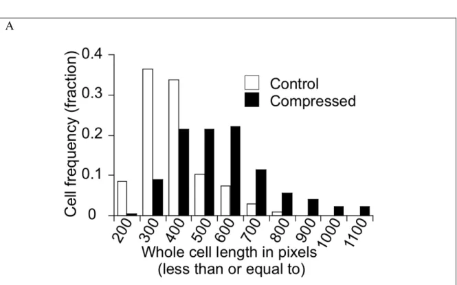
Specific Aim 2a: Characterize the leader cells in the control and compressed cultures. 67NR mammary carcinoma cell line demonstrated coordinated migration behavior (such as migration in a sheet guided by leader cells) in Aim 1b. Using the in vitro scratch-wound assay, which is a useful tool to study collective migration, we stained the cells for actin filament and then quantified the difference in leader-cell formation between the control and compressed cultures. Moreover, we quantified the nuclear offset (an indicator for cell polarization) and cell size/length.
Geometric cues (such as individual cell shape and multi-cellular micro-organization) can modulate cell proliferation [31] and direct the formation of lamellipodia and filopodia [32]. To determine whether geometric cues influence coordinated cell migration with or without compressive stress, we used microfabrication to control the organization of sheet of 67NR mammary carcinoma cells and then quantified the difference in migration rate and shape change of the cell pattern between the control and compressed cultures.
Specific Aim 2c: Examine the role of Rac1 and Cdc42 in compression-induced protrusions and migration.
Rac1 and Cdc42 – members of Rho family small guanosine triphosphate (GTP)-binding proteins (GTPases) – regulate cell shape, cell polarity and formation of protrusions, thereby affecting cell migration[33-35]. Hence, we measured the effect of compressive stress on activation of Rac1 and Cdc42 activity in 67NR mammary carcinoma cells. To determine the significance of Rac 1 and Cdc42 in compression-induced protrusions and migration, we perturbed the activity level of those Rho GTPases proteins using a molecular approach and performed the scratch-wound compression experiment. We quantified the migration rate of the control and compressed cells, and stained the cells for actin filaments to visualize their protrusions.
Specific Aim 3: Determine the effect of compressive stress on cell adhesion and migration (Chapter 4)
For migration to occur, any newly-formed protrusions have to be stabilized by attaching to the substrate surface. In addition, one of the hallmarks characterizing collective cell migration is the preserved cell-cell contact during movement [19,30]. While the
mechanisms of collective migration are less well understood, an understanding of the effect of mechanical stimulation on cell adhesion, including cell-cell and cell-matrix adhesions, during coordinated movement would enable us to define strategies to interfere with cancer cell migration.
Specific Aim 3a: Evaluate the role of cadherin-mediated cell-cell contacts in compression-induced migration
In coordinated migration, most of the adhesive cell-cell couplings are cadherin-mediated but some involve integrin-ECM interactions[36]. To determine whether E-cadherin expression plays a role in compression-associated migration, we treated E-cadherin-expressing normal mammary epithelial cells (MCF10A) and mammary carcinoma cells (MCF7) with an anti-E-cadherin antibody and determined their migration rate with scratch-wound assay in the presence or absence of compressive stress. In addition, we performed ectopic expression of E-cadherin in 67NR mammary carcinoma cells and quantified their motility in response to compression.
Specific Aim 3b: Quantify and characterize the effect of compressive stress on cell-matrix adhesion
Other than cell-cell coadhesion, cell-substrate interaction also plays an important role in modulating cell migration behavior. Therefore, we quantified the difference in the cell-matrix adhesion strength of 67NR mammary carcinoma cells between the control and compressed cultures using a shear detachment assay, and identified the ECM molecule accountable for compression-induced cell-matrix adhesion. Then, we determined the effect of compressive stress on the synthesis and spatial distribution of the determined
respectively. As cell-matrix adhesion involves formation of adhesion sites,
immunostaining of focal adhesion-associated molecules such as focal adhesion kinase (FAK) and vinculin were also performed.
Specific Aim 3c: Assess the role of integrin signaling in compression-induced migration Cell-matrix interaction involves integrin signaling[37,38]. Hence, we identified the integrin subunit involved in compression-induced migration of 67NR mammary carcinoma cells with integrin-blocking antibodies. We also determined the effect of compressive stress on the expression and spatial distribution of the integrin and its associated protein, paxillin, with Western blot and confocal immunofluorescence microscopy, respectively.
Specific Aim 3d: Determine the effect of actomyosin contractility on compression-induced migration.
Cell-matrix adhesion gives rise to intracellular contractile force mediated by actomyosin machinery, which is essential for maturation of focal contacts and stress fiber formation [39]. We examined the requirement of actin-myosin activity in compression-induced migration using molecular (such as dominant-negative RhoA retrovirus) and
pharmacological (such as Rho kinase inhibitor Y-27632, myosin light-chain kinase inhibitor ML-7) approaches and quantified the migration rate with scratch-wound assay.
Specific Aim 4: Develop a preliminary and simple stochastic model to explain the experimental data on compressioninduced coordinated migration (Chapter 5) Our experimental observations on compression-induced coordinated migration of 67NR
of free-cell perimeter (related to the formation rate of protrusions) and cell-cell
interactions would affect cell migration behavior; and (2) externally-applied stress could perturb the protrusion/migration rate. However, definitive experiments to test this hypothesis are elusive. A mathematical model has yet to be developed to describe the relative significance of free-cell perimeter and cell-cell contact on collective cell
migration. Such a model would provide us with insights into the physical underpinnings governing the collective migration induced by compressive stress.
Specific Aim 4a: Simulate cell migration behavior observed in stress-free experiments Based on our experimental observations under stress-free conditions, we developed a stochastic model to simulate 2-dimensional collective migration of cells initially arranged in a square geometry. Each cell is composed of multiple blocks such that their protrusion rates and cell-cell interactions can be determined separately according to the local
microenvironment. Then a force balance - incorporating protrusive force (due to formation of protrusions) and cell-cell interactions - is performed to determine the direction of protrusion and migration.
Specific Aim 4b: Investigate the relative importance of various model parameters, such as free-cell perimeter, cell protrusion length and number of protrusions, in compression-induced coordinated cell migration.
Using the model developed in Aim 4a, we changed one model parameter at a time to determine what model parameter could be influenced by compression. By comparing the simulated migration patterns with our experimental observations of compressed cultures, the model provides an estimate of which critical parameters in the force balance are
Background
Role of mechanical force in normal tissue development and function
Cells are continuously exposed to a variety of mechanical stimuli including hydrostatic pressure, shear, compression and tension. Various lines of investigation have revealed that mechanical stresses play a critical role in normal physiological processes.
I. Force and embryogenesis/differentiation
During embryo development, the normal morphogenic movements generate compressive stress that affects the physical shape of the embryo. To mimic these developmental compressive forces, Drosophila (fruit flies) embryo was deformed by external uniaxial mechanical compression. The externally-applied force was shown to drive nuclear
translocation of the transcriptional factor Armadillo and activate Twist expression, which controls the shape in the early Dropsophila embryo[10].
Mechanical forces are also important to normal tissue-specific development. For example, the major mechanical stimulus to the fetal lung growth is stretch induced by fetal breathing movements[40,41]. Abnormal forces exerted on lung tissues contribute to many pathological conditions such as pulmonary hypoplasia. Other examples are
mechanical stretch-induced hypertrophic responses in cardiac myocytes[42], and hemodynamic forces as regulators for vascular endothelial gene expression[43].
II. Tissue maintenance and modeling
A balance of forces is required to maintain homeostasis in tissues, including bone[5,7] and cartilage [6,44]. For instance, exercise affects joint loading, and thus increases the proteoglycan content of articular cartilage, whereas reduced mobility leads to loss of
proteoglycan content and exacerbates arthritis-associated joint degeneration[45,46]. In addition, microgravity directly affects bone formation and resorption, contributing to severe loss of bone mass during space flight [47]. Similarly, while laminar shear stress induced by blood flow allows normal artery maturation, turbulent shear stress may lead to atherosclerosis[48].
Furthermore, mechanical forces of the same type can produce different responses depending on the magnitude, duration and application mode of loading. One specific example is the effect of compressive stress on the extracellular biosynthesis of
chondrocytes. In vitro studies have shown that static compression on chondrocyte-seeded constructs or cartilage explants inhibits extracellular-matrix (ECM) biosynthesis [44] while dynamic compression stimulates ECM biosynthesis [49].
Experimental models for applying mechanical stimulation to living cells Two main types of mechanical cues have been studied in the area of cell
mechanobiology: (I) matrix stiffness and composition, and (II) external force application. Numerous novel experimental models have been developed to simulate various physical forces on cells in vitro.
I. Matrix stiffness and composition
3D culture systems made of cell-modifiable prefabricated ECM have been commonly used to study epithelial morphogenesis and malignant transformation[15,50]. Matrigel is critical for normal epithelial morphogenesis, but it is very soft with a modulus virtually identical to normal mammary tissue. Therefore, to explore the role of matrix stiffness in mammary tissue behavior, epithelial cells are cultured in Matrigel mixed with different
concentrations of collagen I for the desired the mechanical property[15]. In addition to the biologically-derived ECM, synthetic materials such as polyacrylamide gels
functionalized with ligand of choice (e.g. fibronectin) have been used to illustrate the effect of substrate stiffness on cell phenotype [50,51].
II. Engineered devices for force application
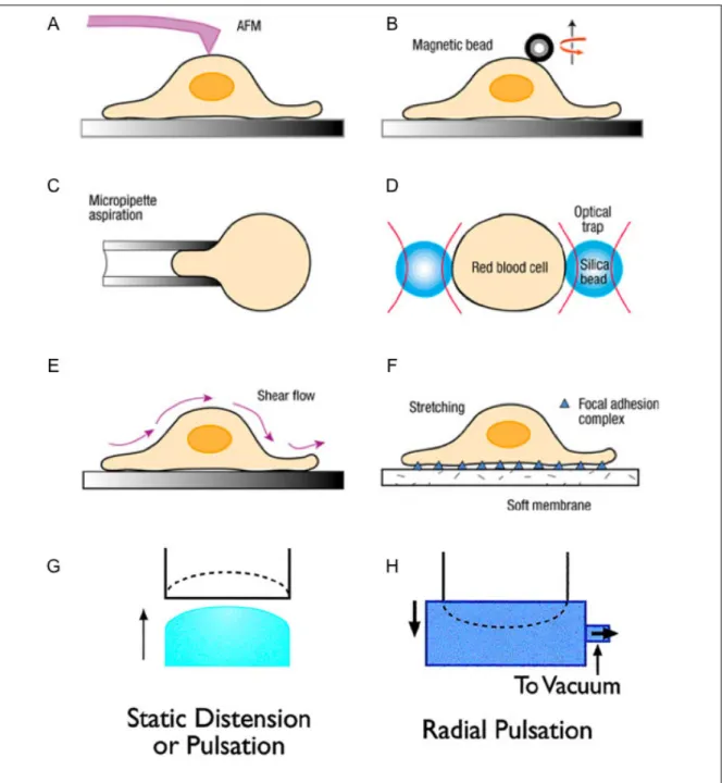
The cellular response to mechanical stimulation depends on the type of force applied, the magnitude, frequency and duration of the applied stimuli. To modulate the temporal, spatial and intensity of physical forces applied to cells, various experimental devices have been developed (Fig. 1.1) and can be categorized into two approaches.
The first approach focuses on the response of individual cells directed to mechanical stimuli (Fig. 1.1A-D). For example, atomic force microscopy (Fig. 1.1A)[52] and
magnetic twisting cytometry (Fig. 1.1B)[53] apply pico- to nano-Newton forces locally to a portion of the cell membrane. Micropipette aspiration (Fig. 1.1C) and optical trapping (Fig. 1.1D) deform an entire cell by applying suction through a micropipette placed on the surface of the cell[54], and directing the beads attached to the cell to move away or closer[55], respectively. It should be noted that these techniques, with appropriate analysis of deformation, can be used to probe the viscoelastic properties of cellular components[52,56,57].
While the previous techniques use sophisticated devices to apply precise forces to
individual cells, cell-cell communication under mechanical stimulation is not considered. Therefore, another approach applies controlled forces to cell monolayers or 3D cultures (ex vivo explant culture or cells embedded in tissue-engineered scaffolds), which mimic
the forces that each cell would experience within their physiological microenvironment (tissues) (Fig.1.1E-H). For instance, flow chambers have been used to apply shear stresses to endothelial cells (Fig. 1.1E). Application of static or cyclic, axial or biaxial strains has been applied to monolayers of cells plated on a deformable membrane (Fig. 1.1F-H) in various organ models such as lung[41,58,59]. Systems for application of compression and hydrostatic pressures have also been developed to study ECM
Figure 1.1. Schematic representations of different experimental techniques used to apply mechanical stimulation to living cells. A, Atomic force microscopy: a sharp tip at the free end of a flexible cantilever generates a local deformation on the cell surface. B, Magnetic twisting cytometry: magnetic beads with functionalized surfaces are attached to a cell and a magnetic field imposes a twisting moment on the beads, thereby deforming a
portion of the cell. C, Micropipette aspiration: a cell is deformed by applying suction through a micropipette. D, Optical trapping: a trap is used with two microbeads attached to the opposite ends of a cell. E, Shear flow: a parallel-plate flow channel applies shear stress to cells cultured as a monolayer. F, Uniaxial stretch: cells are cultured on a thin-sheet polymer substrate, such as silicone, which is stretched uniaxially to deform cells. G, H, Biaxial stretch: cells are cultured on an elastic membrane that is pushed upward (G) or pulled downward by negative pressure (H). (Figures reproduced and modified from refs. [41,57])
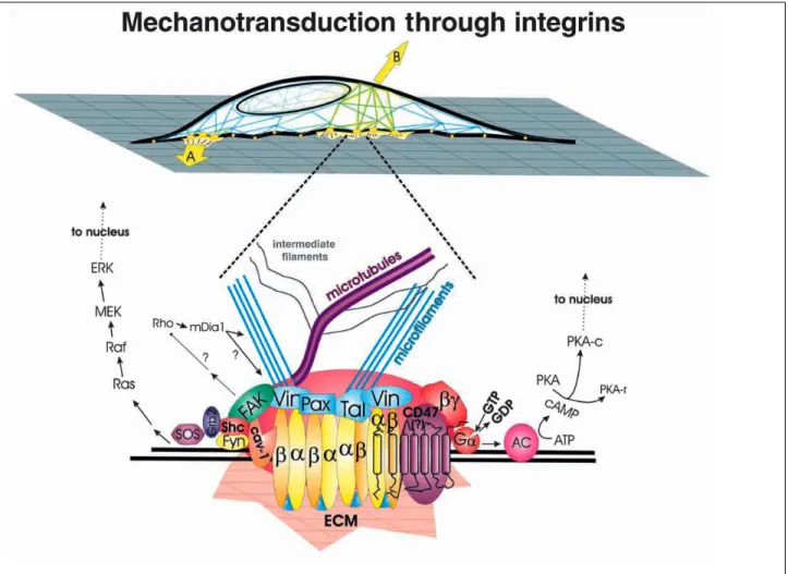
Mechanical models of mechanotransduction
Cells sense and convert mechanical cues into biochemical responses, such as activation of gene transcriptions [63]. Two similar mechanical paradigms – tensegrity[64-66] and adhesion-mediated mechanosensing[67,68]- have been described to explain how cells respond to mechanical stress involving actin cytoskeleton.
Cells do not simply contain viscous cytoplasm surrounded by a plasma membrane, they also contain cytoskeleton – a cellular “skeleton” contained with the cytoplasm- to provide structural support for maintaining cell shape and enabling cellular motion. The tensegrity model suggests that cells exist in a “pre-stress” state, in which their intracellular tension generated in the actin cytoskeleton is balanced by internal microtubule struts and external ECM adhesions. Thus, cellular response to external mechanical loading can vary with the level of intracellular tension in the cell. More importantly, since cell surface-ECM
tensegrity theory indicates that mechanical signals that are transmitted across integrin receptors can be transduced into a chemical response through distortion-dependent changes in cytoskeletal structure either locally at the site of receptor binding or distally at other locations inside the cell [69,70] (Fig. 1.2). Similarly, adhesion-mediated
mechanosensing model suggests that mechanical stimuli transmitted via actin filaments to mechanosensitive layer of focal adhesions triggers the growth of focal adhesions in the direction of the intracellular tension[67].
Figure 1.2. Schematic diagram of how forces applied via ECM (A) or directly to the cell surface (B) transmit across integrins and focal adhesions to induce a biochemical response, respectively. Forces (A) concentrated within the focal adhesion can stimulate clustering of
dimeric (α,β) integrin receptors and induce recruitment of focal adhesion proteins (e.g. vinculin (Vin), paxillin (Pax), talin (Tal)) that connect to cytoskeletal structures (actin filaments and microtubules), thereby activating integrin-associated signaling cascades, such as focal adhesion kinase (FAK). Cell distortion induced by forces (B) increase intracellular tension, which is transmitted to focal adhesions through the cytoskeleton. (Figure reproduced from ref. [65])
Mechanical stress and cancer biology
Historically, research in cancer biology has been focused in genetic or molecular changes in the tumor cells and their cellular responses to extrinsic soluble cues such as growth factors and cytokines. Only in recent years, the importance of mechanical stress in tumor biology has been increasingly appreciated. Indeed, cancer cells in tumors have been shown to experience (i) matrix stiffening due to abundant deposit of collagenous fibers synthesized by activated stromal myofibroblasts[71], (ii) increased interstitial pressure due to a leaky vasculature and poor lymphatic drainage[72,73], and (iii) increased compressive stress due to the expanding tumor mass[3,4]. As cancer cells escape from the tumor and get into blood vessels or lymphatic system, increased hydrostatic pressures enhance tumor cell adhesion to epithelium or extracellular matrix[16-18].
Previous studies have shown the importance of matrix rigidity in tumor development and tumor progression [13-15,74], whereas increased interstitial pressures present significant challenges to drug delivery[75-77] and influence tumor cell proliferation[78]. As for growth-induced compressive stress, little is known about the dynamics of such stress accumulation in tumors or its mechanical impact on tumor pathophysiology.
Growth-induced compressive stress (solid stress) is mechanical compression transmitted through the structural elements of the interstitium and the cells. It is unlikely affected by
interstitial fluid pressures, which is transmitted through intra- and extracellular fluids, under physiological conditions. There have been no physical ways to measure solid stress reliably in tumors in vivo and the heterogeneity of a tumor makes the spatial
quantification of solid stress within the tumor and its surrounding matrix even more challenging.
However, our lab has presented evidence of compressive stress generated by tumor cells in a mouse model that collapsed intratumor blood vessels opened again after killing cancer cells surrounding them [20] (Fig.1.3). A case report also showed that a young adult had visual loss due to the tumor-induced intracranial extrinsic compression of the optic nerve [79].
Figure 1.3. Compressive stress generated by cancer cells collapses blood vessels. In a mouse model, collapsed blood vessels (arrows in panel a) have an open lumen (asterisks in panel b) after relieving compressive forces generated by cancer cells. Scale bars, 50
µm. (Figure reproduced from ref [20])
To better understand the mechanical impact on tumors, our lab has previously
demonstrated that the growth of tumor spheroids in agarose gel is inhibited by increasing gel stiffness[3] and the growth-induced mechanical stress distribution controls tumor spheroid shape[4], as shown in Figure 1.4. Similarly, using multiple-particle tracking, an invading brain tumor spheroid in Matrigel (containing polymer beads as reference markers) has also been shown to exert mechanical pressure and significant traction on its microenvironment[80].
Figure 1.4. Growth-induced mechanical stress distribution controls tumor spheroid shape. A, Agarose gel can fail under tension from growing tumor spheroids (green). Red arrowheads indicate the edge of planar cracks in the agarose gel (BF: bright-field image taken in Nomarski mode). Scale bar = 50 µm. B, Spheroids (green) of different shapes and their surrounding stress fields visualized by micro-beads (red). Scale bar = 150 µm. C, Relationship between local strain in agarose gel (εgel,1,local) and local spheroid
deformation (distseg) for the spheroids (green, inset) shown in A. distseg is the distance of
Correlation between the asymmetry in spheroid shape and in the corresponding strain in the surrounding agarose gel, showing that spheroids are more deformed along the direction of higher stress. Each data point is for one spheroid. R is the linear regression coefficient (p<0.0001). (Figure reproduced from ref[4])
In addition, mathematical models have been recently developed to determine the level of compressive stress induced by tumor growth [81,82]. Using mechanical properties of the tumor and its surrounding tissue, the model predicts equal radial and circumferential stresses at the necrotic center of the spheroid but higher circumferential stress than radial stress near the spheroid boundary (i.e. tumor’s advancing front with rapid cell
proliferation)[81]. Table 1.1 summarizes key studies of mechanical stress in tumor biology.
Growthinduced compressive stress and tumor malignancy?
As mentioned above, cancer cells experience different kind of mechanical stresses. In this thesis, we focused on whether growth-induced compressive stress increases cancer cell motility, because such compressive stress might impose similar selection pressure as demonstrated by oxidative stress[21].
Proliferating tumors rapidly outgrow their blood supply, leaving the cancer cells starved of oxygen – a condition known as hypoxia (oxidative stress). Meanwhile, cancer cells also experience compressive stress generated by uncontrolled cell proliferation in a confined space. Growth-induced compressive stress inhibits the growth of tumor
spheroids in agarose [3,4], whereas a tumor with regions beyond the limits of oxygen diffusion can neither expand beyond a few cubic millimeters in size without an adequate oxygen supply [83]. It is well-established that intratumoral hypoxia (oxidative stress) selects for cancer cells with enhanced migratory and invasive potentials, involving the activation of genes such as Met and CXCR4[21,84-87]. However, little is known about the effect of growth-induced compressive stress on tumor malignancy.
Our lab has previously shown that such compressive stress stimulates hyaluronan synthesis in tumor spheroids [12], of which higher concentrations have been associated with malignant tumors [88-90]. Moreover, other studies have shown that mechanical compression induces Twist gene expression in Drosophila embryo [10] and Twist facilitates metastasis in mice [29]. Taken together, we hypothesized that compressive stress generated by tumor growth promotes a more migratory phenotype in cancer cells.
Year Type of Mechanical
Stress*
Studies Refs
1997 III Growth-induced stress suppresses tumor spheroid growth
[3] 1999 III Neoplastic cell density can affect blood vessel
diameter [91] 2000, 2004, 2006, 2008, 2009
II Increased extracellular hydrostatic pressure
enhances cancer cell adhesion 18,61,9 [16-2-94]
2002 III Solid stress facilitates spheroid formation [12] 2003 III Multiple particle tracking is developed to
measure mechanical stress exerted by the brain tumor to Matrigel
[80]
2003 III A linear poroelasticity model is developed to estimate growth-induced stress
[81] 2004 III Cancer cells compress intratumor vessels [20] 2004 II Shear stress enhances colon cancer cell adhesion [95]
2004, 2005
I Matrix rigidity induces malignant phenotypes [15,24] 2006 II High tumor interstitial fluid pressure can
contribute to increased tumor proliferation
[78] 2009 I Matrix crosslinking forces tumor progression [74] 2009 III Local mechanical stress controls tumor spheroid
size and shape
[4] Table 1.1. Studies of mechanical stress in tumor biology. (*I: Matrix rigidity; II: Fluid pressure (shear/static); III: Solid stress generated by expanding tumor mass)
Cell migration
Cell migration is an important process for many physiological processes such as tissue development, immune response, and cancer metastasis [96,97]. Some cells migrate as individuals (single-cell migration) but many cell types will remain connected and move in groups (collective cell migration) under physiological conditions such as wound repair and even cancer invasion [19].
Single-cell migration has been well-studied and is a highly integrated multistep process, which can be described as follows[96,98]: (i) the cell polarizes and extends
lamellipodial/filopodial protrusions (generally driven by actin polymerization) in the direction of migration guided by external cues such as soluble gradients; (ii) the cell makes adhesions to ECM via adhesion receptors such as integrins to stabilize the protrusions and generate traction on the substrate; (iii) the cell retracts its rear after disassembly of rear adhesions so that it can translocate its cell body forward.
In the classical view of metastasis – a process of tumor cells spreading to other parts of the body, transformation of epithelial-like tumor cells to become mesenchymal is thought to be required for them to migrate as single cells[99]. However, it becomes more
prominent that tumor cells can also invade the surrounding environment in clusters or strands [100]- another mode of movement, termed as collective/coordinated cell
migration. Collective cell movement still retains the principles of single-cell migration, but the main difference is that the cells remain coupled by cell-cell coadhesions (which can be mediated by cadherins [101]or integrins[36]) during collective cell movement and the moving group is usually guided by multiple “leader” cells[19,30,102]. These leader cells at the edge of a group may be polarized by distinct free edge versus cell-cell contact edge within the physically connected sheets of cells[102].
Several in vitro and in vivo experimental systems exist to study collective cell migration[19,103]. The most common 2D in vitro model is scratch-wound assay: an artificial wound generated by mechanical removal of cells from a central region across a confluent monolayer of cells of epithelial cells [33,104]. The assay allows cell-cell and cell-matrix interactions to be studied during the wound closure. In this thesis, we have used scratch-wound assay to quantify the difference in the migration potential of epithelial cancer cells between the control and compressed cultures.
References
1. Jemal, A., R. Siegel, E. Ward, Y. Hao, J. Xu, T. Murray, and M.J. Thun. 2008. Cancer statistics, 2008. CA Cancer J Clin. 58:71-96.
2. Lodish, H., A. Berk, P. Matsudaira, C. Kaiser, M. Krieger, M.P. Scott, S.L. Zipursky, and J. Darnell. 2004. Molecular cell biology. W.H. Freeman and Company. 973 pp.
3. Helmlinger, G., P.A. Netti, H.C. Lichtenbeld, R.J. Melder, and R.K. Jain. 1997. Solid stress inhibits the growth of multicellular tumor spheroids. Nat Biotechnol. 15:778-83.
4. Cheng, G., J. Tse, R.K. Jain, and L.L. Munn. 2009. Micro-environmental mechanical stress controls tumor spheroid size and morphology by suppressing proliferation and inducing apoptosis in cancer cells. PLoS ONE. 4:e4632.
5. Burr, D.B., A.G. Robling, and C.H. Turner. 2002. Effects of biomechanical stress on bones in animals. Bone. 30:781-6.
6. Grodzinsky, A.J., M.E. Levenston, M. Jin, and E.H. Frank. 2000. Cartilage tissue remodeling in response to mechanical forces. Annu Rev Biomed Eng. 2:691-713. 7. Nagatomi, J., B.P. Arulanandam, D.W. Metzger, A. Meunier, and R. Bizios.
2002. Effects of cyclic pressure on bone marrow cell cultures. J Biomech Eng. 124:308-14.
8. Chien, S. 2006. Mechanical and chemical regulation of endothelial cell polarity. Circ Res. 98:863-5.
9. Yao, Y., A. Rabodzey, and C.F. Dewey, Jr. 2007. Glycocalyx modulates the motility and proliferative response of vascular endothelium to fluid shear stress. Am J Physiol Heart Circ Physiol. 293:H1023-30.
10. Farge, E. 2003. Mechanical Induction of Twist in the Drosophila Foregut/Stomodeal Primordium. Current Biology. 13:1365-1377.
11. Nerurkar, N.L., A. Ramasubramanian, and L.A. Taber. 2006. Morphogenetic adaptation of the looping embryonic heart to altered mechanical loads. Dev Dyn. 235:1822-9.
12. Koike, C., T.D. McKee, A. Pluen, S. Ramanujan, K. Burton, L.L. Munn, Y. Boucher, and R.K. Jain. 2002. Solid stress facilitates spheroid formation: potential involvement of hyaluronan. Br J Cancer. 86:947-53.
13. Butcher, D.T., T. Alliston, and V.M. Weaver. 2009. A tense situation: forcing tumour progression. Nat Rev Cancer. 9:108-22.
14. Kumar, S., and V.M. Weaver. 2009. Mechanics, malignancy, and metastasis: the force journey of a tumor cell. Cancer Metastasis Rev. 28:113-27.
15. Paszek, M.J., N. Zahir, K.R. Johnson, J.N. Lakins, G.I. Rozenberg, A. Gefen, C.A. Reinhart-King, S.S. Margulies, M. Dembo, D. Boettiger, D.A. Hammer, and V.M. Weaver. 2005. Tensional homeostasis and the malignant phenotype. Cancer Cell. 8:241-254.
16. Basson, M.D., C.F. Yu, O. Herden-Kirchoff, M. Ellermeier, M.A. Sanders, R.C. Merrell, and B.E. Sumpio. 2000. Effects of increased ambient pressure on colon cancer cell adhesion. J Cell Biochem. 78:47-61.
17. Downey, C., K. Alwan, V. Thamilselvan, L. Zhang, Y. Jiang, A.K. Rishi, and M.D. Basson. 2006. Pressure stimulates breast cancer cell adhesion independently of cell cycle and apoptosis regulatory protein (CARP)-1 regulation of focal
adhesion kinase. Am J Surg. 192:631-5.
18. Craig, D.H., C.P. Gayer, K.L. Schaubert, Y. Wei, J. Li, Y. Laouar, and M.D. Basson. 2009. Increased extracellular pressure enhances cancer cell integrin-binding affinity through phosphorylation of beta1-integrin at threonine 788/789. Am J Physiol Cell Physiol. 296:C193-204.
19. Friedl, P., and D. Gilmour. 2009. Collective cell migration in morphogenesis, regeneration and cancer. Nat Rev Mol Cell Biol. 10:445-57.
20. Padera, T.P., B.R. Stoll, J.B. Tooredman, D. Capen, E. di Tomaso, and R.K. Jain. 2004. Pathology: cancer cells compress intratumour vessels. Nature. 427:695.
21. Bernards, R. 2003. Cancer: cues for migration. Nature. 425:247-8.
22. Dartsch, P.C., and E. Betz. 1989. Response of cultured endothelial cells to mechanical stimulation. Basic Res Cardiol. 84:268-81.
23. Li, J., S. Zhang, J. Chen, T. Du, Y. Wang, and Z. Wang. 2009. Modeled microgravity causes changes in the cytoskeleton and focal adhesions, and decreases in migration in malignant human MCF-7 cells. Protoplasma. 24. Paszek, M.J., and V.M. Weaver. 2004. The tension mounts: mechanics meets
morphogenesis and malignancy. J Mammary Gland Biol Neoplasia. 9:325-42. 25. Alenghat, F.J., and D.E. Ingber. 2002. Mechanotransduction: All Signals Point to
Cytoskeleton, Matrix, and Integrins. Science's STKE. 2002:pe6-.
26. Ingber, D.E. 2003. Tensegrity II. How structural networks influence cellular information processing networks. J Cell Sci. 116:1397-408.
27. Ingber, D.E. 2003. Tensegrity I. Cell structure and hierarchical systems biology. J Cell Sci. 116:1157-73.
28. Wang, N., J.P. Butler, and D.E. Ingber. 1993. Mechanotransduction across the cell surface and through the cytoskeleton. Science. 260:1124-7.
29. Yang, J., S.A. Mani, J.L. Donaher, S. Ramaswamy, R.A. Itzykson, C. Come, P. Savagner, I. Gitelman, A. Richardson, and R.A. Weinberg. 2004. Twist, a Master Regulator of Morphogenesis, Plays an Essential Role in Tumor Metastasis. Cell. 117:927-939.
30. Ilina, O., and P. Friedl. 2009. Mechanisms of collective cell migration at a glance. J Cell Sci. 122:3203-8.
31. Nelson, C.M., R.P. Jean, J.L. Tan, W.F. Liu, N.J. Sniadecki, A.A. Spector, and C.S. Chen. 2005. Emergent patterns of growth controlled by multicellular form and mechanics. Proc Natl Acad Sci U S A. 102:11594-9.
32. Brock, A., E. Chang, C.C. Ho, P. LeDuc, X. Jiang, G.M. Whitesides, and D.E. Ingber. 2003. Geometric determinants of directional cell motility revealed using microcontact printing. Langmuir. 19:1611-7.
33. Nobes, C.D., and A. Hall. 1999. Rho GTPases control polarity, protrusion, and adhesion during cell movement. J Cell Biol. 144:1235-44.
34. Machacek, M., L. Hodgson, C. Welch, H. Elliott, O. Pertz, P. Nalbant, A. Abell, G.L. Johnson, K.M. Hahn, and G. Danuser. 2009. Coordination of Rho GTPase activities during cell protrusion. Nature. 461:99-103.
35. Ridley, A.J. 2001. Rho GTPases and cell migration. J Cell Sci. 114:2713-22. 36. Casey, R.C., K.M. Burleson, K.M. Skubitz, S.E. Pambuccian, T.R. Oegema, Jr.,
L.E. Ruff, and A.P. Skubitz. 2001. Beta 1-integrins regulate the formation and adhesion of ovarian carcinoma multicellular spheroids. Am J Pathol. 159:2071-80.
37. Berrier, A.L., and K.M. Yamada. 2007. Cell-matrix adhesion. J Cell Physiol. 213:565-73.
38. Zamir, E., and B. Geiger. 2001. Molecular complexity and dynamics of cell-matrix adhesions. J Cell Sci. 114:3583-90.
39. Chrzanowska-Wodnicka, M., and K. Burridge. 1996. Rho-stimulated contractility drives the formation of stress fibers and focal adhesions. J Cell Biol. 133:1403-15. 40. Kitterman, J.A. 1996. The effects of mechanical forces on fetal lung growth. Clin
41. Liu, M., A.K. Tanswell, and M. Post. 1999. Mechanical force-induced signal transduction in lung cells. Am J Physiol. 277:L667-83.
42. Sadoshima, J., and S. Izumo. 1997. The cellular and molecular response of cardiac myocytes to mechanical stress. Annu Rev Physiol. 59:551-71.
43. Resnick, N., H. Yahav, L.M. Khachigian, T. Collins, K.R. Anderson, F.C. Dewey, and M.A. Gimbrone, Jr. 1997. Endothelial gene regulation by laminar shear stress. Adv Exp Med Biol. 430:155-64.
44. Chen, A.C., and R.L. Sah. 1998. Effect of static compression on proteoglycan biosynthesis by chondrocytes transplanted to articular cartilage in vitro. J Orthop Res. 16:542-50.
45. Bird, J.L., D. Platt, T. Wells, S.A. May, and M.T. Bayliss. 2000. Exercise-induced changes in proteoglycan metabolism of equine articular cartilage. Equine Vet J. 32:161-3.
46. Haapala, J., M.J. Lammi, R. Inkinen, J.J. Parkkinen, U.M. Agren, J. Arokoski, I. Kiviranta, H.J. Helminen, and M.I. Tammi. 1996. Coordinated regulation of hyaluronan and aggrecan content in the articular cartilage of immobilized and exercised dogs. J Rheumatol. 23:1586-93.
47. Tamma, R., G. Colaianni, C. Camerino, A. Di Benedetto, G. Greco, M. Strippoli, R. Vergari, A. Grano, L. Mancini, G. Mori, S. Colucci, M. Grano, and A. Zallone. 2009. Microgravity during spaceflight directly affects in vitro osteoclastogenesis and bone resorption. FASEB J. 23:2549-54.
48. Davies, P.F., A. Remuzzi, E.J. Gordon, C.F. Dewey, Jr., and M.A. Gimbrone, Jr. 1986. Turbulent fluid shear stress induces vascular endothelial cell turnover in vitro. Proc Natl Acad Sci U S A. 83:2114-7.
49. Kisiday, J.D., M. Jin, M.A. DiMicco, B. Kurz, and A.J. Grodzinsky. 2004. Effects of dynamic compressive loading on chondrocyte biosynthesis in self-assembling peptide scaffolds. Journal of Biomechanics. 37:595-604.
50. Lopez, J.I., J.K. Mouw, and V.M. Weaver. 2008. Biomechanical regulation of cell orientation and fate. Oncogene. 27:6981-93.
51. Ulrich, T.A., E.M. de Juan Pardo, and S. Kumar. 2009. The Mechanical Rigidity of the Extracellular Matrix Regulates the Structure, Motility, and Proliferation of Glioma Cells. Cancer Res. 69:4167-4174.
52. Mathur, A.B., A.M. Collinsworth, W.M. Reichert, W.E. Kraus, and G.A. Truskey. 2001. Endothelial, cardiac muscle and skeletal muscle exhibit different viscous and elastic properties as determined by atomic force microscopy. Journal of Biomechanics. 34:1545-1553.
53. Pommerenke, H., E. Schreiber, F. Durr, B. Nebe, C. Hahnel, W. Moller, and J. Rychly. 1996. Stimulation of integrin receptors using a magnetic drag force device induces an intracellular free calcium response. Eur J Cell Biol. 70:157-64. 54. Hochmuth, R.M. 2000. Micropipette aspiration of living cells. J Biomech.
33:15-22.
55. Gan, Y. 2007. Invited review article: a review of techniques for attaching micro- and nanoparticles to a probe's tip for surface force and near-field optical
56. Bausch, A.R., F. Ziemann, A.A. Boulbitch, K. Jacobson, and E. Sackmann. 1998. Local Measurements of Viscoelastic Parameters of Adherent Cell Surfaces by Magnetic Bead Microrheometry. Biophys. J. 75:2038-2049.
57. Bao, G., and S. Suresh. 2003. Cell and molecular mechanics of biological materials. Nat Mater. 2:715-25.
58. Vanderploeg, E.J., S.M. Imler, K.R. Brodkin, A.J. Garcia, and M.E. Levenston. 2004. Oscillatory tension differentially modulates matrix metabolism and cytoskeletal organization in chondrocytes and fibrochondrocytes. J Biomech. 37:1941-52.
59. Wall, M.E., P.S. Weinhold, T. Siu, T.D. Brown, and A.J. Banes. 2007.
Comparison of cellular strain with applied substrate strain in vitro. J Biomech. 40:173-81.
60. Ragan, P.M., V.I. Chin, H.H. Hung, K. Masuda, E.J. Thonar, E.C. Arner, A.J. Grodzinsky, and J.D. Sandy. 2000. Chondrocyte extracellular matrix synthesis and turnover are influenced by static compression in a new alginate disk culture system. Arch Biochem Biophys. 383:256-64.
61. van Zyp, J.V., W.C. Conway, D.H. Craig, N.V. van Zyp, V. Thamilselvan, and M.D. Basson. 2006. Extracellular pressure stimulates tumor cell adhesion in vitro by paxillin activation. Cancer Biol Ther. 5:1169-78.
62. Flanigan, T.L., D.H. Craig, C.P. Gayer, and M.D. Basson. 2009. 35: Increased Extracellular Pressure and Integrin Phosphorylation Independently Influence Fibroblast Migration. Journal of Surgical Research. 151:187-187.
63. Khachigian, L.M., N. Resnick, M.A. Gimbrone, Jr., and T. Collins. 1995. Nuclear factor-kappa B interacts functionally with the platelet-derived growth factor B-chain shear-stress response element in vascular endothelial cells exposed to fluid shear stress. J Clin Invest. 96:1169-75.
64. Ingber, D.E. 2008. Tensegrity and mechanotransduction. J Bodyw Mov Ther. 12:198-200.
65. Ingber, D.E. 2003. Tensegrity II. How structural networks influence cellular information processing networks. J Cell Sci. 116:1397-408.
66. Ingber, D.E. 2003. Tensegrity I. Cell structure and hierarchical systems biology. J Cell Sci. 116:1157-73.
67. Bershadsky, A., M. Kozlov, and B. Geiger. 2006. Adhesion-mediated
mechanosensitivity: a time to experiment, and a time to theorize. Curr Opin Cell Biol. 18:472-81.
68. Bershadsky, A.D., N.Q. Balaban, and B. Geiger. 2003. Adhesion-dependent cell mechanosensitivity. Annu Rev Cell Dev Biol. 19:677-95.
69. Ingber, D.E. 1997. Tensegrity: the architectural basis of cellular mechanotransduction. Annu Rev Physiol. 59:575-99.
70. Ingber, D. 1991. Integrins as mechanochemical transducers. Curr Opin Cell Biol. 3:841-8.
71. Shao, Z.-M., M. Nguyen, and S. Barsky. 2000. Human breast carcinoma desmoplasia is PDGF initiated. Oncogene. 19:4337-4345.
72. Less, J.R., M.C. Posner, Y. Boucher, D. Borochovitz, N. Wolmark, and R.K. Jain. 1992. Interstitial hypertension in human breast and colorectal tumors. Cancer Res. 52:6371-4.
73. Boucher, Y., and R.K. Jain. 1992. Microvascular pressure is the principal driving force for interstitial hypertension in solid tumors: implications for vascular collapse. Cancer Res. 52:5110-4.
74. Levental, K.R., H. Yu, L. Kass, J.N. Lakins, M. Egeblad, J.T. Erler, S.F. Fong, K. Csiszar, A. Giaccia, W. Weninger, M. Yamauchi, D.L. Gasser, and V.M. Weaver. 2009. Matrix crosslinking forces tumor progression by enhancing integrin
signaling. Cell. 139:891-906.
75. Jain, R.K. 1989. Delivery of novel therapeutic agents in tumors: physiological barriers and strategies. J Natl Cancer Inst. 81:570-6.
76. Jain, R.K. 1994. Barriers to drug delivery in solid tumors. Sci Am. 271:58-65. 77. Minchinton, A.I., and I.F. Tannock. 2006. Drug penetration in solid tumours. Nat
Rev Cancer. 6:583-92.
78. Hofmann, M., M. Guschel, A. Bernd, J. Bereiter-Hahn, R. Kaufmann, C. Tandi, H. Wiig, and S. Kippenberger. 2006. Lowering of tumor interstitial fluid pressure reduces tumor cell proliferation in a xenograft tumor model. Neoplasia. 8:89-95. 79. Hogan, M.C., A. Lee, L.A. Solberg, and S.D. Thomâe. 2002. Unusual
presentation of multiple myeloma with unilateral visual loss and numb chin syndrome in a young adult. Am J Hematol. 70:55-9.
80. Gordon, V.D., M.T. Valentine, M.L. Gardel, D. Andor-Ardo, S. Dennison, A.A. Bogdanov, D.A. Weitz, and T.S. Deisboeck. 2003. Measuring the mechanical stress induced by an expanding multicellular tumor system: a case study. Experimental Cell Research. 289:58-66.
81. Roose, T., P.A. Netti, L.L. Munn, Y. Boucher, and R.K. Jain. 2003. Solid stress generated by spheroid growth estimated using a linear poroelasticity model small star, filled. Microvasc Res. 66:204-12.
82. Sarntinoranont, M., F. Rooney, and M. Ferrari. 2003. Interstitial stress and fluid pressure within a growing tumor. Ann Biomed Eng. 31:327-35.
83. Hanahan, D., and J. Folkman. 1996. Patterns and Emerging Mechanisms of the Angiogenic Switch during Tumorigenesis. Cell. 86:353-364.
84. Buchler, P., H.A. Reber, R.S. Lavey, J. Tomlinson, M.W. Buchler, H. Friess, and O.J. Hines. 2004. Tumor hypoxia correlates with metastatic tumor growth of pancreatic cancer in an orthotopic murine model1. Journal of Surgical Research. 120:295-303.
85. Le, Q.T., N.C. Denko, and A.J. Giaccia. 2004. Hypoxic gene expression and metastasis. Cancer Metastasis Rev. 23:293-310.
86. Rofstad, E.K., and T. Danielsen. 1999. Hypoxia-induced metastasis of human melanoma cells: involvement of vascular endothelial growth factor-mediated angiogenesis. Br J Cancer. 80:1697-707.
87. Zhang, L., and R.P. Hill. 2004. Hypoxia Enhances Metastatic Efficiency by Up-Regulating Mdm2 in KHT Cells and Increasing Resistance to Apoptosis. Cancer Res. 64:4180-4189.
88. Auvinen, P., R. Tammi, J. Parkkinen, M. Tammi, U. Agren, R. Johansson, P. Hirvikoski, M. Eskelinen, and V.-M. Kosma. 2000. Hyaluronan in Peritumoral Stroma and Malignant Cells Associates with Breast Cancer Spreading and Predicts Survival. Am J Pathol. 156:529-536.