ORIGINAL ARTICLE
Validity of the 3D VECTRA photogrammetric surface
imaging system for cranio-maxillofacial
anthropometric measurements
Philipp Metzler&Yi Sun&Wolfgang Zemann&
Alexander Bartella&Marc Lehner&
Joachim Anton Obwegeser&Astrid L. Kruse-Gujer&
Heinz-Theo Lübbers
Received: 19 February 2013 / Accepted: 20 February 2013 / Published online: 5 April 2013 # Springer-Verlag Berlin Heidelberg 2013
Abstract
Purpose The use of three-dimensional (3D) photography for anthropometric measurements is of increasing interest, especially in the cranio-maxillofacial field. Before standard implementation, accurate determination of the precision and accuracy of each system is mandatory.
Methods A mannequin head was labelled with 52 land-marks, and 28 three-dimensional images were taken using a commercially available five-pod 3D photosystem (3D VECTRA; Canfield, Fairfield, NJ) in different head posi-tions. Distances between the landmarks were measured manually using a conventional calliper and compared with the digitally calculated distances acquired from labelling by two independent observers. The experimental set-up accounted for clinical circumstances by varying the posi-tioning (vertical, horizontal, sagittal) of the phantom. Results In the entire calliper measurement data set (n=410), a significant difference (p=0.02) between the directly mea-sured and corresponding virtually calculated distances was found. The mean aberration between both modalities cover-ing all data was 7.96 mm. No differences (p=0.94) between the two groups were found using a cut-off of 10 % (leaving
n=369 distances) due to considerable errors in direct mea-surements and the necessary manual data translation. The mean diversity of both measurement modalities after cut-off was 1.33 mm (maximum, 6.70 mm). Inter-observer analysis of all 1,326 distances showed no difference (p=0.99; max-imal difference, 0.58 mm) in the digital measurements. Conclusion The precision and accuracy of this five-pod 3D photosystem suggests its suitability for clinical applications, particularly anthropometric studies. Three-hundred-and-six-ty degree surface-contour mapping of the craniofacial region within milliseconds is particularly useful in paediatric pa-tients. Proper patient positioning is essential for high-quality imaging.
Keywords 3D . Three-dimensional . Photography . Photometry . Anthropometric . Accuracy . Validation
Introduction
Anthropometry is a powerful tool for the determination of morphological aberrations in, for example, paediatric patients with acquired or congenital growth anomalies [1–4]. Farkas published an atlas focusing on the anthro-pometry of the head and face with standardised and reliably identifiable landmarks, mean values of various measurements, proportions, and indices [5]. This norma-tive database allows for the calculation of standardised (z-) scores based on age-, sex-, and ethnicity-specific characteristics, which further allows for a reliable and objective comparison of surgical techniques, age spans, etc. [6–13].
Three-dimensional (3D) photography permits the quanti-tative assessment of dysmorphology as well as a description of the growth patterns of various craniofacial anomalies on a
P. Metzler (*)
:
W. Zemann:
A. Bartella:
M. Lehner:
J. A. Obwegeser
:
A. L. Kruse-Gujer:
H.-T. LübbersDepartment of Cranio-Maxillofacial and Oral Surgery, University of Zurich, Frauenklinikstrasse 24, 8091 Zurich, Switzerland
e-mail: [email protected] Y. Sun
Oral and Maxillofacial Surgery, St. John’s Hospital,
Schiepse bos 6, 3600 Genk, Belgium Y. Sun
continuum. Exact measurements are irreplaceable in cranio-facial surgical assessment, planning, and follow-up [7]. The non-radiologic approach of 3D photography allows for ar-bitrary measurements and therefore represents a useful tool for craniofacial anthropometric evaluation, especially in paediatric patients [14].
In principle, not only the surface but also the under-lying bone can be 3D-reconstructed sufficiently using imaging techniques such as computed tomography. However, this is accompanied by X-ray exposition, and its consecutive use is not suitable for short-interval follow-up studies. Especially in the infant grow-ing phase, this is not acceptable. In addition, many imaging techniques require several seconds for data acquisition, and so general anaesthesia is required in infants, who by nature are not able to hold one position for this amount of time. Therefore, soft tissue acquisi-tion using digital image processing techniques such as 3D photography is of increasing interest [14–17]. This field has developed rapidly within the last decade not only in medicine, but also in physics, engineering and material science.
Commercially available 3D photosystems capture excel-lent 3D surface data within milliseconds. Patients' compli-ance is no longer as critical, and numerous datasets can be acquired and the most appropriate of these selected later. Furthermore, a coordinate system and various measuring tools allow for the acquisition of reliable, true-to-scale data. Observer errors, a common problem in direct anthropome-try, are reduced. The greatest advantage of 3D photography may its enabling perfect archiving of 3D surface data for further analysis, making any retrospective analysis based on 3D imaging as valid as if it were derived from a prospective setting. The advantages of this technique are undeniable, but its potential has not yet been fully integrated into most clinics.
Much data exist in the literature, which focuses on an-thropometric studies in the field of cleft and craniofacial
anomalies [7, 18–20]. Several techniques have been de-scribed and their advantages and disadvantages have been reported, but no reliable comparison has been performed due to heterogeneity in study design or data assessment method. 3D photography, with its above-mentioned advan-tages, may overcome this problem and facilitate identifica-tion of favourable treatment strategies for anomalies on an individual basis.
However, before implementation in further research, each system must be evaluated in terms of its accuracy, precision and reliability [16]. Various studies have fo-cused on these parameters using various 3D photosys-tems [16, 21, 22]. Nevertheless, no evaluation of the above-mentioned questions utilising the currently avail-able five-pod 3D photosystems for fully textured 360° surface contour mapping of the craniofacial area has been reported.
Materials and methods
Model
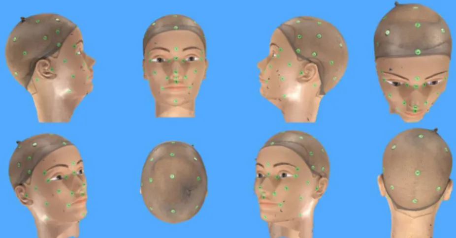
A plastic mannequin head was labelled with 52 artificial landmarks covering all craniofacial anatomic regions (Fig. 1). This set-up was chosen because it excludes the known technical errors associated with involuntary facial movements and hair volume [17].
Data acquisition
A total of 410 direct distances between the labelled landmarks on the mannequin head were acquired using a standardised clinical sliding-and-spreading calliper. One observer performed all measurements twice in a single session. The median of the measurements for each distance was accepted as the real distance between the two labels.
Fig. 1 Virtual model of the mannequin head as acquired by the 3D VECTRA photosystem (Canfield, Fairfield, NJ)
The 3D photogrammetric data were acquired under clinical lighting using the 3D VECTRA photosystem (Canfield, Fair-field, NJ). This system comprises five pod-mounted cameras oriented (Fig. 2). The captured data set was saved and converted into a 3D model for further evaluation. A natural head position (Frankfurt horizontal line parallel to the floor) was used for data acquisition [23]. Various positions following the standardised protocol of Lübbers et al. [16] were chosen to simulate clinical conditions (Table1). The system was cali-brated and was performed before each capture process to simulate the clinical follow-up of a patient.
Data processing
Further data processing was performed on a standard desk-top computer using the corresponding imaging software (Mirror) of the 3D photosystem. Two observers digitised the landmarks on the surface of the 3D model and the x, y and z coordinates of each data set. These markings were exported to an Excel® for Mac 2011 file (Microsoft® Cor-poration, Redmond, WA) for further processing and evalu-ation. Failed landmarks (blurred, double pictures, not captured) were excluded (Fig. 3). The distances between all landmarks were calculated using the following formula: d ¼p2 ffiffiffiffiffiffiffiffiffiffiffiffiffiffiffiffiffiffiffiffiffiffiffiffiffiffiffiffiffiffiffiffiffiffiffiffiΔx2þ Δy2þ Δz2, which yields the direct 3D
dis-tance between two coordinates [17,24–26]. Target variables
1. Accuracy (defined as the agreement between digital measurements and the true [direct] anthropometric measurements);
2. Inter-observer error (inaccuracies among multiple ob-servers during repeated digital measurements of the same data set);
3. Object positioning and recalibration.
Null hypothesis and data analysis
To assess the above-mentioned parameters, the following null hypotheses were defined:
1. The distances measured by the calliper do not differ from those derived from the virtual model.
2. The distances derived from the labelling by Observer 1 do not differ from those derived from the labelling by Observer 2.
3. The distances between any of the models measured by the same observer do not differ.
Statistical tools
The acquired data were analysed using descriptive statistics and parametric Student's t tests using Excel® for Mac 2011 (Microsoft® Corporation, Redmond, WA). Significance was considered at values of p<0.05.
Results
A total of 410 direct distances were measured (each twice, for a total of 820 calliper measurements). The two observers labelled a total of 28 virtual models. Altogether, 2,912 labels were to be placed. All distances between the labels were calculated, resulting in a total of 74,256 virtual distances. Only a small number of labels could not be placed due to issues with the 3D image (Fig.3).
Accuracy
A total of 410 distances between the landmarks were mea-sured directly and compared with the corresponding virtually calculated distances in the 3D data sets. In the whole data set, a significant difference (p=0.02) between the measured and calculated distances was found. The mean aberration between the measurement modalities using all data was 7.96 mm, with a maximum of 177.97 mm. Non-significant differences (p=0.94) were found between both groups using a cut-off of 10 % of the most unreliable data, due to considerable errors in direct measurement and data translation.
Furthermore, the mean diversity of both measurements was 1.33 mm, with a maximum of 6.70 mm. The allocation of both groups is shown in Fig.4.
Inter-observer error
Inter-observer error is a result of inaccuracies among repeat-ed digital measurements of the same identical 3D model taken by multiple observers. Virtual measurements did not differ significantly (p=0.99) between the two observers. If a
Fig. 2 Set-up of the 3D photosystem and mannequin head labelled with 52 landmarks covering all craniofacial regions
mean was calculated for the same distance in all 28 virtual models labelled by an observer, the maximum difference among all 1,326 measured distances was 0.58 mm (mean, 0.11 mm; SD, 0.10 mm).
Object positioning and recalibration
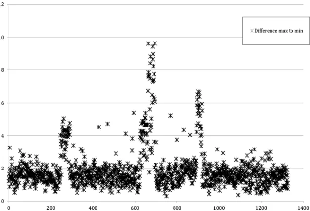
According to the study protocol, recalibration was performed between each positioning of the mannequin head. There were no significant differences among any of these virtual measure-ment data sets. If each of the 1,326 distances was compared over all 56 models (28 each observed by the two observers), the maximum difference between two corresponding land-marks (inside the data block of the total 74,256 distances) was 9.63 mm. The mean of these maximum differences in all 1,326 corresponding distances was 1.91±0.42 mm (Fig.5).
Failed or missing landmarks were detected in some data sets (Fig. 3). The latter were excluded from the data analysis.
Discussion
Interest in 3D surface imaging, such as laser scanning [27,28], structured light [29,30], and 3D photogrammetry [15,16,22], as a radiation-free morphologic assessment modality, is in-creasing. Three-dimensional photography in its various appli-cations (medicine, physics, engineering, etc.) allows digitalisation of the true dimensions and proportions of subjects within milliseconds. Therefore, this technique is becoming increasingly more important in clinical documentation, moni-toring, and characterisation of facial morphologies/anomalies, especially in paediatrics. Furthermore, 3D-photography-acquired data sets allow for individual production of skull orthoses for the correction of deformational plagiocephaly and are replacing the more conventional plaster casts.
In addition, because of their true-to-scale mapping, further anthropometric and forensic approaches are of increasing interest. Arbitrary measurements in a calibrated system allow valid data to be obtained. A variety of 3D photogrammetric systems have been evaluated with a focus on precision and accuracy, which is mandatory before implementation in clin-ical practice or research. To the authors' knowledge, no study has evaluated the precision and accuracy of the five-pod VECTRA 3D system (Canfield, USA). Unlike most other systems, VECTRA 3D enables fully textured 360° surface contour mapping of the craniofacial area.
Including all measurements, a discrepancy between the directly measured and corresponding virtually calculated dis-tances was found (p=0.02). Further, a marked mean distance aberration of 7.96 mm underlined the incongruence of both measurement modalities. Errors in direct measurement and data translation were evident. Therefore, a cut-off of 10 % of the least reliable measurement data was used to exclude obvi-ous mistakes in calliper measurements and data translation (which must be performed manually on calliper measure-ments). After application of this cut-off, no differences
Fig. 3 Exemplary selection of failed and missing landmarks. The submental and occipital regions are missing. Double picture Nos. 4 and 6 and blurred landmark No. 11
Table 1 Data acquisition protocol according to Lübbers et al. [16]
Study number Anterior–posterior positioning Vertical positioning Horizontal positioning Number of acquired data sets
1 Direct calliper measurements
2 Neutral Neutral Neutral 1
3 Neutral 10 degrees down Neutral 1
4 Neutral Neutral −5 to +5 degrees 11 (in 1-degree steps)
5 Neutral Neutral −30 to +30 degrees 13 (in 5-degree steps)
6 15 cm posterior Neutral Neutral 1
Fig. 5 Maximum inter-observer differences in virtual measurements. Three marked outliers due to incorrect labelling of individual landmarks with consequential incorrect calculation of distances are shown (x axis, no of distances; y axis, difference in millimeter)
Fig. 4 Differences in direct and virtual measurements with and without a cut-off of the least reliable 10 % of measurements (x axis, no. of
between the two groups were found (p=0.94). The mean difference in both measurement modalities was 1.33 mm, showing high concurrency. Therefore, if only the best 90 % of the direct measurements are utilised, the first null hypothesis can be accepted. The data suggest that the 3D photosystem provides an accurate representation of reality because no other hypothesis explains 90 % of the data fitting with high concur-rency. However, systematic errors can definitely be excluded. As mentioned above, direct measurements seem more prone to errors and inaccuracies compared with data ac-quired digitally. We strongly believe that this because of the major downsize of direct anthropometry. Especially regarding retrospective evaluation of clinical data, around 10 % of measurements might be inaccurate. In addition, it is important to note that this value was derived in a mannequin setting, in which measurements are rather easy to obtain. Unfortunately, in the clinical setting identifying an appro-priate cut-off is problematic, as in the present study. This is especially frustrating because 90 % of the data obtained by direct anthropometry seems to be valid.
The inter-observer reliability of the 3D models showed high agreement, with a maximum difference in mean dis-tances of 0.58 mm (mean, 0.11 mm; SD, 0.10 mm). The maximum differences in the distances reported by the two observers in all data sets showed a mean of 1.91 mm, which was rated as clinically acceptable. As a major advantage of indirect anthropometry, individual outliers can be identified (Fig.5). Due to the accuracy and precision of similar sys-tems [14–17], we suggest that imprecise labelling of three landmarks by one or both observers resulted in the discrep-ancies. In a retrospective study, statistical tools allow iden-tification of outliers within a large data set. Because only the labelling, and not the original 3D model, is affected by the error, the virtual data sets allow arbitrary re-measuring and data correction. This underlines the suitability of such a system in terms of the reliability and validity of anthropo-metric data analysis, comparison, and evidence.
Registration error is caused primarily by variability in the capturing system between two captures. In our study, there were no significant differences in measurements between calibration and various positions simulating a continuous follow-up. Previous studies showed that this error class is negligible [16, 17]. Nevertheless, although minimal differ-ences in the phantom head positioning were identified, occa-sional missing regions or failed landmarks occurred. In our opinion, failed landmarks (blurred, double-pictured) are due to misassembling of the mapped surface in the post-processing digital 3D reconstruction. Furthermore, missing regions can be caused by malpositioning of the object outside the focus of the system.
Therefore, for clinical implementation, multiple pictures should be acquired and the most appropriate used for further data recording and analysis. Because of the short time frame
necessary for a picture (we calculate∼15 s until the system is ready again), this is not a problem even in a clinical setting with young children.
The measured errors, single or in aggregation, were small and therefore not clinically relevant. Systemic errors due to capturing, post-processing, and registration can be neglected due to the above-mentioned minimal total error. Altogether, the system showed high precision and accuracy for the determination of landmarks and further measurements. Fur-ther, repeatability was excellent due to the high consistency of the system and the high inter-observer agreement. There-fore, null hypotheses 2 and 3 can be accepted.
Our protocol using an inanimate mannequin head allows determination of the precision and accuracy of 3D photo-systems, as reported previously [16,21,31]. Studies includ-ing human subjects are of limited validity because the results can be influenced by involuntary facial movements [17] or variation in hair volume. These types of studies were not discussed in this paper because they cannot be compared with our data due to differences in methodology.
Previous studies using mannequin models to determine precision and accuracy of 3D photosystems showed excellent results. Lübbers et al. used a two-pod photosystem (3dMD Inc., Atlanta, GA) and reported a precision greater than that required for clinical use [16]. A technical validation of the Di3D stereophotogrammetry surface imaging system (Dimen-sional Imaging, Glasgow), performed by Winder et al. showed a repeatability error (variance) of 0.0016 mm and a mean error of 0.6 mm in linear compared with manual measurements [31]. Weinberg et al. showed an error of <1 mm in a compar-ison of two digital systems (Genex FaceCam 250; Kensington, MD and 3dMD MU-4 Imaging System; Atlanta, GA) versus direct measurements [21]. Compared with all other systems evaluated previously, the current 3D system showed favourable precision and accuracy. Further, using this five-pod system, a 360° surface contour map that captured all craniofacial landmarks with adequate quality could be gener-ated. Calibration and operation of the system in clinical cir-cumstances as well the post-processing software are easy to handle and can be sufficiently incorporated into the clinical routine. The automation of data exportation could be in-creased because at present all data must be copied manually into a datasheet. A direct export function to, for example, Microsoft® Excel® would be favourable.
Our data suggest that future studies using this five-pod 3D photo assessment system are warranted. Nevertheless, some basic requirements should be noted: (1) the calibration procedure recommended by the manufacturer must be performed before each photo session; (2) the object must be positioned precisely within the focus of the system; and (3) for valid data acquisition, each system must be calibrated using a standardised labelled mannequin head to minimise errors between various systems or in post-processing.
Although this system allows reliable measurement and generation of objective data from a static model head, additional variables must be considered before clinical implementation.
The following parameters must be considered to quantify measurement errors in human subjects and interpret the data correctly:
(i) Subjects' involuntary facial movements can influence anthropometric measurements [17].
(ii) Young children may not be sufficiently compliant for accurate 3D photogrammetric data acquisition, espe-cially due to involuntary movements.
(iii) Variation in hair volume results in considerable bias in cranial vault measurements.
Conclusions
The high precision and accuracy of this five-pod 3D photo-system suggest its suitability for clinical use and application, especially for anthropometric studies. Three-hundred-and-sixty degree surface-contour mapping of the craniofacial region within milliseconds is particularly advantageous in paediatric patients. Proper patient positioning is essential for high-quality imaging. Images should be checked carefully in terms of completeness of the chin region, blurred landmarks, etc., to take advantage of the possibility of later data acqui-sition. Further studies should focus on minimising error due to involuntary facial movements and hair volume.
Acknowledgments The authors would like to thank Philippe
Halioua for taking of photographs.
Conflicts of interest The authors declare that they have no conflicts
of interest.
References
1. Meintjes EM, Douglas TS, Martinez F, Vaughan CL, Adams LP, Stekhoven A et al (2002) A stereo-photogrammetric method to measure the facial dysmorphology of children in the diagnosis of
fetal alcohol syndrome. Med Eng Phys 24(10):683–689, Epub
2002/12/04
2. Farkas LG, James JS (1977) Anthropometry of the face in lateral
facial dysplasia: the unilateral form. Cleft Palate J 14(3):193–199,
Epub 1977/07/01
3. Farkas LG, Kolar JC, Munro IR (1985) Craniofacial dispropor-tions in Apert's syndrome: an anthropometric study. Cleft Palate J
22(4):253–265, Epub 1985/10/01
4. Kolar JC, Farkas LG, Munro IR (1985) Surface morphology in Treacher Collins syndrome: an anthropometric study. Cleft Palate J
22(4):266–274, Epub 1985/10/01
5. Farkas LG (ed) (1994) Anthropometry of the head and face. Raven Press, New York
6. Kolar JC (1993) Methods in anthropometric studies. Cleft Palate
Craniofac J 30(4):429–431, Epub 1993/07/01
7. Kolar JC, Salter EM, Weinberg SM (2010) Preoperative craniofa-cial dysmorphology in isolated sagittal synostosis: a
comprehen-sive anthropometric evaluation. J Craniofac Surg 21(5):1404–
1410, Epub 2010/09/22
8. Kolar JC (2011) An epidemiological study of nonsyndromal
craniosynostoses. J Craniofac Surg 22(1):47–49, Epub 2010/12/29
9. Metzler P, Zemann W, Jacobsen C, Gratz KW, Obwegeser JA (2013) Postoperative cranial vault growth in premature sagittal
craniosynostosis. J Craniofac Surg 24(1):146–149, Epub 2013/
01/26
10. Metzler P, Zemann W, Jacobsen C, Gratz KW, Obwegeser JA (2013) Cranial vault growth patterns of plagiocephaly and trigonocephaly patients following fronto-orbital advancement: a long-term anthropometric outcome assessment. J Craniomaxillofac
Surg. doi:10.1016/j.jcms.2012.11.035
11. Fearon JA, Ruotolo RA, Kolar JC (2009) Single sutural craniosynostoses: surgical outcomes and long-term growth. Plast Reconstr Surg 123(2):635–642, Epub 2009/02/03
12. Farkas LG, Hreczko TA, Kolar JC, Munro IR (1985) Vertical and horizontal proportions of the face in young adult North American Caucasians: revision of neoclassical canons. Plast Reconstr Surg 75(3):328–338, Epub 1985/03/01
13. Farkas LG, Hreczko TM, Katic MJ, Forrest CR (2003) Proportion indices in the craniofacial regions of 284 healthy North American white children between 1 and 5 years of age. J Craniofac Surg 14(1):13–28, Epub 2003/01/25
14. Metzler P, Bruegger LS, Kruse Gujer AL, Matthews F, Zemann W, Graetz KW et al (2012) Craniofacial landmarks in young children: how reliable are measurements based on 3-dimensional imaging? J Craniofac Surg 23(6):1790–1795, Epub 2012/11/14
15. Ort R, Metzler P, Kruse AL, Matthews F, Zemann W, Gratz KW et al (2012) The reliability of a three-dimensional photo system-(3dMDface-) based evaluation of the face in cleft Lip infants. Plast Surg Int 2012:138090, Epub 2012/08/25
16. Lubbers HT, Medinger L, Kruse A, Gratz KW, Matthews F (2010) Precision and accuracy of the 3dMD photogrammetric system in craniomaxillofacial application. J Craniofac Surg 21(3):763–767, Epub 2010/05/21
17. Lubbers HT, Medinger L, Kruse AL, Gratz KW, Obwegeser JA, Matthews F (2012) The influence of involuntary facial movements on craniofacial anthropometry: a survey using a three-dimensional
photographic system. Br J Oral Maxillofac Surg 50(2):171–175,
Epub 2011/01/18
18. Christofides EA, Steinmann ME (2010) A novel anthropometric
chart for craniofacial surgery. J Craniofac Surg 21(2):352–357,
Epub 2010/02/27
19. Wilbrand JF, Wilbrand M, Pons-Kuehnemann J, Blecher JC, Christophis P, Howaldt HP et al (2011) Value and reliability of anthropometric measurements of cranial deformity in early
child-hood. J Craniomaxillofac Surg 39(1):24–29, Epub 2010/04/27
20. Farkas LG, Forrest CR (2006) Changes in anthropometric values of paired craniofacial measurements of patients with right coronal
synostosis. Ann Plast Surg 56(4):427–430, Epub 2006/03/25
21. Weinberg SM, Naidoo S, Govier DP, Martin RA, Kane AA, Marazita ML (2006) Anthropometric precision and accuracy of digital three-dimensional photogrammetry: comparing the Genex and 3dMD imaging systems with one another and with direct
anthropometry. J Craniofac Surg 17(3):477–483, Epub 2006/06/14
22. Wong JY, Oh AK, Ohta E, Hunt AT, Rogers GF, Mulliken JB et al (2008) Validity and reliability of craniofacial anthropometric mea-surement of 3D digital photogrammetric images. Cleft Palate
Craniofac J 45(3):232–239, Epub 2008/05/03
23. Kau CH, Zhurov A, Bibb R, Hunter L, Richmond S (2005) The investigation of the changing facial appearance of identical twins
employing a three-dimensional laser imaging system. Orthod
Craniofac Res 8(2):85–90, Epub 2005/05/13
24. Luebbers HT, Messmer P, Obwegeser JA, Zwahlen RA, Kikinis R, Graetz KW et al (2008) Comparison of different registration methods for surgical navigation in cranio-maxillofacial surgery. J
Craniomaxillofac Surg 36(2):109–116, Epub 2008/02/19
25. Marmulla R, Muhling J, Eggers G, Hassfeld S (2005) Markerless patient registration. A new technique for image-guided surgery of
the lateral base of the skull HNO 53(2):148–154, Markerlose
Registrierung der Patientenlage. Ein neues Verfahren zur bildgestutzten Chirurgie der lateralen Schadelbasis
26. Fitzpatrick JM, West JB (2001) The distribution of target registra-tion error in rigid-body point-based registraregistra-tion. IEEE Trans Med
Imaging 20(9):917–927, Epub 2001/10/05
27. Kovacs L, Zimmermann A, Brockmann G, Baurecht H, Schwenzer-Zimmerer K, Papadopulos NA et al (2006) Accuracy and precision of
the three-dimensional assessment of the facial surface using a 3-D
laser scanner. IEEE Trans Med Imaging 25(6):742–754
28. Bardouille T, Krishnamurthy SV, Hajra SG, D'Arcy RC (2012) Improved localization accuracy in magnetic source imaging using
a 3-D laser scanner. IEEE Trans Biomed Eng 59(12):3491–3497,
Epub 2012/10/04
29. Ma L, Xu T, Lin J (2009) Validation of a three-dimensional facial scanning system based on structured light techniques. Comput
Methods Programs Biomed 94(3):290–298, Epub 2009/03/24
30. Zhang X, Li Y, Zhu L (2012) Color code identification in
coded structured light. Appl Opt 51(22):5340–5356, Epub
2012/08/04
31. Winder RJ, Darvann TA, McKnight W, Magee JD, Ramsay-Baggs P (2008) Technical validation of the Di3D stereophotogrammetry
surface imaging system. Br J Oral Maxillofac Surg 46(1):33–37,
![Table 1 Data acquisition protocol according to Lübbers et al. [16]](https://thumb-eu.123doks.com/thumbv2/123doknet/14812149.611765/4.892.76.826.106.279/table-data-acquisition-protocol-according-to-lübbers-et.webp)