Lukas Hechelhammer Christian W. A. Pfirrmann Marco Zanetti Juerg Hodler Norbert Boos Marius R. Schmid Received: 30 January 2006 Revised: 8 June 2006 Accepted: 23 June 2006
Published online: 3 October 2006 # Springer-Verlag 2006
Imaging findings predicting the outcome
of cervical facet joint blocks
Abstract To determine which cross-sectional imaging findings predict the short-term outcome of cervical facet joint blocks (FJB) and to evaluate the effect of combined intra-/periarticular versus periarticular injection on pain. Fifty facet joints in 37 patients were included in the study. Single, unilat-eral FJBs in 24 patients, and bilatunilat-eral single level FJBs in 13 patients were performed, respectively. In all pa-tients, pain relief was assessed using a visual analogue scale. All computed tomography (CT) examinations were blindly reviewed by two radiologists. Osteoarthritis was rated using the Kellgren classification. The presence of combined intra-/periarticular vs. sole periarticular injection of contrast was evaluated. Kellgren Grades 0 (n=23), 1 (n=5), 2 (n=3), 3 (n=9), and
4 (n=10) were found. Mean pain relief after injection was 35% (range: 0– 100%). 40% of all injections were combined intra-/periarticular. There was neither a statistically significant difference between pain relief and combined intra-/periarticular versus sole periarticular injection (p=0.64) nor the grade of osteoarthritis (p=0.49). Pain relief after cervical FJBs does not correlate with morpho-logic alterations seen on CT. Periar-ticular FJBs are not less successful than combined intra-/periarticular FJBs.
Keywords Spine . Interventions . CT fluoroscopy
Introduction
Imaging guided percutaneous cervical facet joint proce-dures have been shown to be useful in facet joint degeneration (cervical facet syndrome) [1, 2] and in chronic post-traumatic neck pain [3]. Either injection of corticosteroids and/or local anaesthetics into or around the facet joints[1], or percutaneous radio-frequency neurotomy of the facet joint (two adjacent medial branches of the dorsal rami) [2,3] have been described.
There is still disagreement as to whether intra- or periarticular facet joint injection are preferred in facet syndrome [4, 5]. Roy et al. [1] found no significant difference in pain relief comparing intra- and periarticular lumbar facet joint infiltration. To our knowledge, no such investigation has yet been performed in the cervical spine. Another controversially discussed subject are
morpholog-ical criteria to predict the success rate in facet syndrome [6– 8]. Some studies investigated criteria to predict treatment response of lumbar but not cervical facet joint injections [6–9].
The purpose of this investigation was to determine which cross-sectional imaging findings predict the short term outcome of cervical facet joint blocks (FJB) and to evaluate the effect of combined intra-/periarticular versus sole periarticular injection on pain.
Materials and methods
Patients
Between March 1998 and March 2005, 726 cervical facet joint blocks in 378 patients were performed under L. Hechelhammer .
C. W. A. Pfirrmann . M. Zanetti . J. Hodler . M. R. Schmid (*) Radiology, University of Zurich, Balgrist Hospital, CH-8008 Zurich, Switzerland e-mail: [email protected] Tel.: +41-44-3863306 Fax: +41-44-3863319 L. Hechelhammer
Institute of Diagnostic Radiology, University Hospital Zurich, Zurich, Switzerland N. Boos
Center for Spinal Surgery,
University of Zurich, Balgrist Hospital, CH-8008 Zurich, Switzerland
computed tomography (CT) guidance in our institution. Most of these FJBs were multi-segmental and bilateral. The inclusion criteria for this investigation were chosen in order to allow comparison of CT facet joint morphology and the effect of FJBs. Only those interventions which fulfilled the following criteria were included in this retrospective study: (1) consecutive patients who underwent CT-guided cervi-cal facet joint injections, (2) patients referred by experi-enced physicians for single level injections (unilateral or bilateral), (3) patients had to be able to rate pain relief separately for the left and right side, respectively. The criteria were fulfilled by 24 patients with single FJBs (and an additional 13 patients with bilateral single-level FJBs (26 facet joints). Therefore, a total of 50 facet joints in 37 patients were included in our study. Twenty-five patients were woman (mean age 55 years; range 19–92 years) and 12 men (mean age 57 years; range 34–97 years). All patients were referred to the FJB either by a rheumatolo-gist, chiropractor or spine surgeon. Degenerative facet joint syndrome was the assumed diagnosis in all patients.
Our institutional review board does not require its approval or informed consent for the review of patient records or images. Patient rights are protected by a law that requires patients to be informed at the time of examination about the possibility that their medical records and radiographs will be reviewed for scientific purposes.
Computed Tomography
In all patients, CT images were obtained immediately before injection. These CT images were performed in the prone position and served primarily for planning of the procedure. These CT examinations were secondarily used in our study for morphological evaluation of the facet joints. All CT images were obtained on a continuous-rotation fan-beam geometry CT scanner (Somatom Plus 4,
CARE Vision; Siemens Medical Systems, Erlangen, Germany). After determination of the injection height on a lateral scout view, an acquisition using a slice thickness of 1 mm, table feed of 1.3 mm and reconstruction interval of 2 mm, and bone algorithm as well as bone windowing (center 450 HU and window 1800 HU) was performed. The FJB was performed immediately after this acquisition.
Injection technique
All cervical FJBs were performed under CT fluoroscopic guidance. CT fluoroscopic images were acquired with a rotation speed of 0.75 seconds per rotation, a tube current of 50 mA, a voltage of 120 kV, and a collimation of 5 mm. Images were calculated at a rate of six images per second with a 256×256 matrix and displayed in full resolution (1,024×1,024 matrix) on an in-room monitor. The patients were placed in the prone position. After skin anaesthesia with approximately 1 ml mepivacain hydrochloride 2% (Scandicain®, AstraZeneca, London, UK) either a poste-rior, postero-lateral or lateral approach towards the dorsal or dorso-lateral border of the cervical facet joint was chosen. Either a 6 cm/23 G needle (Sterican®, Braun AG, Melsungen, Germany) or a 9 cm/21 G spinal needle (Becton Dickinson SA, Madrid, Spain) was used. While advancing the needle, 1 to 2 ml of mepivacain hydrochlo-ride 2% (Scandiacin®, AstraZeneca, London, UK) were injected. As soon as the needle tip reached the facet joint, 0.5 ml of iodinated contrast agent (Iopamidol 200 mg iodine/ml, Iopamiro 200®, Bracco, Milan, Italy) was injected to document either a combination of intra-/ periarticular or a sole periarticular distribution. 1.5 ml of mepivacain hydrochloride 2% (Scandiacin®, AstraZeneca, London, UK) mixed with 40 mg of a crystalline triamcinolon (Kenacort®; Dermapharm, Munich, Germany) were then injected.
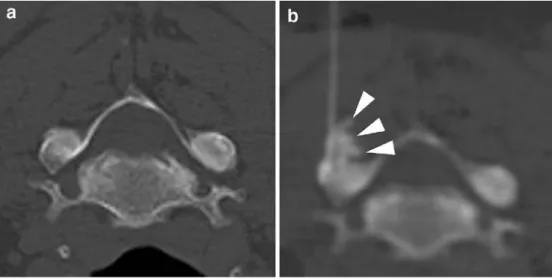
Fig. 1 48 year-old female with neck pain since 4 months and 100% pain relief after sole pe-riarticular FJB. Axial CT image (a) of the left cervical facet joint C2/C3 without signs of osteo-arthritis. CT fluoroscopy image after periarticular facet injection (b) shows contrast distribution immediately posterior of the facet joint (white arrowheads)
Evaluation of pain relief
All patients prospectively assessed pain relief on a visual analogue scale of 10 cm length without and sub-divisions ranging from no change of the initial pain (0%) to complete pain relief (100%). The evaluation of pain relief was performed 15 to 20 minutes after the end of the FJB by a technician educated with regard to the meaning of visual analogue scales. More than 50% of pain reduction was regarded as a successful cervical FJB.
Image evaluation
Two experienced musculoskeletal radiologists reviewed the CT images in consensus on a PACS Workstation. Only the injected facet joints were graded for osteoarthritis according to Kellgren et al. [10]. The following grades of osteoarthritis (OA) are differentiated in this score: (Grade 1) definite absence of osteoarthritic changes on CT; (Grade 2) probably absence of osteoarthritic changes on CT; (Grade 3) definite minimal osteoarthritic CT; (Grade 4) definite moderate osteoarthritic CT; (Grade 5) definite
severe osteoarthritic CT. All factors contributing to this grading score (e. g., osteophytes, periarticular ossicles, subchondral bone sclerosis, subchondral pseudocystic changes and altered bone shape) were recorded separately. The contrast medium injection was classified as either combined intra-/periarticular (contrast medium visible within the joint space) or sole periarticular (no contrast medium visible within the joint space) application (Figs.1 and2). Since the capacity of a facet joint is far below 2 ml, a sole intraarticular FJB can almost be ruled out and all injections showing contrast material within the facet joint were included in the combined intra-/periarticular FJBs.
Statistical analysis
Analyses were performed with SPSS 12.2 for Windows (SPSS Inc., Chicago, Illinois). Mann–Whitney U test was used for non-parametric test to assess significant differ-ences of the morphologic criteria, gender, and contrast distribution regarding pain reduction. Multivariate logistic regression and receiver-operating curves for analysis of different morphologic factors were calculated.
Fig. 2 60 year-old male patient having neck pain since 3 months and 100% pain relief after combined intra-/periarticular FJB. Axial CT image (a) of left cervical facet joint C5/C6 showing inferior articular pro-cess of C5 (black curved arrow) and superior articular process of C6 (white curved arrow) with normal articular cartilage (black arrowhead) and without any signs of Osteoarthritis. CT fluo-roscopy image after intra-/peri-articular facet injection (b) shows contrast distribution within the joint capsule filling the anterior (white arrowhead) and posterior (white arrow) re-cess of the joint space
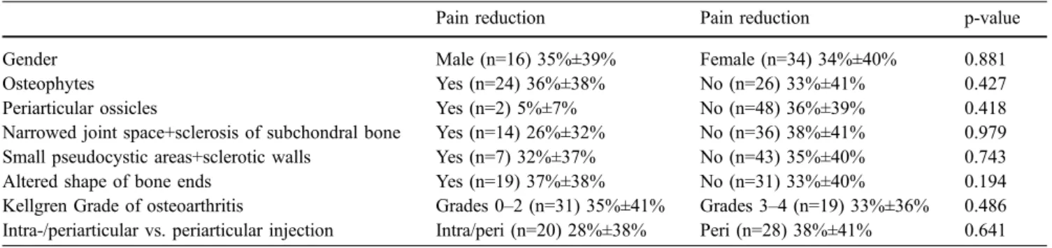
Table 1 Statistical analyisis of gender, morphological criteria, joint injection, and pain relief
Pain reduction Pain reduction p-value
Gender Male (n=16) 35%±39% Female (n=34) 34%±40% 0.881
Osteophytes Yes (n=24) 36%±38% No (n=26) 33%±41% 0.427
Periarticular ossicles Yes (n=2) 5%±7% No (n=48) 36%±39% 0.418
Narrowed joint space+sclerosis of subchondral bone Yes (n=14) 26%±32% No (n=36) 38%±41% 0.979 Small pseudocystic areas+sclerotic walls Yes (n=7) 32%±37% No (n=43) 35%±40% 0.743
Altered shape of bone ends Yes (n=19) 37%±38% No (n=31) 33%±40% 0.194
Kellgren Grade of osteoarthritis Grades 0–2 (n=31) 35%±41% Grades 3–4 (n=19) 33%±36% 0.486 Intra-/periarticular vs. periarticular injection Intra/peri (n=20) 28%±38% Peri (n=28) 38%±41% 0.641 N=number of cervical facet joints. Mean pain relief (%)±standard deviation (%)
Results
Results are presented in Table1. With regard to gender and age, no statistically significant differences in immediate pain reduction were found.
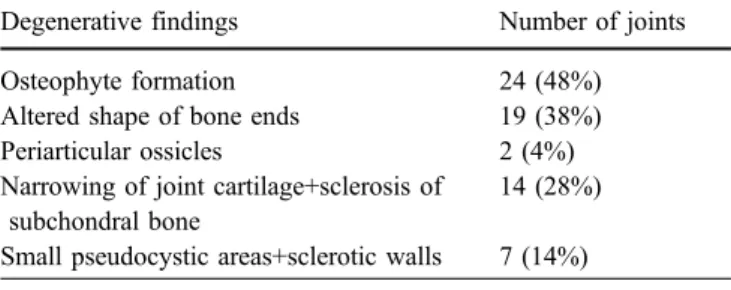
Evaluating CT images, following grades of osteoarthritis were diagnosed (n=50): Grade 0 (n=23), Grade 1 (n=5), Grade 2 (n=3), Grade 3 (n=9), and Grade 4 (n=10). Injected facet joints were located at the following levels C2/3 (n=13), C3/4 (n=16), C4/5 (n=8), C5/6 (n=12), and C6/7 (n=1), respectively. Twenty-four left- and twenty-six right-sided injections were performed, respectively. Numbers of morphological criteria contributing to the grading of facet joint osteoarthritis are summarized in Table2.
Mean pain reduction for all facet joint injections (n=50) was 35 (SD±40%; range 0–100%). No statistically signif-icant differences in pain reduction were found for any of the analyzed morphological CT signs (Table1).
Combined Intra-/periarticular vs. sole periarticular FJBs and pain reduction
Consensus analysis of the CT fluoroscopy images after contrast injection revealed combined intra-/periarticular cervical FJBs in 20 and sole periarticular FJBs in 28 joints. In two joints assessment for intra- or periarticular application was not possible because no contrast agent was applied due to prior allergic reaction to iodinated contrast agents. No statistically significant difference in pain reduction (p=0.64) was found for combined intra-/ periarticular and sole periarticular injections with mean pain decrease of 28% (SD±38%; range.0–100%) and 38% (SD±41%, range:0–100%), respectively (Table1). Figures3 and 4 demonstrate combined intra-/periarticular FJBs in two different patients with severe osteoarthritis without (Fig.3) and with (Fig.4) pain reduction after injection.
Logistic regression of osteoarthritis grade, side, gender, injection level, age of the patients and combined intra-/ periarticular or sole periarticular injections showed no statistically significant differences.
Receiver operating characteristic curves of the different morphological signs of osteoarthritis and their relation to the pain relief is displayed in Fig. 5. The curves of the analyzed different morphologic criteria are grouped tightly around the reference line (chance diagonal).
Discussion
Cervical facet syndrome may be caused by degeneration, inflammatory processes or trauma. Different therapeutic approaches exist [1–3]. Due to the lack of morphological predictors, cervical facet joints are selected for injection based on clinical examination [1] or on diagnostic medial Table 2 Morphologic criteria contributing to osteoarthritis grading
Degenerative findings Number of joints
Osteophyte formation 24 (48%)
Altered shape of bone ends 19 (38%)
Periarticular ossicles 2 (4%)
Narrowing of joint cartilage+sclerosis of subchondral bone
14 (28%) Small pseudocystic areas+sclerotic walls 7 (14%) Note: total number of analyzed joints (n=50)
Fig. 3 A 48-year-old female patient having neck pain for six months without pain relief after combined intra-/periarticular FJB. Axial CT image (a) of right cervical facet C3/C4 with severe osteoarthritis (Kellgren Grade 4) shows osteophytes (white
arrow-heads) and subchondral cysts (black arrowhead). Lateral approach to the facet joint. CT fluoroscopy before (b) and after (c) combined intra-/periarticular FJB shows some contrast media within the joint space in the posterior joint recess (curved white arrow)
branch block infiltration [11]. Reliable morphological predictors would make diagnostic medial branch block unnecessary or limit this procedure to non-distinctive cases.
In the current study no statistically significant relation-ship between the degree of osteoarthritis seen on CT and pain relief after cervical facet joint injection was found. Similar investigations regarding facet joint morphology and facet joint blocks have been performed in the lumbar spine. In 1980, in a study investigating CT morphology and pain relief after facet join injection in the lumbar spine a
relationship between facet joint morphology and pain relief after facet joint block was suggested [9]. In this study, four patients with joint narrowing, subchondral irregularities or erosions diagnosed on CT had complete pain relief after a facet joint block. The six patients with normal or only mildly hypertrophic facet joints had no pain relief after injection. A more recent study demonstrated a low positive predictive value for CT as a diagnostic test for lumbar facet syndrome [6]. Scoring of the lumbar facet joints for joint space narrowing, sclerosis, subchondral erosions, cysts and osteophytes in 63 patients showed no significant correla-Fig. 4 FJB in a 97 year-old
male patient having neck pain for more than two years results in an 80% pain relief after combined intra-/periarticular facet injection. Axial CT image (a) of left cervical facet C4/C5 shows severe osteoarthritis (Kellgren Grade 4) with osteo-phytes (white arrowheads) and subchondral cysts (black ar-rowhead). CT fluoroscopy image (b) after combined intra-/ periarticular FJB shows contrast accumulation in the anterior (white arrow) and posterior joint recess (curved white arrow)
Fig. 5 ROC-Curve of the dif-ferent morphological criteria contributing to the grading of facet joint osteoarthritis and re-lation to pain relief. Asymptotic 95% Confidence Interval: 0.303–0.761
tion with pain relief after intraarticular facet joint block. To our knowledge, no similar studies investigating cervical facet syndrome have been performed.
CT and MR imaging have been shown to be identical in the identification of facet joint degeneration in the lumbar spine [12]. Therefore, MR imaging probably does not add more information regarding the prediction of success of facet joint blocks. On the other hand, MR imaging, especially when short inversion time inversion-recovery sequences are performed, is able to identify inflammation related bone marrow and soft tissue edema. This could be helpful to identify inflammatory changes of facet joints. But since only few patients in our study group were additionally examined by MR imaging, the prediction of facet joint block success by MR imaging was not evaluated in our investigation.
By injection of an irritating substance (11% saline) into the lumbar facet joints, low back pain as well as referring pain in the gluteal and sacroiliac region could be provoked [13]. Aprill and Bogduk showed similar effects for arthrography of cervical facets [14]. Intraarticular lidocaine and hydrocortisone injections lower the nerve activity in irritated facet joints of rabbits [15]. On the other hand periarticular injection of 0.4 ml of 6% saline solution was able to create the same radiating pain as an intraarticular injection in normal volunteers did [16]. Large volumes injected intraarticularly may cause capsule rupture and fluid leakage into the epidural space, which can in part account for pain response [17]. In addition, after removal of the needle, injected substances may flow back through the
injection canal into the periarticular tissue. Therefore, purely intraarticular injections are probably not achievable in the cervical spine. The intraarticular injections included into our investigation may rather be combined intra-/ periarticular infiltrations. In our study group, no significant difference between cervical facet injections with or without intraarticular portion of infiltrated substances on pain response was found.
There are study limitations to consider: Our analysis is based on the pain relief reflecting mostly the effect of the injected mepivacaine. Analysis of pain relief after a few months and correlation with morphology may have resulted in different findings. Our study goal was not the evaluation of long term outcome of cervical facet joint blocks but of the finding of signs that indicate that facet joints are symptomatic or not and may, therefore, react to imaging guided injections. The fact that multilevel injections were excluded may be criticized. However, we were looking for a model which could be used to explain the relationship between morphology and pain relief. This would not be possible in multilevel injections because facet joint degeneration tends to be highly variable between levels.
In conclusion, our study suggests that pain relief after cervical facet joint injection does not correlate with morphologic alterations seen on CT. The pain relief of cervical facet joint blocks does not depend on the distribution of the infiltrated substances (intra-/periarticular or periarticular).
References
1. Roy DF, Fleury J, Fontaine SB, Dussault RG (1988) Clinical evaluation of cervical facet joint infiltration. Can Assoc Radiol J 39:118–120
2. Mikeladze G, Espinal R, Finnegan R, Routon J, Martin D (2003) Pulsed radiofrequency application in treatment of chronic zygapophyseal joint pain. Spine J 3:360–362
3. Lord SM, Barnsley L, Wallis BJ, McDonald GJ, Bogduk N (1996) Per-cutaneous radio-frequency neurotomy for chronic cervical zygapophyseal-joint pain. N Engl J Med 335: 1721–1726
4. Lynch MC, Taylor JF (1986) Facet joint injection for low back pain. A clinical study. J Bone Joint Surg Br 68: 138–141
5. Lilius G, Laasonen EM, Myllynen P, Harilainen A, Gronlund G (1989) Lumbar facet joint syndrome. A ran-domised clinical trial. J Bone Joint Surg Br 71:681–684
6. Schwarzer AC, Wang SC, O’Driscoll D, Harrington T, Bogduk N, Laurent R (1995) The ability of computed tomo-graphy to identify a painful zygapoph-ysial joint in patients with chronic low back pain. Spine 20:907–912
7. Lewinnek GE, Warfield CA (1986) Facet joint degeneration as a cause of low back pain. Clin Orthop Relat Res 216–222
8. Helbig T, Lee CK (1988) The lumbar facet syndrome. Spine 13:61–64 9. Carrera GF (1980) Lumbar facet joint
injection in low back pain and sciatica: preliminary results. Radiology 137:665–667
10. Kellgren JH, Lawrence JS (1957) Ra-diological assessment of osteo-arthro-sis. Ann Rheum Dis 16:494–502 11. Barnsley L, Lord S, Bogduk N (1993)
Comparative local anaesthetic blocks in the diagnosis of cervical zygapophysial joint pain. Pain 55:99–106
12. Weishaupt D, Zanetti M, Boos N, Hodler J (1999) MR imaging and CT in osteoarthritis of the lumbar facet joints. Skeletal Radiol 28(4):215–219, Apr
13. Hirsch C, Ingelmark BE, Miller M (1963) The anatomical basis for low back pain. Studies on the presence of sensory nerve endings in ligamentous, capsular and intervertebral disc struc-tures in the human lumbar spine. Acta Orthop Scand 33:1–17
14. Aprill C, Bogduk N (1992) The prev-alence of cervical zygapophyseal joint pain. A first approximation. Spine 17:744–747
15. Cavanaugh JM, Ozaktay AC,
Yamashita HT, King AI (1996) Lumbar facet pain: biomechanics, neuroanato-my and neurophysiology. J Biomech 29:1117–1129
16. McCall IW, Park WM, O’Brien JP (1979) Induced pain referral from pos-terior lumbar elements in normal sub-jects. Spine 4:441–446
17. Moran R, O’Connell D, Walsh MG (1988) The diagnostic value of facet joint injections. Spine 13:1407–1410