HAL Id: inserm-00472414
https://www.hal.inserm.fr/inserm-00472414
Submitted on 19 Mar 2012
HAL is a multi-disciplinary open access
archive for the deposit and dissemination of
sci-entific research documents, whether they are
pub-lished or not. The documents may come from
teaching and research institutions in France or
abroad, or from public or private research centers.
L’archive ouverte pluridisciplinaire HAL, est
destinée au dépôt et à la diffusion de documents
scientifiques de niveau recherche, publiés ou non,
émanant des établissements d’enseignement et de
recherche français ou étrangers, des laboratoires
publics ou privés.
The involvement of Fyn kinase in resumption of the first
meiotic division in mouse oocytes.
Mattan Levi, Bernard Maro, Ruth Shalgi
To cite this version:
Mattan Levi, Bernard Maro, Ruth Shalgi. The involvement of Fyn kinase in resumption of the first
meiotic division in mouse oocytes.. Cell Cycle, Taylor & Francis, 2010, 9 (8), pp.1577-89.
�inserm-00472414�
Cell Cycle 9:8, 1577-1589; April 15, 2010; © 2010 Landes Bioscience
RepoRt RepoRt
*Correspondence to: Ruth Shalgi; Email: shalgir@post.tau.ac.il Submitted: 10/22/09; Revised: 01/25/10; Accepted: 01/25/10
Previously published online: www.landesbioscience.com/journals/cc/article/11299
Introduction
Meiosis of mammalian oocytes starts during embryonic life and arrests around birth, at prophase of the first meiotic divi-sion, characterized by the presence of a germinal vesicle (GV). Meiotic arrest lasts at least until puberty, when selected oocytes resume their first meiotic division (RMI) and are ovulated after being exposed to the midcycle surge of luteinizing hormone (LH).1 RMI is manifested by chromosomes condensation, GV
breakdown (GVBD), formation of the spindle and its migration towards the oocyte cortex, segregation of homologous chromo-somes, extrusion of the first polar body (PBI) and an arrest at metaphase of the second meiotic division (MII).2
Progression through meiotic and mitotic divisions is con-trolled by maturation promoting factor (MPF), a heterodimer that consists of the cyclin-dependent kinase I (p34cdc2) and a
The involvement of Fyn kinase in resumption
of the irst meiotic division in mouse oocytes
Mattan Levi, Bernard Maro† and Ruth Shalgi*
Department of Cell and Developmental Biology; Sackler Faculty of Medicine; tel Aviv University; Ramat-Aviv, tel-Aviv Israel
†Current address: CNRS; paris, France
Key words: meiosis, oocyte, Fyn, GVBD, exit from metaphase, polar body
Abbreviations: AI, anaphase of the first meiotic division; APC, anaphase promoting complex; CA, constitutively active;
CLSM, confocal laser-scanning microscope; cRNA, complementary RNA; DIC, differential interference contrast; DN, dominant negative; ECL, enhanced chemiluminescence; GV, germinal vesicle; GVBD, germinal vesicle breakdown; hCG, human chorionic
gonadotrophin; LH, luteinizing hormone; MI, metaphase of the first meiotic division; MII, metaphase of the second meiotic division; MPF, maturation promoting factor; MTs, microtubules; PBI, polar body I; PMSG, pregnant mares’ serum gonadotrophin;
RMI, resumption of the first meiotic division; SDS-PAGE, sodium dodecyl sulfate polyacrylamide gel electrophoresis; SFKs, Src family kinases; TI, telophase of the first meiotic division; WB, western blot; WT, wild type; χ2, chi-square
regulatory subunit (cyclin-B).2-4 MPF activity is low in oocytes
at the GV stage. The intra-oocyte drop in cAMP concentration during GVBD5,6 leads to an increase in MPF activity and thus to
the formation of the spindle. MPF activity stays elevated during the first meiotic division (MI). Degradation of cyclin-B accom-panies the metaphase-anaphase transition and the extrusion of PBI,7 followed by formation of the MII spindle and a second
mei-otic arrest.8,9 Release from MII arrest is triggered by an increase in
intracellular Ca2+ concentration induced by the fertilizing
sperma-tozoon. Inactivation of MPF during exit from both MI and MII stages is controlled by the anaphase promoting complex (APC), a multi-subunit E3 ligase that ubiquitinates cyclin-B, thereby tar-geting it for degradation by the 26S proteasome.3,8,10,11
Src family kinases (SFKs) are nine non-receptor protein tyrosine kinases sharing a homologous structure.12 Members of
the SFKs are able to compensate for one another.13,14 Src, Yes, Fyn
the process of resumption of the irst meiotic division (RMI) in mammalian oocytes includes germinal vesicle breakdown (GVBD), spindle formation during irst metaphase (MI), segregation of homologous chromosomes, extrusion of the irst polar body (pBI) and an arrest at metaphase of the second meiotic division (MII). previous studies suggest a role for Fyn, a non-receptor Src family tyrosine kinase, in the exit from MII arrest. In the current study we characterized the involvement of Fyn in RMI. Western blot analysis demonstrated a signiicant, proteasome independent, degradation of Fyn during GVBD. Immunostaining of ixed oocytes and confocal imaging of live oocytes microinjected with Fyn complementary RNA (cRNA) demonstrated Fyn localization to the oocyte cortex and to the spindle poles. Fyn was recruited during telophase to the cortical area surrounding the midzone of the spindle and was then translocated to the contractile ring during extrusion of pBI. GVBD, exit from MI and pBI extrusion were inhibited in oocytes exposed to the chemical inhibitor SU6656 or microinjected with dominant negative Fyn cRNA. None of the microinjected oocytes showed misaligned or lagging chromosomes during chromosomes segregation and the spindle migration and anchoring were not afected. However, the extruded pBI was of large size. Altogether, a role for Fyn in regulating several key pathways during the irst meiotic division in mammalian oocytes is suggested, particularly at the GV and metaphase checkpoints and in signaling the ingression of the cleavage furrow.
three hours after milrinone was washed out (Fig. 1A). Milrinone (Primacor) is a phosphodiesterase III inhibitor that causes high levels of cAMP in the oocytes cytoplasm48 and thus inhibits the
intra-oocyte drop in cAMP concentration leading to GVBD.5,6
No other significant changes were observed during the next 8 hours of RMI in vitro (Fig. 1B), when the majority of the oocytes reached the MII stage. Fyn degradation was correlated with the process of GVBD (Fig. 1C). The phenomena of Fyn degradation during in vitro maturation was also observed in in vivo maturation (Fig. 1D). In order to examine whether the decrease in the amount of Fyn during RMI involves the pro-teasome pathway we used two propro-teasome inhibitors, MG132 (50 µM) and Lactacystin (50 µM).49,50 As indicated in the
litera-ture, the proteasome is essential in mouse oocytes for the exit from MI and for PBI extrusion, but not for the process of GVBD.50
Hence, we assessed by light microscopy in our system, the influ-ence of proteasome inhibition on the two processes of PBI and GVBD. Fourteen hours after removal of oocytes from milrinone, none of the proteasome-inhibitors treated oocytes extruded PBI (0/71 of the MG132-treated oocytes; 0/60 of the Lactacystin-treated oocytes), while 91.4% (64/70) of the control oocytes extruded PBI. Exposing milrinone-treated oocytes to the protea-some inhibitors at the indicated concentrations did not affect the rate of GVBD (96.7, 95.3 and 96.7% for control, MG132-treated and Lactacystin-treated oocytes, respectively; 150 oocytes in each treatment). Though our results confirm previous published studies regarding the role of the proteasome in meiosis, our cur-rent data indicated that inhibition of the proteasome did not affect the degradation of Fyn and the amount of Fyn in oocytes matured in vitro for three hours was similar in treatment and control groups (Fig. 1E).
Localization of Fyn during RMI. Localization of Fyn within
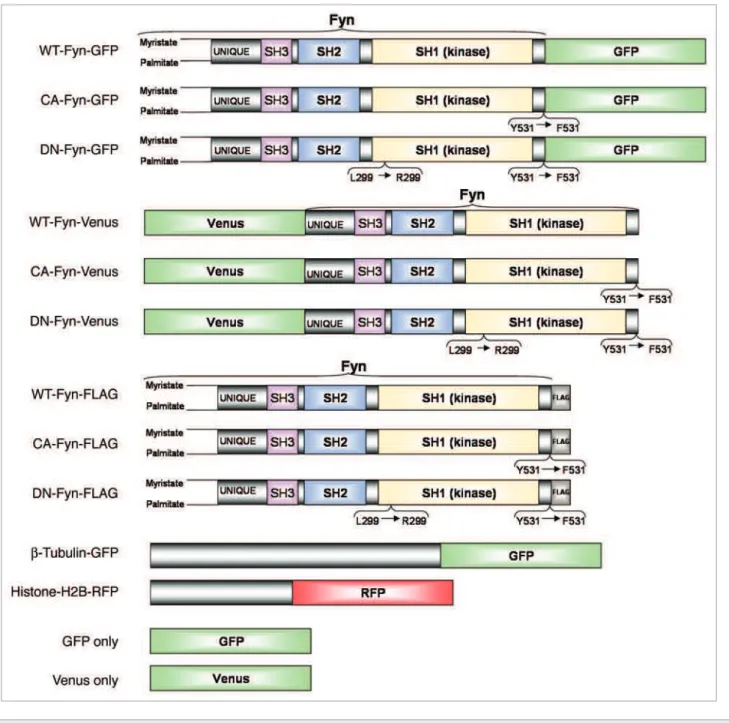
in vitro matured oocytes was examined by microinjection of cRNA of Fyn-GFP or Fyn-Venus (Fig. 2) into live oocytes or by confocal laser-scanning microscope (CLSM) imaging of fixed oocytes immunostained with rabbit anti-Fyn antibody. Immunostaining showed homogenous localization of Fyn throughout the oocyte cytoplasm and concentration at the spindle and cortex areas (Fig. 3Bb). In order to track temporal changes of Fyn localization within in vitro matured oocytes, the localization of Fyn was examined by imaging live oocytes with spinning-disk confocal microscopy, ensuring minimal laser exposure and thus minimum damage to the oocytes. Two cRNA constructs with opposite conjugated-fluorophore orientations, wildtype Fyn-GFP (WT-Fyn-GFP; fluorophore at the carboxy-terminus of Fyn;
Fig. 2) or wildtype Fyn-Venus (WT-Fyn-Venus; fluorophore at the
amino-terminus of Fyn; Fig. 2), mixed with Histone-H2B-RFP cRNA, were microinjected into live GV oocytes. The carboxy-terminal orientation of the GFP-conjugate allowed the trans-lated Fyn-GFP protein to be myristoytrans-lated and palmitoytrans-lated on the free amino-terminus, leading to an increased hydrophobic-ity of the protein and contributing to its membrane-association (Fig. 3Aa–e). The orientation of the Venus-conjugate at the amino-terminus (Fig. 2) obstructed the translated Fyn protein from being myristoylated and palmitoylated, but allowed its localization to the meiotic spindle (Fig. 3Aa’–e’). Concordantly, and Yrk (a non mammalian SFK) are found in a broad range of
tissues, including oocytes,1,15-21 while Lck, Fgr, Lyn, Hck and Blk
are restricted to cells of the haematopoietic lineages.22 Activated
SFKs catalyze the transfer of γ-phosphates from ATP molecules to tyrosine residues of target proteins, thereby transmitting extra-cellular signals downstream to extra-cellular components.22
Fyn is a 59 kDa SFK involved in a vast variety of cell functions, including cell cycle,22 apoptosis,23 adhesion,22 control of Sertoli
cell structure,24 B-cell activation25 and growth factor-induced
mitogenesis.26-28 Several studies suggest an involvement of Fyn in
the regulation of microtubules (MTs) organization,16,17,21,29,30,32-34
while according to others Fyn also affects actin-dependent pro-cesses.24,30,35-42 Furthermore, Fyn-/- female mice show decreased
fertility.14,32
In our previous studies we have determined the expression and localization of Fyn, Yes and Src in rat oocytes fixed at the MII stage. Fyn is localized to the oocytes cytoplasm, cortex and spindle, Yes to the cytoplasm and the cortex, whereas Src is found only at the cytoplasm.20,43,44 Fyn coimmunopercipitates with
β-tubulin and phosphorylates β-tubulin in vitro,44 indicating an
association with the oocyte MTs.
We have further demonstrated that the release from MII arrest in rat oocytes activated by SrCl2, puromycin or ionomycin is inhibited in the presence of SFKs inhibitors, PP2 or SU6656.20,45,46
As we have recently shown, the same applies to mouse oocytes.47
Moreover, inhibition of SFKs by SU6656 also inhibited the deg-radation of cyclin-B and the release from MII arrest in fertil-ized rat oocytes.45 Altogether, these observations imply a role for
SFKs in general and for Fyn in particular, in the signal leading to cyclin-B degradation downstream to the Ca2+ elevation in
mam-malian oocytes during the exit from MII arrest.
Although the importance of SFKs in regulating cell cycle of somatic cells and in activating mammalian oocytes is determined, the data regarding their role in RMI is still limited. Inhibition of SFKs by PP2 caused inhibition of GVBD and the exit from MI.21
Inhibiting SFKs with either siRNA knockdown or gene deletion also inhibited the exit from MI, but had no effect on the process of GVBD or extrusion of PBI.32 Though both studies indicate
defects in chromosome alignment in some fixed oocytes, it is still unclear whether the defected alignment inhibits the exit from MI. The limited evidence implicating SFKs in RMI encour-aged us to elucidate the role of SFKs, particularly Fyn, in RMI in oocytes, by several approaches including live cell confocal imaging, immunoblotting, immunostaining, microinjection of selective complementary RNAs (cRNAs) and the use of chemical inhibitors.
Results
Fyn degradation during RMI. Changes in the amount of the
endogenous Fyn protein (59 kDa) within oocytes during RMI in vitro were recorded by sodium dodecyl sulfate 10% polyacryl-amide gel electrophoresis (SDS-PAGE) and western blot (WB) analysis. ERK1/2 (42 kDa) or p34cdc2 (34 kDa) served as ref-erence for the amount of total oocytes proteins analyzed in all experimental groups. The amount of Fyn decreased considerably
(DN-Fyn; Suppl. movie 3) proteins. Interestingly, the WT-Fyn-Venus protein (Fig. 3Aa’–e’; Suppl. movie 4) was homogenously distributed throughout the spindle whereas the DN-Fyn-Venus protein was specifically localized at the poles of the meiotic spindle in mouse oocytes (Fig. 3Ba). Moreover, the expressed WT-Fyn-Venus protein showed higher concentration at the spindle area in MI and MII rat oocytes (Fig. 3Ca and b, respectively).
Experimentally expressed GFP-only and Venus-only, which served as negative-controls, were homogenously localized throughout the oocyte cytoplasm (Fig. 3Da and b, respectively). The amount of WT-Fyn-GFP protein, translated six hours after microinjection of their cRNA into GV oocytes (85 kDa;
Fig. 3E2), was measured by SDS-PAGE and WB analysis and
was found to be 6.7 ± 0.8 times higher then the amount of the the combined localization of the two injected exogenous Fyn
con-jugates, matched the signal detected in oocytes immunostained for endogenous Fyn (Fig. 3Bb), i.e., homogenous localization of Fyn throughout the cytoplasm and concentration at the cortex and spindle areas.
WT-Fyn-GFP was recruited to the cortical area designated for PBI extrusion, 15 minutes prior to PBI extrusion, whereas 15 minutes after PBI extrusion, the kinase resumed a uniform distribution pattern throughout the cortex (Fig. 3Aa–c; Suppl.
movie 1). Fyn was also detected at the cleavage furrow area
during PBI extrusion (Suppl. movie 1). The localization of WT-Fyn-GFP protein (Fig. 3Aa–e; Suppl. movie 1) during the whole RMI process was similar to that of constitutively active Fyn (CA-Fyn; Suppl. movie 2) and dominant negative Fyn
Figure 1. Degradation of Fyn during RMI. oocytes were lysed and subjected to SDS-pAGe and WB for detection of Fyn protein (59 kDa). p34cdc2 (34 kDa) or eRK1/2 (42 kDa) served as indicators for the number of oocytes in all experimental groups. (A) Batches of 12, 25 and 50 (A1, A2 and A3, respectively) GV mouse oocytes and batches of 50 oocytes cultured for 0.5, 1.5 and 3 hours (A4, A5 and A6, respectively) after milrinone wash out. (B) Batches of 50 mouse oocytes, before (B1) or after 4, 8 and 12 hours of culture after release from milrinone (B2, B3 and B4, respectively). (C) the rela-tive percent of GV oocytes in each lane, as a function of RMI in vitro culture period, are presented. (D) Batches of 12, 25 and 50 (D1, D2 and D3 respec-tively) GV mouse oocytes and 50 oocytes matured in vitro (C4; 12 hours after milrinone wash out) or matured in vivo (C5; 14 hours after hCG injection). three independent experiments were performed. A representative experiment is presented. (e) Batches of 12, 25 and 50 (e1, e2 and e3 respectively) GV mouse oocytes collected immediately or oocytes after 3 hours culture without milrinone (e3) or after 3 hours culture without milrinone, but with 50 µM MG132 (e4) or 50 µM Lactacystin (e5).
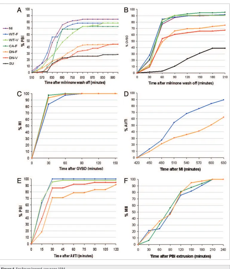
span elapsed from milrinone wash out to PBI-extrusion was lon-ger in oocytes subjected to SU6656 (657 min), or microinjected with DN-Fyn-FLAG (655 min) or DN-Fyn-Venus cRNA (713 min) than in non-injected oocytes (611 min) or WT-Fyn-FLAG cRNA injected oocytes (604 min; p < 0.05; two-sample t-test for unequal sample sizes and unequal variances).
In the attempt to evaluate the involvement of Fyn in specific cell cycle events during RMI we followed developmental stages of the oocytes according to the next criteria: the disappearance of the GV as seen at the DIC channel was defined as GVBD stage; the appearance of condensed chromosomes at the 561 nm chan-nel and of non-organized spindle at the 488 nm chanchan-nel were defined as MI stage (assessed only in the FLAG-injected oocytes); the appearance of segregated chromosomes at the 561nm channel endogenous Fyn protein (59 kDa) in non-injected GV oocytes
(Fig. 3E1).
The effect of Fyn inhibition on duration and rate of RMI.
First, we examined the involvement of Fyn in the entire process of RMI of GV oocytes by assessing PBI extrusion, scored via the Differential Interference Contrast (DIC) channel. Inhibition of Fyn significantly inhibited the duration and rate of RMI (Fig. 4A). Only 25, 44.4 and 44.8% of the SU6656-exposed, DN-Fyn-FLAG (Fyn conjugated a tag of 8 amino acid FLAG had already been constructed in our lab; Fig. 2) and DN-Fyn-Venus cRNA microinjected oocytes extruded the PBI, respec-tively, compared to 84, 77.9, 72.7 and 78% of the control, WT-Fyn-FLAG, CA-Fyn-FLAG and WT-Fyn-Venus cRNA microinjected oocytes, respectively. Moreover, the average time
Figure 3. Localization of Fyn during RMI. (A) Spinning-disk confocal images of representative live mouse oocytes microinjected with Histone-H2B-RFp (red) for chromosomes detection, together with one of two cRNA constructs of Wt-Fyn, conjugated to luorophores with opposite orientations: GFp luorophore at the carboxy-terminus of Fyn (Aa–e; green) or Venus luorophore at the amino-terminus of Fyn (Aa’–e’; green). Injected oocytes were cultured for six hours in M2 medium in the presence of milrinone, to allow translation of the cRNA, and were then transferred into milrinone-devoid M2 culture medium. Four-dimensional (x, y, z, t) images in three channels (488 nm, 561 nm and DIC) were acquired using the Volocity 5 software at intervals of 15 or 30 minutes with an automatic movement of the objective in the z-axis (∆z = 5 µm) over a total depth of 45 µm. Scale bar, 10 µm. (B) Confocal images of representative MII mouse oocytes microinjected with (a) Histone-H2B-RFp (red) together with DN-Fyn-Venus (green) or stained (b) rabbit anti-Fyn antibody (1:100), followed by goat anti-rabbit AlexaFlour488-conjugated secondary antibody (1:400; green) mixed with DNA-specif-ic luorochrome (Hoechst 33342; blue). (C) Spinning-disk confocal images of representative live, in vitro matured rat MI (Ca) or MII (Cb) oocytes mDNA-specif-icro- micro-injected with Wt-Fyn-Venus cRNA. (D) Spinning-disk confocal images of representative live mouse oocytes (control) micromicro-injected with GFp-only (Da; green) or Venus-only (Db; green) mixed with Histone-H2B-RFp (red) cRNA at the MII stage after RMI in vitro. (e) SDS-pAGe and WB analysis indicat-ing the amount of endogenous Fyn (59 kDa) in 50 non-injected GV oocytes (e1) and of exogenous Fyn-GFp (85 kDa; e2), six hours after microinjection of cRNA into 8 GV oocytes. A representative experiment is presented.
cRNA microinjected oocytes, respectively—all statistically simi-lar (p > 0.05). The average length of MII-spindles were 24.2 ± 0.72, 23.2 ± 0.9 and 23 ± 0.61 µm in WT-Fyn-FLAG, CA-Fyn-FLAG and DN-Fyn-CA-Fyn-FLAG cRNA microinjected oocytes, respec-tively—all statistically similar (p > 0.05).
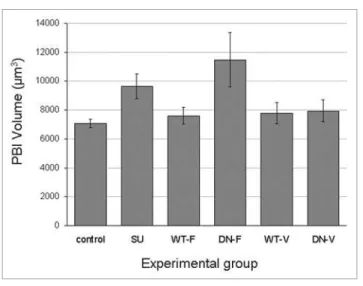
In order to examine the effect of Fyn inhibition on the volume of the extruded PBI, groups of GV mouse oocytes were subjected to several treatments: non-injected oocytes incubated in the pres-ence or abspres-ence (control) of 10 µM SU6656 (added seven hours after oocytes were removed from milrinone); oocytes microin-jected with WT-Fyn-FLAG, DN-Fyn-FLAG, WT-Fyn-Venus or DN-Fyn-Venus cRNA. The average volume of PBI was sig-nificantly larger (p < 0.05) in SU6656-treated oocytes (9644.2 ± 854.5 µm3) and in DN-Fyn-FLAG cRNA microinjected oocytes
(11462.1 ± 1879.7 µm3) than in the non-injected control oocytes
(7058.3 ± 310.9 µm3) and in the WT-Fyn-FLAG (7588.3 ± 585.4
µm3), WT-Fyn-Venus (7760.5 ± 758.5 µm3) or DN-Fyn-Venus
cRNA microinjected oocytes (7916.2 ± 773.1 µm3; Fig. 6).
Discussion
The involvement of Fyn in GVBD. One can assume that the
mei-otic stages in which Fyn is involved are those that exhibit changes in the expression of Fyn. Therefore, we examined, by SDS-PAGE and WB analysis, the changes in the amount of endogenous Fyn protein during RMI in vitro and in vivo. The amount of Fyn decreased considerably during RMI and correlated with GVBD. More important, when Fyn was inhibited by a chemical inhibitor, or by microinjection of DN-Fyn cRNA, the process of GVBD was inhibited. The effect of SU6656, that inhibits Fyn, Yes and Src, on GVBD was more dramatic, though it is possible that SU6656 inhibits other related kinases in the oocytes, as indi-cated in somatic cells.51 These results can imply that Fyn takes
part in the signal to GVBD, however, because the developmental stages later than GVBD seemed to be normal in oocytes microin-jected with CA-Fyn-FLAG, the requirement for Fyn degradation once the GVBD was already triggered, would therefore not seem to be absolute, but only a coincidence. Studies of the involve-ment of SFKs in GVBD in mouse oocytes show contradicting data: Zheng et al.21 showed that mouse oocytes matured in the
presence of another SFK inhibitor, PP2, were unable to undergo GVBD, whereas McGinnis et al.32 showed that SKI606, a
chemi-cal inhibitor of SFKs, induced GVBD in mouse oocytes cultured under conditions that promote metaphase arrest. Differences in the affinity and selectivity among the various inhibitors, SKI606, PP2 and SU6656, may account for the contradictory data.
We ruled out the involvement of the proteasome in the down-regulation of Fyn since we have shown that the amount of Fyn in in vitro matured mouse oocytes was similar in the absence or presence of proteasome inhibitors. It seems therefore, that Fyn is downregulated or degraded via other pathways, such as the cal-pain, endosome or lysosome pathways as suggested for Fyn in other cell types.38,39,52,53
The localization of Fyn at the meiotic spindle. Activated
Src-family kinases are possibly associated with the meiotic spin-dle during meiosis.16 Differences in intracellular localization of
was defined as either anaphase or telophase of the first meiotic division (AI/TI; assessed only in the FLAG injected oocytes); the appearance of a spindle at the 488 nm channel after PBI extrusion was defined as MII stage (assessed only in the FLAG-injected oocytes).
Inhibition of Fyn significantly inhibited GVBD (Fig. 4B). Only 38.7%, 75% and 67.1% of the SU6656-treated, DN-Fyn-FLAG and DN-Fyn-Venus cRNA microinjected oocytes com-pleted GVBD, respectively, compared to 90.4%, 91.7%, 95.3% and 90.4% of the control, WT-Fyn-FLAG, CA-Fyn-FLAG and WT-Fyn-Venus cRNA microinjected oocytes, respectively. Moreover, the average duration of the transition from GV to GVBD in SU6656-treated oocytes (127 min) and Fyn-FLAG cRNA injected oocytes (81 min) was statistically longer than that of non-injected control oocytes (59 min), WT-Fyn-FLAG cRNA injected oocytes (56 min), WT-Fyn-Venus (59 min) and CA-Fyn-FLAG (62 min; p < 0.05). However, we can not con-clude that the average duration of the transition from GV to GVBD was affected by the microinjected DN-Fyn-Venus cRNA (68 min) because it was measured in only 74 oocytes.
Inhibition of Fyn inhibited the exit from MI. Only 62.6% of the DN-Fyn-FLAG injected oocytes went through the transition from MI to AI/TI, compared to 89.4% of the WT-Fyn-FLAG injected oocytes (p < 0.05; Fig. 4D). More important, none of the oocytes showed misalignment of the chromosomes during MI stage or lagging chromosomes during chromosomes segre-gation. Moreover, the dynamics of the timing of exit from MI was not affected (Fig. 4D), nor were the spindle migration and anchoring to the cortex.
Inhibition of Fyn prolonged the process of the PBI extrusion. Although the timing of the start of the ingression of the cleav-age furrow was similar in all groups, the avercleav-age time needed to complete the extrusion of the PBI in DN-Fyn-FLAG cRNA was longer than the WT-Fyn-FLAG cRNA microinjected oocytes (38 min, as compared to 20 mim; p < 0.05; Fig. 4E).
Inhibition of Fyn had no significant effect (p > 0.05) on the transition from GVBD to MI (Fig. 4C) or from PBI to MII (Fig. 4F) and showed no effect on anchoring of the MII spindle to the cortex.
The effect of Fyn inhibition or activation on spindle length and PBI volume. Inhibition of SFKs by SU6656 results in
reduc-tion in size of the spindle structure in MII rat oocytes.44,47 The
current study shows that the inhibition of SFKs in mouse oocytes causes a reduction of the spindle poles structure (Fig. 5Ab). However, we did not observe a significant effect on the spindle structure in MII oocytes fixed six hours after microinjection of DN-Fyn-FLAG (Fig. 5Ac).
In order to examine a potential effect of Fyn inhibition or activation on the length of the spindle during RMI, GV mouse oocytes were microinjected with cRNA of Histone-H2B-RFP and β-tubulin-GFP mixed with either WT-Fyn-FLAG, CA-Fyn-FLAG or DN-Fyn-CA-Fyn-FLAG. Spindle length was defined as the dis-tance between its two poles. Spindles length (Fig. 5B) and shape were similar in all the experimental groups. The average length of MI-spindles was 26.3 ± 0.71 (mean ± SE), 25.2 ± 1.05 and 25.7 ± 0.55 µm in WT-Fyn-FLAG, CA-Fyn-FLAG and DN-Fyn-FLAG
disruption and reduction in size of the MII spindle structure, reduction in spindle size, misalignment of the chromosomes and appearance of MTs throughout the cytoplasm in a dose-response manner.44,47 The exposure of MII rat oocytes to a
tubu-lin-depolymerizing agent, nocodazole, caused disappearance of the various SFKs could indicate their unique roles during
meio-sis. Immunostaining of rat MII oocytes showed that Fyn, Src and Yes are localized in the cytoplasm, while Yes and Fyn are concentrated at the cortex and Fyn concentrates at the meiotic spindle as well.43,44,47 Inhibition of SFKs by SU6656 results in
Figure 4 (See previous page). the efect of inhibition or activation of Fyn on duration and rate of RMI. Groups of mouse oocytes at the GV stage were subjected to several treatments: non-injected oocytes incubated in the presence of 10 µM SU6656 (SU; 132 oocytes; black) or its absence
(NI, non-injected control; 125 oocytes; purple); oocytes microinjected with cRNA of Histone-H2B-RFp and β-tubulin-GFp mixed with Wt-Fyn-FLAG (Wt-F; 97 oocytes; blue), CA-Fyn-FLAG (CA-F; 121 oocytes; dark green) or DN-Fyn-FLAG (DN-F; 170 oocytes; orange); oocytes microinjected with cRNA of Histone-H2B-RFp mixed with Wt-Fyn-Venus (Wt-V; 83 oocytes; pale green) or DN-Fyn-Venus (DN-V; 74 oocytes; red). oocytes were incubated in M2 medium in the presence of 1 µM milrinone for six hours to allow translation of the cRNA. Milrinone was washed out and oocytes were imaged at intervals of 15 minutes by spinning-disk confocal microscopy. Non-injected oocytes were kept in the incubator and served as a control for the RMI in vitro process. Four-dimensional (x, y, z, t) image stacks in a three channels (488 nm, 561 nm and DIC) were processed and analyzed by the Volocity 5 software. Several parameters were scored: the transition from GV to pBI as observed through the DIC channel was classiied as complete RMI (A); the disappearance of the GV as observed through the DIC channel was classiied as GVBD stage (B); the appearance of condensed chromosomes as observed through the 561 nm channel and of non-organized spindle as observed through the 488 nm channel was classiied as metaphase of the irst meiotic division (MI; C); the appearance of segregated chromosomes as observed through the 561 nm channel was classiied as either anaphase or telophase of the irst meiotic division (AI and tI, respectively; D); the appearance of extruded polar body as observed through the DIC channel was classiied as pBI stage (e); the appearance of a spindle as observed through the 488 nm channel after pBI extrusion was classiied as metaphase of the second meiotic division (MII; F).
Figure 5. efect of SFKs inhibition on spindle length. (A) CLSM images of representative ixed MII mouse oocyte after incubation in the absence (a) or presence (b) of 10 µM SU6656 for 30 minutes or after microinjection of DN-Fyn-FLAG cRNA and incubation for six hours (c). oocytes were ixed and permeabilized and then exposed to rabbit anti-Fyn antibody (1:100), followed by goat anti-rabbit AlexaFlour488-conjugated secondary antibody (1:400; green). Chromosomes were detected by a DNA-speciic luorochrome (Hoechst 33342; blue). Scale bar, 10 µm. three independent experiments were performed. (B) Mouse oocytes at the GV stage were microinjected with Histone-H2B-RFp and β-tubulin-GFp cRNA mixed with Wt-Fyn-FLAG cRNA (Wt; 30 oocytes), CA-Fyn-FLAG cRNA (CA; 16 oocytes) or DN-Fyn-FLAG cRNA (DN; 68 oocytes). oocytes were incubated for six hours in M2 me-dium in the presence of 1 µM milrinone to allow translation of the cRNA. Milrinone was washed out and oocytes were imaged by spinning-disk confo-cal microscopy. the distance between the MI and MII spindle-poles was measured in the 3D images acquired by the 488 nm channel using Volocity 5 software and was scored as spindle length. each bar represents mean ± Se.
at the poles and/or by phosphorylating tyrosine residues of other proteins.
In contrast to cultured somatic cells, poleward flux in mouse eggs is critically dependent upon kinesin 5 and its inhibition leads to spindle shortening by depolymerization of the poles.57
In the current study, a similar phenomenon of reduction of the spindle length caused by shortening of the spindle poles was observed when SFKs were inhibited with 10 µM SU6656. This could imply that SFKs take part in spindle elongation at the poles area. The existence of functional overlapping or compensatory relationship among the various SFKs, regarding their involve-ment in spindle stabilization can explain how no effect on spindle characterization was observed when Fyn alone was inhibited by DN-Fyn cRNA, but a significant effect was observed when all SFKs-Fyn, Yes and Src-were inhibited by SU6656.
The involvement of Fyn in the exit from metaphase. Our
previous studies showed that the degradation of cyclin-B and the release from MII arrest in rat oocytes was inhibited in the pres-ence of SFKs inhibitors.20,45,46 As we have recently shown, the same
applies to mouse oocytes.47 Moreover, Microinjection of CA-Fyn
protein into rat oocytes caused release from MII arrest in a dose-depended manner.44 Microinjection of CA forms of either Fyn
or Yes cRNA caused release from MII arrest. Microinjection of DN forms of either Fyn or Yes cRNA inhibited SrCl2- or sperm-induced exit from MII arrest in rat oocytes.45 In the present study,
inhibition of Fyn by microinjecting DN-Fyn-FLAG cRNA into mouse oocytes resulted in inhibition of the exit from MI. Our results imply the involvement of Fyn not is being specific for the exit from MII, but rather to an exit from metaphase in general. However, it is also possible that overexpression of DN-Fyn can also inhibit other related SFKs in the oocytes. Furthermore, the exit from MI and MII arrest in fixed mouse oocytes was inhibited also in Fyn-/- mice oocytes and in oocytes exposed to the SFKs
inhibitors, SKI606 or PP2.16,21,32 A possible mechanism for a role
of SFKs in somatic cell mitosis has been suggested based on stud-ies of SFKs in cells arrested during mitosis after exposure to the tubulin depolymerizer, nocodazole, or in cells microinjected with SFKs inhibitory antibodies.22,57-60 The metaphase-stage elevated
activity of Fyn, Yes and Src correlates with phosphorylation of Src by p34cdc2 kinase at several serine and threonine residues. However, an antibody that recognizes Src alone did not prevent cell division; only the cross-inhibitory peptide antibodies against Fyn, Yes and Src were effective. These studies, combined with our results, suggest a possible negative feedback mechanism dur-ing the cell cycle: accumulation of cyclin-B durdur-ing metaphase leads to high activity of p34cdc2, which, in-turn, phosphorylates SFKs and induces their elevated activity. High activity of SFKs can participate in induction of the degradation of cyclin-B and the exit from metaphase arrest.
The involvement of Fyn in PBI extrusion. Various
stud-ies have demonstrated the involvement of Fyn in several actin functions, including cytokinesis in somatic cells.24,37-42 However,
in some cases it was hard to distinguish between cell cycle and cytokinesis. Lou et al.61 demonstrated a disruption in the
orga-nization of microvilli, cortical granules and filamentous actin at the cortical area overlaying the meiotic spindle in MII mice the spindle and abolished Fyn labeling at the spindle area. After
washing the nocodazole off, the oocytes re-exhibited a fully recovered spindle formation accompanied by Fyn labeling.44
The current study demonstrates Fyn localization at the area of meiotic MI and MII spindles of mouse oocytes matured in vitro and immunostained with anti-Fyn antibodies or microinjected with WT-Fyn-Venus while the localization of DN-Fyn-Venus was at the spindle poles. The differences in the spindle localiza-tion between the DN- and the WT-forms of Fyn-Venus could be explained by the allosteric open-conformation of the DN form that allows its concentration at the poles, compared to the closed conformation of the WT form that enables a homogenous localization at the spindle. This could imply the importance of the SH2 domain of Fyn for its association with key factors on the spindle poles during metaphase. Although meiotic spindle assembly in mammalian oocytes occurs in the absence of cen-trioles,54 it can be established by the presence of γ-tubulin at the
spindle poles.55 Fyn co-localizes with both mitotic spindle and
poles in T lymphocytes56 and coimmunoprecipitates through its
SH2 and SH3 domains with γ-tubulin in activated mast cells.34
Pretreatment of mast cells with PP2 inhibits the formation of MTs and reduce the amount of tyrosine-phosphorylated-pro-teins in γ-tubulin complexes, suggesting that SFKs are involved in the initial stages of MTs formation.34 other findings
demon-strated an elevated level of phosphorylated tyrosine at the poles of MII spindles in mouse oocytes, which eventually disappeared as MII progressed to AII.16 It is possible that phosphorylated
Fyn is one of the factors responsible for the high level of phos-phorylated tyrosine at the spindle poles, either by accumulating
Figure 6. efect of Fyn inhibition or activation on pBI volume. Mouse oocytes at the GV stage were subjected to several treatments: non-injected oocytes incubated in the presence of 10 µM SU6656 (SU; 48 oo-cytes; SU6656 was added seven hours after removal of milrinone from culture medium) or its absence (NI—non-injected control; 42 oocytes); oocytes microinjected with cRNA of Wt-Fyn-FLAG (Wt-F; 49 oocytes), DN-Fyn-FLAG (DN-F; 52 oocytes), Wt-Fyn-Venus (Wt-V; 33 oocytes) or DN-Fyn-Venus (DN-V; 25 oocytes). oocytes were imaged by spinning-disk confocal microscopy. the volume of pBI was measured in the DIC channel using Volocity 5 software [Volume of oblate spheroid = 4/3 x π x (length)2 x height]. each bar represents mean ± Se.
Materials and Methods
Animals. OF1 female mice (7–9 weeks-old; Charles River
Laboratories International, Chatillon-sur-Chalaronne, France) and Wistar Han IGS female rats (3–4 weeks-old; Charles River Laboratories International) were housed in air conditioned, light controlled animal facilities. Animal care was in accordance with institutional guidelines and was approved by the local authori-ties. Some female mice were superstimulated with 5 IU of preg-nant mares’ serum gonadotrophin (PMSG; Syncro-Part, Sanofi, France) and superovulated with 5 IU of human chorionic gonad-otrophin (hCG; Sigma Chemical Co., St. Louis, MO) 48–54 hours later.
Oocytes collection and culture. Non-stimulated female mice
and rats were sacrificed and their ovaries removed into prewarmed (37°C) M2 medium (M-7167; Sigma) supplemented with 4 mg/ml bovine serum albumin (BSA; Sigma). GV oocytes arrested at prophase I were released from the follicles into M2 medium containing 1 µM milrinone (Sigma). Milrinone (Primacor) is a phosphodiesterase III inhibitor that causes high levels of cAMP in the oocytes cytoplasm48 and thus inhibits the
intra-oocyte drop in cAMP concentration leading to GVBD.5,6
Oocytes were then cultured under mineral oil (Sigma) at 37°C. RMI was synchronized in all oocytes by transferring them into milrinone-free M2 medium.
Mature, ovulated MII mouse oocytes were removed from the oviductal ampullae into M2 medium 14 hours after hCG administration. Cumulus cells were removed by hyaluronidase (H-3631; Sigma).
Chemical inhibitors. A selective SFKs inhibitor (SU6656;
Calbiochem, San Diego, CA) was added to the mouse oocytes culture medium.20 SU6656 is known to inhibit Fyn, c-Src and
c-Yes51 by diffusing into cells, attaching to the ATP binding
pocket of SFKs and hence acting as a competitive inhibitor. We used SU6656 at a concentration of 10 µM since this was the effective concentration that blocked activation of rat20 and
mouse47 oocytes.
Mouse oocytes were also incubated in the presence of the pro-teasome catalytic activity inhibitors (50 µM MG132 or 50 µM Lactacystin; Calbiochem).
WB analysis. Batches of oocytes were collected as described
by Talmor-Cohen et al.20 Proteins were separated by SDS-PAGE.
oocyte exposed to SKI606 or in Fyn-null oocytes. By using live image confocal microscopy of oocytes microinjected with cRNA of WT-Fyn-GFP, CA-Fyn-GFP or DN-Fyn-GFP, we were able to examine the exact process of cytokinesis and show that all forms of Fyn were recruited to the cortical differentiation area designated for PBI extrusion, already 15 minutes prior to extru-sion. Fyn was concentrated at the cleavage furrow and cortical area surrounding the meiotic spindle midzone during TI and at the cleavage furrow area during PBI extrusion. Fyn resumed a uniform distribution pattern throughout the cortex 15 minutes after PBI extrusion. The summery of these observations is illus-trated in Figure 7. Moreover, inhibition of Fyn by microinjection of DN-Fyn-FLAG cRNA did not change the starting time of the cleavage furrow ingression, but caused a significant increase in the total duration needed for the ingression and the completion of the extrusion of PBI, compared to WT-Fyn-FLAG cRNA. We also observed a considerable influence of Fyn inhibition on the volume of the extruded PBI. The average volume of PBI was larger in oocytes exposed to SU6656 and in DN-Fyn-FLAG microinjected oocytes than in the non-injected, control oocytes or in oocytes microinjected with WT-Fyn-FLAG, WT-Fyn-Venus or DN-Fyn-Venus cRNA. The fact that the protein translated from the micro-injected DN-Fyn-FLAG cRNA and concentrated at the cortex of the oocytes,45 caused a more significant delay in PBI extrusion and
extrusion of a bigger PBI than proteins translated from microin-jected WT-Fyn-Venus or DN-Fyn-Venus cRNA that localize at the cytoplasm, could suggest that cortical Fyn is the main contrib-utor to the cleavage furrow ingression. It is possible that inhibition of Fyn by the cortical DN-Fyn-FLAG cRNA extends the duration of the cleavage furrow ingression; as a result the area restricted for formation of the cleavage furrow is more extended and a larger PBII is extruded. In conclusion, the recruitment of Fyn to the area designated for PBI extrusion, the delay in timing of the extru-sion process and the extruextru-sion of a larger PBI by DN-Fyn-FLAG injected oocytes, demonstrate the involvement of cortical Fyn in the regulation of the process of PBI extrusion, possibly by facili-tating the organization and function of actin-filaments and/or related molecular machinery in the area of the cleavage furrow.
Altogether, we propose that during RMI in mammalian oocytes Fyn takes part in regulating several key signaling path-ways leading to the exit from GV and MI and in the ingression of the cleavage furrow.
Figure 7. Changes in the localization of cortical Fyn during RMI. Cortical Fyn is recruited to the cortical diferentiation area designated for pBI extru-sion during AI stage. Fyn is concentrated to the cortical area around the midzone of the meiotic spindle during tI stage and at the cleavage furrow area during extrusion of pBI. After pBI extrusion, Fyn resumes a uniform distribution pattern throughout the cortex.
(New England Biolabs, Ipswich, MA) and cRNA transcripts were prepared in vitro using T3 mMESSAGE mMACHINE kit (Ambion, Austin, TX). The cRNA transcripts were then purified on RNeasy columns (Qiagen, Duesseldorf, Germany) and eluted by nuclease free water to a final concentration of 0.4–1 µg/ml. Aliquots of 4 µl were stored at -80°C.
The expression of Fyn protein translated in GV oocytes injected with Fyn cRNA, was measured by both WB analysis and Reticulocyte Lysate System.45
Histone-H2B-RFP cRNA and β-tubulin-GFP cRNA68-70
were donated by Dr. Marie-Helene Verlhac, Pierre and Marie Curie University, Paris, and were used for imaging of chromo-somes and MTs, respectively.
The in vitro synthesized cRNA of Fyn-GFP or Fyn-Venus, mixed with Histone-H2B-RFP, or of Fyn-FLAG mixed with Histone-H2B-RFP and β-tubulin-GFP were microinjected into the cytoplasm of oocytes at the GV stage at a volume of ∼5% of the oocyte volume, using the Eppendorf micromanipulator sys-tem (Leitz, Heidelberg, Germany).
Live cell imaging. Oocytes injected with cRNA were
cul-tured for 6 hours in M2 medium supplemented with milri-none to allow protein translation. For imaging, oocytes were put into oil covered, 10 µl medium within a specially designed chamber, maintained at 37°C on a heated microscope stage. Signal was recorded via three channels (488 nm, 561 nm and DIC) at intervals of 15 or 30 minutes by a C910 CCD digital-camera (Hamamatsu, Hamamatsu City, Japan) attached to an advanced confocal inverted microscope (Axiovert 200M; Carl Zeiss MicroImaging) with a CSU10 Spinning Disk head (Yokogawa, Tokyo, Japan), operated by Volocity 5 software (Improvision, Coventry, England). Three-dimensional images were created by the movement of the objective along the z-axis (∆z = 5 µm) over a total depth of 45 µm. Four-dimensional (x, y, z, t) image stacks were processed and analyzed by the Volocity 5 software.
Fyn inhibition and activation. In order to characterize the
involvement of Fyn in RMI, we employed several approaches for inhibiting or activating Fyn within oocytes. Mouse oocytes were subjected to several treatments: (1) GV oocytes incubated in the presence or absence (control) of 10 µM SU6656. (2) GV Oocytes microinjected with Histone-H2B-RFP cRNA and β-tubulin-GFP cRNA mixed with either WT-Fyn-FLAG cRNA, CA-Fyn-FLAG cRNA or DN-Fyn-FLAG cRNA. As shown pre-viously, Fyn-FLAG concentrates at the cortex and spindle area of oocytes.45 (3) GV oocytes microinjected with Histone-H2B-RFP
cRNA mixed with WT-Fyn-Venus cRNA or with DN-Fyn-Venus cRNA. This was preformed to examine whether the opposite ori-entation of the tag in Fyn-Venus that localizes homogenously to the oocyte’s cytoplasm, acts different then Fyn-FLAG during oocyte RMI. Since Fyn-Venus localizes to the spindle, we did not co-inject it with β-tubulin-GFP.
Statistical analysis. Data were evaluated by Pearson’s χ2
(chi-square) test for independence or by independent, two-sample t-test for unequal sample sizes and unequal variances. p < 0.05 was considered significant.
Approximate molecular masses were determined by comparison with the migration of prestained protein standards (Amersham, Buckinghamshire, UK). WB analysis was performed with either rabbit anti-Fyn IgG (1:200; sc-16; Santa Cruz, Santa Cruz, CA), rabbit anti-p34cdc2 IgG (1:1,000; c-19; Santa Cruz) or rabbit ERK1/2 (1:1,000; sc-94; Santa Cruz). Bound anti-bodies were recognized by goat anti-rabbit antibody conjugated to horseradish peroxidase (1:10,000; 111-035-003; Jackson Immunoresearch Laboratories, West Grove, PA). Detection was performed by an enhanced chemiluminescence (ECL; Pierce, Rockford, IL, USA). Intensity of bands was quantified and analyzed using ImageJ software (National Institutes of Health, Bethesda, MD, USA).
Immunofluorescence staining and CLSM. Oocytes were
fixed and permeabilized as described elsewhere43 and were then
incubated in the presence of rabbit anti-Fyn (1:100; sc-16; Santa Cruz) or mouse anti-β-tubulin (1:5,000; T8328; Sigma) primary antibodies, prior to incubation with AlexaFlour488-conjugated goat anti-rabbit (1:400; A11034; Invitrogen—Molecular Probes, Carlsbad, CA) or Cy-3-conjugated donkey anti-mouse (1:400; 715-165-151; Jackson Immunoresearch Laboratories) secondary antibodies, respectively, mixed with Hoechst 33342 (1 µg/ml; Sigma).
Stained oocytes were visualized and photographed with CLSM, Zeiss-LSM410 (Carl Zeiss MicroImaging, Oberkochen, Germany), equipped with a 25 mW krypton-argon laser (488 nm and 568 nm lines), a 10 mW helium-neon laser (633 nm line) and an UV laser (Coherent Laser Group, Santa Clara, CA). A 40X NA/1.2 planapochromat water-immersion lens (Axiovert 135 M; Zeiss) was used for all imaging.
Preparation and microinjection of cRNA. The full-length
sequences of human cDNA encoding the WT, CA or DN forms of Fyn kinase (I.M.A.G.E. clone ID number 3613878, ResGen, Carlsbad, CA), inserted into pCMV-Tag 4A with an in-frame FLAG at the 3'-end (Stratagene, Cedar Creek, TX) had already been constructed in our lab (Fig. 2).46 As indicated in the
litera-ture, a point mutation at Y531 in the regulation site of Fyn opens the conformation of Fyn kinase and activates it by exposing the SH1 and SH2 domains to the cytoplasm.12,62-65 A point mutation
at L299 in the ATP binding site of the kinase makes Fyn a kinase-dead mutant, capable of inhibiting endogenous Fyn.12,28,62,63,66,67
Furthermore, the activity of the different forms of Fyn had previ-ously been assessed by an in vitro autophosphorylation activity of Fyn. The purified protein of the CA-Fyn-FLAG was highly autophosphorylated whereas the WT-Fyn-FLAG and DN-Fyn-FLAG had only a minimal autophosphorylation activity or none at all.46 WT-Fyn, DN-Fyn and CA-Fyn were amplified and
inserted into pRN-N1, with an in-frame GFP at the 3'-end. The 3'-end orientation of FLAG and GFP epitopes allowed eukaryotic post translational modifications, i.e., amino-terminal myristoy-lation and palmitoymyristoy-lation of Fyn within oocytes, thus enabling Fyn membrane targeting. Fyn conjugated to Venus at the 5'-end was also constructed in pSPE3 vectors in order to examine the cytoplasmic localization of Fyn. All plasmids were sequenced and analyzed. They were linearized using SfiI restriction enzyme
Note
Supplementary materials can be found at:
www.landesbioscience.com/supplement/LeviCC9-8-Sup.pdf www.landesbioscience.com/supplement/LeviCC9-8-Sup1.avi www.landesbioscience.com/supplement/LeviCC9-8-Sup2.avi www.landesbioscience.com/supplement/LeviCC9-8-Sup3.avi www.landesbioscience.com/supplement/LeviCC9-8-Sup4.avi Acknowledgements
This work was supported by a grant from the Israel Science Foundation to R. Shalgi and by the Eshkol scholarship granted by the Israel Ministry of Science, Culture and Sport to M. Levi. We thank Dr. J.Z. Kubiak for critical reading of the paper. We thank Dr. M.H. Verlhac, Pierre and Marie Curie University, Paris, for the Histone-H2B-RFP and β-tubulin-GFP plasmids and for the anti-ERK1/2 antibody. We also thank R. Kaplan-Kraicer for her help in preparing the manuscript. This work is a partial fulfill-ment of the requirefulfill-ments for the Ph.D. degree of M. Levi at the Sackler Faculty of Medicine, Tel-Aviv University.
References
1. Mehlmann LM. Stops and starts in mammalian oocytes: recent advances in understanding the regulation of mei-otic arrest and oocyte maturation. Reproduction 2005; 130:791-9.
2. Dekel N. Protein phosphorylation/dephosphorylation in the meiotic cell cycle of mammalian oocytes. Rev Reprod 1996; 1:82-8.
3. Jones KT. Turning it on and off: M-phase promoting factor during meiotic maturation and fertilization. Mol Hum Reprod 2004; 10:1-5.
4. Morgan DO. Cyclin-dependent kinases: engines, clocks and microprocessors. Annu Rev Cell Dev Biol 1997; 13:261-91.
5. Dekel N, Beers WH. Rat oocyte maturation in vitro: relief of cyclic AMP inhibition by gonadotropins. Proc Natl Acad Sci USA 1978; 75:4369-73.
6. Sela-Abramovich S, Galiani D, Nevo N, Dekel N. Inhibition of rat oocyte maturation and ovulation by nitric oxide: mechanism of action. Biol Reprod 2008; 78:1111-8.
7. Brunet S, Pahlavan G, Taylor S, Maro B. Functionality of the spindle checkpoint during the first meiotic division of mammalian oocytes. Reproduction 2003; 126:443-50.
8. Fang G, Yu H, Kirschner MW. Control of mitotic transitions by the anaphase-promoting complex. Philos Trans R Soc Lond B Biol Sci 1999; 354:1583-90. 9. Kubiak JZ, Weber M, de Pennart H, Winston NJ,
Maro B. The metaphase II arrest in mouse oocytes is controlled through microtubule-dependent destruction of cyclin B in the presence of CSF. EMBO J 1993; 12:3773-8.
10. Nixon VL, Levasseur M, McDougall A, Jones KT. Calcium oscillations promote APC/C-dependent cyclin B1 degradation during metaphase arrest and comple-tion of meiosis in fertilizing mouse oocytes. Curr Biol 2002; 12:746-50.
11. Peters JM. The anaphase-promoting complex: proteol-ysis in mitosis and beyond. Mol Cell 2002; 9:931-43. 12. Sato KI, wasaki T, Hirahara S, Nishihira Y, Fukami
Y. Molecular dissection of oocyte fertilization signal-ing with the aid of tyrosine kinase-specific inhibitor and activator strategies. Biochim Biophys Acta 2004; 1697:103-21.
13. Roche S, Fumagalli S, Courtneidge SA. Requirement for Src family protein tyrosine kinases in G2 for fibro-blast cell division. Science 1995; 269:1567-9. 14. Stein PL, Vogel H, Soriano P. Combined deficiencies
of Src, Fyn and Yes tyrosine kinases in mutant mice. Genes Dev 1994; 8:1999-2007.
15. Kinsey WH. Biphasic activation of Fyn kinase activity upon fertilization of the sea urchin oocyte. Dev Biol 1996; 174:281-7.
16. McGinnis LK, Albertini DF, Kinsey WH. Localized activation of Src-family protein kinases in the mouse egg. Dev Biol 2007; 306:241-54.
17. Meng L, Luo J, Li C, Kinsey WH. Role of Src homolo-gy 2 domain-mediated PTK signaling in mouse zygotic development. Reproduction 2006; 132:413-21.
18. Sato KI, Aoto M, Mori K, Akasofu S, Tokmakov AA, Sahara S, et al. Purification and characterization of a Src-related p57 protein tyrosine kinase from Xenopus oocytes. J Biol Chem 1996; 271:13250-7.
19. Schartl M, Barnekow A. Differential expression of the cellular src gene during vertebrate development. Dev Biol 1984; 105:415-22.
20. Talmor-Cohen A, Matar-Tomashov R, Eliyahu E, Shapira R, Shalgi R. Are Src family kinases involved in cell cycle resumption of rat egg? Reproduction 2004; 127:455-63.
21. Zheng KG, Meng XQ, Yang Y, Yu YS, Liu DC, Li YL. Requirements of Src family kinase during meiotic maturation in mouse oocyte. Mol Reprod Dev 2007; 74:125-30.
22. Thomas SM, Brugge JS. Cellular functions regulated by Src family kinases. Annu Rev Cell Dev Biol 1997; 13:513-60.
23. Resh MD. Fyn, a Src family tyrosine kinase. Int J Biochem Cell Biol 1998; 30:1159-62.
24. Maekawa M, Toyama Y, Yasuda M, Yagi T, Yuasa S. Fyn tyrosine kinase in Sertoli cells is involved in mouse spermatogenesis. Biol Reprod 2002; 66:211-21. 25. Horikawa K, Nishizumi H, Umemori H, Aizawa S,
Takatsu K, Yamamoto T. Distinctive roles of Fyn and Lyn in IgD- and IgM-mediated signaling. Int Immunol 1999; 11:1441-9.
26. Kypta RM, Goldberg Y, Ulug ET, Courtneidge SA. Association between the PDGF receptor and members of the src family of tyrosine kinases. Cell 1990; 62:481-92.
27. Twamley-Stein GM, Kypta RM, Hall B, Courtneidge SA. Association of Fyn with the activated platelet-derived growth factor receptor: requirements for bind-ing and phosphorylation. Oncogene 1992; 7:1893-901.
28. Twamley-Stein GM, Pepperkok R, Ansorge W, Courtneidge SA. The Src family tyrosine kinases are required for platelet-derived growth factor-mediated signal transduction in NIH 3T3 cells. Proc Natl Acad Sci USA 1993; 90:7696-700.
29. Campbell KS, Cooper S, Dessing M, Yates S, Buder A. Interaction of p59fyn kinase with the Dynein light chain, Tctex-1 and colocalization during cytokinesis. J Immunol 1998; 161:1728-37.
30. Krause M, Sechi AS, Konradt M, Monner D, Gertler FB, Wehland J. Fyn-binding protein (Fyb)/SLP-76-associated protein (SLAP), Ena/Vasodilator-stimulated phosphoprotein (VASP) proteins and the Arp2/3 com-plex links T cell receptor (TCR) signaling to the actin cytoskeleton. J Cell Biol 2000; 149:181-94. 31. Martin-Cofreces NB, Sancho D, Fernandez E,
Vicente-Manzanares M, Gordon-Alonso M, Montoya MC, et al. Role of Fyn in the rearrangement of tubulin cytoskeleton induced through TCR. J Immunol 2006; 176:4201-7.
32. McGinnis LK, Kinsey WH, Albertini DF. Functions of Fyn kinase in the completion of meiosis in mouse oocytes. Dev Biol 2009; 327:280-7.
33. Nishida KS, Yamasaki Y, Ito K, Kabu K, Hattori T, Tezuka H, et al. FcRI-mediated mast cell degranulation requires calcium-independent microtubule-dependent translocation of granules to the plasma membrane. J Cell Biol 2005; 170:115-26.
34. Sulimenko V, Draberova E, Sulimenko T, Macurek L, Richterova V, Draber P. Regulation of microtubule formation in activated mast cells by complexes of gamma-tubulin with Fyn and Syk kinases. J Immunol 2006; 176:157243-53.
35. Badour K, Zhang J, Shi F, Leng Y, Collin M, Siminovitch KA. Fyn and PTP-PEST-mediated regu-lation of Wiskott-Aldrich syndrome protein (WASp) tyrosine phosphorylation is required for coupling T cell antigen receptor engagement to WASp effector func-tion and T cell activafunc-tion. J Exp Med 2004; 199:99-112.
36. Lannutti BJ, Blake N, Gandhi MJ, Reems JA, Drachman JG. Induction of polyploidization in leuke-mic cell lines and primary bone marrow by Src kinase inhibitor SU6656. Blood 2005; 105:3875-8. 37. Ng M, Chang F, Burgess D. Movement of membrane
domains and requirement of membrane signaling mol-ecules for cytokinesis. Dev Cell 2005; 9781-90. 38. Sandilands E, Brunton VG, Frame MC. The
mem-brane targeting and spatial activation of Src, Yes and Fyn is influenced by palmitoylation and distinct RhoB/RhoD endosome requirements. J Cell Sci 2007; 120:2555-6.
39. Sandilands E, Frame MC. Endosomal trafficking of Src tyrosine kinase. Trends Cell Biol 2008; 18:322-9. 40. Samayawardhena LA, Kapur R, Craig AW. Involvement
of Fyn kinase in Kit and integrin-mediated Rac activa-tion, cytoskeletal reorganization and chemotaxis of mast cells. Blood 2007; 109:3679-86.
41. Thomas SM, Soriano P, Imamoto A. Specific and redundant roles of Src and Fyn in organizing the cytoskeleton. Nature 1995; 376:267-71.
42. Yasunaga M, Yagi T, Hanzawa N, Yasuda M, Yamanashi Y, Yamamoto T, et al. Involvement of Fyn tyrosine kinase in progression of cytokinesis of B lymphocyte progenitor. J Cell Biol 1996; 132:91-9.
43. Talmor A, Kinsey WH, Shalgi R. Expression and immunolocalization of p59c-Fyn tyrosine kinase in rat oocyte. Dev Biol 1998; 194:38-46.
44. Talmor-Cohen A, Matar-Tomashov R, Tsai WB, Kinsey WH, Shalgi R. Fyn kinase-tubulin interaction during meiosis of rat oocytes. Reproduction 2004; 128:1-8. 45. Tomashov-Matar R, Levi M, Shalgi R. The
involve-ment of Src family kinases (SFKs) in the events leading to resumption of meiosis. Mol Cell Endocrinol 2008; 282:56-62.
46. Tomashov-Matar R, Levi M, Tchetchik D, Kraicer-Kaplan R, Shalgi R. The role of Src family kinases in egg activation. Dev Biol 2007; 312:77-89.
47. Levi M, Shalgi R. The role of Fyn kinase in the release from metaphase in mammalian oocytes. Mol Cell Endocrinol 2009; 314:228-33
48. Reis A, Chang HY, Levasseur M, Jones KT. APCcdh1 activity in mouse oocytes prevents entry into the first meiotic division. Nat Cell Biol 2006; 8:539-40.
64. Colognato H, Ramachandrappa S, Olsen IM, ffrench-Constant C. Integrins direct Src family kinases to regu-late distinct phases of oligodendrocyte development. J Cell Biol 2004; 167:365-75.
65. Laursen LS, Chan CW, ffrench-Constant C. An inte-grin-contactin complex regulates CNS myelination by differential Fyn phosphorylation. J Neurosci 2009; 29:9174-85.
66. Shima T, Okumura N, Takao T, Satomi Y, Yagi T, Okada M, et al. Interaction of the SH2 domain of Fyn with a cytoskeletal protein, beta-adducin. J Biol Chem 2001; 276:42233-40.
67. Osterhout DJ, Wolven A, Wolf RM, Resh MD, Chao MV. Morphological differentiation of oligodendrocytes requires activation of Fyn tyrosine kinase. J Cell Biol. 68. Ledan E, Polanski Z, Terret ME, Maro B. Meiotic
maturation of the mouse oocyte requires an equilib-rium between cyclin B synthesis and degradation. Dev Biol 2001; 232:400-13.
69. Tsurumi C, Hoffmann S, Geley S, Graeser R, Polanski Z. The spindle assembly checkpoint is not essential for CSF arrest of mouse oocytes. J Cell Biol 2004; 167:1037-50.
70. Brunet S, Polanski Z, Verlhac MH, Kubiak JZ, Maro B. Bipolar meiotic spindle formation without chromatin. Curr Biol 1998; 8:1231-4.
56. Ley SC, Marsh M, Bebington CR, Proudfoot K, Jordan P. Distinct intracellular localization of Lck and Fyn protein tyrosine kinases in human T lymphocytes. J Cell Biol 1994; 125:639-49.
57. Fitzharris G. A shift from kinesin 5-dependent meta-phase spindle function during preimplantation devel-opment in mouse. Develdevel-opment 2009; 136:2111-9. 58. Fumagalli S, Totty NF, Hsuan JJ, Courtneidge SA. A
target for Src in mitosis. Nature 1994; 368:871-4. 59. Shalloway D, Shenoy S. Oncogene kinases in mitosis.
Adv Cancer Res 1991; 57:185-225.
60. Taylor SJ, Shalloway D. Src and the control of cell divi-sion. BioEssays 1996; 18:9-11.
61. Luo J, McGinnis LK, Kinsey WH. Fyn kinase activ-ity is required for normal organization and functional polarity of the mouse oocyte cortex. Mol Reprod Dev 2009; 76:819-31.
62. Tsujikawa K, Ichijo T, Moriyama K, Tadotsu N, Sakamoto K, Sakane N, et al. Regulation of Lck and Fyn tyrosine kinase activities by transmembrane pro-tein tyrosine phosphatase leukocyte common antigen-related molecule. Mol Cancer Res 2002; 2:155-63. 63. Suetsugu S, Tezuka T, Morimura T, Hattori M,
Mikoshiba K, Yamamoto T, et al. Regulation of actin cytoskeleton by mDab1 through N-WASP and ubiq-uitination of mDab1. Biochem J 2004; 384:1-8. 49. Huo LJ, Fan HY, Zhong ZS, Chen DY, Schatten H,
Sun QY. Ubiquitin-proteasome pathway modulates mouse oocyte meiotic maturation and fertilization via regulation of MAPK cascade and cyclin B1 degrada-tion. Mech Dev 2004; 121:1275-87.
50. Josefsberg LB, Galiani D, Dantes A, Amsterdam A, Dekel N. The proteasome is involved in the first meta-phase-to-anaphase transition of meiosis in rat oocytes. Biol Reprod.
51. Blake RA, Broome MA, Liu X, Wu J, Gishizky M, Sun L, et al. SU6656, a selective src family kinase inhibitor, used to probe growth factor signaling. Mol Cell Biol 2000; 20:9018-27.
52. Carragher NO, Westhoff MA, Riley D, Potter DA, Dutt P, Elce JS, et al. v-Src-induced modulation of the Calpain-Calpastatin proteolytic system regulates transformation. Mol Cell Biol 2002; 22:257-69. 53. Rao N, Miyake S, Reddi AL, Douillard P, Ghosh
AK, Dodge IL, et al. Negative regulation of Lck by Cbl ubiquitin ligase. Proc Natl Acad Sci USA 2002; 99:3794-9.
54. Szollosi D, Calarco P, Donahue RP. Absence of centri-oles in the first and second meiotic spindles of mouse oocytes. J Cell Sci 1972; 11:521-41.
55. Barrett SL, Albertini DF. Allocation of gamma-tubulin between oocyte cortex and meiotic spindle influ-ences asymmetric cytokinesis in the mouse oocyte. Biol Reprod 2007; 76:949-57.