The Fen1 extrahelical 3¢-¯ap pocket is conserved
from archaea to human and regulates DNA substrate
speci®city
Erica Friedrich-Heineken and Ulrich HuÈbscher*
Institute of Veterinary Biochemistry and Molecular Biology, University of ZuÈrich, Winterthurerstrasse 190, CH 8057 ZuÈrich, Switzerland
Received February 20, 2004; Revised March 24, 2004; Accepted April 12, 2004
ABSTRACT
Fen1 is a key enzyme for the maintenance of genetic stability in archaea and eukaryotes and is classi®ed as a tumor suppressor. Very recent structural data obtained from Archaeoglobus fulgidus Fen1 suggest that an extrahelical 3¢-¯ap pocket is respon-sible for substrate speci®city, by binding to the unpaired 3¢-¯ap and by opening and kinking the DNA. Since the extrahelical 3¢-¯ap pocket in archaeal Fen1 contains seven amino acids that are conserved to a great extent in human Fen1, we have mutated the four conserved or all seven amino acids in the human Fen1 extrahelical 3¢-¯ap pocket to alanine. Our data suggest that the human extra-helical 3¢-¯ap pocket mutants have lost substrate speci®city to the double-¯ap DNA. Moreover, loss of high af®nity for the unpaired 3¢-¯ap suggests that the extrahelical 3¢-¯ap pocket is essential for recognition and processing of the `physiological' template. Human PCNA could stimulate the human Fen1 extrahelical 3¢-¯ap pocket mutants but not restore their speci®city. Thus the substrate speci®-city of Fen1 has been functionally conserved over a billion years from archaea to human.
INTRODUCTION
Flap endonuclease 1 (Fen1) belongs to a family of structure-speci®c nucleases that are evolutionarily conserved between Archaea and Eukarya (reviewed in 1±4). Its ¯ap endonuclease activity as well as its 5¢®3¢ exonuclease activity allows Fen1 to remove the RNA primers during lagging strand synthesis and damaged DNA fragments in various DNA repair pathways (reviewed in 4). Based on protein sequence comparison and biochemical assays, two major conserved motifs, the N (N-terminal) and I (intermediate) motifs, were found to be essential for the nuclease activities of Fen1 (3,5). A third motif at the C-terminal end is involved in the interaction with proliferating cell nuclear antigen (PCNA) (3,6,7). All crystal structures of Fen1 homologs solved to date (8±11) showed a conserved helical arch located above the globular domain that
contains the active site. This ¯exible loop, in addition to the catalytic site, was shown to be essential for ¯ap cleavage (12) and it has been proposed that the 5¢-end of the DNA ¯ap could thread through the hole of the loop tracking the length of the 5¢-tail (13).
The cleavage rate on a single-5¢-¯ap substrate, which is usually used to measure Fen1 endonuclease activity, is signi®cantly increased by the presence of a 1 bp overlap between the upstream and downstream duplex regions. This stimulatory effect has been reported previously for Fen1 homologs from Archaea to Eukarya (13±19). When the cleavage speci®city of Fen1 nucleases was analyzed on a single-5¢-¯ap substrate, the heterogeneity in cleavage position encountered (either at the junction or 1 nt into the annealed region) seemed to be inconsistent with the proposed roles of Fen1 in DNA replication and repair. In the case of the substrate with an overlap between the upstream and down-stream duplexes (also called the double-¯ap substrate), the site of cleavage was found to be exclusively 1 nt into the annealed region (16,18,19), allowing DNA ligase I to seal the resulting nicks (16,18). In contrast, a portion of the products from the `traditional' single-5¢-¯ap substrate was not ligated. This led to the proposition that there is a region or pocket in the enzyme that speci®cally recognizes that 3¢-¯ap nucleotide (16). A study of the T5 5¢-nuclease also suggested that there could be room for an additional base at the primer terminus (10), and analysis of the interaction between the 5¢-nuclease domain of bacterial DNA polymerase I and different 3¢-tailed substrates also supported the binding of 1±2 nt at the 3¢-terminus into an enzyme binding pocket (20).
Two additional ®ndings suggest that a double-¯ap is formed and cleaved during eukaryotic DNA replication in vivo. Firstly, a rad27p G240D mutant (rad27p is the Saccharomyves cerevisiae homolog of Fen1) exhibited very low endo- and exonuclease activities on a conventional single-¯ap substrate, however, it ef®ciently cleaved a double-¯ap substrate (18). The mutant showed a mutator phenotype but it could grow with almost equal ef®ciency as the wild-type. Secondly, a dual mode of binding was shown for human Fen1 to a double-¯ap substrate at a 1:1 molar ratio (19), whereas a 100-fold excess of Fen1 to a single-¯ap substrate was required to obtain complex formation.
Very recently, the structure of the archaeon Archaeoglobus fulgidus Fen1 bound to a duplex DNA was solved, and FRET
*To whom correspondence should be addressed. Tel: +41 1 6355472; Fax: +41 1 6356840; Email: [email protected]
DOI: 10.1093/nar/gkh576
experiments and modeling suggest that A.fulgidus Fen1 binds to the unpaired DNA 3¢-end (3¢-¯ap), opens and kinks the DNA and promotes conformational closing of the ¯exible helical clamp to facilitate 5¢ cleavage speci®city (11). This led us to analyze whether the extrahelical 3¢-¯ap pocket is also conserved in human Fen1 and to study the effect of replacing the seven amino acids, which comprise this extrahelical 3¢-¯ap pocket, by alanine.
MATERIALS AND METHODS Nucleic acid substrates
Oligonucleotides used to prepare the substrates for Fen1 nuclease assays were purchased from Microsynth GmbH (Balgach, Switzerland) and their sequences are listed in Table 1. They were labeled at the 5¢-end in a buffer containing 70 mM Tris±HCl (pH 7.6), 10 mM MgCl2, 5 mM DTT, an
equimolar amount of [g-32P]ATP to the 5¢-end to be labeled
and T4 polynucleotide kinase (New England Biolabs) for 45 min at 37°C. T4 polynucleotide kinase was heat-inactivated at 80°C for 15 min and free ATP was removed on MicrospinÔ G-25 columns. To generate the substrates for the nuclease assays, the appropriate oligonucleotides were mixed in a 1:1 molar ratio in a buffer containing 50 mM Tris±HCl (pH 7.4), 150 mM NaCl, heated to 75°C and slowly cooled to room temperature. The substrates for the electromobility shift assays were prepared according to the same protocol.
Enzymes and proteins
Human Fen1 cDNA was cloned into the pET 23d vector (Novagen) (6). The DP mutant was puri®ed as described previously (6). Wild-type and mutant proteins were over-expressed in Escherichia coli strain BL21(DE3)pLysS as
histidine-tagged proteins and puri®ed to near homogeneity using nickel charged metal chelating resin (HiTrap; Amersham Pharmacia Biotech) and SP Sepharose using fast protein liquid chromatography. Human PCNA was produced in E.coli using the plasmid pT7/hPCNA and puri®ed to homogeneity as described (21).
Generation of two extrahelical 3¢-¯ap pocket mutant Fen1 enzymes
Mutagenic PCR was performed using DyNAzymeÔ (Finnzymes) and the primers listed in Table 2. Following digestion of the parental, non-mutated pET23d vector tem-plate with DpnI (New England Biolabs), mutated plasmids were transformed into E.coli and plasmid DNA was isolated. To introduce all the desired mutations, this protocol was repeated several times. Finally, DNA sequencing reactions were performed to verify introduction of the desired mutations and absence of additional mutations.
Fen1 nuclease assay
Assay conditions were identical to those described previously (19). The enzyme titration assays were performed in a ®nal volume of 12.5 ml containing the following ingredients: 40 mM Tris±HCl (pH 7.5), 10 mM MgCl2, 1 mM dithiothreitol,
200 mg/ml bovine serum albumin and 50 fmol DNA substrate. After addition of wild-type (wt) and mutant Fen1, reactions were incubated for 15 min at 30°C and stopped with 2.53 stop buffer (95% formamide, 20 mM EDTA, 0.05% each bromophenol blue and xylene cyanol). Products were separ-ated on 19% denaturing polyacrylamide gels and visualized by autoradiography. For all experiments, the product size was determined by using appropriate radioactively labeled oligo-nucleotides (nt) of identical sequence to the template. For PCNA stimulation of Fen1, reactions were performed as
Table 1. Oligonucleotides used to create the Fen1 substrates used in this study
Oligonucleotide Size Sequence (5¢®3¢) Upstream primers
Up1 30mer TCGAGGTCGACGGTATCGATAAGCTTGATA
Up2 31mer TCGAGGTCGACGGTATCGATAAGCTTGATAC
Up3 31mer TCGAGGTCGACGGTATCGATAAGCTTGATAT
Up4 32mer TCGAGGTCGACGGTATCGATAAGCTTGATACG
Downstream primers
Dn1 19mer TCGAATTCCTGCAGCCCGG
Dn2 39mer GTCATGATAGATCTGATCGCTCGAATTCCTGCAGCCCGG
Template
T 49mer CCGGGCTGCAGGAATTCGATATCAAGCTTATCGATACCGTCGACCTCGA
Unannealed nucleotides are in bold.
Table 2. Primers used to create the Fen1 mutants used in this study
Mutant Amino acid Sequence (5¢®3¢)
LTFR L53 GGGTGGGGATGTGGCGCAGAATGAGGAGGG T61 GGAGGGTGAGACCGCTAGCCACCTGATGGG F316 R320 GTGTGGTGAAAAGCAGGCCTCTGAGGAGGCAATCCGCAGTGGG LQTKFSR L53 Q54 CGCCAGGGTGGGGATGTGGCCGCGAATGAGGAGGGTGAGACC T61 GGAGGGTGAGACCGCTAGCCACCTGATGGG K314 F316 S317 R320 GTTCATGTGTGGTGAAGCGCAGGCCGCGGAGGAGGCAATCCGCAGTGGGG Mutated nucleotides are in bold.
described above except that the buffer was replaced by 50 mM
Bis±Tris (pH 6.5) and 100 mM NaCl was added (22). For calculation of PCNA stimulation, the gels were quanti®ed on aPhosphorImager using the ImageQuant software (Molecular Dynamics). The SEM was calculated from the results of three independent experiments.
Electrophoresis mobility shift assay (EMSA) for Fen1 Assay conditions were the same as described previously (19). The binding reactions were carried out in a ®nal volume of 20 ml containing 50 mM Tris±HCl (pH 8.0), 10 mM NaCl, 5 mM EDTA, 100 mg bovine serum albumin, 4% Ficoll, 50 fmol labeled substrate and the indicated amounts of wt or mutant Fen1. After incubation for 10 min at room temperature, reactions were loaded on 6% polyacrylamide gels containing 0.53 TBE and run ®rst at 50 V for 45 min and then at 100 V for 90 min. Finally, the bands were visualized by autoradio-graphy.
Pull down assay with His6±Fen1
Aliquots of 8 mg Fen1 wt or mutant proteins were bound on Ni2+beads. To prevent non-speci®c binding, the beads were
pretreated with 10 mg Sf9 cell extract prior to the addition of 460 ng PCNA in 50 mM Tris±HCl (pH 8.0), 150 mM NaCl, 0.025% NP-40 and incubated for 2 h at 4°C. The beads were then washed ®ve times with the same buffer, but in addition 60 mM imidazole was included. Bound proteins were eluted by boiling in SDS±PAGE loading buffer and resolved on a 12% SDS±PAGE gel. PCNA was detected by western blot analysis using the PC10 antibody (Santa Cruz Biotechnology).
RESULTS
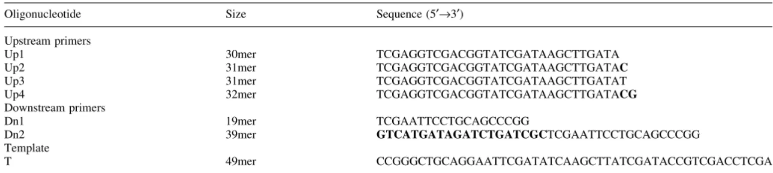
The amino acids in the extrahelical 3¢-¯ap pocket of human Fen1 are conserved from archaea to human Modeling of the extrahelical 3¢-¯ap pocket from the structure of the archaeon A.fulgidus Fen1 (11) suggests that this region might be very similar in human Fen1, since seven amino acids within this extrahelical 3¢-¯ap pocket are conserved in Fen1 from Archaea (Methanococcus janaschii, Pyrococcus abyssi and A.fulgidus), to two yeasts (Saccharomyces cerevisiae and Schizosaccaromyces pombe), to a ¯y (Drosophila melanoga-ster), to a frog (Xenopus laevis) and to human (Homo sapiens) (Fig. 1A and B). The human amino acids L53, Q54, T61, K314, F316, S317 and R320 are all in the extrahelical 3¢-¯ap
Figure 1. The amino acids in the extrahelical 3¢-¯ap pocket of human Fen1. (A) Amino acid alignments of different Fen1 proteins. HSA, Homo sapiens; DME, Drosophila melanogaster; XLA, Xenopus laevis; SCE, Saccharomyces cerevisiae; SPO, Schizosaccharomyces pombe; AFU, Archaeoglobus fulgidus; PAB, Pyrococcus abyssi; MJA, Methanococcus janaschii. Identical amino acids are shaded black, similar amino acids grey. The asterisks indicate the seven amino acids in the extrahelical 3¢-¯ap pocket of human Fen1: L53, Q54, T61, K314, F316, S317 and R320. (B) The 3-dimensional structure of human Fen1 was modeled with the Swiss-Pdb viewer according to the structure of M.janaschii Fen1 (9). (C) Two mutant human Fen1 proteins were designed: Fen1 (LTFR), where the four conserved amino acids (L53, T61, F316 and R320) were mutated to alanine; Fen1 (LQTKFSR) where the seven amino acids L53, Q54, T61, K314, F316, S317 and R320 were mutated to alanine. Mutagenesis, recombinant production and puri®cation were carried out as described in Materials and Methods. (Left) Fen1 (LTFR) and Fen1 wt, 5 and 10 mg each; (right) Fen1 (LQTKFSR) and Fen1 wt, 5 and 10 mg each were analyzed by 10% SDS±PAGE.
pocket (Fig. 1B) and correspond to the amino acids L47, K48, T55, H308, F310, S311 and R314 of A.fulgidus (see ®g. 1C in 11). We have constructed two mutants of human Fen1. First, the four conserved amino acids L53, T61, F316 and R320 and, second, the seven amino acids L53, Q54, T61, K314, F316, S317 and R320 (Fig. 1A) were mutated to alanine, since these amino acids are important for A.fulgidus Fen1 binding to the unpaired DNA 3¢-end. The two mutants carrying a His tag, called Fen1 (LTFR) and Fen1 (LQTKFSR), respectively, were produced in bacteria and puri®ed to apparent homogeneity (Fig. 1C). The preparations were free of contaminating nucleases, as tested on linear and supercoiled plasmid DNA (data not shown).
Human Fen1 extrahelical 3¢-¯ap pocket mutants have reduced exonuclease activities and a less stringent cleavage pattern
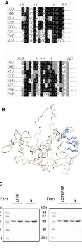
First we tested exonuclease activities on a nicked DNA substrate and on a nicked substrate containing an unpaired 3¢-end. From Figure 2 it is evident that both mutants had a greatly reduced activity compared to Fen1 wt (compare lanes 4±6 to 7±9 and 10±12, respectively). In addition, both mutants synthesized a two base product compared to the one base product of Fen1 wt. Furthermore, the activity of Fen1 (LQTKFSR) is considerably lower than the four amino acid mutant Fen1 (LTFR). This result is surprising since we would expect no effect on a substrate lacking a ¯ap when the 3¢-¯ap pocket is mutated. This might be explained by a tendency of Fen1 wt to bind to the end of the nick and to induce a 3¢-¯ap. A similar activity pro®le was found with the unpaired DNA 3¢-end substrate (Fig. 2, lanes 13±22) and the previously observed stimulation of Fen1 wt by the unpaired 3¢-¯ap (19) is lost in both mutants. In summary, the two extrahelical 3¢-¯ap pocket mutants have reduced exonuclease activities, which are coupled with a loss of product speci®city. The effect was more pronounced with Fen1 (LQTKFSR), suggesting that the extrahelical 3¢-¯ap pocket is important for the exonuclease activity of human Fen1.
Human Fen1 extrahelical 3¢-¯ap pocket mutants have reduced endonuclease activities and a less stringent cleavage pattern
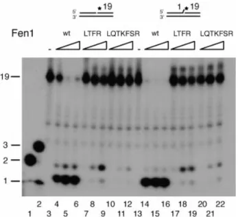
It has been found that Fen1 has high cleavage speci®city for double-¯ap DNA substrates and this speci®city suggests that Fen1 contains a region or pocket that can speci®cally bind to the 3¢-¯ap (16). This has been con®rmed at the structural level for archaeal Fen1, where this binding induces a kinking of the DNA substrate (11). We therefore next tested different ¯ap DNA substrates and compared the two extrahelical 3¢-¯ap Fen1 mutants with their wild-type counterpart. Figure 3A shows that Fen1 wt has a clear preference for double-¯ap substrates (compare lanes 4±6 with lanes 8±10 and 12±14 in Fig. 3A). A 2 nt non-complementary ¯ap mainly resulted in a 1 nt longer product (22 nt), likely due to the transition to the optimal one nt 3¢-¯ap (lanes 16±18). When the four amino acid extrahelical 3¢-¯ap pocket mutant Fen1 (LTFR) was tested with four DNA substrates we found that, ®rst, >100 times more molecules of Fen1 were required for cutting, second, two bands appeared with all four substrates tested and, third, the previously observed stimulation by the unpaired 3¢-¯ap (19) is lost (compare lanes 4 and 8 in Fig. 3B with lanes 4 and 8 in
Fig. 3A). With the `physiological' complementary 3¢-¯ap substrate the product was mainly 1 nt longer (22 nt) than with Fen1 wt and only a small proportion of the correct 21 nt product was made (compare lanes 12±14 of Fig. 3A and B). For the single-¯ap and non-complementary 3¢-¯ap substrates two products appeared (21 and 22 nt), whereas the wild-type showed a clear preference for the 21 nt product (compare lanes 4±6 and 8±10 of Fig. 3B with lanes 4±6 and 8±10 of Fig. 3A). This suggests that substrate speci®city was severely hampered in this mutant. Finally, the seven amino acid extrahelical 3¢-¯ap pocket mutant Fen1 (LQTKFSR) showed a similar altered cleavage pattern to the Fen1 (LTFR) mutant and with the `physiological' complementary 3¢-¯ap substrate the product was exclusively 1 nt longer (22 nt) than with Fen1 wt (lanes 12±14 of Fig. 3C). Only the 2 nt non-complementary 3¢-¯ap substrate showed the expected product of 22 nt (lanes 16±18 in Fig. 3C). These data suggest that replacement of only the four conserved amino acids L53, T61, F316 and R320 resulted in a severe loss of substrate speci®city, while replacement of all seven conserved amino acids L53, Q54, T61, K314, F316, S317 and R320 resulted in complete loss of the 3¢-¯ap speci®city.
Human Fen1 extrahelical 3¢-¯ap pocket mutants have lost the speci®c binding to double-¯ap DNA
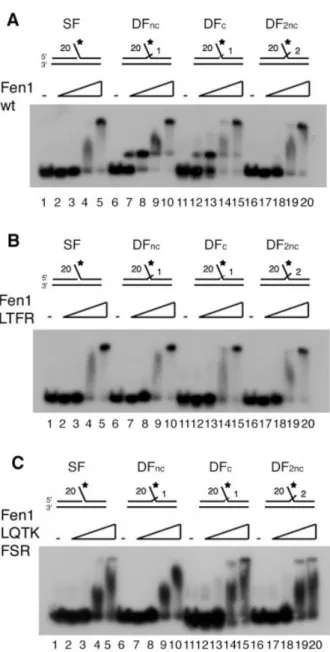
EMSA recently showed two binding modes of human Fen1, but only with the double-¯ap DNA structure containing a 3¢-¯ap (19). Using gel ®ltration experiments we could show that the faster migrating complex had a molecular weight which corresponded to a 1:1 complex between Fen1 and the double-¯ap DNA (data not shown). Again as expected, Fen1 wt
Figure 2. Both human Fen1 extrahelical 3¢-¯ap pocket mutants have reduced exonuclease activities and a less stringent cleavage pattern. Fen1 exonuclease activities were determined as described in Materials and Methods on a nicked (lanes 3±12) and on a 3¢-overhang nicked (lanes 13±22) DNA substrate. Lanes 1 and 2, oligonucleotide (nt) markers of 1, 2 and 3 nt; lanes 3 and 13, no enzyme control; lanes 4±6 and 14±16, 5, 50 and 500 ng Fen1 wt; lanes 7±9 and 17±19, 5, 50 and 500 ng Fen1 (LTFR); lanes 10±12 and 20±22, 5, 50 and 500 ng Fen1 (LQTKFSR). DNA substrate and product size (in nt) are indicated on the left of the ®gure.
revealed two binding modes on both double-¯ap substrates (Fig. 4A, lanes 7±10 and 12±15). This dual binding mode was lost with both extrahelical 3¢-¯ap pocket mutants (Fig. 4B and C). In contrast to a Fen1 mutant lacking the C-terminal 20
amino acids, which is completely unable to bind DNA (6), these two mutants have not lost the property of general DNA binding, but rather DNA-speci®c binding. Although the slightly weaker binding of the Fen1 (LTQKFSR) mutant to
Figure 3. Both human Fen1 extrahelical 3¢-¯ap pocket mutants have reduced endonuclease activities and a less stringent cleavage pattern. (A) The amounts of Fen1 wt indicated at the top of the autoradiogram were tested on a ¯ap (SF) (lanes 3±6), on a non-complementary double-¯ap (DFnc) (lanes 7±10), on a complementary double-¯ap (DFc) (lanes 11±14) and on a two 3¢-nucleotide overhang (DF2nc) (lanes 15±18) DNA substrate as outlined in Materials and Methods. Lanes 1 and 2 contain oligonucleotide (nt) markers of 20 and 22 nt and 21 and 23 nt, respectively. (B) The amounts of Fen1 (LTFR) indicated at the top of the autoradiogram were tested on a ¯ap (SF) (lanes 3±6), on a non-complementary double-¯ap (DFnc) (lanes 7±10), on a complementary double-¯ap (DFc) (lanes 11±14) and on a two 3¢-nucleotide overhang (DF2nc) (lanes 15±18) DNA substrate as outlined in Materials and Methods. Lanes 1 and 2 contain oligonucleotide (nt) markers of 20 and 22 nt and 21 and 23 nt, respectively. (C) Shown on the top of the autoradiogram, the indicated amounts of Fen1 (LQTKFSR) were tested on a ¯ap (SF) (lanes 3±6), on a non-complementary double-¯ap (DFnc) (lanes 7±10), on a complementary double-¯ap (DFc) (lanes 11±14) and on a two 3¢-nucleotide overhang (DF2nc) (lanes 15±18) DNA substrate as outlined in Materials and Methods. Lanes 1 and 2 contain oligonucleotide (nt) markers of 20 nt and 22 nt, or 21 nt and 23 nt, respectively. Product size (in nt) is indicated on the left of the ®gure.
the different DNA ¯ap substrates may indicate a perturbation of its structure by the seven amino acid replacements, our data in Figures 2 and 3 show that this mutant is still enzymatically active. Therefore, we conclude that the Fen1 (LQTKFSR) mutant has not lost its general DNA binding ability. These
data suggest that the mutant Fen1 proteins could still bind to DNA but were no longer able to properly accommodate the 3¢-¯ap and kink the DNA substrate. Fluorescent resonance energy transfer (FRET) experiments with A.fulgidus Fen1 are in agreement with these observations (11). In conclusion, the extrahelical 3¢-¯ap pocket mutant Fen1 enzymes were no longer able to distinguish the `physiological' substrate from other ones.
Human Fen1 extrahelical 3¢-¯ap pocket mutants can interact and are stimulated by PCNA, but the normal cleavage pattern cannot be restored by PCNA
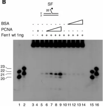
PCNA has been shown to bind to Fen1 and to stimulate its activity by >50 times under physiological salt conditions (22± 25). We therefore tested the two extrahelical 3¢-¯ap pocket mutants for their capacity to bind PCNA. Both mutants bound PCNA, the four amino acid mutant Fen1 (LTFR) as strongly as Fen1 wt, while the binding of the seven amino acid mutant Fen1 (LQTKFSR) to PCNA was reduced by 50% (Fig. 5A). These binding experiments were nicely con®rmed in a PCNA stimulation assay (Fig. 5B±D). On a single-¯ap DNA substrate, Fen1 wt was stimulated by PCNA in a dose-dependent manner and this stimulation was speci®c, since BSA showed no stimulatory effect (Fig. 5B, compare lanes 5±9 with lanes 10±14). Both extrahelical 3¢-¯ap pocket mutants were stimulated by PCNA (Fig. 5C), although 100 times more Fen1 (LTFR) or 250 times more Fen1 (LQTKFSR) had to be included to see a similar effect (compare Fig. 5C with B). Quanti®cation of the stimulation showed that the four amino acid mutant Fen1 (LTFR) was stimulated by PCNA to the same extent as Fen1 wt, whereas the seven amino acid mutant Fen1 (LQTKFSR) was stimulated to a lesser extent (Fig. 5D), probably due to its reduced interaction with PCNA (Fig. 5A). Finally, we analyzed whether PCNA could restore the substrate speci®city of the mutant Fen1 enzymes. As seen from Figure 5B, Fen1 wt mainly synthesized a product of 21 nt, while the two mutants produced about equal amounts of the 21 and 22 nt cleavage products (Fig. 5C). These data are in agreement with those seen in Figure 3. In summary, the extrahelical 3¢-¯ap pocket mutant Fen1 enzymes could be stimulated by PCNA but the substrate speci®city could not be restored.
DISCUSSION
The substrate speci®city of Fen1 is evolutionarily conserved
The data presented in this work clearly indicate that the substrate speci®city of Fen1 is conserved through evolution in an extraordinary way and that this conservation is seen in the structural similarity of the hydrophobic wedge in general and the extrahelical 3¢-¯ap pocket in particular. The biochemical and functional data presented in this paper suggest that the extrahelical 3¢-¯ap pocket, as structurally identi®ed in the archaeon A.fulgidus Fen1 (11), appears to be almost identical to human Fen1. The observation that a 1 nt double-¯ap structure is the optimal substrate for human Fen1 and that with this substrate a DNA±protein complex could be observed at a 1:1 molar ratio (19 and data not shown) suggested that a double-¯ap substrate containing a 1 nt 3¢-tail is the in vivo
Figure 4. Both human Fen1 extrahelical 3¢-¯ap pocket mutants have lost speci®c binding to double-¯ap DNA. The EMSA assays were carried out as outlined in Materials and Methods. (A) Aliquots of 0, 5, 50, 200 and 400 ng Fen1 wt were tested on a ¯ap (SF) (lanes 1±5), on a non-complementary double-¯ap (DFnc) (lanes 6±10), on a complementary double-¯ap (DFc) (lanes 11±15) and on a two 3¢-nucleotide overhang (DF2nc) (lanes 16±20) DNA substrate. (B) Aliquots of 0, 5, 50, 200 and 400 ng Fen1 (LTFR) were tested on a ¯ap (SF) (lanes 1±5), on a non-complementary double-¯ap (DFnc) (lanes 6±10), on a complementary double-¯ap (DFc) (lanes 11±15) and on a two 3¢-nucleotide overhang (DF2nc) (lanes 16±20) DNA substrate. (C) Aliquots of 0, 5, 50, 200 and 400 ng Fen1 (LQTKFSR) were tested on a ¯ap (SF) (lanes 1±5), on a non-complementary double-¯ap (DFnc) (lanes 6±10), on a complementary double-¯ap (DFc) (lanes 11±15) and on a two 3¢-nucleotide overhang (DF2nc) (lanes 16±20) DNA substrate.
substrate for human Fen1. This has been described previously
for the Fen1 homolog in yeast, rad27p (18). This double-¯ap substrate could also re¯ect an in vivo intermediate betweentwo conformations that would be interchangeable from a
Figure 5. Both human Fen1 extrahelical 3¢-¯ap pocket mutants can interact and be stimulated by PCNA, but the normal cleavage pattern cannot be restored. (A) Fen1 wt and mutant proteins (8 mg) were incubated with PCNA (460 ng) and pull-down experiments performed as outlined in Materials and Methods. PCNA was detected by western blot analysis. Lane1, Fen1 (LQTKFSR) and PCNA; lane 2, Fen1 (LTFR) and PCNA; lane 3, Fen1 DP (Fen1 lacking the PCNA binding domain) and PCNA, negative control; lane 4, Fen1 wt and PCNA, positive control; lane 5, binding of PCNA to the beads, non-speci®c control; lane 6, 46 ng PCNA. (B) Stimulation of Fen1 wt by PCNA and BSA in the presence of the single-¯ap DNA substrate was performed as outlined in Materials and Methods. Lanes 1 and 2, oligonucleotide (nt) markers of 20 and 22 nt and 21 and 23 nt, respectively; lane 3, no enzyme control; lane 4, PCNA control; lanes 5±9, 0, 25, 50, 100 and 250 ng PCNA tested with 1 ng Fen1 wt; lanes 10±14, 0, 25, 50, 100 and 250 ng BSA tested with 1 ng Fen1 wt, negative control; lanes 15 and 16, oligonucleotide (nt) markers of 20 and 22 nt and 21 and 23 nt, respectively. (C) Cleavage speci®city of the two extrahelical 3¢-¯ap pocket mutants in the presence of PCNA. Reactions were performed as outlined in Materials and Methods. Lanes 1 and 2, oligonucleotide (nt) markers of 20 and 22 nt and 21 and 23 nt, respectively; lane 3 and 11, no enzyme control; lanes 4±8, 0, 25, 50, 100 and 250 ng PCNA tested with 100 ng Fen1 (LTFR); lane 12, PCNA control; lanes 13±17, 0, 25, 50, 100 and 250 ng PNA tested with 250 ng Fen1 (LQTKFSR); lanes 9 and 10, oligonucleotide (nt) markers of 20 and 22 nt and 21 and 23 nt. Product size in nucleotides is indicated on the left of each gel. (D) Stimulation of Fen1 wt and the two extrahelical 3¢-¯ap mutants by PCNA. The bar graph documents the maximal stimulation 6 SEM of Fen1 wt (1 ng), Fen1 (LTFR) (100 ng) and Fen1 (LQTKFSR) (250 ng) endonuclease activity by saturating amounts of PCNA. The stimulation was calculated as the quotient between the activity in the presence and absence of PCNA.
double-¯ap to a single-¯ap containing a +1 5¢-¯ap, pro-vided the single 3¢-¯ap is complementary. The 3¢-terminal nucleotide is recognized by a region or hydrophobic pocket in the Fen1 protein (11,16) that could accommodate a mono-nucleotide. Our results suggest that, as proposed recently for the archaeal A.fulgidus Fen1, binding to the 3¢-¯ap anchors the DNA in a de®ned orientation and positions the scissile phosphate near the active site. The kink promotes con-formational closing of the ¯exible helical clamp and thus can facilitate the cleavage speci®city at the 5¢-¯ap to be cleaved.
Implications of a double-¯ap structure for DNA transactions carried out by Fen1 in vivo
A double-¯ap DNA structure ensures speci®c recognition of an unpaired 3¢ nucleotide at a junction in DNA replication. Due to the universal directionality of any DNA polymerase, replication at the lagging strand is discontinuous (reviewed in 26). This occurs every 200 bases, thus resulting in more than 107Okazaki fragment maturation events per replication of the
human genome. During their maturation, the DNA polymerase d holoenzyme performs strand displacement synthesis and the Okazaki fragment is processed by the concerted action of replication protein A, PCNA, Fen1 and subsequent ligation by DNA ligase I (27,28). `Freezing' of the double-¯ap structure for Fen1 is likely guaranteed by the concerted action of these proteins, but how they are positioned around PCNA is not known. In Sulfolobus solfataricus, where PCNA is a heterotrimer, one subunit binds Fen1, the second DNA polymerase and the third DNA ligase (29). This suggests that kinking the DNA at the physiological double-¯ap DNA would guarantee concomitant binding of Fen1 endonuclease, DNA polymerase d and DNA ligase I to precisely coordinate DNA synthesis, ¯ap cutting and DNA ligation. A frozen double-¯ap structure would be the optimal contribution from the DNA side to precisely perform the many millions of Okazaki fragment processing events.
In long patch base excision repair it has been proposed that DNA ligase I can act as a patch size mediator (30) that can obviously determine proper Fen1 positioning for 5¢-¯ap cleavage in the presence of the DNA polymerase d holoenzyme (DNA polymerase d, PCNA and replication factor C) (31). Again, `freezing' of the double-¯ap DNA would guarantee proper ligation, regardless of the size of the base excision repair patch.
Uncovering how cleavage speci®city is guaranteed by means of the extrahelical 3¢-¯ap pocket which positions the DNA to allow precise cleavage may explain how Fen1 deals with its paramount role in DNA replication and in preventing a situation in the genome that leads to unwanted genetic exchanges and eventually to a cancerous phenotype.
ACKNOWLEDGEMENTS
We thank M.O.Hottiger for critically reading and Heather Owen for proofreading the manuscript. This work was supported by the Swiss National Science Foundation (grant 31.061361.00) and by the Kanton of ZuÈrich.
REFERENCES
1. Bambara,R.A., Murante,R.S. and Henricksen,L.A. (1997) Enzymes and reactions at the eukaryotic DNA replication fork. J. Biol. Chem., 272, 4647±4650.
2. Lieber,M.R. (1997) The FEN-1 family of structure-speci®c nucleases in eukaryotic DNA replication, recombination and repair. Bioessays, 19, 233±240.
3. Shen,B., Qiu,J., Hos®eld,D. and Tainer,J.A. (1998) Flap endonuclease homologs in archaebacteria exist as independent proteins. Trends Biochem. Sci., 23, 171±173.
4. Henneke,G., Friedrich-Heineken,E. and Hubscher,U. (2003) Flap endonuclease 1: a novel tumour suppresser protein. Trends Biochem. Sci., 28, 384±390.
5. Harrington,J.J. and Lieber,M.R. (1994) Functional domains within FEN-1 and RAD2 de®ne a family of structure-speci®c endonucleases: implications for nucleotide excision repair. Genes Dev., 8, 1344±1355. 6. Stucki,M., JoÂnsson,Z.O. and HuÈbscher,U. (2001) In eukaryotic ¯ap
endonuclease 1, the C terminus is essential for substrate binding. J. Biol. Chem., 276, 7843±7849.
7. Warbrick,E., Lane,D.P., Glover,D.M. and Cox,L.S. (1997) Homologous regions of Fen1 and p21Cip1 compete for binding to the same site on PCNA: a potential mechanism to co-ordinate DNA replication and repair. Oncogene, 14, 2313±2321.
8. Hos®eld,D.J., Mol,C.D., Shen,B. and Tainer,J.A. (1998) Structure of the DNA repair and replication endonuclease and exonuclease FEN-1: coupling DNA and PCNA binding to FEN-1 activity. Cell, 95, 135±146. 9. Hwang,K.Y., Baek,K., Kim,H.Y. and Cho,Y. (1998) The crystal structure of ¯ap endonuclease-1 from Methanococcus jannaschii. Nature Struct. Biol., 5, 707±713.
10. Ceska,T,A., Sayers,J.R., Stier,G. and Suck,D. (1996) A helical arch allowing single-stranded DNA to thread through T5 5¢- exonuclease. Nature, 382, 90±93.
11. Chapados,B.R., Hos®eld,D.J., Han,S., Qiu,J., Yelent,B., Shen,B. and Tainer,J.A. (2004) Structural basis for FEN-1 substrate speci®city and PCNA-mediated activation in DNA replication and repair. Cell, 116, 39±50.
12. Storici,F., Henneke,G., Ferrari,E., Gordenin,D.A., HuÈbscher,U. and Resnick,M.A. (2002) The ¯exible loop of human FEN1 endonuclease is required for ¯ap cleavage during DNA replication and repair. EMBO J., 21, 5930±5942.
13. Murante,R.S., Rust,L. and Bambara,R.A. (1995) Calf 5¢ to 3¢ exo/ endonuclease must slide from a 5¢ end of the substrate to perform structure-speci®c cleavage. J. Biol. Chem., 270, 30377±30383. 14. Harrington,J.J. and Lieber,M.R. (1995) DNA structural elements required
for FEN-1 binding. J. Biol. Chem., 270, 4503±4508.
15. Lyamichev,V., Brow,M.A., Varvel,V.E. and Dahlberg,J.E. (1999) Comparison of the 5¢ nuclease activities of taq DNA polymerase and its isolated nuclease domain. Proc. Natl Acad. Sci. USA, 96, 6143±6148. 16. Kaiser,M.W., Lyamicheva,N., Ma,W., Miller,C., Neri,B., Fors,L. and
Lyamichev,V.I. (1999) A comparison of eubacterial and archaeal structure-speci®c 5¢-exonucleases. J. Biol. Chem., 274, 21387±21394. 17. Xu,Y., Grindley,N.D. and Joyce,C.M. (2000) Coordination between the
polymerase and 5¢-nuclease components of DNA polymerase I of Escherichia coli. J. Biol. Chem., 275, 20949±20955.
18. Kao,H.I., Henricksen,L.A., Liu,Y. and Bambara,R.A. (2002) Cleavage speci®city of Saccharomyces cerevisiae ¯ap endonuclease 1 suggests a double-¯ap structure as the cellular substrate. J. Biol. Chem., 277, 14379±14389.
19. Friedrich-Heineken,E., Henneke,G., Ferrari,E. and HuÈbscher,U. (2003) The acetylatable lysines of human Fen1 are important for endo- and exonuclease activities. J. Mol. Biol., 328, 73±84.
20. Xu,Y., Potapova,O., Leschziner,A.E., Grindley,N.D. and Joyce,C.M. (2001) Contacts between the 5¢ nuclease of DNA polymerase I and its DNA substrate. J. Biol. Chem., 276, 30167±30177.
21. Schurtenberger,P., Egelhaaf,S.U., Hindges,R., Maga,G., JoÂnsson,Z.O., May,R.P., Glatter,O. and HuÈbscher,U. (1998) The solution structure of functionally active human proliferating cell nuclear antigen determined by small-angle neutron scattering. J. Mol. Biol., 275, 123±132. 22. JoÂnsson,Z.O., Hindges,R. and HuÈbscher,U. (1998) Regulation of DNA
replication and repair proteins through interaction with the front side of proliferating cell nuclear antigen. EMBO J., 17, 2412±2425.
23. Li,X., Li,J., Harrington,J., Lieber,M.R. and Burgers,P.M. (1995) Lagging strand DNA synthesis at the eukaryotic replication fork involves binding
and stimulation of FEN-1 by proliferating cell nuclear antigen. J. Biol. Chem., 270, 22109±22112.
24. Wu,X., Li,J., Li,X., Hsieh,C.L., Burgers,P.M. and Lieber,M.R. (1996) Processing of branched DNA intermediates by a complex of human FEN-1 and PCNA. Nucleic Acids Res., 24, 2036±2043.
25. Chen,U., Chen,S., Saha,P. and Dutta,A. (1996) p21Cip1/Waf1 disrupts the recruitment of human Fen1 by proliferating-cell nuclear antigen into the DNA replication complex. Proc. Natl Acad. Sci. USA, 93, 11597±11602.
26. HuÈbscher,U. and Seo,Y.S. (2001) Replication of the lagging strand: a concert of at least 23 polypeptides. Mol. Cells, 12, 149±157. 27. Maga,G., Villani,G., Tillement,V., Stucki,M., Locatelli,G.A., Frouin,I.,
Spadari,S. and HuÈbscher,U. (2001) Okazaki fragment processing: modulation of the strand displacement activity of DNA polymerase delta by the concerted action of replication protein A, proliferating cell nuclear
antigen and ¯ap endonuclease-1. Proc. Natl Acad. Sci. USA, 98, 14298±14303.
28. Ayyagari,R., Gomes,X.V., Gordenin,D.A. and Burgers,P.M. (2003) Okazaki fragment maturation in yeast. I. Distribution of functions between FEN1 and DNA2. J. Biol. Chem., 278, 1618±1625.
29. Dionne,I., Nookala,R.K., Jackson,S,P., Doherty,A.J. and Bell,S.D. (2003) A heterotrimeric PCNA in the hyperthermophilic archaeon Sulfolobus solfataricus. Mol. Cell, 11, 275±282.
30. Pascucci,B., Stucki,M., JoÂnsson,Z.O., Dogliotti,E. and HuÈbscher,U. (1999) Long patch base excision repair with puri®ed human proteins. DNA ligase I as patch size mediator for DNA polymerases delta and epsilon. J. Biol. Chem., 274, 33696±33702.
31. Jin,Y.H., Ayyagari,R., Resnick,M.A., Gordenin,D.A. and Burgers,P.M. (2003) Okazaki fragment maturation in yeast. II. Cooperation between the polymerase and 3¢-5¢-exonuclease activities of Pol delta in the creation of a ligatable nick. J. Biol. Chem., 278, 1626±1633.