HAL Id: hal-01628496
https://hal.sorbonne-universite.fr/hal-01628496
Submitted on 3 Nov 2017
HAL is a multi-disciplinary open access
archive for the deposit and dissemination of
sci-entific research documents, whether they are
pub-lished or not. The documents may come from
teaching and research institutions in France or
abroad, or from public or private research centers.
L’archive ouverte pluridisciplinaire HAL, est
destinée au dépôt et à la diffusion de documents
scientifiques de niveau recherche, publiés ou non,
émanant des établissements d’enseignement et de
recherche français ou étrangers, des laboratoires
publics ou privés.
Distributed under a Creative Commons Attribution| 4.0 International License
Generation of an induced pluripotent stem cell (iPSC)
line from a patient with autosomal dominant retinitis
pigmentosa due to a mutation in the NR2E3 gene
Angélique Terray, Amélie Slembrouck, Céline Nanteau, Christel Chondroyer,
Christina Zeitz, José-Alain Sahel, Isabelle Audo, Sacha Reichman, Olivier
Goureau
To cite this version:
Angélique Terray, Amélie Slembrouck, Céline Nanteau, Christel Chondroyer, Christina Zeitz, et al..
Generation of an induced pluripotent stem cell (iPSC) line from a patient with autosomal dominant
retinitis pigmentosa due to a mutation in the NR2E3 gene. Stem cell research, Elsevier, 2017, 24,
pp.1-4. �10.1016/j.scr.2017.08.003�. �hal-01628496�
Lab Resource: Stem Cell Line
Generation of an induced pluripotent stem cell (iPSC) line from a patient
with autosomal dominant retinitis pigmentosa due to a mutation in the
NR2E3 gene
Angélique Terray
a, Amélie Slembrouck
a, Céline Nanteau
a, Christel Chondroyer
a, Christina Zeitz
a,
José-Alain Sahel
a,b,c, Isabelle Audo
a,b, Sacha Reichman
a, Olivier Goureau
a,⁎
a
Institut de la Vision, Sorbonne Universités, UPMC Univ Paris 06, INSERM UMR_S968, CNRS UMR7210, 75012 Paris, France
b
Centre d'Investigation Clinique 1423, INSERM-Center Hospitalier National d'Ophtalmologie des Quinze-Vingts, 75012 Paris, France
c
Department of Ophthalmology, University of Pittsburgh School of Medicine, Pittsburgh, PA 15213, USA
a b s t r a c t
a r t i c l e i n f o
Article history: Received 3 February 2017
Received in revised form 28 July 2017 Accepted 3 August 2017
Available online 05 August 2017
A human iPSC line was generated fromfibroblasts of a patient affected with autosomal dominant Retinitis Pigmentosa (RP) carrying the mutation p.Gly56Arg in the NR2E3 gene. The transgene-free iPSCs were generated with the human OSKM transcription factors using the Sendai-virus reprogramming system. iPSCs contained the expected c.166GNA substitution in exon 2 of NR2E3, expressed the expected pluripotency markers, displayed in vivo differentiation potential to the three germ layers and had normal karyotype. This cellular model will provide a powerful tool to study the pathogenesis of NR2E3-associated RP.
Resource table.
© 2017 Published by Elsevier B.V. This is an open access article under the CC BY-NC-ND license (http:// creativecommons.org/licenses/by-nc-nd/4.0/).
Resource utility
This NR2E3-86-iPS cell line constitutes a unique tool to study the pathogenesis of NR2E3-associated Retinitis Pigmentosa (RP); essentially by generating photoreceptor cells carrying the mutation in order to de-termine pathogenic mechanism underlying RP caused by mutation in this photoreceptor-specific gene.
Resource details
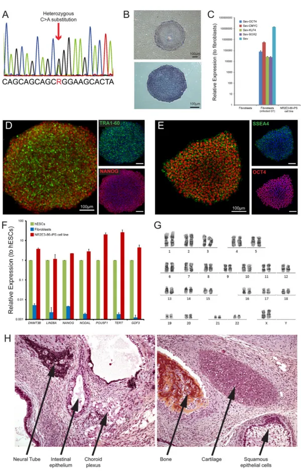
NR2E3 (Nuclear Receptor subfamily 2 group E member 3) is a mem-ber of the nuclear hormone receptor superfamily of ligand-activated transcription factors, whose expression is restricted to photoreceptors and essential for proper rod and cone photoreceptor differentiation (Mollema and Haider 2010). Most of the disorders linked to mutations in this gene have a recessive mode of inheritance and lead to enhanced S-cone sensitivity syndrome, including Goldmann-Favre syndrome (Schorderet and Escher 2009). Only one mutation (p.Gly56Arg) in the first zinc-finger of the DNA-binding domain of NR2E3 causes autosomal dominant Retinitis Pigmentosa (RP) with a relatively high prevalence (3 to 3.5%) in Europe (Audo et al. 2012).
In this study, skinfibroblasts from a 49-year-old woman were reprogrammed into iPSCs using non-integrative Sendai viruses
containing the reprogramming factors, OCT3/4, SOX2, CMYC, KLF4. The presence of the mutation (c.166GNA p.Gly56Arg) in the derived NR2E3-86 iPS cell line was confirmed by Sanger sequencing (Fig. 1A). NR2E3-86 iPS cell colonies displayed a typical ES-like colony morpholo-gy and growth behavior and they stained positive for alkaline phospha-tase activity (Fig. 1B). We confirmed the clearance of the vectors and the ⁎ Corresponding author.
E-mail address:olivier.goureau@inserm.fr(O. Goureau).
Unique stem cell line identifier IDVi001-A Alternative name(s) of stem cell line NR2E3-86-iPS Institution Institut de la Vision
Contact information of distributor Olivier Goureau,olivier.goureau@inserm.fr
Type of cell line iPSC
Origin Human
Additional origin info Age: 49-year old Sex: female Cell Source Dermalfibroblasts Method of reprogramming Transgene free (Sendai Virus) Associated disease Retinitis Pigmentosa (RP) Gene/locus NR2E3 (c.166GNA; p.Gly56Arg) Method of modification N/A
Gene correction NO Name of transgene of resistance N/A Inducible/constitutive system N/A Date archived/stock date Dec. 22, 2016 Cell line repository/bank Not applicable
Ethical approval Approval by French regulatory agencies: CPP Ile de France (2012-A01333-40; P12-02) and the ANSM (B121362-32)
http://dx.doi.org/10.1016/j.scr.2017.08.003
1873-5061/© 2017 Published by Elsevier B.V. This is an open access article under the CC BY-NC-ND license (http://creativecommons.org/licenses/by-nc-nd/4.0/). Contents lists available atScienceDirect
Stem Cell Research
exogenous reprogramming factor genes by qPCR after ten passages (Fig. 1C). Immunofluorescence analysis revealed expression of transcription factors OCT4 and NANOG, and surface markers SSEA4 and TRA1-60, characteristics of pluripotent stem cells (Fig. 1D-E). Flow cytometry
confirmed the expression of the pluripotent markers SSEA4 and TRA1-81 (Supplementary Fig. 1A). The endogenous expression of the pluripotency associated transcription factors DNMT3B, LIN28A, NANOG, NODAL, POU5F1 (OCT4), TERT and GDF3 evaluated by RT-qPCR was
comparable with levels observed in human ES cells (hESCs) (Fig. 1F). The NR2E3-86 iPS cell line displayed a normal karyotype (46, XX) after more than twenty culture passages (Fig. 1G). The identity of the iPS cell line with parentalfibroblasts and genomic integrity was con-firmed by SNP genotyping (Supplementary Fig. 1B). Teratoma assays showed the presence of normal differentiation towards endodermal, ec-todermal and mesodermal layers (Fig. 1H). NR2E3-86 iPS cell line was negative for Mycoplasma contamination (Supplementary Fig. 1C). Taken together, we have successfully reprogrammed p.Gly56Arg NR2E3 dermalfibroblasts into iPSCs that can be used to generate photo-receptors in order to study pathogenic mechanism underlying RP caused by mutation in the NR2E3 gene (Table 1).
Materials and methods
Humanfibroblast cultures and reprogramming
Small pieces of punch biopsy placed into a T25 cm2culture dish were exposed to a minimal amount of media Dulbecco's Modified Eagle's me-dium (DMEM) with 10% fetal calf serum (FCS), 1 mM some-dium pyruvate, Amphotericinβ (Fungizone®) and penicillin–streptomycin (all from ThermoFischer Scientific), and incubated at 37 °C in 5% CO2in a
humid-ified incubator. The following day, 2 ml of fresh medium was added. When sufficient fibroblast outgrowth had occurred, cells were washed in PBS, dissociated using TrypLE Express (Thermo Fischer Scientific) and re-plated at a split ratio of 1:3. Humanfibroblasts at 50–80% of con-fluence were transduced using the CytoTune Sendai reprogramming vectors Oct4, Klf4, Sox2 and c-Myc (ThermoFischer Scientific) and cul-tured for 6 days infibroblast medium before to be plate on mitomycined human foreskin (MHF)-seeded dishes. The day after, thefibroblast me-dium was replaced with the iPS meme-dium, corresponding to ReproStem medium (ReproCELL, Ozyme) supplemented with 10 ng/ml of human recombinant FGF2 (PreproTech France). The emergent hiPSC colonies were picked under a stereomicroscope according to their hESC-like col-ony morphology and expanded on feeder layers. After generation of a frozen stocks, hiPSCs were cultured on MHF feeder layers or preferen-tially adapted and cultured in feeder free conditions (Reichman et al. 2017) on truncated recombinant human vitronectin-coated dishes with Essential 8™ medium (both from ThermoFischer Scientific). Ab-sence of mycoplasma contamination was verified by the MycoAlert™ Mycoplasma Detection Kit (selective biochemical test of mycoplasma enzymes) according to the manufacturer's instructions (Lonza).
After ten passages, the clearance of the exogenous reprogramming factors and Sendai virus genome was confirmed by qPCR following the manufacturer's instructions (ThermoFischer Scientific) (Table 2).
Mutation analysis
Genomic DNA from hiPSCs was extracted with Nucleospin Tissue Kit (Macherey-Nagel) according to the manufacturer instruction. PCR am-plification flanking exon 2 of NR2E3 gene (Table 2) was performed using HOT FIRE Pol DNA Polymerase (Solis BioDyne). PCR products were sequenced using BigDye® Terminator v1.1 Cycle Sequencing Kit (ThermoFischer Scientific) on a 3730 DNA analyzer (Applied Biosystems).
Karyotype analyses
Conventional cytogenetic analysis was performed as described pre-viously (Reichman et al. 2014). Molecular karyotype was analyzed by SNP genotyping using Illumina's Infinium HumanCore-24 Bead Chips (Illumina, Inc., San Diego, USA) at Integragen (Evry, France). Processing was performed on genomic DNA following the manufacturer's proce-dures. LogR ratio and B allele plots were generated in GenomeStudio software (Illumina, Inc.).
In vivo pluripotency analysis by teratoma formation assay
Teratoma assays were performed as described previously (Reichman et al. 2014).
Real-Time PCR analysis
Total RNAs were extracted using Nucleospin RNA II kit (Macherey-Nagel) and cDNA synthesized using the QuantiTect reverse transcription kit (Qiagen) following manufacturer's recommendations. qPCR analysis was performed in three minimum independent biological experiments with custom TaqMan® Array 96-Well Fast plates (Thermo Fischer Scientific) according to the manufacturer's protocol as described previ-ously (Reichman et al. 2014) (Table 2) .
Flow cytometry
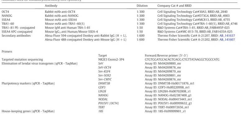
Cells were detached with Accutase solution and harvested for quan-titative analysis byflow cytometry (Cytomics FC500 MCL; Beckman Coulter) by staining the TRA-1-81, and SSEA-4 antibodies (Table 2) and data were analyzed with FlowJo software.
Alkaline phosphatase and immunofluorescence staining
Staining offixed hiPSCs was performed as described previously (Reichman et al. 2014) (Table 2).
Table 1
Characterization and validation.
Classification Test Result Data
Morphology Photography AP staining
hESC-like morphology Positive
Fig. 1, panel B
Phenotype qPCR Expression of pluripotency markers: DNMT3B, LIN28A, NANOG, NODAL, POU5F1, TERT, GDF3
Fig. 1, panel F
Immunohistochemistry Expression of pluripotency markers: OCT4, NANOG, SSEA4 and TRA1–60
Fig. 1, panels D and E
Flow cytometry SSEA-4:97.8% and TRA1-81: 95.1% Supplementary Fig. 1A Genotype Karyotype (G-banding) and resolution 46XX,
Resolution 450–500
Fig. 1, panel G
SNP array analysis Genomic integrity Supplementary Fig. 1B Identity SNP array analysis Genomic integrity and identity (parentalfibroblasts
and the respective iPS cell line)
Supplementary Fig. 1B
Mutation analysis (IF APPLICABLE) Sequencing Heterozygous (GNA) Fig. 1, panel A Microbiology and virology Mycoplasma Mycoplasma testing by luminescence: Negative Supplementary Fig. 1C Differentiation potential Teratoma formation Representation of all three germ layers formation Fig. 1, panel H Donor screening (OPTIONAL) N/A
Supplementary data to this article can be found online athttp://dx. doi.org/10.1016/j.scr.2017.08.003.
Acknowledgements
We are grateful to Dr. S. Mohand-Said and D. Dagostinoz (CIC1423, Hôpital des Quinze-vingts) for their help in patient recruitments. We thank Dr. S. Aractingi and I. Naoura (INSERM UMRS_938, Hôpital Saint-Antoine, Paris) for skin biopsies. We thank L. Riancho for FACS analysis, Dr. N. Oudrhiri and Prof. A. Bennaceur (Service d'hématologie cytogénétique GHU Paris-Sud APHP, INGESTEM ANR Programme Investissements d'Avenir) for the conventional cytogenetic analysis and Dr. O. Feraud and Prof. F. Griscelli (ESTeam Paris Sud/U935 INGESTEM ANR Programme Investissement d'Avenir) for the teratoma assay. This work was supported by grants from the ANR [GPiPS: ANR-2010-RFCS005] and SANOFI-FOVEA to O.G. It was also performed in the frame of the LABEX LIFESENSES [ANR-10-LABX-65] supported by the ANR within the Investissements d'Avenir programme [ANR-11-IDEX-0004-02]. A. Terray was supported by Regional Council of Ile-de-France (DIM Biothérapies) and by Fondation de Ile-de-France (Berthe Fouassier grant).
Author disclosure statement
There are no competingfinancial interests in this study.
References
Audo, I., Bujakowska, K.M., Léveillard, T., Mohand-Saïd, S., Lancelot, M.-E., Germain, A., Antonio, A., Michiels, C., Saraiva, J.-P., Letexier, M., Sahel, J.-A., Bhattacharya, S.S., Zeitz, C., 2012. Development and application of a next-generation-sequencing (NGS) approach to detect known and novel gene defects underlying retinal diseases. Orphanet J. Rare Dis. 7:8.http://dx.doi.org/10.1186/1750-1172-7-8.
Mollema, N., Haider, N.B., 2010. Focus on molecules: nuclear hormone receptor Nr2e3: impact on retinal development and disease. Exp. Eye Res. 91:116–117.http:// dx.doi.org/10.1016/j.exer.2010.04.013.
Reichman, S., Terray, A., Slembrouck, A., Nanteau, C., Orieux, G., Habeler, W., Nandrot, E.F., Sahel, J.-A., Monville, C., Goureau, O., 2014. From confluent human iPS cells to self-forming neural retina and retinal pigmented epithelium. Proc. Natl. Acad. Sci. U. S. A. 111:8518–8523.http://dx.doi.org/10.1073/pnas.1324212111.
Reichman, S., Slembrouck, A., Gagliardi, G., Chaffiol, A., Terray, A., Nanteau, C., Potey, A., Belle, M., Rabesandratana, O., Duebel, J., Orieux, G., Nandrot, E.F., Sahel, J.-A., Goureau, O., 2017. Generation of storable retinal organoids and retinal pigmented ep-ithelium from adherent human iPS cells in xeno-free and feeder-free conditions. Stem Cells 35:1176–1188.http://dx.doi.org/10.1002/stem.2586.
Schorderet, D.F., Escher, P., 2009. NR2E3 mutations in enhanced S-cone sensitivity syn-drome (ESCS), Goldmann-Favre synsyn-drome (GFS), clumped pigmentary retinal de-generation (CPRD), and retinitis pigmentosa (RP). Hum. Mutat. 30:1475–1485.
http://dx.doi.org/10.1002/humu.21096. Table 2
Reagents details.
Antibodies used for immunocytochemistry/flow cytometry
Antibody Dilution Company Cat # and RRID
OCT4 Rabbit mAb anti-OCT4 1:300 Cell Signaling Technology Cat#30A3, RRID:AB_2840 NANOG Rabbit mAb anti-NANOG 1:300 Cell Signaling Technology Cat#D73G4, RRID:AB_4903 SSEA4 Mouse mAb anti-SSEA4 1:300 Cell Signaling Technology Cat#MC813, RRID:AB_4755 TRA1–60 Mouse mAb anti-TRA1–60(S) 1:300 Cell Signaling Technology Cat#TRA-1-60(S), RRID:AB_4746 TRA1–81 PE- conjugated Mouse IgM anti Human TRA-1-81 1:50 R&D Systems Cat#TRA-1-81, RRID:AB_FAB8495P-025 SSEA4 APC-conjugated Mouse IgG3anti Human/Mouse SSEA-4 1:50 R&D Systems Cat#MC-813-70, RRID:AB_FAB1435A-025
Secondary antibodies Alexa Fluor 594-conjugated Donkey anti-Rabbit IgG (H + L), 1:600 Thermo Fisher Scientific Cat# A-21207, RRID:AB_141637
Alexa Fluor 488-conjugated Donkey anti-Mouse IgG (H + L) 1:600 Thermo Fisher Scientific Cat# A-21202, RRID:AB_141607
Primers
Target Forward/Reverse primer (5′-3′)
Targeted mutation sequencing NR2E3 Exons2-3P4 GTGTGGATGCACAGTGAGG/CTGTTATAAGGCTGGCCATG Elimination of Sendai virus transgenes (qPCR - TaqMan) SeV
SeV-OCT4 Sev-KLF4 Sev-SOX2 Sev-CMYC Assay ID: Mr04269880_mr Assay ID: Mr04269878_mr Assay ID: Mr04269879_mr Assay ID: Mr04269881_mr Assay ID: Mr04269876_mr Pluripotency markers (qPCR - TaqMan) DNMT3B
GDF3 LIN28 NANOG NODAL POU5F1 (OCT4) TERT Assay ID: DNMT3B-Hs00171876_m1 Assay ID: GDF3-Hs00220998_m1 Assay ID: LIN28A-Hs00702808_s1 Assay ID: NANOG-Hs02387400_g1 Assay ID: NODAL-Hs00415443_m1 Assay ID: POU5F1-Hs00999632_g1 Assay ID: TERT-Hs00972656_m1 House-keeping genes (qPCR - TaqMan) 18S Assay ID: 18S-Hs99999901_s1