Augmented venous return for minimally invasive open heart surgery with
selective caval cannulation
David Jegger, Hendrik T. Tevaearai*, Judith Horisberger, Xavier M. Mueller, Yves Boone,
Nicolas Pierrel, Isabelle Seigneul, Ludwig K. von Segesser
Department of Cardiovascular Surgery, University Hospital (CHUV), Rue du Bugnon 46, CH-1011, Lausanne, Switzerland Received 28 April 1999; received in revised form 21 June 1999; accepted 22 June 1999
Abstract
Objective: Minimally invasive open heart surgery involves limited intrathoracic cannulation sites necessitating cardiopulmonary bypass to be initiated via peripheral access using percutaneous cannulae with the tip placed into the right atrial cavity. However, surgery involving the opening of the right heart obliges the surgeon to maintain the end of the cannulae into the vena cavae. The impeded venous return due to the smaller diameter may be alleviated by inserting a centrifugal pump in the venous line. Methods: Right anterior mini-thoracotomy and exposure of the femoral site were performed before the patient was heparinized. Cannulation of the femoral artery, the inferior vena cava via the femoral vein and the superior vena cava through the mini-thoracotomy was performed and cardiopulmonary bypass was initiated. Venous drainage was augmented with the centrifugal pump. Cardiac arrest was provoked and both vena cavae were snared before performing the intracardiac procedure. Results: Twenty consecutive patients were operated on using this technique (15 males/®ve females; age: 44:8 ^ 14:3 years; bodyweight: 73:5 ^ 15:1 kg; body surface area: 1:8 ^ 0:2 m2; theoretical blood ¯ow rate: 4:4 ^ 0:5 l/min). The cannula sizes were
21:9 ^ 2:2 Fr for the femoral artery, 26:5 ^ 1:7 Fr for the inferior vena cava and 23:8 ^ 2:5 Fr for the superior vena cava. Venous drainage through the single inferior vena cava cannula was 2:1 ^ 0:6 l/min (48:8 ^ 13:3% of the theoretical ¯ow). Adding the superior vena cava cannula increased the venous ¯ow to 3:1 ^ 0:4 l/min (70:7 ^ 9:6% of the theoretical value, P , 0:005). The use of the centrifugal pump increased the ¯ow to 4:1 ^ 0:6 l/min (93:4 ^ 8:9% of the theoretical ¯ow, P , 0:001) with a mean inlet negative pressure of 269:1 ^ 10:2 mmHg. The mean bypass time was 64:0 ^ 24:6 min for a mean operative time of 226:3 ^ 61:0 min. Minimum venous saturation was 69:4 ^ 8:5%. Conclusions: Despite the smaller diameter of the vena cavae compared to the right atrium, and a smaller internal diameter of percutaneous cardiopulmonary bypass cannulae compared to classic ones; the centrifugal pump improves the venous drainage signi®cantly so that minimally invasive open heart procedures can be performed under optimal and safe perfusion conditions. q 1999 Elsevier Science B.V. All rights reserved.
Keywords: Thoracic surgery; Surgical procedures/minimally invasive; Cardiopulmonary bypass; Extracorporeal circulation; Centrifugal; Kinetic/drainage
1. Introduction
Over the past 10 years most of the surgical specialties have moved towards minimally invasive operations with rapid progress in developments of new instrumentation, reduction in hospital stay, reduction in postoperative discomfort and consecutive expansion of surgical indica-tions. Classic open heart surgery requiring cardiopulmonary bypass (CPB) is generally performed through a full median sternotomy which provides maximum area for surgical maneuvers. This approach also offers the easiest access for optimal cannulae placement. Minimally invasive Cardiac Surgery recently followed. Reduction of tissue
trauma is evident as seen in leg wounds where minimally invasive harvesting of the great saphenous vein was performed for Coronary Artery Bypass Graft (CABG) surgery [1]. Anastamosis of the left internal mammary artery (LIMA) to the left anterior descending coronary artery (LAD) through a mini thoracotomy [2] was ®rst performed in 1995 as well as the thoracoscopic closure of a patent ductus arteriosus (PDA) [3]. Other intracardiac procedures were then performed through a mini thoracot-omy such as a mitral valve procedure [4] and an atrial septal defect (ASD) closure [5]. Alternative cannulation sites can be chosen for patients having reoperations, ascending aortic aneurysm or dissections, and aneurysms of the descending thoracic aorta, patients requiring support angioplasty or extra-corporeal membrane oxygenation, or patients necessi-tating CPB for severe accidental hypothermia [6,7]. These
1010-7940/99/$ - see front matter q 1999 Elsevier Science B.V. All rights reserved. PII: S1010-7940(99)00228-6
www.elsevier.com/locate/ejcts
* Corresponding author. Tel: 141-21-314-2280; fax: 141-21-314-2278. E-mail address: [email protected] (H.T. Tevaearai)
sites include ilio-femoral artery and vein, or less frequent axilloaxillary usage [8].
Access for minimally invasive open heart surgery poses the greatest limitation for the surgical team and involves restricted intrathoracic cannulation sites. Peripheral cannula-tion of the ilio-femoral artery and vein is therefore often preferred [9]. Percutaneous cannulae are longer and have smaller internal diameters compared to standard CPB cannu-lae. This causes ¯ow restrictions which is disadvantageous for the surgical team. Minimally invasive surgery where the right heart is not opened such as CABG procedures, can be performed with a single venous cannula introduced via the femoral vein until the tip is positioned into the right atrial cavity. In these circumstances, a centrifugal pump (CF pump) placed on the venous line prior to the cardiotomy reservoir has proven its ef®ciency in providing additional ¯ow to the heart lung machine providing a kinetic assisted venous drainage [10]. However, open heart procedures invol-ving opening of the right and sometimes the left atrium for example the closure of an ASD, preclude the positioning of the venous cannula into the atrial cavity. The tip of the cannu-lae thus have to be positioned in the venae cavae. Because of their smaller diameter compared to the right atrial cavity, the venous return to the heart lung machine may further be restricted. We investigated the addition of a CF pump into
the venous line and prospectively analysed the potential bene®ts of active venous drainage of both vena cavae during minimally invasive open heart surgery.
2. Materials and methods 2.1. Perfusion technique
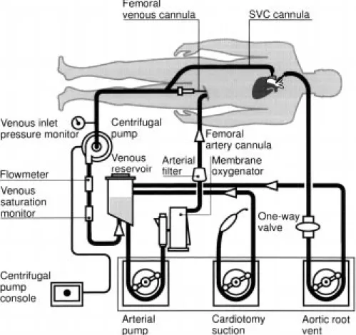
The perfusion circuit layout is represented in Fig. 1. An arterial roller pump (Jostra Medizintechnik AG, Hirrlingen, Germany), a hollow ®bre membrane oxygenator (Dideco, Mirandola, Italy), a conventional tubing circuit (Dideco, Mirandola, Italy), a Biomedicus CF pump console (Medtro-nic Biomedicus, Eden Prairie, MN, USA) and an air-oxygen blender (Sechrist Industries, Inc., Anaheim, CA, USA) are used in all of the procedures. The venous CF pumphead is either a Biomedicus BP-80 (Medtronic Biomedicus, Eden Prairie, MN, USA) or a Jostra `Rota¯o' (Jostra, Medizin-technik AG, Hirrlingen, Germany). The extracorporeal circuit is primed with 1000 ml Ringer lactate, 500 ml Haes, 100 mg heparin (Liquemin, Hoffmann-La Roche, Basel, Switzerland), an antibiotic and 1 million IU Aproti-nin (Trasylol, Bayer, ZuÈrich, Switzerland) following the departmental protocol. The CF pump is placed in the venous
line with a negative pressure monitoring site at its inlet (DLP, Grand Rapids, MI, USA) to monitor the mean nega-tive pressure generated by the CF pump. A ¯ow meter connector is inserted between the CF pump outlet and the inlet of the venous reservoir (Medtronic Biomedicus, Eden Prairie, MN, USA). The mixed venous oxygen saturation is measured with an OxySat Meter (Bentley, Baxter, Irvine, CA, USA).
2.2. Surgical technique
A right anterior mini-thoracotomy is performed at the 4th intercostal space. The pericardium is opened anteriorly to the phrenic nerve to expose the right atrium and both venae cavae. Simultaneously, the femoral site is prepared to expose the femoral artery and femoral vein.
The patient is anticoagulated with 3 mg/kg heparin (Liquemin, Hoffmann-La Roche, Basel, Switzerland) before cannulation of the femoral artery. The length of the inferior vena cava (IVC) venous cannula is estimated to position its tip into the mid IVC via the femoral vein. Subsequent super-ior vena cava (SVC) cannulation occurs via the mini thor-acotomy. The two cavae cannulae are joined to each other with a `Y' connector which makes up the venous inlet to the CPB circuit. CPB is initiated with passive venous drainage via the IVC cannula alone, and a ®rst ¯ow is noted. The SVC cannula is then unclamped and the new venous ¯ow is noted. Finally, the CF pump is started and its speed is increased progressively to reach the maximum active venous return, while maintaining a negative pressure between 250 and 280 mmHg. Again the ¯ow rate is recorded. Ventricular ®brillation is electrically induced (Fibrillator Fi10M, Stockert Instrumente GmbH, Munich,
Germany) before both vena cavae are snared and the atrium opened. The intracardiac procedure is performed under direct and video assisted vision [5]. After careful deairing maneuvers by way of a vent placed in the ascending aorta, external de®brillation is performed (CodeMaster de®brilla-tor, Hewlett-Packard Company, Andover, USA). The wean-ing process started by ®rst stoppwean-ing the centrifugal pump and then reducing the arterial ¯ow rate while impeding the venous return.
The following parameters were continually recorded: arterial pump ¯ow rate, negative pressure at the venous inlet to the CF pump dome, mean arterial pressure (MAP) and mixed venous oxygen saturation.
Data were analysed using the Student's t-test and statis-tical signi®cance was assumed when P was ,0.05. 3. Results
Twenty consecutive patients involving cannulation of both vena cavae and active venous drainage using this tech-nique are reported (18 patent foramen ovale closures, 1 mitral valve replacement and 1 ASD closure). They consisted of 15 males and ®ve females with a mean age of 44:8 ^ 14:3 years. The weight was 73:5 ^ 15:1 kg (min: 51.8 kg, max: 114 kg), the mean body surface area (BSA) was 1:8 ^ 0:2 m2(min: 1.5 m2, max: 2.3 m2) and the mean theoretical blood ¯ow was 4:4 ^ 0:5 l/min (min: 3.6 l/min, max: 5.5 l/min). The cannulae sizes was 26:5 ^ 1:7 Fr for the IVC, 23:8 ^ 2:5 Fr for the SVC and 21:9 ^ 2:2 Fr for the femoral artery.
Mean arterial pump ¯ow through the single femoral cannula was 2:1 ^ 0:6 l/min or 48:8 ^ 13:3% of the
theore-Fig. 2. Means and standard deviations for one cannula (A), two (p) and two plus the CF pump (B) during CPB. P , 0:005 for two cannulae vs. one cannula (*) and P , 0:001 for two cannulae plus the CF pump vs. two cannulae without the CF pump (**).
tical ¯ow (Fig. 2). Adding the SVC cannula increased the mean arterial pump ¯ow to 3:1 ^ 0:4 l/min or 70:7 ^ 9:6% of the theoretical value (Fig. 2) (P , 0:005). Adding the CF pump increased the mean arterial pump ¯ow to 4:1 ^ 0:6 l/ min or 93:4 ^ 8:9% of the theoretical ¯ow (Fig. 2) (P , 0:001) with an overall mean inlet negative pressure of 269:1 ^ 10:2 mmHg. The mean CPB time was 64:0 ^ 24:6 min for a mean operative time of 226:3 ^ 61:0 min. Minimum venous saturation during the running of the CPB was 69:4 ^ 8:5%. The cardiac arrest time was 31:4 ^ 18:5 min. All patients underwent successful CPB weaning with minimal inotropic support and were transferred to the inten-sive care unit in a stable condition. There were no compli-cations reported, no postoperative organ dysfunction, nor any other complication related to this surgical technique. Echocardiographic control at day 7 was normal in every patient.
4. Discussion
The small thoracotomy performed during minimally invasive cardiac surgery procedures presents limited access for cannulation when CPB is required. Under these circum-stances, the preferred alternative is the femoral or iliac approach for venous and arterial cannulation. This techni-que has proven its ef®ciency for other procedures such as certain dif®cult reoperations [6,7], ascending aortic aneur-ysm or dissection, or cases where the extent of arteriosclero-sis contra-indicates the possibility of a direct aortic cannulation because of the high risk of emboli dislodgement [8]. It is also a convenient access for extra-corporeal membrane oxygenation, severe accidental hypothermia rescue or support angioplasty. In these situations, the percu-taneous venous cannula is inserted until its tip is positioned into the right atrium to obtain a maximum blood drainage from the patient to the heart lung machine. Nevertheless, the venous return is usually limited compared to direct cannula-tion of the right atrium in the classic sternotomy approach. The length of the percutaneous cannula and its internal diameter are the major factors responsible for the ¯ow impe-diment although parameters not related to the cannula like the central venous pressure (CVP), also in¯uence the drai-nage of blood into the cardiotomy reservoir. These limita-tions imply a suboptimal venous drainage which may consequently impede arterial perfusion. In order to compen-sate the reduced venous return, the insertion of the CF pump in the venous line was proposed. Bench tests demonstrated that the active ¯ow is not in¯uenced by the ®lling pressure as long as the tip of the cannula is maintained into a large reservoir such as the right atrium [11]. In these cases situa-tions, the risk of atrial wall collapsing is very unlikely even with the negative pressure induced around the cannula by the CF pump.
In our study, the surgical procedures involved the actual (ASD closure) or potential (left atriotomy) opening of the
right atrium. Therefore drainage of both vena cavae was necessary. The positioning of two cannulae through the mini-thoracotomy would limit the surgeons access to the heart, overcrowd the operating ®eld and limit the degree of maneuverability in this restricted operating area. Recently, a two-stage cannula was designed to permit the drainage of both vena cavae through a single femoral access (CarpentierTM Bi-caval femoral cannula, Medtronic DLP, Grand Rapids, MI, USA) [12]. In our patients, the drainage of the IVC was performed through a peripheral approach whereas the SVC was drained with a second cannula placed into the mini-thoracotomy. In vitro analysis demonstrated that the drainage through a cannula positioned into a collap-sible tube such as the vena cava is highly dependent on the ®lling pressure [11]. It is, in addition, signi®cantly in¯u-enced by the ratio between the outer diameter of the cannula and the diameter of the drained cavity because; whatever the size of the cannula, the ¯ow is reduced when this ratio is higher than 50% [11]. This would suggest that the use of a CF pump may not be applicable in those situations as the negative pressure induced by the CF pump would provoke the collapsing of the venous wall. We nevertheless observed a signi®cant augmentation of the venous return with the use of the CF pump. The passive venous drainage of both vena cavae was limited to approximately 3.0 l/min or 70% of the theoretical ¯ow which is insuf®cient to ensure adequate systemic perfusion. The usage of the CF pump signi®cantly augmented the venous return to approximately 4 l/min corresponding to approximately 95% of the theoretical blood ¯ow. The CF pump therefore allowed augmentation of roughly 25% of the venous return to the heart lung machine. In this situation, the systemic perfusion was adequate as demonstrated by the normal mixed venous oxygen saturation values we observed in every patient.
The continual monitoring of the negative pressure in the venous line is mandatory. A range of 250 to 280 mmHg was suggested [10] as higher values may provoke a cavita-tion phenomenon or unnecessary hemolysis [13]. In addi-tion, because of the small diameter of the vein compared to that of the cannula, the vein wall may be trapped onto the cannula tip, which would in turn create temporary chattering of the venous line. The elevated CVP, i.e. a high ®lling pressure allows to balance this phenomenon and prevents from vein collapse. However, because of the arti®cial nega-tive pressure created by the CF pump at the tip of the venous cannula, it is not possible to monitor the CVP during the procedure. Therefore, a special attention has to be paid to variations in the negative pressure as it may correspond to the chattering of the venous walls and indicate that the CF pump speed has to be adapted. In our patients, the negative pressure was maintained around 270 mmHg, and no collap-sing or other technical problems were observed.
The use of a CF pump was already documented in PortAccess cardiac surgery providing total CPB support and cardiac decompression [10,14]. In these studies, the tip of the venous cannula was placed into the right atrial
cavity and an augmentation of blood ¯ow of 20±40% was shown when used optimally.
Minimally invasive cardiac surgery is just beginning and the entire therapeutic team has to adapt to the new surgical condition. Because of the limited access, monitoring of the heart during the procedure obliges one to employ remote systems. CF pump is one example that has been added to ensure an optimal CPB. The running of the CPB procedure is nevertheless unchanged. Once the CF pump speed is optimally regulated and the negative pressure in the venous line is within its limits, no further adjustments need to be made. Other additional instruments such as the endoscope recently appeared helpful to further increase the quality of surgery [15].
We demonstrate in our study that the use of a CF pump for kinetic assisted venous drainage can also be applied in cardiac surgery involving the opening of the right atrium where a venous cannula must be inserted into each vena cava. Adequate mixed venous oxygen saturation, ®lling pressure and continual monitoring of the negative pressure render this technique totally safe and adapted to suit mini-mally invasive open heart surgery.
References
[1] Tevaearai HT, Mueller XM, von Segesser LK. Minimally invasive harvest of the saphenous vein for coronary artery bypass grafting. Ann Thorac Surg 1997;63:119±121.
[2] BenettiFJ,BallesterC.Useofthoracoscopyandaminimalthoracotomy in mammary-coronary bypass to left anterior descending artery without extracorporeal circulation. J Cardiovasc Surg 1995;36:159±161. [3] Laborde F, Folliguet T, Batisse A, Didie A, da-Cruz E. Carbognani,
D. Video-assisted thoracoscopic surgical interruption: the technique of choice for patent ductus arteriosus. Routine experience in 230 pediatric cases. J Thorac Cardiovcasc Surg 1995;110:1681-1684.
[4] Carpentier A, Loulmet D, Carpentier A, Le Bret E, Haugades B, Dassier P, Guibourt P. Chirurgie aÁ coeur ouvert par videÂo-chirurgie et mini-thoracotomie. Premier cas (valvuloplastie mitrale) opeÂre avec succeÁs. C R Acad Sci III 1996;319:219±223.
[5] Tevaearai HT, Mueller XM, Ruchat P, Hurni M, Stumpe F, Fischer A, von Segesser LK. Fermeture chirurgicale `minimale invasive' de Foramen Ovale Permeable. Swiss Surg 1998;S2:14±17.
[6] Fried DW, Zombolas TL, Weiss SJ. Single pump mechanically aspi-rated venous drainage (SPMAVD) for cardiac reoperation. Perfusion 1995;10:327±332.
[7] Jones RE, Fitzgerald D, Cohn LH. Reoperative cardiac surgery using a new femoral venous right atrial catheter. J Card Surg 1990;5:170± 173.
[8] Bichell DP, Balaguer JM, Aranki SF, Couper GS, Adams DH, Rizzo RJ, Collins JJ, Cohn LH. Axilloaxillary cardiopulmonary bypass: a practical alternative to femorofemoral bypass. Ann Thorac Surg 1997;64:702±705.
[9] Wechsler, A.S. Cardiopulmonary bypass through alternative routes Ð thoracic incisions. Proceedings of the Pathophysiology and techni-ques of Cardiopulmonary bypass symposium. XVI symposium brochure. San Diego, CA, 1996. pp. 138-141.
[10] Toomasian JM, McCarthy JP. Total extrathoracic cardiopulmonary support with kinetic assisted venous drainage: experience in 50 patients. Perfusion 1998;13:137±143.
[11] Wenger RK, Bavaria JE, Ratcliffe MB, Bogen D, Edmunds LH. Flow dynamics of peripheral venous catheters during extracorporeal membrane oxygenation with a centrifugal pump. J Thorac Cardiovasc Surg 1988;96:478±484.
[12] Loulmet DF, Carpentier A, Cho PW, Berrebi A, d'Atellis N, Austin AB, Couetil JP, Lajos P. Less invasive techniques for mitral valve surgery. J Thorac Cardiovasc Surg 1998;115:772±779.
[13] Vertrees RA, Brownstein L, Gilbert JL, Cernalanu AC, Delrossi AJ. Centrifugal pump induced alterations in red cell morphology. Proceedings of the Pathophysiology and Techniques in Cardiopul-monary Bypass Symposium. San Diego, CA, 1992.
[14] Solomon L, Sutter FP, Goldman SM, Mitchell JM, Casey K. Augmen-ted femoral venous return. Ann Thorac surg 1993;55:1262±1263. [15] Tevaearai HT, Mueller XM, Stumpe F, Ruchat P, von Segesser LK.
Advantages of a modi®ed gastroscope for video-assisted internal mammary artery harvesting. Ann Thorac Surg 1999;67:872±873.