M A J O R A R T I C L E
Whole Genome Pyrosequencing of Rare
Hepatitis C Virus Genotypes Enhances Subtype
Classi
fication and Identification of Naturally
Occurring Drug Resistance Variants
Ruchi M. Newman,1,aThomas Kuntzen,2,4,aBrian Weiner,1Andrew Berical,2Patrick Charlebois,1Carla Kuiken,3Donald G. Murphy,5Peter Simmonds,6Phil Bennett,7Niall J. Lennon,1Bruce W. Birren,1Michael C. Zody,1Todd M. Allen,2,band Matthew R. Henn1,b
1Broad Institute of MIT and Harvard, Cambridge, and2Ragon Institute of MGH, MIT, and Harvard, Boston, Massachusetts;3Theoretical Biology and
Biophysics Group, Los Alamos National Laboratory, New Mexico;4Department of Gastroenterology and Hepatology, University Hospital Zurich,
Switzerland;5Laboratoire de Santé Publique du Québec, Institut National de Santé Publique du Québec, Canada; and6Centre for Infectious Diseases,
University of Edinburgh, and7Micropathology, University of Warwick Science Park, Coventry, United Kingdom
Background. Infection with hepatitis C virus (HCV) is a burgeoning worldwide public health problem, with 170 million infected individuals and an estimated 20 million deaths in the coming decades. While 6 main geno-types generally distinguish the global geographic diversity of HCV, a multitude of closely related subgeno-types within these genotypes are poorly defined and may influence clinical outcome and treatment options. Unfortunately, the paucity of genetic data from many of these subtypes makes time-consuming primer walking the limiting step for sequencing understudied subtypes.
Methods. Here we combined long-range polymerase chain reaction amplification with pyrosequencing for a rapid approach to generate the complete viral coding region of 31 samples representing poorly defined HCV subtypes. Results. Phylogenetic classification based on full genome sequences validated previously identified HCV subtypes, identified a recombinant sequence, and identified a new distinct subtype of genotype 4. Unlike conventional sequenc-ing methods, use of deep sequencsequenc-ing also facilitated characterization of minor drug resistance variants within these uncommon or, in some cases, previously uncharacterized HCV subtypes.
Conclusions. These data aid in the classification of uncommon HCV subtypes while also providing a high-resolution view of viral diversity within infected patients, which may be relevant to the development of therapeutic regimens to minimize drug resistance.
Keywords. Hepatitis C virus; pyrosequencing; subtype classification; drug resistance mutations; viral diversity.
Hepatitis C virus (HCV) is an enveloped, positive-strand RNA virus belonging to theHepacivirus genus in the Flaviviridae family. Seven confirmed genotypes (1–7) are generally distinguished by phylogenetic methods and pair-wise distance calculations [1,2]. On
the basis of full genome nucleotide sequences, HCV genotypes diverged from each other by a pair-wise dis-tance of >30%. Individual genotypes can be further divided into more closely related subtypes that di-verged by a pair-wise distance of 15%–30%. All viral genotypes retain their repertoire of colinear structural and nonstructural genes, thereby facilitating prelimi-nary genotype classification on the basis of partial genome sequences of short fragments (approximately 300–400 nucleotides) in the structural core/E1 region and the nonstructural NS5B region [3]. However, full genome sequences remain indispensible for the detec-tion of genome recombinadetec-tion events.
While only recombinant HCV genotype 2 k/1b viruses have been found to actively circulate in the
Received 2 April 2012; accepted 8 August 2012; electronically published 6 November 2012.
a
R. M. N. and T. K. contributed equally to this study.
b
T. M. A. and M. R. H. contributed equally to this study.
Correspondence: Ruchi M. Newman, PhD, Broad Institute of MIT & Harvard, 7 Cambridge Center, Cambridge, MA 02142 ([email protected]). The Journal of Infectious Diseases 2013;208:17–31
© The Author 2012. Published by Oxford University Press on behalf of the Infectious Diseases Society of America. All rights reserved. For Permissions, please e-mail: [email protected].
DOI: 10.1093/infdis/jis679
population so far [4–7], recombinants of genotypes 2/5, 2b/1b, 2b/1a, and 2i/6p have been detected in single isolates from humans [8–11]. Furthermore, the frequency of HCV intergen-otype and intragenintergen-otype recombination may be underestimat-ed because of the lack of robust detection methods [12,13]. Although genotyping based on core/E1 or NS5B sequences has resulted in the provisional classification of a large number of subtype variants within each genotype, at least 1 but prefer-ably ≥2 full genome sequences are required to confirm subtype designation [2,12].
Accurate genotype and subtype classification is clinically important because major genotypes differ considerably in their response rates to treatment with pegylated interferon and ribavirin and with directly acting antiviral drugs (DAAs) that are designed mainly against genotype 1 isolates. The limited efficacy of DAAs against other genotypes has been shown for genotype 3 [14], and treatment response rates for drug regi-mens containing boceprevir (an NS3 protease inhibitor) or BMS-790052 (an NS5A inhibitor) were found to vary even between subtypes 1a and 1b [15, 16]. Different baseline fre-quencies of drug resistance mutations may account for these differences [17], and such variation might become more sig-nificant if interferon-free regimens containing 1 or more DAAs could indeed displace the current standard of care [18]. Until clinically tolerable DAAs with high barriers to resistance against the complete spectrum of HCV genotypes become available [18,19], accurate subtype determination and, possi-bly, drug resistance profiling of the individual’s viral popula-tion may guide the optimal choice of drugs. Full genome deep sequencing, as performed in this study, allows the identi fica-tion of drug resistance mutafica-tions across the genome that may exist as dominant or minor variants in treatment-naive pa-tients, thereby informing the design of therapeutic regimens. METHODS
Sample Collection
Plasma samples obtained from HCV-infected patients in Canada and the United Kingdom were received with no ac-companying patient-associated demographic or clinical infor-mation. This study was approved by the Massachusetts General Hospital Review Board and was granted exemption by the Massachusetts Institute of Technology and Massachusetts General Hospital Review Boards as discarded diagnostic samples. Preliminary HCV genotypes were assigned by the clinical centers, and all samples were thought to represent poorly characterized variants of genotypes 2–4.
Polymerase Chain Reaction (PCR) Amplification, Viral Sample Preparation, and Genome Sequencing
RNA isolation, complementary DNA generation, and PCR amplification within the 5′ untranslated region (UTR) and 3
small sequence islands, followed by amplification of 2 hemi-genome fragments that covered the entire protein coding region, were performed as previously described [20], using primers described inSupplementary Table 1. The 5′ UTR and small sequence islands underwent conventional Sanger se-quencing. Pooled hemi-genome PCR products were sequenced by Roche/454, and a de novo assembly was generated as previ-ously described [21]. Any ambiguities in 454 assemblies were resolved using Sanger sequencing. All full genome sequences used in this study have been submitted to Genbank (accession numbers JX227949–JX227979).
Alignments
All unique, nonrecombinant, full-length HCV genomes found in the Los Alamos Hepatitis C Sequence Database (http://hcv. lanl.gov), along with sequences generated in this study, were used for analyses. For core/E1-NS5B analyses, the Los Alamos National Laboratory (LANL) HCV database was searched for all isolates with available sequences for both core/E1 and NS5B (nucleotide positions 869–1292 and 8276–8615, accord-ing to genotype 1a reference strain H77). The intersection of isolates containing both of these sequences was examined on a genotype and subtype level.
Multiple alignments were performed in MUSCLE v3.8.31 [22], using default parameters with 5 iterative refinements. A total of 193 full-length genomes (excluding recombinants) were trimmed to the coding sequence producing a master alignment of 9323 bases (including gaps). Any gaps in the triplet of nucleotides underlying codons were removed from the entire alignment in a greedy fashion, thereby conservative-ly eliminating any alignment artifacts. For the genotyping fragment trees that included 300 viral strains for which both regions of core/E1 (869–1292) and NS5B (8276–8615) were available, these regions were concatenated into an alignment of 779 nucleotides (including gaps). No gap stripping was per-formed on the genotyping fragment sequences.
Identification of Recombinants
The recombination detection program (RDP) [23] was run on all full-length genome sequences representing all available ge-notypes and subtypes. Any sequences that produced consis-tently low P values among the RDP’s multiple tests for recombination were subject to further analysis. These potential recombinant sequences were further validated with SimPlot v3.5.1 [24], in which all possible combinations of major and minor parent strains were run against the recombinant strain.
Construction of Phylogenetic Trees
The randomized axelerated maximum likelihood program [25] was run with 1000 bootstraps to construct a single tree for the 193 genomes mentioned above, using the cleaned alignment containing 3753 bases. The smaller genotyping
trees of 300 concatenated fragments spanning 779 nucleotides were used in the same manner. The GTRGAMMA model was chosen as the model of nucleotide substitution. The single best tree from the 1000 bootstraps is shown, along with the total percentage (0%–100%) of bootstrap support for each branch.
Computation of Pair-Wise Intersubtype and Intrasubtype Divergence
Pair-wise distances for all 193 full-length nonrecombinant genomes were calculated in MEGA v.5.05 [26].
The PhyloPlace tool was used to compute the branching index over the length of our 2 putative subtype 4v genomes (http://hcv.lanl.gov/content/sequence/phyloplace/PhyloPlace. html).
Variant Calling in Mixed Populations
Prior to variant calling, correction of known error modes in Roche/454 read data was performed with the RC454 program [21]. Reliable and sensitive detection of variants in a mixed population and analysis of intrahost diversity was then accom-plished using the VPhaser tool [27].
RESULTS
Phylogenetic Classification of HCV Genotypes Based on Full-Genome Sequences
To better characterize global HCV genotype diversity, sequenc-es reprsequenc-esenting the entire HCV protein-coding region and por-tions of the 5′ and 3′ UTR were generated from plasma samples acquired from 54 patients in Canada or the United Kingdom. Preliminary HCV genotyping based on core/E1 or NS5B sequences indicated that these individuals were infected with poorly characterized subtypes of genotypes 2, 3, or 4. Use of a hemi-genome amplification approach [20], coupled with high-throughput pyrosequencing technology, enabled genera-tion of sequence reads covering the entire coding region without the need for traditional primer-walking methods. The success rate for amplification of both hemi-genomes from the 54 samples attempted was 83%, with 31 samples being used for sequencing. A recently developed algorithm allowed de novo assembly of all 31 genomes, despite the lack of full-length refer-ence sequrefer-ences for these poorly characterized genotypes [21]. Genome sequences were aligned to all available full-length sequences from the National Center for Biotechnology Infor-mation and LANL public databases, including select represen-tatives of the commonly found 1a or 1b subtypes.
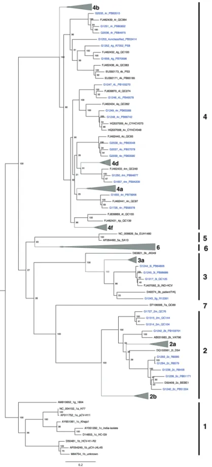
Phylogenetic analysis revealed that several of the new se-quences clustered with subtypes for which only 1 full-length genome sequence had been reported (2c, 2k, 3i, 4g, 4l, 4m, 4n, 4o, and 4r), thereby strengthening these phylogenetic group-ings (Figure 1), with pair-wise distance measurements con-firming their distinction from other subtypes (Table 1).
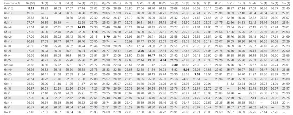
Sequences provisionally assigned to the 2 m and 3 g subtypes on the basis of partial sequence grouped accordingly, formally establishing these as distinct HCV subtypes (Figures1and2). Likewise, quantification of relatedness to all known subtypes by branching index analysis (Supplementary Figure 1), coupled with 100% bootstrap support by phylogeny (Figure1) and pair-wise distance measurements (Table 1), clearly grouped 2 newly described genotype 4 sequences, G1248/ PB66742 and G1249/PB65568, to near full-length genomes from the recently identified 4v provisional subtype [28], thereby confirming 4v as a distinct subtype of genotype 4.
In addition to validating previously identified HCV genetic subtypes, phylogenetic analysis also identified a novel geno-type 4 variant. While isolate G1253/PB53414 grouped with ge-notype 4 sequences in the full-genome phylogenetic tree (Figure 1), bootstrap resampling strongly supported G1253/ PB53414 as a unique subtype as compared to genotypes 4g and 4k (Figure1). Similarly, G1253/PB53414 was genetically distinct from subtypes 4h, 4g, and 4k in the core/E1-NS5B tree (Figure2). In addition, nucleotide sequence divergence of G1253/PB53414 from all other genotype 4 full genome se-quences was >17.7% (Table 1). These lines of evidence strongly suggest that G1253/PB53414 represents a novel subtype of a genotype 4. This new genotype 4 variant could be designated as subtype 4w, but since only 1 example of this subtype has been sequenced, accepted convention for assign-ing provisional subtypes calls for designation of G1253/ PB53414 as an unclassified genotype 4 variant.
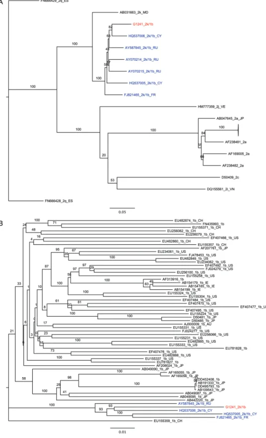
One recombinant sequence was also identified. SimPlot analysis showed G1241/PB41880 to be a recombinant between subtypes 2k (K1-S2/AB031663) and 1b (Vat-96/D50485), sharing a recombination break point with 2k/1b recombi-nants previously identified in St. Petersburg, CRF_01_1b2k (Figure3). Despite high sequence identity (93%–94%), G1241/ PB41880 is not identical to previously reported 2k/1b recom-binants. Nevertheless, separate phylogenetic analysis of 2k and 1b components of the new strain identified here along with other nonrecombinant 2k and 1b sequences confirm that there is 100% bootstrap support for G1241/PB41880 grouping with all other reported 2k/1b strains (Figure4).
In the case of subtypes 2 m and 3 g, for which no full genome sequences were available in public databases, classification of these new sequences on the basis of genotyping regions alone was clearly supported (Figure2). As expected, in instances where both full genome and partial genome data were available, bootstrap support for subtype classification was stronger in full-genome trees as compared to partial genome trees (Figures1and2).
Prevalence of Dominant, Naturally Occurring Polymorphisms at Positions Associated with Drug Resistance
Alignments of these full genome sequences allowed us to in-vestigate the prevalence of dominant amino acid substitutions
Figure 1. Maximum likelihood tree of 30 complete, nonrecombinant hepatitis C virus coding-region nucleotide sequences generated as part of this study (highlighted in blue), and 163 sequences from public sequence repositories. Bootstrap resampling (1000 replications) support values are shown at nodes. The tree is rooted using genotype 1 sequences, and all horizontal branch lengths are drawn to a scale of nucleotide substitutions per site.
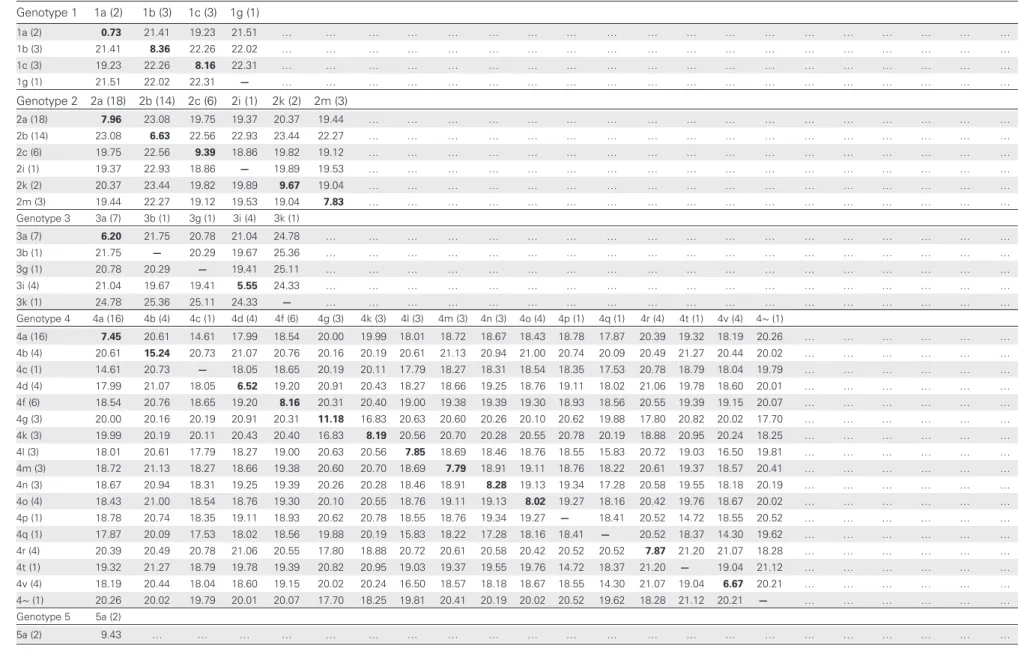
Table 1. Average Internucleotide and Intranucleotide Divergence Among Hepatitis C Virus Subtypes Genotype 1 1a (2) 1b (3) 1c (3) 1g (1) 1a (2) 0.73 21.41 19.23 21.51 … … … … 1b (3) 21.41 8.36 22.26 22.02 … … … … 1c (3) 19.23 22.26 8.16 22.31 … … … … 1g (1) 21.51 22.02 22.31 — … … … … Genotype 2 2a (18) 2b (14) 2c (6) 2i (1) 2k (2) 2m (3) 2a (18) 7.96 23.08 19.75 19.37 20.37 19.44 … … … … 2b (14) 23.08 6.63 22.56 22.93 23.44 22.27 … … … … 2c (6) 19.75 22.56 9.39 18.86 19.82 19.12 … … … … 2i (1) 19.37 22.93 18.86 — 19.89 19.53 … … … … 2k (2) 20.37 23.44 19.82 19.89 9.67 19.04 … … … … 2m (3) 19.44 22.27 19.12 19.53 19.04 7.83 … … … … Genotype 3 3a (7) 3b (1) 3g (1) 3i (4) 3k (1) 3a (7) 6.20 21.75 20.78 21.04 24.78 … … … … 3b (1) 21.75 — 20.29 19.67 25.36 … … … … 3g (1) 20.78 20.29 — 19.41 25.11 … … … … 3i (4) 21.04 19.67 19.41 5.55 24.33 … … … … 3k (1) 24.78 25.36 25.11 24.33 — … … … … Genotype 4 4a (16) 4b (4) 4c (1) 4d (4) 4f (6) 4g (3) 4k (3) 4l (3) 4m (3) 4n (3) 4o (4) 4p (1) 4q (1) 4r (4) 4t (1) 4v (4) 4∼ (1) 4a (16) 7.45 20.61 14.61 17.99 18.54 20.00 19.99 18.01 18.72 18.67 18.43 18.78 17.87 20.39 19.32 18.19 20.26 … … … … 4b (4) 20.61 15.24 20.73 21.07 20.76 20.16 20.19 20.61 21.13 20.94 21.00 20.74 20.09 20.49 21.27 20.44 20.02 … … … … 4c (1) 14.61 20.73 — 18.05 18.65 20.19 20.11 17.79 18.27 18.31 18.54 18.35 17.53 20.78 18.79 18.04 19.79 … … … … 4d (4) 17.99 21.07 18.05 6.52 19.20 20.91 20.43 18.27 18.66 19.25 18.76 19.11 18.02 21.06 19.78 18.60 20.01 … … … … 4f (6) 18.54 20.76 18.65 19.20 8.16 20.31 20.40 19.00 19.38 19.39 19.30 18.93 18.56 20.55 19.39 19.15 20.07 … … … … 4g (3) 20.00 20.16 20.19 20.91 20.31 11.18 16.83 20.63 20.60 20.26 20.10 20.62 19.88 17.80 20.82 20.02 17.70 … … … … 4k (3) 19.99 20.19 20.11 20.43 20.40 16.83 8.19 20.56 20.70 20.28 20.55 20.78 20.19 18.88 20.95 20.24 18.25 … … … … 4l (3) 18.01 20.61 17.79 18.27 19.00 20.63 20.56 7.85 18.69 18.46 18.76 18.55 15.83 20.72 19.03 16.50 19.81 … … … … 4m (3) 18.72 21.13 18.27 18.66 19.38 20.60 20.70 18.69 7.79 18.91 19.11 18.76 18.22 20.61 19.37 18.57 20.41 … … … … 4n (3) 18.67 20.94 18.31 19.25 19.39 20.26 20.28 18.46 18.91 8.28 19.13 19.34 17.28 20.58 19.55 18.18 20.19 … … … … 4o (4) 18.43 21.00 18.54 18.76 19.30 20.10 20.55 18.76 19.11 19.13 8.02 19.27 18.16 20.42 19.76 18.67 20.02 … … … … 4p (1) 18.78 20.74 18.35 19.11 18.93 20.62 20.78 18.55 18.76 19.34 19.27 — 18.41 20.52 14.72 18.55 20.52 … … … … 4q (1) 17.87 20.09 17.53 18.02 18.56 19.88 20.19 15.83 18.22 17.28 18.16 18.41 — 20.52 18.37 14.30 19.62 … … … … 4r (4) 20.39 20.49 20.78 21.06 20.55 17.80 18.88 20.72 20.61 20.58 20.42 20.52 20.52 7.87 21.20 21.07 18.28 … … … … 4t (1) 19.32 21.27 18.79 19.78 19.39 20.82 20.95 19.03 19.37 19.55 19.76 14.72 18.37 21.20 — 19.04 21.12 … … … … 4v (4) 18.19 20.44 18.04 18.60 19.15 20.02 20.24 16.50 18.57 18.18 18.67 18.55 14.30 21.07 19.04 6.67 20.21 … … … … 4∼ (1) 20.26 20.02 19.79 20.01 20.07 17.70 18.25 19.81 20.41 20.19 20.02 20.52 19.62 18.28 21.12 20.21 — … … … … Genotype 5 5a (2) 5a (2) 9.43 … … … … Pyr osequenci ng of Rar e HCV Genotypes
•
JID 2013:208 (1 July)•
21Table 1 continued. Genotype 6 6a (16) 6b (1) 6c (1) 6d (1) 6e (4) 6f (3) 6g (2) 6h (1) 6i (3) 6j (2) 6k (4) 6l (2) 6m (4) 6n (4) 6o (2) 6p (1) 6q (1) 6r (1) 6s (1) 6t (4) 6u (1) 6v (1) 6w (1) 6a (16) 5.32 19.92 26.53 27.07 27.14 27.02 27.09 26.99 26.85 27.04 26.76 26.18 26.68 26.98 26.09 26.14 25.60 26.87 27.14 27.09 26.36 26.77 27.40 6b (1) 19.92 — 26.54 26.85 26.98 26.96 26.85 27.03 27.40 26.93 26.89 26.71 26.56 26.83 26.41 26.22 25.90 26.63 27.10 26.84 26.64 26.80 27.31 6c (1) 26.53 26.54 — 20.89 22.45 22.40 25.02 26.47 25.70 26.26 25.09 25.38 25.42 25.48 21.68 21.46 21.19 22.39 25.40 22.32 25.38 26.30 26.07 6d (1) 27.07 26.85 20.89 — 22.59 22.79 25.43 26.47 26.32 26.31 26.11 25.78 25.61 25.50 22.39 22.32 21.75 22.36 24.83 22.42 25.16 26.64 26.54 6e (4) 27.14 26.98 22.45 22.59 14.49 22.99 25.46 26.46 26.24 26.24 26.43 25.96 26.27 25.88 21.84 21.80 21.17 23.54 25.21 22.19 25.53 27.24 26.01 6f (3) 27.02 26.96 22.40 22.79 22.99 4.14 25.15 26.50 26.44 26.09 25.91 25.61 25.72 25.75 22.43 22.86 21.64 17.26 25.25 22.61 25.59 26.36 25.93 6g (2) 27.09 26.85 25.02 25.43 25.46 25.15 6.70 26.74 26.98 26.77 26.71 25.98 26.58 26.33 25.68 25.57 24.52 25.76 26.25 25.48 26.74 27.31 24.69 6h (1) 26.99 27.03 26.47 26.47 26.46 26.50 26.74 — 20.99 20.47 23.00 22.59 22.63 22.38 26.08 26.12 25.07 26.59 26.96 26.48 26.55 26.52 27.29 6i (3) 26.85 27.40 25.70 26.32 26.24 26.44 26.98 20.99 5.13 17.04 23.50 22.63 22.51 22.68 25.76 25.25 24.60 26.39 26.67 25.97 26.40 26.29 27.23 6j (2) 27.04 26.93 26.26 26.31 26.24 26.09 26.77 20.47 17.04 3.26 23.25 22.44 22.79 22.58 26.30 26.05 24.75 26.46 26.70 26.14 25.89 26.45 27.00 6k (4) 26.76 26.89 25.09 26.11 26.43 25.91 26.71 23.00 23.50 23.25 10.47 19.00 21.42 21.54 26.13 25.60 24.95 26.38 26.35 26.19 25.66 26.30 26.55 6l (2) 26.18 26.71 25.38 25.78 25.96 25.61 25.98 22.59 22.63 22.44 19.00 4.94 21.26 20.93 25.74 25.33 24.26 25.78 25.96 25.53 25.46 25.74 26.72 6m (4) 26.68 26.56 25.42 25.61 26.27 25.72 26.58 22.63 22.51 22.79 21.42 21.26 3.30 18.82 25.30 25.16 24.51 25.76 26.27 25.57 25.43 25.74 26.91 6n (4) 26.98 26.83 25.48 25.50 25.88 25.75 26.33 22.38 22.68 22.58 21.54 20.93 18.82 5.03 25.08 24.86 23.93 25.47 26.27 25.61 25.47 26.18 26.85 6o (2) 26.09 26.41 21.68 22.39 21.84 22.43 25.68 26.08 25.76 26.30 26.13 25.74 25.30 25.08 7.52 19.54 20.81 22.81 24.70 21.27 25.30 25.87 25.71 6p (1) 26.14 26.22 21.46 22.32 21.80 22.86 25.57 26.12 25.25 26.05 25.60 25.33 25.16 24.86 19.54 — 20.94 22.70 25.09 21.39 25.56 26.47 26.00 6q (1) 25.60 25.90 21.19 21.75 21.17 21.64 24.52 25.07 24.60 24.75 24.95 24.26 24.51 23.93 20.81 20.94 — 21.53 23.84 20.32 25.25 24.94 24.59 6r (1) 26.87 26.63 22.39 22.36 23.54 17.26 25.76 26.59 26.39 26.46 26.38 25.78 25.76 25.47 22.81 22.70 21.53 — 24.76 22.79 25.86 26.57 25.97 6s (1) 27.14 27.10 25.40 24.83 25.21 25.25 26.25 26.96 26.67 26.70 26.35 25.96 26.27 26.27 24.70 25.09 23.84 24.76 — 25.01 25.88 27.02 26.39 6t (4) 27.09 26.84 22.32 22.42 22.19 22.61 25.48 26.48 25.97 26.14 26.19 25.53 25.57 25.61 21.27 21.39 20.32 22.79 25.01 7.51 25.71 26.02 25.75 6u (1) 26.36 26.64 25.38 25.16 25.53 25.59 26.74 26.55 26.40 25.89 25.66 25.46 25.43 25.47 25.30 25.56 25.25 25.86 25.88 25.71 — 24.58 27.14 6v (1) 26.77 26.80 26.30 26.64 27.24 26.36 27.31 26.52 26.29 26.45 26.30 25.74 25.74 26.18 25.87 26.47 24.94 26.57 27.02 26.02 24.58 — 27.20 6w (1) 27.40 27.31 26.07 26.54 26.01 25.93 24.69 27.29 27.23 27.00 26.55 26.72 26.91 26.85 25.71 26.00 24.59 25.97 26.39 25.75 27.14 27.20 — Data are no. of base differences per sequence (divided by total no. of positions [ie, 9314] in the master alignment) from between sequences. The analysis involved 193 nucleotide sequences. All ambiguous positions were removed for each sequence pair. Numbers in parentheses next to subtype names indicate no. of genomes of each subtype included in pair-wise analysis. Bold text shows distances within a given subtype. Subtypes with <2 sequences for comparison are denoted by—.
22
•
JID 2013:2 08 (1 July)•
Ne wman et alFigure 2. Maximum likelihood tree based on concatenated core/E1 (869–1292 on H77 reference) and NS5B (8276–8615 on H77 reference) nucleotide sequences. Hepatitis C virus sequences are designated by accession number or sequence ID, followed by subtype designation and unique name. Sequences for the 30 nonrecombinant isolates described in this study are colored in blue. Bootstrap resampling (1000 replications) support values are shown at nodes. The tree is rooted using genotype 1 sequences, and all horizontal branch lengths are drawn to a scale of nucleotide substitutions per site.
among rare HCV subtypes and identify those known to be as-sociated with resistance to NS3 protease and NS5B polymerase inhibitors (Table 2). In NS3, the V36L substitution that confers resistance to telaprevir [29] was found to be dominant in 100% of genotype 2–5 sequences analyzed, the single geno-type 7 sequence analyzed, and 20% of genogeno-type 6 sequences analyzed, while the A39V substitution responsible for resis-tance to ACH-806 [30], was dominant in 100% of genotype 2 sequences analyzed. Moreover, all genotype 3 sequences and the 1 available genotype 7 sequence analyzed contained a D to Q substitution at amino acid 168, a site at which other muta-tions conferring up to 10 000-fold resistance to TMC-435 have been described in HCV genotype 1 [31]. Additionally, a D168E substitution known to confer 40-fold resistance to TMC-435 was found in 1 of 2 genotype 5 sequences and in 2 of 62 genotype 6 sequences [31]. In rare instances, we also identified genomes with dominant drug resistance mutations at positions C16, T54, A156, or V170 (Table2).
In NS5B, the M414L substitution conferring resistance to the polymerase inhibitor A-782759 was found in 53% of geno-type 4 sequences spanning multiple subgeno-types. The S282T mu-tation associated with resistance to the nucleoside analog mericitabine and the nucleotide analog PSI-7977 [37] was found to be dominant in only one of the 191 sequences ana-lyzed, a previously described genotype 4a isolate (ED43) from Egypt [32]. A small number of genomes harboring dominant drug resistance mutations at positions N411, M423, P495, and G554 were also identified (Table2).
Combinations of dominant resistance mutations in NS3 and/or NS5B were found in genotype 2 (NS3:V36L and A39V), genotype 3 (NS3:V36L, and D168Q), and genotype 4 (NS3:V36L and M414L) sequences, suggesting possible resis-tance to multiple antiviral drugs (telaprevir/ACH806, telapre-vir/TCM-435, and telaprevir/A-782759, respectively). Further examination of the highly polymorphic residue 414 of NS5B in genotype 4 sequences revealed a subtype-specific pattern
Figure 3. Recombination similarity plot of the 2k/1b recombinant genome. Colored lines indicate the percentage identity (y-axis) of G1253 to each of 9 full-length nonrecombinant genomes across the entire genome (x-axis).
Figure 4. Phylogenetic analysis of genotype 2k and genotype 1b sequences composing the G1241/PB41880 2k/1b recombinant. Hepatitis C virus sequences are designated by accession number, followed by subtype. A, Maximum likelihood tree of 2k nucleotide sequences corresponding to nucleo-tides 1–3291 of the G1241/PB41880 open reading frame (ORF). B, Maximum likelihood tree of 1b nucleotide sequences corresponding to nucleotides 3292–8926 of the G1241/PB41880 ORF. Genomes corresponding to other confirmed 2k/1b recombinants are colored in blue, while G1241/PB41880 is colored in red. Bars, 0.2 nucleotide substitutions per site. Black text denotes sequences from nonrecombinant genomes.
Table 2. Dominant Mutations Associated With Resistance to NS3 Protease and NS5B Polymerase Inhibitors in Complete Genome Sequences of Hepatitis C Virus Genotypes 1–7 Protein Amino Acid Position Dominant Amino Acid Mutant Amino Acid Drug
Substitution (% of Genomes), by Genotype Genotype 1 (n = 9) Genotype 2 (n = 43) Genotype 3 (n = 14) Genotype 4 (n = 62) Genotype 5 (n = 2) Genotype 6 (n = 61) Genotype 7 (n = 1) NS3 16 C S ACH806 ▪ A (77)/S (2)/T (21) T (100) A (100) T (100) T (100) NS3 36 V AMLG Telaprevir, boceprevir, SCH900518 ▪ L (100) L (100) L (100) L (100) V (75)/L (20)/ I (5) L (100) NS3 39 A V ACH806 ▪ V (98)/I (2) A (65)/S (14)/T (21) A (92)/T (8) ▪ A (97)/T (1.5)/ D (1.5) S (100) NS3 41 Q R Boceprevir, ITMN-191 ▪ ▪ ▪ ▪ ▪ ▪ ▪ NS3 43 F SC ITMN-191, telaprevir, boceprevir ▪ ▪ ▪ ▪ ▪ ▪ ▪ NS3 54 T AS Telaprevir, boceprevir, SCH900518 T (89)/S (11) ▪ ▪ T (97)/S (3) ▪ ▪ G (100) NS3 109 R K SCH6 ▪ ▪ ▪ R (100) ▪ ▪ ▪ NS3 138 S T ITMN-191 ▪ ▪ ▪ S (98)/F (2) ▪ S (98.5)/F (1.5) ▪ NS3 155 R K,T,I,M, G,L,S,Q Telaprevir, boceprevir, TMC 435, R7227, MK 7009, BI 201335, SCH900518, BILN-2061 ▪ ▪ ▪ ▪ ▪ ▪ ▪ NS3 156 A STVI Telaprevir, boceprevir, R7227, SCH900518, BILN-2061 ▪ ▪ ▪ ▪ ▪ A (98.5)/V (1.5) ▪ NS3 168 D QAVET TMC 435, R7227, MK 7009, BI201335, BILN-2061 ▪ ▪ Q (100) ▪ D (50)/E (50) D (97)/E (3) Q (100) NS3 170 V AT Telaprevir, boceprevir V (56)/I (44) I (95)/V (2.5) /unresolved (2.5) I (71)/V (29) V (97)/I (3) I (50)/V (50) V (65.5)/A (1.5)/I (33) ▪ NS3 489 S L ITMN-191 S (67)/V (33) V (100) V (100) S (89)/A (9)/V (2) V (100) V (85)/I (15) ▪ NS5B 95 H Q A-782759 ▪ ▪ ▪ ▪ ▪ H (97)/C (1.5)/R (1.5) ▪ NS5B 96 S T R1479 ▪ ▪ ▪ ▪ ▪ ▪ ▪ NS5B 282 S T 2 ’C-methyl-ribonucleosides ▪ ▪ ▪ S (98)/T (2) ▪ ▪ ▪ NS5B 316 C Y HCV-796, A-837093 C (89)/N (11) C (98)/W (2) ▪ C (87)/N (8)/H (5) ▪ ▪ ▪ 26
•
JID 2013:2 08 (1 July)•
Ne wman et alTable 2 continued. Protein Amino Acid Position Dominant Amino Acid Mutant Amino Acid Drug
Substitution (% of Genomes), by Genotype Genotype 1 (n = 9) Genotype 2 (n = 43) Genotype 3 (n = 14) Genotype 4 (n = 62) Genotype 5 (n = 2) Genotype 6 (n = 61) Genotype 7 (n = 1) NS5B 365 S TA HCV-796 ▪ ▪ ▪ ▪ ▪ ▪ ▪ NS5B 411 N S A-782759 ▪ ▪ N (93)/S (7) ▪ ▪ ▪ ▪ NS5B 414 M LT A-782759 ▪ Q (98)a ▪ L (53)/V (32)/I (13)/Q (2) ▪ M (98.5)/A (1.5) ▪ NS5B 423 M TVI AG-021541 ▪ ▪a ▪ ▪ I (100) ▪ ▪ NS5B 448 Y HC 782759, A-837093 ▪ ▪ a ▪ ▪ ▪ ▪ ▪ NS5B 495 P LA JTK-109 ▪ ▪a,b ▪c ▪e ▪ P (98.5)/L (1.5) ▪ NS5B 554 G DS A-837093 ▪ G (74)/S (21)a,b ▪ c,d ▪e,f ▪ ▪ C (100)
NS5B 559 D GSN A-837093 ▪ D (93)/H (2)a,b ▪c,d ▪e,f ▪ ▪ ▪
Filled squares indicate that the dominant residue found in genotype 1 is found in 100% of the genomes analyzed for any given genotype. Substitutions known to confer drug resistance are in bold, and substitutions of unknown impact are in italics.
Abbreviation: AA, amino acid.
a
No data at this position for G1727_2m_QC76.
b
No data at this position for DQ430815_2b_TN9-0FL.
c
No data at this position for AY956467_3a_32E and DQ430819_3a_TN78-0.
d
No data at this position for G1245_3i_PB98686.
e
No data at this position for DQ988079_4a_Eg12 and DQ988073_4a_Eg2.
f
No data at this position for EF589160_4f_IFBT84, DQ988074_4a_Eg3, DQ988075_4a_Eg4, DQ988076_4a_Eg7, DQ988077_4a_Eg9, DQ988078_4a_Eg10, and G1726_4n_PB58378.
Pyr osequenci ng of Rar e HCV Genotypes
•
JID 2013:208 (1 July)•
27whereby all subtype 4g, 4k-4r, and 4v genomes and 3 of 4 subtype b sequences had the M414L mutation reported to confer resistance to A-782759, while the remaining subtypes (a, c, d, f, and t) harbored multiple variants of unknown phe-notype (M414I, M414Q, or M414V; Table2and Supplemen-tary Table 2).
In addition to known resistance changes, other dominant substitutions of unknown resistance status were also common at key sites in NS3 and NS5B (Table2). The dominant residue at amino acid 16 of NS3 was cysteine, in genotype 1, but was alanine, in genotypes 2 (74.4%) and 5 (100%), and threonine, in genotypes 4 and 6 (both 100%). The dominant NS3 V170 residue found in genotype 1 was an isoleucine, in 95% of ge-notype 2 and 71% of gege-notype 3 sequences. Isoleucine was also found at this position in genotypes 4–6, although at lower frequency (3%–50%). All genotype 2 and 3 sequences had a valine at residue 489 in NS3, rather than the serine found in genotype 1. Finally, the M414 in NS5B was replaced with glu-tamine, in 100% of genotype 2 sequences, and with isoleucine, in 32% of genotype 4 sequences.
Analysis of Viral Populations for Minor Variants at Drug Resistant Sites
The average depth of coverage generated by pyrosequencing across all 31 samples ranged from 2 to 3200 across the com-plete HCV genome and from 22 to 3041 at the drug resistance sites analyzed in NS3 and NS5B. This led to the identification of minor variants at drug resistance sites in NS3 in 7 of 31 samples. Minor variants ranged in frequency, from 0.10% to 7.78% (Table 3). Only the dominant amino acid was detected in the remaining 24 sequences, despite 500–900-fold sequence coverage. Even within genomes with extremely high sequence coverage, no minor variants were detected at known drug re-sistance sites in NS5B. It is possible that sequencing genomes to higher coverage could result in identification of additional low-frequency mutations at these drug resistance sites. Fur-thermore, we cannot exclude the possibility that some muta-tions were not detected because of primer selection bias.
The viral population of 3 individuals contained single low-frequency polymorphisms in NS3 that have been associated with resistance to BI-201335 or SCH-900518 (G1251/ PB80852:D168E, G1314/QC104:A156V, and G2035/PB83515: D168E). One individual (G2036/PB84975) had multiple mu-tations (A29V, T54A, A156T, and V170A) associated with resistance to the protease inhibitors ACH806, telaprevir, boce-previr, SCH-900518, or R7227. The viral populations also con-tained variants of unknown phenotype at key sites in NS3 (Table 3). One individual, G2037/PB57078, harbored low-fre-quency HCV variants L36P/R altering the dominant leucine that is associated with drug resistance to mutations of unknown drug response status.
DISCUSSION
In this study, we report the use of pyrosequencing combined with a hemi-genome amplification approach to rapidly quence, assemble, and classify 30 complete HCV genome se-quences of poorly characterized HCV subtypes (2c, 2k, 2m, 3g, 3i, 4g, 4l, 4m, 4n, 4o, 4r, and 4v) and 1 intergenotypic recombinant (2k/1b) for a better view of global sequence di-versity, enabling formal confirmation of the provisionally clas-sified HCV subtypes 2c, 3g, and 4v and the identification of a novel variant of genotype 4. Together with sequences available from public databases, these new data enhance our knowledge of potential drug resistance mutations, especially for subtypes of genotypes 2 and 4 that are mainly found on the African continent [2]. Pyrosequencing of HCV hemi-genomes, without previous knowledge of target sequence or use of time-consuming and labor-intensive methods such as primer walking, allowed a resolution assessment of both high-frequency and low-high-frequency drug resistance mutations across the full viral genome in infected individuals.
The importance of accurate HCV genotyping, with at least 1 full genome sequence required to resolve discrepancies in HCV subtype classification, has been emphasized for the study of HCV evolution and potential associations with treat-ment response to DAAs [2,12, 28, 33–37]. Unlike core-E1/ NS5B genotyping, analysis of full genome sequences allows identification of divergence across all regions of the genome, thereby facilitating parsing of closely related, yet distinct subtype sequences. This is further evidenced in our phyloge-netic analysis by the increase in bootstrap support for subtype assignments based on full genome sequences as compared to assignments based on core-E1/NS5B sequences. As a result, we are able to provide definitive confirmation of several provi-sionally classified subtypes and to identify a potential novel subtype of genotype 4. We expect that as more full genome sequences become available, new HCV subtypes will be teased apart from existing ones and that additional nomenclature cri-teria will be necessary.
The ability to robustly identify and classify genotype recom-binants in full genome sequences is also critical in determin-ing the prevalence and clinical relevance of such genetic events. The genotype 2k/1b recombinant sequenced here, G1241/PB41880, appears to be a member of the same circulat-ing recombinant form as that identified in St. Petersburg [4, 5]. However, despite sharing the same mosaic structure as the St. Petersburg isolate, phylogenetic and break point site evi-dence suggests that G1241/PB41880 is likely to be an indepen-dently transmitted and diverged copy of the previously observed recombinant type, rather than a novel recombinant of similar parental strains.
Dominant drug resistance mutations in response to treat-ment with DAAs have been extensively studied in genotype 1
Table 3. Minor Variants at Amino Acids (AAs) Associated With Resistance to NS3 Protease Inhibitors in the Viral Population of Patients Infected With Hepatitis C Virus
Position
Dominant AA in Genotype 1
Mutant AA in
Genotype 1 Drug Sample Genotype
Dominant AA (Frequency) Minor AA (Frequency) Fold-Sequence Coverage 16 C S ACH806 G2035/PB83515 4r T (98.86) A (1.14) 2458
36 V AMLG Telaprevir, boceprevir, SCH900518 G2037/PB57078 4o L (99.51) P (0.2)/R (0.10) 3030
39 A V ACH806 G2036/PB84975 4r A (99.39) V (0.61) 2300
43 F SC ITMN-191, telaprevir, boceprevir G2038/PB63548 4o F (98.21) L (1.79) 2345
F SC ITMN-191, telaprevir, boceprevir G2039/PB63590 4o F (98.77) L (1.23) 2690
F SC ITMN-191, telaprevir, boceprevir G2037/PB57078 4o F (99.34) L (0.66) 3041
54 T AS Telaprevir, boceprevir, SCH900518 G2036/PB84975 4r T (99.55) P (0.45)/A (0.11) 2537
109 R K SCH6 G2036/PB84975 4r R (99.89) Q (0.11) 2717 155 R K,T,I,M,G,L, S,Q Telaprevir, boceprevir, TMC 435, R7227, MK 7009, BI 201335, SCH900518, BILN-2061 G2036/PB84975 4r R (99.89) W (0.11) 2846
156 A STVI Telaprevir, boceprevir, R7227,
SCH900518, BILN-2061
G1314/QC104 2m A (92.22) V (7.78) 90
A STVI Telaprevir, boceprevir, R7227,
SCH900518, BILN-2061 G2036/PB84975 4r A (99.86) T (0.14) 2066 168 D QAVET TMC 435, R7227, MK 7009, BI201335, BILN-2061 G1251/PB80852 4r D (94.64) E (5.36) 56 D QAVET TMC 435, R7227, MK 7009, BI201335, BILN-2061 G2035/PB83515 4r D (99.7) E (0.3) 2109 170 V AT Telaprevir, boceprevir G2036/PB84975 4r V (99.61) A (0.39) 2818
Substitutions known to confer drug resistance are in bold, and substitutions of unknown impact are in italics.
Pyr osequenci ng of Rar e HCV Genotypes
•
JID 2013:208 (1 July)•
29infection, but data on drug resistance mutations in other geno-types remain scarce [38–40]. Furthermore, very few studies explore sensitive techniques for the identification of minor drug resistance variants in viral populations [41]. The applica-tion of pyrosequencing to poorly characterized HCV genotypes allowed the identification of naturally occurring genotype and subtype-specific drug resistance mutations to mutations confer-ring resistance to DAAs, as well as a high-resolution view of intrapatient sequence diversity. In these poorly described HCV genotypes, wefind a genotype and subtype-specific distribution of dominant changes in key resistance sites in NS3 and NS5B proteins, as well as minor variants in NS3. While mutations at drug resistance sites in NS3 in genotypes 2–5 have been previ-ously reported [39,40], this study extends the analysis to resis-tance mutations in NS5B and includes genotype 6 and 7 sequences in the analysis.
The availability of full genome sequences also allowed the identification of genotype 4 subtypes that harbor resistance mu-tations in both NS3 (V36L) and NS5B (M414L) that have been associated with increased resistance to treatment with telaprevir and A-782759. The presence of both of these dominant resis-tance mutations in subtype 4g, 4k-4r, and 4v virus isolated from infected patients suggests that these patients might respond poorly to treatment with both drugs, although this remains to be tested. Furthermore, thefinding that these 2 changes are found only in a subset of genotype 4 subtypes reinforces the impor-tance of subtype classification in helping to predict clinical re-sponse to treatment, as has recently been found for treatment response rates to DAAs for HCV subtypes 1a and 1b [15,16].
It is important to note that for a significant proportion of amino acid changes identified here, their impact on drug resis-tance is unknown. Moreover, given the significant nucleotide differences between HCV genotypes and subtypes, it is unclear whether the drug resistance mutations that have been extensively characterized in genotype 1 would have the same impact on resistance in other genotypic backgrounds. A recent in vitro pilot study on the activity of the NS5A inhibitor BMS-790052 in genotype 4, however, found overlapping resistance profiles between HCV genotypes 1 and 4 [42], suggesting that mutations analyzed here may also be important for treatment-response rates in areas with a high HCV prevalence, such as Africa, where HCV genotype 4 subtypes are prevalent. It is also possible that previously uncharacterized mutations such as the D168Q mutation prevalent in 100% of HCV genotype 3 sequences may help explain the poor treatment response of this genotype to TMC-435 [43]. It will be important to study the consequence of these changes in the various genetic back-grounds on replication and resistance to antiviral drugs.
Taken together, our results indicate that deep sequencing of full genomes from HCV-infected patients can be an ef fici-ent method to classify virus subtypes and may help to inform treatment strategies. Appraisal of consensus full genome
sequence from HCV-infected individuals can be used to iden-tify the presence of dominant antiviral drug resistance muta-tions before initiation of a treatment regimen, while deep sequence data can be used to identify resistance mutations present at low frequency in the viral population of the same individual. Our approach identified minor drug-resistant vari-ants in a subset of the treatment-naive patients tested, while a recent report describing the use of Illumina sequencing to identify DAA resistance mutations in treatment-naive geno-type 1b–infected patients showed the presence of DAA resis-tant mutations in both NS3 and NS5B in up to 74.1% of the patients tested [44], suggesting that these low-level mutations may be highly prevalent in selected HCV subtypes. This infor-mation can be used to study the impact of dominant or sub-dominant mutations on treatment response rates for future interferon-free treatment regimens with DAAs and to eventu-ally steer selection of appropriate single or combinatorial anti-viral therapies in order to minimize treatment failure due to preexisting drug resistance mutations among viral subtypes. Supplementary Data
Supplementary materialsare available atThe Journal of Infectious Diseases
online (http://jid.oxfordjournals.org/). Supplementary materials consist of data provided by the author that are published to benefit the reader. The posted materials are not copyedited. The contents of all supplementary data are the sole responsibility of the authors. Questions or messages regarding errors should be addressed to the author.
Notes
Acknowledgments. We thank the Broad Institute’s Sequencing and Biological Samples Repository Platforms for their assistance in generation of genomic data and sample handling.
Disclaimer. The funders had no role in study design, data collection and analysis, decision to publish, or preparation of the manuscript.
Financial support. This work was supported by the National Institute of Allergy and Infectious Diseases, National Institutes of Health, Depart-ment of Health and Human Services (contract HHSN272200900018C to B. W. B., contract HHSN272200900006C to B. W. B., grant R01-AI067926 to T. M. A., and grant U19-AI082630 to T. M. A.; and the Deutsche For-schungsgemeinschaft (grant DFG KU2250/1-1 to T. K.).
Potential conflicts of interest. All authors: No reported conflicts. All authors have submitted the ICMJE Form for Disclosure of Potential Conflicts of Interest. Conflicts that the editors consider relevant to the content of the manuscript have been disclosed.
References
1. Murphy DG, Chamberland J, Dandavino R, Sablon E. A new geno-type of hepatitis C virus originating from Central Africa. Hepatology 2007; 46 (Suppl 1):623A.
2. Simmonds P, Bukh J, Combet C, et al. Consensus proposals for a unified system of nomenclature of hepatitis C virus genotypes. Hepa-tology (Baltimore, Md).2005; 42:962–73.
3. Murphy DG, Willems B, Deschênes M, Hilzenrat N, Mousseau R, Sabbah S. Use of sequence analysis of the NS5B region for routine gen-otyping of hepatitis C virus with reference to C/E1 and 5′ untranslated region sequences. Journal of Clinical Microbiology2007; 45:1102–12. 4. Kalinina O, Norder H, Magnius LO. Full-length open reading frame
of a recombinant hepatitis C virus strain from St Petersburg: proposed
mechanism for its formation. Journal of General Virology 2004; 85:1853–7.
5. Kalinina O, Norder H, Mukomolov S, Magnius LO. A natural
inter-genotypic recombinant of hepatitis C virus identified in
St. Petersburg. Journal of Virology2002; 76:4034–43.
6. Raghwani J, Thomas XV, Koekkoek SM, et al. The origin and evolu-tion of the unique HCV circulating recombinant form 2k/1b. Journal of Virology2011; 86:2212–20.
7. Moreau I, Hegarty S, Levis J, et al. Serendipitous identification of natural intergenotypic recombinants of hepatitis C in Ireland. Virolo-gy Journal2006; 3:95.
8. Bhattacharya D, Accola MA, Ansari IH, Striker R, Rehrauer WM. Naturally occurring genotype 2b/1a hepatitis C virus in the United States. Virology Journal2011; 8:458.
9. Kageyama S, Agdamag DM, Alesna ET, et al. A natural inter-genotyp-ic (2b/1b) recombinant of hepatitis C virus in the Philippines. Journal of Medical Virology2006; 78:1423–8.
10. Legrand-Abravanel F, Claudinon J, Nicot F, et al. New natural inter-genotypic (2/5) recombinant of hepatitis C virus. Journal of Virology 2007; 81:4357–62.
11. Noppornpanth S, Lien TX, Poovorawan Y, Smits SL, Osterhaus AD-MEHaagmans BL. Identification of a naturally occurring recombinant genotype 2/6 hepatitis C virus. Journal of Virology2006; 80:7569–77. 12. Kuiken C, Simmonds P. Nomenclature and numbering of the hepatitis
C virus. Methods in Molecular Biology2009; 510:33–53.
13. Morel V, Fournier C, François C, et al. Genetic recombination of the hepa-titis C virus: clinical implications. Journal of Viral Hepahepa-titis2011; 18:77–83. 14. Foster GR, Hezode C, Bronowicki JP, et al. Telaprevir alone or with peginterferon and ribavirin reduces HCV RNA in patients with chronic genotype 2 but not genotype 3 infections. Gastroenterology 2011; 141:881–889 e1.
15. Fridell RA, Wang C, Sun J-H, et al. Genotypic and phenotypic analy-sis of variants reanaly-sistant to Hepatitis C Virus nonstructural protein 5A replication complex inhibitor BMS-790052 in humans: in vitro and in vivo correlations. Hepatology2011; 54:1924–35.
16. Hazuda D, Ogert R, Howe J, et al. Analysis of resistance associated variants by HCV genotype 1 subtype (1a/1b) in phase 3 (SPRINT-2/ RESPOND-2) boceprevir plus standard of care clinical trials. In: Sixth International AIDS Society Conference on HIV Pathogenesis, Treat-ment and Prevention, Rome, Italy,2011.
17. Kuntzen T, Timm J, Berical A, et al. Naturally occurring dominant resistance mutations to hepatitis C virus protease and polymerase in-hibitors in treatment-naive patients. Hepatology2008; 48:1769–78. 18. Lok AS, Gardiner DF, Lawitz E, et al. Preliminary study of two
antivi-ral agents for hepatitis C genotype 1. New England Journal of Medi-cine2012; 366:216–24.
19. Gane EJ, Stedman CA, Hyland RH, et al.. Once daily PSI-7977 plus RBV: pegylated interferon-ALFA not required for complete rapid viral response in treatment-naive patients with HCV GT2 or GT3. In: The Liver Meeting 2011: American Association for the Study of Liver Dis-eases (AASLD) 62nd Annual Meeting, Boston, MA,2011.
20. Kuntzen T, Berical A, Ndjomou J, et al. A set of reference sequences for the hepatitis C genotypes 4d, 4f, and 4k covering the full open reading frame. Journal of Medical Virology2008; 80:1370–8. 21. Henn MR, Boutwell CL, Charlebois P, et al. Whole genome deep
se-quencing of HIV-1 reveals the impact of early minor variants upon immune recognition during acute infection. PLoS Pathogens2012; 8: e1002529.
22. Edgar RC. MUSCLE: multiple sequence alignment with high accuracy and high throughput. Nucleic Acids Research2004; 32:1792–7. 23. Martin DP, Lemey P, Lott M, Moulton V, Posada D, Lefeuvre P.
RDP3: aflexible and fast computer program for analyzing recombina-tion. Bioinformatics2010; 26:2462–3.
24. Lole KS, Bollinger RC, Paranjape RS, et al. Full-length human immu-nodeficiency virus type 1 genomes from subtype C-infected sero-converters in India, with evidence of intersubtype recombination. Journal of Virology1999; 73:152–160.
25. Stamatakis A. RAxML-VI-HPC: maximum likelihood-based phyloge-netic analyses with thousands of taxa and mixed models. Bioinfor-matics2006; 22:2688–90.
26. Tamura K, Peterson D, Peterson N, Stecher G, Nei M, Kumar S. MEGA5: molecular evolutionary genetics analysis using maximum likelihood, evolutionary distance, and maximum parsimony methods. Molecular Biology and Evolution2011; 28:2731–9.
27. Macalalad AR, Zody MC, Charlebois P, et al. Highly sensitive and spe-cific detection of rare variants in mixed viral populations from mas-sively parallel sequence data. PLoS Comput Biol2012; 8:e1002417. 28. Demetriou VL, Kostrikis LG. Near-full genome characterization of
un-classified hepatitis c virus strains relating to genotypes 1 and 4. Journal of Medical Virology2011; 83:2119–27.
29. Bartels DJ, Zhou Y, Zhang EZ, et al. Natural prevalence of hepatitis C virus variants with decreased sensitivity to NS3.4A protease inhibitors in treatment-naive subjects. Journal of Infectious Diseases 2008; 198:800–7.
30. Yang W, Zhao Y, Fabrycki J, et al. Selection of replicon variants resistant to ACH-806, a novel hepatitis C virus inhibitor with no cross-resistance to NS3 protease and NS5B polymerase inhibitors. An-timicrobial Agents and Chemotherapy2008; 52:2043–52.
31. Lenz O, Verbinnen T, Lin TI, et al. In vitro resistance profile of the hepatitis C virus NS3/4A protease inhibitor TMC435. Antimicrobial Agents and Chemotherapy2010; 54:1878–87.
32. Chamberlain RW, Adams N, Saeed AA, Simmonds P, Elliott RM. Complete nucleotide sequence of a type 4 hepatitis C virus variant, the predominant genotype in the Middle East. Journal of General Vi-rology1997; 78 (Pt 6):1341–7.
33. Cummings MD, Lindberg J, Lin TI, et al. Induced-fit binding of the macrocyclic noncovalent inhibitor TMC435 to its HCV NS3/NS4A protease target. Angew Chem Int Ed Engl2010; 49:1652–5.
34. Li C, Lu L, Wu X, et al. Complete genomic sequences for hepatitis C virus subtypes 4b, 4c, 4d, 4g, 4k, 4l, 4m, 4n, 4o, 4p, 4q, 4r and 4t. Journal of General Virology2009; 90:1820–6.
35. Pang PS, Planet PJ, Glenn JS. The evolution of the major Hepatitis C genotypes correlates with clinical response to interferon therapy. PLoS ONE2009; 4:e6579.
36. Reesink HW, Fanning GC, Farha KA, et al. Rapid HCV-RNA decline with once daily TMC435: a phase I study in healthy volunteers and hepatitis C patients. Gastroenterology2010; 138:913–21.
37. Soriano V, Vispo E, Poveda E, et al. Directly acting antivirals against hepatitis C virus. J Antimicrob Chemother2011; 66:1673–86. 38. Akhavan S, Schnuriger A, Lebray P, Benhamou Y, Poynard T,
Thi-bault V. Natural variability of NS3 protease in patients infected with genotype 4 hepatitis C virus (HCV): implications for antiviral treat-ment using specifically targeted antiviral therapy for HCV. J Infect Dis 2009; 200:524–7.
39. Lopez-Labrador FX, Moya A, Gonzalez-Candelas F. Mapping natural polymorphisms of hepatitis C virus NS3/4A protease and antiviral re-sistance to inhibitors in worldwide isolates. Antiviral Therapy2008; 13:481–94.
40. Vallet S, Viron F, Henquell C, et al. NS3 protease polymorphism and natural resistance to protease inhibitors in French patients infected with HCV genotypes 1-5. Antiviral Therapy2011; 16:1093–1102. 41. Lauck M, Alvarado-Mora MV, Becker EA, et al. Analysis of hepatitis
C virus intra-host diversity Across The Coding Region by Ultra-Deep pyrosequencing. Journal of Virology2012; 86:3952–60.
42. Wang C, Jia L, Huang H, et al. In vitro activity of BMS-790052 on hepatitis C virus genotype 4 NS5A. Antimicrobial Agents and Chemo-therapy2011; 55:3795–802.
43. Moreno C, Berg T, Tanwandee T, et al. Antiviral activity of TMC435 monotherapy in patients infected with HCV genotypes 2 to 6: TMC435-C202, a phase IIa, open-label study. Journal of Hepatology 2012; 56:1247–53.
44. Nasu A, Marusawa H, Ueda Y, et al. Genetic heterogeneity of hepatitis C virus in association with antiviral therapy determined by ultra-deep sequencing. PLoS One2011; 6:e24907.