Synergistic effect of low dose Cyclosporine A and human interleukin 10
overexpression on acute rejection in rat lung allotransplantation
*Jaroslaw Pierog
a, Amiq Gazdhar
a, Uz Stammberger
a, Matthias Gugger
a,b, Steven Hyde
a,c,
Iacob Mathiesen
a,d, Tomasz Grodzki
a,e, Ralph A. Schmid
a,*
aDivision of General Thoracic Surgery, University Hospital, CH-3010 Bern, Switzerland bDepartment of Pathology, University of Bern, Switzerland
cJohn Radcliff Hospital, University of Oxford, UK dInovivo Oslo Research Park, Oslo, Norway
eRegional Hospital for Lung Diseases, Szczecin-Zdunowo, Poland
Received 22 September 2004; received in revised form 7 February 2005; accepted 9 March 2005; Available online 9 April 2005
Abstract
Objective: Electroporation mediated transfer of plasmid DNA into peripheral muscle results in high transfection efficiency. The aim of this study was to investigate the effect of gene transfer of human IL-10 (hIL-10) into the tibialis anterior muscle (MTA) in combination with low dose Cyclosporine A (CsA) on acute rejection of lung allografts in the rat. Methods: Lung allotransplantation was performed from male BN donor to male Fisher F344 rats. Gene transfer was achieved by intramuscular injection into the MTA of the recipient followed by electroporation (4!20 ms impulses at 200 V/cm) 24 h prior to the transplantation. Group A (nZ5) received CsA (2.5 mg/kg bw ip) for 5 days post-transplant and group B (nZ5) 2.5 mg of PCIK hIL-10 (plasmid expression vector containing human CMV immediate early gene promoter and enhancer) and a low dose CsA (2.5 mg/kg bw i.p.). Graft function was assessed by blood gas at day 5 after exclusion of the native lung. Animals were sacrificed and blood was drawn to measure serum hIL-10 levels (ELISA) and tissue was sampled for histological grading of rejection. Results: Local expression of hIL-10 was confirmed at the mRNA level by in situ hybridization. All group A control animals showed severe signs of rejection. At day 5 all grafts in group B showed good gas exchange mean PaO2 233G123 mmHg, vs 44G8 mmHg in group A. Histological examination revealed moderate to severe rejection in all animals in group A (IIIB, ISHLT) in contrast to low moderate rejection in group B (II–IIIA). hIL-10 serum levels on day 5 were 14G7 pg/ml in group B vs. 0 in group A. Conclusions: Electroporation mediated hIL-10 overexpression in a peripheral muscle of the recipient in combination with low dose CsA reduces acute rejection in this model of rat lung allotransplantation.
Q2005 Elsevier B.V. All rights reserved.
Keywords: Organ transplantation; In vivo electroporation; Gene transfer
1. Introduction
Allotransplantation of whole organs is an established therapeutic option in end-stage disease. Much of the progress in this field was made by the development and improvement of immunosupressive agents such as Cyclos-porine A (CsA), aziathioprine, tacrolimus, mycofenolate mofetil, which are commonly used in the treatment of acute rejection[1]. But still, administration of immunosupressive agents such as CsA is associated with adverse effects and high dosage of immunosupression in the initial phase after transplantation increases the risk of infections[2].
Acute rejection is a severe immunological process and the immunomodulatory and anti inflammatory properties of IL-10 ameliorates this reaction [3]. IL-10 is a pleiotropic cytokine. It inhibits the synthesis of cytokines by TH1 cells activated by monocyte/macrophage antigen-presenting cells [4]. In addition it has been shown that IL-10 inhibits monocyte/macrophage dependent T-cell activation and antigen presentation [3,5], as well as alloreactivity in vivo[6].
Gene transfer defined as the temporary introduction of genes that results in transient gene expression and production of a functional gene product to modify tissue responses, might be an effective strategy to decrease transplant rejection. Electroporation mediated gene trans-fer to the skeletal muscle has evolved as a safe and efficient method with very promising results[7]. The aim of this study is to evaluate the effect of hIL-10 overexpression in peripheral muscle tissue of the recipient in combination with subtherapeutic doses of CsA to reduce acute rejection in a fully mismatched orthotopic rat lung transplantation model.
www.elsevier.com/locate/ejcts
1010-7940/$ - see front matter Q 2005 Elsevier B.V. All rights reserved. doi:10.1016/j.ejcts.2005.03.008
*
Presented at the joint 18th Annual Meeting of the European Association for Cardio-thoracic Surgery and the 12th Annual Meeting of the European Society of Thoracic Surgeons, Leipzig, Germany, September 12–15, 2004.
* Corresponding author. Tel.: C41 31 632 2330; fax: C41 31 632 2327. E-mail address: ralph.schmid@insel.ch (R.A. Schmid).
2. Materials and methods 2.1. Plasmid
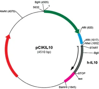
Plasmid pCik IL-10 was constructed by inserting hIL-10 c DNA (537 bp) into a unique backbone of pCIk driven by human CMV early promoter enhancer at Not K1 and Nhe 1 site[8](Fig. 1). The plasmid was purified and produced in the quantity required at (Plasmid Factory Gmbh &Co, Bielefeld Germany).
2.2. Experimental groups
Left lung allotransplantation was performed in rats with major full mismatch and one minor immunological mismatch (Donor: Brown-Norway; Recipient: Fischer F344; obtained from Harlan Netherlands B.V., Ad Horst, Netherlands). Two groups (nZ5) were studied: Group A animals received sub therapeutic doses of CsA 2.5 mg/kg/day i.p., starting the day of transplantation. Group B (nZ5) received the same dose of CsA per day in combination with transduction of 2.5 mg hIL-10, plasmid DNA (pCik hIL-10). In the second group, pCiK hIL-10 was injected to the tibialis anterior muscle followed by electroporation, 24 h prior to transplantation.
CsA (Sandimmunw) was provided by Novartis Pharma AG,
Basel Switzerland and diluted in castor oil in a ratio of 1:9. All animals received human care in compliance with the ‘Principles of Laboratory Animal Care’, formulated by the National Society for Medical Research, and ‘Guide for the Care and Use of Laboratory Animals prepared by the institute of Laboratory Animal Research and published by the National Institutes of Health (NIH Pub. No. 86-23, revised 1985)’. The protocol was approved by the local animal study committee. 2.3. Operative procedure
2.3.1. Donor
The animal is anaesthetized in a glass chamber by inhaling 4% Halotane (SIGMA, Buchs, Switzerland). Thiopental
(Pentotalw
, Abbot AG, Baar, Switzerland) at a dosage of 50 mg/kg body weight (b.w.) is administered i.p. Heparine (Liqueminw
, Roche Pharma, Rheinach, Switzerland) is administered by injection into the penile vein (500 IU/kg b.w.). A trachostomy is performed and the animal is ventilated with a 14 GA catheter (Insytew, Becto Dickinson,
Sandy UT USA) with 100% oxygen, a breathing frequency of 100/min, and tidal volume of 8 ml/kg body weight by a rodent ventilator (Model 683, Harvard Apparatus, South Natick MA USA). After cutting the inferior vena cava and left appendix of the heart, a small silicon hose is inserted into the main pulmonary artery (PA) via an incision in the right ventricle. Both lungs are flushed with 20 ml of LPD solution (Perfadexw,Vitrolife Pharmaceutics, Uppsala Sweden) at
20 cm H2O pressure. The trachea is then tied with the lungs in end respiratory position. The heart-lung block is removed and the left lung is separated ex vivo. Small plastic cuffs are placed around the PA and the left pulmonary vein (PV), the vessels are everted and tied on the cuffs and fastened with 8–0 monofilament thread (Surgiprow
, USSC, Norwalk CT USA). The graft is stored in LPD solution at 10 8C until implantation.
2.3.2. Recipient
The recipient is anaesthetized by breathing 4% Halotane in the glass chamber. Pentobarbital (Nembutalw
, Abbot AG, Baar Switzerland) at a dosage of 50 mg/kg is administered i.p. Dissection of the left tibialis anterior muscle (MTA) is performed. The muscle is injected with 2.5 mg of pCikhIL-10 with Omnican30 insulin syringe (Braun, Switzerland), and subsequently placed between the plate electrodes. 4!20 ms impulses at 200 V/cm are applied with the INOVIO Pulse Generator (Inovio Oslo).
After 24 h, the recipient is anaesthetized by breathing 4% Halotane in the glass chamber again. Intubation is carried out using a 14 GA catheter placed into the trachea. Anaesthesia is maintained with Halotane at 2.5%. The recipient is ventilated with 1 cm water PEEP, a breathing frequency of 100/min and a tidal volume of 8 ml/kg body weight by rodent ventilator. A left thoracotomy is per-formed. The left hilum is dissected and micro clips are put on the left pulmonary artery (PA) and the left pulmonary vein (PV). The left main bronchus is ligated with 6–0 polyfilament thread (Sofsilkw
, USSC, Norwalk CT USA) and cut. An incision is made in both PV and PA. The vessels are flushed with saline solution. The cuffs of the donor lung are inserted into the recipient’s vessels, and 6-0 polyfilament ligatures (Sofsilkw
) are placed around the cuffs and tied. The native PA and PV are cut off beyond the anastomoses and the native lung is removed. A 9-0 monofilament running over-and-over con-tinuous suture (Monosofw, Tyco Healthcare, Wollerau
Switzerland) is employed for the bronchial anastomosis. Ventilation is started. The microclips are removed to allow retrograde and subsequently anterograde perfusion of the graft. A small chest drain is inserted into the left pleural space and the thoracotomy is closed with three layers of continuous sutures. The chest drainage is removed after spontaneous breathing is restored, followed by extubation.
Fig. 1. The plasmid expression vector pCiK IL-10 constructed by inserting 541 bp hIL-10 cDNA.
2.4. Statistics
For continuous data all values are given as meanGSD. Data were log-transformed and the t-test performed on these log-transformed data. A P-value less than 0.05 were considered significant. The STATISTICA 5.1 software (StatSoftw, Tulsa, OK) was used.
3. Assessment 3.1. Graft function
Five days after the transplantation, the recipient is preanaesthetized in a glass chamber inhaling 4% Halotane, Thiopental (50 mg/kg bw) is administered i.p. The animal is ventilated via a tracheostomy with the Harvard Rodent Ventilator with FIO2Z1.0, a frequency of 100 breaths/min, and a tidal volume of 8 ml/kg. A thoraco-laparotomy in the anterior midline is done. Dissection of the right hilum is performed. Microvascular clips are put on the right main bronchus and right PA in order to ventilate and perfuse only the isolated left lung graft. Five minutes after the occlusion, 1 ml of blood is aspirated from the aortic arch to a syringe (Radiometer Pico 50, Copenhagen, Denmark) for blood gas assessment (Radiometer ABL 700 Serie, Copenhgen, Den-mark) and 1 ml of blood for the measurement of the serum hIL-10 levels is collected from the inferior vena cava. Subsequently, the inferior vena cava and left appendix of the heart are incised and a small silicon hose is inserted into the main PA via an incision in the right ventricle. The lungs are then flushed with 20 ml of 0.9% saline solution under pressure of 20 cm H2O. The heart-lung block is explanted and tissue samples collected.
3.2. Histology
After explantation the graft is isolated and inflated to total capacity with 4% formaline and placed in a container with the same fixative for 12 h. After paraffin fixation the sections were cut and routine haematoxylin and eosin staining performed. The histological assessment was done by a trained lung pathologist in blinded fashion according to the Working Formulation for the Classification of Pulmonary Allograft Rejection of the International Society for Heart and Lung Transplantation[9].
3.3. hIL-10 expression 3.3.1. ELISA
Circulating serum levels of hIL-10 were measured using hIL-10 immunoassay kit (R&D Systems, Abingdon UK) following the manufacturers instructions. The antibody is specific and no cross-reaction with rat IL-10 occurs. 1 ml blood was drawn from the inferior vena cava on day 5 at sacrifice, centrifuged, and stored at K20 8C till the measurements. In order to assess the level of hIL-10 at the time of transplantation two separate animals were injected with pCikhIL-10 followed by electroporation as described above, as the loss of 1 ml blood at the time of transplan-tation results in a high mortality of the animals.
3.3.2. In situ hybridization
The hIL-10 cDNA fragment (537 bp) was subcloned into blue script vector (Stratagene, Europe The Netherlands). The plasmid containing the cDNA probe inserts was linear-ized using the restriction endonucleases Not-I for in vitro transcription of the sense strand and HindIII for the antisense strand. Single-stranded sense and antisense digoxigenin (DIG)-labeled cRNA probes were transcribed in vitro using 1ug of linearized template and 40 units of the appropriate polymerase (T7 RNA polymerase for the sense strand, SP6 RNA polymerase for the antisense strand) with DIG-labeled uridine triphosphate (UTP) using the DIG RNA Labeling Kit according to the manufacturer’s directions (Roche Diagnos-tics, Basel Switzerland).
To localize the expression and distribution of hIL-10 after in vivo electroporation in the skeletal muscle we performed in situ hybridization to detect the expression at mRNA level. Tissue sections were deparaffinized in a xylene series and then rehydrated through a decreasing ethanol series diluted in dimethyl pyrocarbonate-treated water. Sections were then subjected to Proteinase K treatment at 37 8C for 10 min. After washing with 1XPBS sections were treated with acetic anhydride for 10 min at room temperature. The slides were washed again with 1XPBS and prehybridized for 2 h at 55 8C in the hybridization solution (Sigma/Aldrich USA) and t-RNA before hybridization with 100 ng in 55 ul of the sense or antisense cRNA DIG labelled probes in a humid chamber at 55 8C for 18 h. Hybridized slides were treated in increasingly stringent pre-heated 2!SSCC50% formamide, 2!SSC, 0.2% SDS for 15 min at 37 8C, followed by two washes with 0.1!SSC, 0.2% SDS for 15 min at 60 8C. Detection was carried out by incubation with an alkaline phosphatase conjugated anti-DIG antibody using nitroblue tetrazolium chloride and 5-bromo-4-chloro-3-indolyl phosphate according to the manufacturer’s instructions (Roche Diagnostics, Basel Switzerland).
4. Results
4.1. Characterisation of experimental groups
Donors and recipients weighed 220–240 g, 240G20 g with no statistical difference between groups. The transplan-tation procedure was performed within a mean 180G15 min. Warm ischemic time was 30G5 min, without statistical differences between groups.
4.2. Graft function
PaO2 (meanGSD) levels in arterial blood, obtained 5 min after right hilar crossclamping in group B (233G123 mmHg) were significantly higher than those in group A (44G 8 mmHg) (PZ0.0091).
4.3. Rejection grading
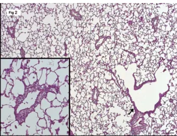
The histopathological assessment demonstrated improve-ment in lung architecture in the treated animals as seen by the rejection score of II–III A in group B (Fig. 2) as compared to III B in group A (Fig. 3,Table 1).
4.4. ELISA
Sustained levels of human IL-10 in the circulation of pCik hIL-10 transfected rats were detected. Serum levels of hIL-10 increased significantly after electroporation mediated hIL-10 transfection 74G4 pg/ml at 24 h and 14G7 pg/ml at day 5, in group B. No hIL-10 was detected in the serum of group A animals at either time point (Fig. 4). 4.5. Local expression of hIL-10 in the tibialis anterior muscle The expression, histologic localization, and the persist-ence of hIL-10 in the transduced muscle was determined by
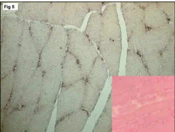
in situ hybridization. An antisense probe to hIL-10 detected transcripts in muscle fibers and in macrophages. The sense probe showed no signal, indicating the specificity of the signal for hIL-10 (Fig. 5).
5. Discussion
The present study demonstrates a synergistic effect of hIL-10 overexpression and sub therapeutic doses of Cyclos-porine A (CsA). The treated animals showed improved graft function and reduced histologic rejection on day 5 post-transplantation in this model of lung allopost-transplantation.
The idea to reduce the alloimmune response after organ transplantation by overexpression of immunosuppressive proteins has been pursued by a number of groups[10,11], however, the technique so far was limited, first by the low efficiency of gene transfer, and second by temporary expression of the transgene. Previous studies evaluated the effect on acute rejection by local expression of transgene in the graft [11]. This may be more effective if only a low expression is desired. In addition, if the transduction is done at the time of organ harvest, in vivo or ex vivo, it is difficult to get a homogenous expression in the graft tissue which might limit the effect substantially. On the other hand the graft might be damaged by the transduction itself, as viral vectors cause inflammatory responses and the most effective non viral vectors, as for example PEI, are rather toxic for the lungs[12]. In contrast, peripheral transduction in the muscle results in high expression of the transgene in the recipient and is technically easy to achieve.
Fig. 2. The transplanted lung of treated animals (group B) shows mostly clear alveoli. The perivascular and the peribronchiolar infiltrate (arrow) is still visible at low magnification, but it is clearly less dense and the alveolar epithelium is unchanged (inset). (Hematoxylin&eosin, bar 100 mm, inset 50 mm).
Fig. 3. A representative histological section of the graft of group A, at day 5. The dense infiltrate around the pulmonary blood vessels and the bronchioles is visible at low magnification. Moderately increased macrophages and a faint edema occupy the alveoli. Inset: high magnification reveals up to five layers of a lymphocytic infiltrate (arrow) around a pulmomary vein. Note the increased number of endothelial cells. The infiltrate reaches focally the alveolar interstitium (arrowhead). The alveolar epithelial cells are increased in number. Histological assessment revealed low moderate rejection in group B II–IIIA (Fig. 2). In contrast to moderate to severe rejection in all animals in group A IIIB–IV (Fig. 3).
Table 1
Low dose CsA hIL-10CCsA Vascular A3 (A2–A3) A2 (A2–A3) Airway B3 (B2–B3) B2 (B2–B3)
Histological assessment of the grafts for rejection scoring using the ISHLT formulation was implied. Although the difference is not significant yet is considerable since the functional parameters as the PaO2 shows significant improvement.
Fig. 4. hIL-10 expression and localization Levels of circulating hIL-10 in the serum of the rat at day 1 (74G4 pg/ml) and at day 5 after electroporation mediated gene transfer (14G7 pg/ml) (meanGSD). The hIL-10 levels in rat control serum was 0.
Electroporation mediated gene transfer has emerged as a promising gene delivery method. The technique is not only available at low cost, but its easiness of application in addition to its safety of production of the plasmid constructs are unique. Several reports show that it is one of the most efficient non-viral strategies [7], increasing reporter gene and therapeutic gene expression by several orders of magnitude over conventional non viral and viral vectors (Gazdhar et al, in press). In vivo electroporation is a physical method of gene transfer. The plasmid DNA is injected followed by delivery of a series of electric pulses of short duration using suitable electrodes. In preliminary studies we observed hIL-10 transduction alone was not consistently effective in reducing the acute rejection in lung transplan-tation model in spite of high hIL-10 serum levels.
IL-10 is a potent suppressor of TNFa Interleukin-1b, interleukin-6, interleukin-12, and interferong production and may facilitate the induction of tolerance after allogenic transplantation[13,14]. Both CsA and IL-10 act synergisti-cally inhibiting T cell activation and IL-2 and TNFa production. CsA inhibits calcineurin; IL-10 inhibits both Th1 cell proliferation and other proinflammatory cytokines. Thus, each drug acts via an independent pathway yet exert a complimentary immunosuppressive effect, but timing of gene transfer and the route of administration may play a key role. Our preliminary data showed that IL-10 overexpression at the time or after transplantation had little effect or was even detrimental, as reported earlier by other groups
[15–17]. In contrast previous reports show that IL-10 treatment of the recipient prior to transplantation enhances graft survival of heart allografts in mice[18], and rat liver allografts[19], and was effective in studies of bone marrow transplantation [20]. Very high doses of IL-10 have also proved to be deleterious as reported earlier[16], hence the optimal dosage is crucial.
In a study by Itano et al. the use of lipid mediated viral IL-10 transduction to the lung graft reduced rejection[21]. In our study hIL-10 was used which is active in the rat due to receptor homology[22,23]and which also results in higher serum concentrations than viral IL-10 as latter has been
shown to be retained in the tissue compartments[24]. The sustained levels of circulating hIL-10 in the blood in the present experiments highlights the possibility to use the muscle for secretion of proteins into circulation thus opening a large field of therapeutic options[25].
In summary, our observations support the key role of IL-10 during the initial events of lung transplantation. The time course of the hIL-10 serum levels in the present experiment shows peak levels at 24 h, gradually decreasing over the next five days. This fast increase of circulating hIL-10 achieved with the CMV promoter seems to be ideal for the prevention of acute rejection, however; other constructs that are currently evaluated by our group have to be employed for the maintenance of the transgene expression over longer time (Gazdhar et al. in press). In a parallel study conducted by our group, (Tavakoli et al in press) it has been shown that in cardiac transplantation hIL-10 alone, without additional CsA, is effective to reduce acute rejection in the same strain combination. In renal allografts, however, IL-10 has a detrimental effect[3].
In conclusion, we have demonstrated that electropora-tion mediated gene transfer of hIL-10 to skeletal muscle of the recipient in combination with low dose CsA improves lung allograft function, and reduces histologic rejection. This approach may even have clinical potential, as the method is easily applicable and may help to modify current methods of immunosupression in the early phase after transplantation.
Acknowledgements
This study was supported by a grant from the swiss science foundation SNF 3200-06304, R.A. Schmid. We also thank Prof Robert Friis Department of Clinical Research University of Bern and Dr Ian Pringle John Radcliff Hospital, University of Oxford, UK for their valuable support.
References
[1] Zuckermann A, Reichenspurner H, Birsan T, Treede H, Deviatko E, Reichart B, Klepetko W. Cyclosporine A versus tacrolimus in combination with mycophenolate mofetil and steroids as primary immunosuppression after lung transplantation: one-year results of a 2-center prospective randomized trial. J Thorac Cardiovasc Surg 2003;4:891–900.
[2] Parekh K, Trulock E, Patterson GA. Use of cyclosporine in lung transplantation. Transplant Proc 2004;2:318S–322.
[3] Moore KW, de Waal Malefyt R, Coffman RL, O’Garra A. Interleukin-10 and the interleukin-10 receptor. Annu Rev Immunol 2001;683–765. [4] de Waal Malefyt R, Abrams J, Bennett B, Figdor CG, de Vries JE.
Interleukin 10(IL-10) inhibits cytokine synthesis by human monocytes: an autoregulatory role of IL-10 produced by monocytes. J Exp Med 1991;5: 1209–20.
[5] Howard M, O’Garra A, Ishida H, de Waal Malefyt R, de Vries J. Biological properties of interleukin 10. J Clin Immunol 1992;4:239–47.
[6] Delvaux A, Donckier V, Bruyns C, Florquin S, Gerard C, Amraoui Z, Abramowicz D, Goldman M, Velu T. Effects of systemic administration of rIL-10 in an in vivo model of alloreactivity. Transplantation 1994;8: 972–4.
[7] Mathiesen I. Electropermeabilization of skeletal muscle enhances gene transfer in vivo. Gene Ther 1999;4:508–14.
[8] Gill DR, Smyth SE, Goddard CA, Pringle IA, Higgins CF, Colledge WH, Hyde SC. Increased persistence of lung gene expression using plasmids containing the ubiquitin C or elongation factor 1alpha promoter. Gene Ther 2001;20:1539–46.
Fig. 5. mRNA expression and localization in the myocytes and macrophages in the M. tibialis anterior of a group B animal by in situ hybridization. Inset (Negative control sense probe).
[9] Yousem SA, Berry GJ, Cagle PT, Chamberlain D, Husain AN, Hruban RH, Marchevsky A, Ohori NP, Ritter J, Stewart S, Tazelaar HD. Revision of the 1990 working formulation for the classification of pulmonary allograft rejection: Lung Rejection Study Group. J Heart Lung Transplant 1996; 1(Pt 1):1–15.
[10] Shinozaki K, Yahata H, Tanji H, Sakaguchi T, Ito H, Dohi K. Allograft transduction of IL-10 prolongs survival following orthotopic liver transplantation. Gene Ther 1999;5:816–22.
[11] Hong YS, Laks H, Cui G, Chong T, Sen L. Localized immunosuppression in the cardiac allograft induced by a new liposome-mediated IL-10 gene therapy. J Heart Lung Transplant 2002;11:1188–200.
[12] Uduehi AN, Stammberger U, Kubisa B, Gugger M, Buehler TA, Schmid RA. Effects of linear polyethylenimine and polyethylenimine/DNA on lung function after airway instillation to rat lungs. Mol Ther 2001;1:52–7. [13] Levings MK, Sangregorio R, Galbiati F, Squadrone S, de Waal Malefyt R,
Roncarolo MG. IFN-alpha and IL-10 induce the differentiation of human type 1 T regulatory cells. J Immunol 2001;9:5530–9.
[14] Fiorentino DF, Zlotnik A, Mosmann TR, Howard M, O’Garra A. IL-10 inhibits cytokine production by activated macrophages. J Immunol 1991; 11:3815–22.
[15] Li W, Fu F, Lu L, Narula SK, Fung JJ, Thomson AW, Qian S. Differential effects of exogenous interleukin-10 on cardiac allograft survival: inhibition of rejection by recipient pretreatment reflects impaired host accessory cell function. Transplantation 1999;9:1402–9.
[16] Li W, Lu L, Li Y, Fu F, Fung JJ, Thomson AW, Qian S. High-dose cellular IL-10 exacerbates rejection and reverses effects of cyclosporine and tacrolimus in Mouse cardiac transplantation. Transplant Proc 1997;1–2: 1081–2.
[17] Qian S, Li W, Li Y, Fu F, Lu L, Fung JJ, Thomson AW. Systemic administration of cellular interleukin-10 can exacerbate cardiac allograft rejection in mice. Transplantation 1996;12:1709–14. [18] Li W, Fu F, Lu L, Narula SK, Fung JJ, Thomson AW, Qian S. Recipient
pretreatment with mammalian IL-10 prolongs mouse cardiac allograft survival by inhibition of anti-donor T cell responses. Transplant Proc 1999;1–2:115.
[19] Zou XM, Yagihashi A, Hirata K, Tsuruma T, Matsuno T, Tarumi K, Asanuma K, Watanabe N. Downregulation of cytokine-induced neutro-phil chemoattractant and prolongation of rat liver allograft survival by interleukin-10. Surg Today 1998;2:184–91.
[20] Holler E, Roncarolo MG, Hintermeier-Knabe R, Eissner G, Ertl B, Schulz U, Knabe H, Kolb HJ, Andreesen R, Wilmanns W. Prognostic significance of increased IL-10 production in patients prior to allogeneic bone marrow transplantation. Bone Marrow Transplant 2000;3:237–41. [21] Itano H, Mora BN, Zhang W, Ritter JH, McCarthy TJ, Yew NS,
Mohanakumar T, Patterson GA. Lipid-mediated ex vivo gene transfer of viral interleukin 10 in rat lung allotransplantation. J Thorac Cardiovasc Surg 2001;1:29–38.
[22] Rachmawati H, Beljaars L, Reker-Smit C, Van Loenen-Weemaes AM, Hagens WI, Meijer DK, Poelstra K. Pharmacokinetic and biodistribution profile of recombinant human interleukin-10 following intravenous administration in rats with extensive liver fibrosis. Pharm Res 2004;11: 2072–8.
[23] Ward H, Vigues S, Poole S, Bristow AF. The rat interleukin 10 receptor: cloning and sequencing of cDNA coding for the alpha-chain protein sequence, and demonstration by western blotting of expression in the rat brain. Cytokine 2001;5:237–40.
[24] Minter RM, Ferry MA, Rectenwald JE, Bahjat FR, Oberholzer A, Oberholzer C, La Face D, Tsai V, Ahmed CM, Hutchins B, Copeland 3rd EM, Ginsberg HS, Moldawer LL. Extended lung expression and increased tissue localization of viral IL-10 with adenoviral gene therapy. Proc Natl Acad Sci USA 2001;1:277–82.
[25] Tollefsen S, Vordermeier M, Olsen I, Storset AK, Reitan LJ, Clifford D, Lowrie DB, Wiker HG, Huygen K, Hewinson G, Mathiesen I, Tjelle TE. DNA injection in combination with electroporation: a novel method for vaccination of farmed ruminants. Scand J Immunol 2003;3:229–38.
Appendix A. Conference discussion
Dr N. Daddi (Perugia, Italy): I have mainly two questions to ask you. The first one is about the method. When you transfect the muscle with electroporation, did you also check the Human IL-10 concentration in both lungs and in other organs as heart, liver and kidney?
The second question is on the gene you adopted. The molecular structure of Human IL-10 is similar to cellular IL-10 produced in the animal. The anti-inflammatory activity of cellular IL-10 is known by downregulating several pro-inflammatory cytokines and natural killer cell activity. Conversely, this potent anti-inflammatory cytokine might have an important role in the differentiation of mastocytes.
Did you consider other gene vectors as a plasmid encoding viral IL-10? This cytokine is structurally similar to cellular IL-10. It inhibits as well the activity of the cytokines and downregulates the expression of major histocompat-ibility complex II on the surface of monocytes but does not possess the costimulatory effect on mastocytes.
Dr Pierog: We measured overexpression of human IL-10 only in the electroporated muscle and in circulating blood. We didn’t measure the expression in the lung or in the liver. The human IL-10 was distributed to the bloodstream systematically. Regarding to your second question, we decided to use only human interleukin-10 because it results in higher serum concentration and makes it available systematically, whereas viral IL-10, is retained in the tissue compartments.
Dr D. Van Raemdonck (Leuven, Belgium): You’ve shown a nice drop in serum levels of interleukin-10 from post transplant day 1 to day 5. Can you also give us the figure on the level pre transplantation. Was there an increase in interleukin-10?
Dr Pierog: This expression was according to the CMV promoter/enhancer included in the plasmid. This promoter allows high expression which occurs 24 hours following gene transfer, the levels were measured before transplan-tation, and then the expression gradually decreases between day 1 and day 5. This pattern of the expression was confirmed also in other experiments performed in our laboratories.
Dr G. Laufer (Innsbruck, Austria): You’re basically augmenting your immunosuppression by this immunosuppressive cytokine. What would be the consequences for infection? I mean, if you have high levels of IL-10, that might also impair the resistance to viral infection, susceptibility to viral infection and viral disease early after transplantation.
Dr Pierog: We didn’t assess susceptibility for infection. I can say that I performed many lung transplantations with using this regimen of immuno-suppression and I have not seen any infections during the post operative course because the recipients were sacrificed after 5 days.
Dr D. Van Raemdonck (Leuven, Belgium): My final question would be, you did this gene transfer 24 hours prior to transplantation, how would you translate it to the clinical practice? How do we know 24 hours in advance that there will be a donor?
Dr Pierog: In the literature it has been demonstrated that human interleukin-10 administered after lung transplantation had no beneficial effect on lung transplantation. Even the rejection score may be worse after administration of human interleukin-10 following lung transplantation. In contrast, administration of human interleukin-10 prior to implantation has a beneficial effect. The mechanism of this phenomenon has not been elucidated so far.
Dr Van Raemdonck: But 24 hours seems a long period in advance. Have you looked at 12 or 6 hours prior to transplant?
Dr Pierog: This is an experimental setting for a proof of principle. The recipients were electroporated 24 hours prior to implantation because of the expression pattern provided by the CMV promoter of the plasmid, and because it is known that the levels of pro inflammatory cytokines like IL-2 and INFg attain the peak between 6 to 12 hours after transplantation, hence in order to achieve high expression of human IL-10 in the bloodstream at this time point a 24-hour interval might be ideal.