REVIEW ARTICLE
Fractional photothermolysis
—an update
Inja Bogdan Allemann&Joely Kaufman
Received: 30 June 2009 / Accepted: 27 August 2009 / Published online: 29 September 2009
# Springer-Verlag London Ltd 2009
Abstract The novel concept of non-ablative fractional photothermolysis was introduced to the market in 2003 as an answer to the need for effective, yet low risk, resurfacing techniques. Unlike conventional ablative and non-ablative lasers, fractional ablative and non-ablative photothermolysis treats only a fraction of the skin, leaving up to a maximum of 95% of the skin uninvolved. The undamaged surrounding tissue allows for a reservoir of viable tissue, permitting rapid epidermal repair. Non-ablative fractional photother-molysis is currently approved by the US Food and Drug Administration (FDA) for the treatment of pigmented lesions, periorbital rhytides, skin resurfacing, melasma and soft tissue coagulation, acne and surgical scars, and actinic keratoses. However, its off-label use is clearly more extended. In 2007 the concept was further devel-oped, and ablative fractional photothermolysis was intro-duced, using an erbium yttrium aluminium garnet (Er: YAG) or carbon dioxide laser. These devices are FDA cleared to treat wrinkles, rhytides, furrows, fine lines, textural irregularities, pigmented lesions and vascular dys-chromia. In this review we discuss the two concepts, their technical details and clinical indications, and we describe the current literature available.
Keywords Ablative fractional photothermolysis . Non-ablative fractional photothermolysis . Laser
The novel concept of fractional photothermolysis (FP) was first reported in 2003, in its basic application [1]. In 2004 and 2005 the first full reports and applications for photoaging by Manstein et al. followed [2, 3]. This technology has been developed as a logical next step, triggered by the undesirably risky, though very effective, ablative technologies and the limited efficacy of the non-ablative laser modalities. In contrary to the conventional laser technologies, which target an entire area of the skin, FP treats only fractions of the skin, by inducing small three-dimensional zones of thermal damage, referred to as “Microscopic Thermal Zones” (MTZs). The surrounding tissue is not involved, which allows fast epidermal repair via migration of the surrounding viable cells, hence the name fractional. Depending on the chosen parameters of energy per MTZ and the density of MTZs per square centimeter, anywhere from 3–40% of the skin can be covered with each treatment. The MTZs are usually smaller than 400 µm in diameter and can penetrate the skin to varying depths of up to 1,300 µm, depending on the wavelength, pulse energy, and device chosen. As the energy of each spot increases, the size/diameter of the MTZ increases, as well as the depth of penetration. Thus, energy is chosen based on the desired depth of treatment. Densities can be reported as either percent coverage, or MTZs per square centimeter. The terminologies may be congruent at low energies, but, as the energy increases and the MTZ increases, these parameters may differ significantly. In order to translate one devices density settings into another, a consistent density parameter chosen. Percent coverage is more translatable, as all MTZ sizes, and individual spots, differ, not only between devices but also within a single device. The target chromophore, for all of the fractional devices on the market at this time, is water. This allows selective thermal damage to various water-containing struc-tures, such as collagen, blood-vessels and epidermal kerati-nocytes [3]. Thermal damage is thus induced in the
I. Bogdan Allemann (*)
Dermatologische Klinik, UniversitätsSpital Zürich, Gloriastrasse 31,
8091 Zürich, Switzerland e-mail: [email protected] J. Kaufman
Cosmetic Medicine and Research Institute, University of Miami,
4701 N. Meridian Av., Miami Beach, FL 33140, USA
epidermis and dermis, leading to epidermal turnover and collagen induction. Fractional technologies can be divided into two main categories, based on the wavelength’s affinity for water. Those devices with wavelengths that are highly absorbed by water are termed ablative. These include both erbium yttrium aluminium garnet (Er:YAG; 2,940 nm) or yttrium ycandium gallium garnet (YSGG; 2,790 nm) and carbon dioxide (CO2; 10,600 nm) lasers. Those with
wavelengths only moderately absorbed are ‘non-ablative’ (1,410 nm, 1,440 nm, 1,540 nm, 1,550 nm). Histologically, with the non-ablative variety, one can observe a column-like denaturation of the epidermis and dermis, a disruption of the dermo-epidermal junction, with subepidermal clefting within the MTZ, and an intact stratum corneum. The surrounding tissue is unharmed. The thermally destroyed tissue becomes replaced by keratinocytes that migrate from the surrounding healthy tissue within the first 24 hours [4]. The button-shaped necrotic tissue, which is called Microscopic Epidermal Necrotic Debris (MEND), is eliminated transepidermally by the keratinocytes, and its migration upwards and through the stratum corneum is facilitated by the subepidermal clefting. This migration is called the MEND-shuttle, literally‘shuttling’ dermal contents out of the skin. Within each amount of MEND, elastic tissue, melanin and other dermal contents are found. The clinical outcome of this process is slight scaling and bronzing of the skin, which occurs roughly after 1 week. The replacement of the MTZs with new collagen occurs within 3–6 months.
The results of the ablative fractional technologies have also been examined. With the ablative approach, histologically one can see ablated micro-columns, varying in thickness and depth depending on pulse width and wavelength used. A thin layer of eschar lines the cavity, which is consistent with ablative laser treatment. Around these cavities annular coagulation zones of varying thickness, which represent denatured collagen, can be observed. Owing to the ablative character of the procedure, the stratum corneum is mostly absent, contrary to the situation in non-ablative fractionated technology (Fig.1) [5]. Reepithelialization of the coagulation zones occurs rapidly, within 48 hours [5,6].
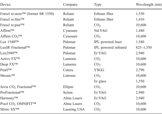
The first medical laser utilizing fractional photothermolysis technology and consequently the most studied one is the 1,550 nm non-ablative fractional device (Fraxel™ re:store, Reliant Technologies, San Diego, CA, USA). This device uses an erbium-doped fiber laser to produce laser light at a non-ablative wavelength of 1,550 nm. It is not selective for the other chromophores in the skin, such as melanin or hemoglobin [7]. The handpiece is handled in a scanning mode and utilizes an intelligent optical tracking system (IOTS), which monitors the treatment area and allows the system to track handpiece velocity, ensuring a unvarying number and pattern of MTZs. Histological studies using this device show that full epidermal healing occurs within the first 24 hours after the procedure [4]. This rapid barrier restoration lowers the risk of infection, prolonged erythema and other complications frequently associated with previous types of resurfacing procedures. This has been supported by a study showing unchanged transepidermal water loss (TEWL) within the MTZ [2]. Most of the data discussed below have been obtained from studies with this device. Currently, various laser technologies by different producers that are based on the FP technique exist. They differ in wavelength, pulse energy and damage pattern. For example, the Lux 1,540 nm laser (Palomar Medical Technologies, Burlington, MA, USA) and the Affirm 1,440 nm neodymium yttrium aluminum garnet (Nd:YAG; Cynosure Inc., Westford, MA, USA) use stamping technology instead of scanning mode. There are few studies comparing the different fractional non-ablative devices, making it difficult to state definitively which system is superior. Their mechanisms of action and theoretical basis of function are similar in nature (Table1).
During the treatment patients feel slight pain. To ease the pain the skin surface is cooled or topical anaesthesia is used for pain control. Directly after the treatment with non-ablative devices, one can observe erythema and edema, which usually lasts for 1–3 days, again depending on the treatment parameters used. It is followed by a bronzing and slight scaling of the skin, which reflects the elimination of the MENDs. As for ablative fractional treatment, in addition to
Fig. 1 a Schematic histology of non-ablative and ablative FP. Column-like denaturation of the epidermis and dermis, with a disruption of the dermo-epidermal junction, clefting within the MTZ,
and an intact stratum corneum. The surrounding tissue is unharmed. b Schematic histology of ablative FP. Ablated micro-columns, lined by a thin layer of eschar and with annular coagulation zones
edema and erythema, which last longer, bleeding and/or oozing can be observed, and downtime can lasts up to 14 days [8].
As for most laser interventions, the ideal candidate is a person with Fitzpatrick skin type I–III. Yet, as the wavelengths of these devices are not absorbed by melanin, non-ablative fractional systems can be used for most skin types, and, in fact, non-ablative FP has been shown to be effective and safe in darker skin types (IV, V, and VI). A recent pilot study even showed ablative CO2FP to be safe for Fitzpatrick phototypes
IV and V [9]. However, the possibility of post-inflammatory pigment alterations in darker skin types should not be neglected and should thus be accounted for by adjustment of treatment settings accordingly. Further, unlike traditional ablative resurfacing, non-ablative fractional photothermolysis (NAFP) has been shown to be safe to use on locations other than the face, and no side-effects such as scarring have been reported so far [10]. For ablative fractional photothermolysis (AFP) however, first reports of side-effects and complications when used off-face or in locations with thin skin, such as the eyelids, have recently been published. Both publications reported scarring after the use of a CO2 ablative fractional
device [11,12]. More data are needed for it to be determined whether AFP, especially CO2AFP, should be used at all off
the face and in areas of thin skin. For NAFP and AFP the patient’s medical history prior to FP should include history of herpes labialis, as FP can trigger reactivation of herpes simplex infection. Furthermore, one should inquire about post-inflammatory hyperpigmentation, tendency for hyper-trophic scarring or keloids and retinoid use (oral or topical). There are varying recommendations on the use of oral
retinoids, from pausing them 6–12 months prior to FP to not stopping them at all [13].
A recent study investigated the short-term side effects after FP. They showed that 100% of patients had transient erythema, 82% had edema, 86.6% felt dry skin, 60% experienced flaking, and 26.6% bronzing [14]. Those more ‘obligatory’ side-effects (pain, erythema, edema) are stronger and persist longer with higher densities. Increased density seems to be more likely to produce edema, erythema, and even post-inflammatory hyperpigmentation than does increased energy [10]. However, patient satisfaction was shown to be significantly greater for patients treated with higher fluences, but not for patients treated with higher densities [10]. Graber et al. retrospectively evaluated 961 treatments with a 1,550 nm erbium-doped laser for side-effects and complications [15]. They showed that, overall, only 7.6% of treatments resulted in complications, the most frequent of them being acneiform eruptions (1.87%) and herpes simplex reactivations (1.77%). No difference in side effects was observed for different skin types and body locations, except for post-inflammatory hyperpigmentation, which occurred more frequently in darker skin types. A simple decrease in treatment density (percentage coverage) can make this procedure safe for this population.
The ability of both non-ablative and ablative FP to induce collagen formation has been proven histologically via demonstration of an increase in collagen type 3 production after treatment. Collagen production and remodeling clinically should result in an improvement in rhytides. This, however, is not always the case [4]. Let us explore the clinical data
Device Company Type Wavelength (nm)
Fraxel re:store™ (former SR 1550) Reliant Erbium fiber 1,550
Fraxel re:fine™ Reliant Erbium fiber 1,410
Fraxel re:pair™ Reliant CO2 10,600
Affirm™ Cynosure Nd:YAG 1,440
Affirm CO2™ Cynosure CO2 10,600
Lux 1540™ Palomar IPL powered laser 1,540
LuxIR Fractional™ Palomar IPL powered infrared 825–1,350
Lux2940™ Palomar Er:YAG 2,940
Active FX™ Lumenis CO2 10,600
Deep FX™ Lumenis CO2 10,600
Pearl™ Cutera YSGG 2,790
Mosaic™ Lutronic CO2 10,600
Er glass 1,550
Juvia CO2Fractional™ Ellipse CO2 10,600
ProFractional™ Sciton Er:YAG 2,940
Harmony™ Alma Lasers Er:YAG 2,940
Pixel CO2OMNIFIT™ Alma Lasers CO2 10,600
Mixto SX™ Lasering USA CO2 10,600
Table 1 Ablative and non-ablative fractional lasers currently marketed (IPL intense pulsed light)
available on non-ablative FP. Manstein et al. treated lateral peri-orbital rhytides in 30 subjects with four non-ablative FP treatments over a period of 2–3 weeks [2]. Using the Fitzpatrick wrinkle score, two independent dermatologists assessed the treatment effect by blinded analysis of the pre-treatment photographs and 1-month and 3-month follow-up photographs. A significant improvement 3 months after treatment could be demonstrated. Periorbital treatments were well tolerated.
FP seems to be effective in the treatment of fine and moderate facial wrinkles, less so, however, for deep lines. In a recent study 50 patients with cutaneous photodamage, rhytides, and dyspigmentation were treated three times with the 1,550 nm non-ablative fractional device at intervals of 3–4 weeks. Clinical improvement was assessed visually, using comparative photographs at baseline and 3 months, 6 months, and 9 months after treatment. At all time points, clinical improvement of the face, as well as non-facial skin, could be shown. In 73% of those treated on the face and 55% of those treated off face, an improvement in photodamage of at least 51% to 75% after 9 months was demonstrated [16]. Another study examined the effects of FP on photoaged hands. Ten patients (skin phototypes II to IV) were randomized to receive treatment on either the right or left hand. A total of five treatments to one hand was performed (1,550 nm Fraxel™, 8–9 mJ per MTZ and density of 2,500 MTZs per square centimeter). Clinical assessment by patients and physician after 1 month and 3 months showed a mean improvement of 51% to 75% in skin pigmentation and 25% to 50% in skin wrinkling. An increased density of dermal collagen was shown histologically both 1 month and 3 months after treatment when compared with that in baseline biopsies; no statistical analysis was presented on those findings though [17]. For non-ablative FP the histologic collagen production does seem to correlate with a clinical improvement of fine lines.
Non-ablative FP is FDA-approved for the treatment of pigmented lesions, periorbital rhytides, skin resurfacing, melasma and soft tissue coagulation, acne and surgical scars, and actinic keratoses [18]. Though these are the ‘approved’ uses, clearly, there is an even wider array of clinical indications.
Melasma is a difficult to treat pigmentary disorder with modest success rates and a high number of recurrences after most treatment modalities. Fontana Masson staining of histological sections taken after non-ablative FP have demonstrated melanin within the MENDs created. This melanin originates not only from the epidermis, but also from misplaced dermal melanin, leading to the conclusion that FP seems to be a convincing therapeutic option for the treatment of melasma [7]. It has also provided a well-needed option for the treatment of dermal melasma. In 2005 Tannous and Astner first reported on the treatment of
melasma with the 1,550 nm laser [19]. They treated a Caucasian female patient (skin type II to III) with epidermal and dermal melasma twice with full-face fractional resurfacing three weeks apart. Six months after treatment they demonstrated a marked reduction in epidermal and dermal pigment, as assessed by Wood’s lamp examination and comparative photography.
Rokhsar and Fitzpatrick conducted a pilot study, treating ten female patients (Fitzpatrick skin types III–V) suffering from melasma with the 1,550 nm fractional laser at 1- to 2-week intervals for four to six times [20]. They used 6 mJ to 12 mJ per MTZ and 2,000 to 3,500 MTZs per square centimeter. Three months after treatment they were evaluated visually, according to the percentage of lightening of original pigmentation. They showed 75–100% clearing in 60% of patients, one patient showed 50–75% of clearance and 30% of patients achieved less than 25% of clearance. Although rather darker skin types had been treated (Fitzpatrick skin types III–V), only one patient showed post-inflammatory hyperpigmentation. The follow-up period of this study was too short for recurrence to be assessed. Another study supported FP as a good treatment option for melasma also in darker skinned patients [21].
Recently, a study examined the histologic and ultrastructural changes of melasma after treatment with FP. Goldberg et al. demonstrated a decrease in melanocytes and a relative absence of melanin in the surrounding keratinocytes in post-treatment specimens obtained 3 months after final treatment in comparison with the pre-treatment ones, assessed by light and electron microscopy [22]. Investigator assess-ment showed that the clinical improveassess-ment was good for six subjects with skin type III and fair for four subjects with skin type IV. It is critical to understand that patients with melasma are at high risk of hyperpigmentation with any form of treatment. Fluences and densities used in patients with melasma should be conservative. Treatments should be placed further apart than the original publications indicate, with ideal interval times in the range of 4–6 weeks [17–19]. However, more studies with larger patient populations and long-term follow-up periods (>6 months) are needed to finally assess the true value of FP in the treatment of melasma. At this time, FP should not be used as first line therapy for the treatment of melasma.
As with the treatment of rhytides, atrophic scars such as acne scars should also improve after FP therapy, due to new collagen formation. Moreover, the undesirable side effects of the very effective original ablative laser resurfacing fostered the use of FP in the treatment of scars, and, so far, various studies have examined its effect on different scar types. For acne scars, ten patients treated with non-ablative FP for up to three times at 2–3 week intervals all showed clinical improvement 4 weeks after treatment [23]. In a
larger study by Alster et al. 53 patients with atrophic scars were treated with non-ablative FP on a monthly basis for two or more treatments. Nearly 90% of the patients showed an improvement of 51–75% after three treatments. Mean improvement scores increased proportionately with each successive laser session [24]. A recent study of 27 Korean patients (Fitzpatrick skin types IV or V) reported a marked improvement in the appearance of acne scars 3 months after treatment and proved non-ablative FP to be effective and safe also for darker skin types [25].
Another pilot study of seven patients and four treatments at 4-week intervals with the 1,550 nm laser (7–20 mJ, total density 1,000–2,500 MTZs per square centimeter) demon-strated improvements of 1–75% in scar hypopigmentation in six of seven patients [26]. In another case report a white patient with a surgical scar on the chin was treated with the 1,550 nm Fraxel™ in a single treatment session, using pulse energy of 8 mJ and density of 2,000 MTZs per square centimeter. A greater than 75% clinical improvement of the scar was demonstrated 2 weeks after treatment [27]. Thus, FP seems to be a valuable treatment modality for atrophic and potentially hypopigmented scars as well as for surgical scars. In addition to the above-mentioned clinical indications, there are various anecdotal reports on the effect of FP on various other skin disorders, suggesting the potential use of FP for further indications. Again, controlled studies with larger patient populations are needed to offer valid indications and treatment recommendations for FP.
With regard to vascular indications, Glaich et al. reported on the positive effect of FP on matted telangiectasias on the thigh of a female patient, skin phototype III, after five successive treatments at monthly intervals with the 1,550 nm fractional laser [28]. Good results were also shown for pigmentary disorders other than melasma. Two male patients with a Becker`s nevus on the chest and on the cheek, respectively, were treated monthly with the 1,550 nm device with energy densities from 6–10 mJ, final densities of 2,000–3,048 MTZs per square centimeter, and a total of five and six treatments, respectively. Both patients showed a greater than 75% improvement 1 month after treatment; hypertrichosis remained unaltered [29]. In a Japanese patient a successful treatment of nevus of Ota on the face could be achieved with the 1,440 nm Nd: YAG laser (Affirm) with fluences from 3–5–4.0 J/cm2
, 1 cm spot size, single pass with 20% overlap per treatment. The authors performed two treatments 4 weeks apart. After the first treatment there was only an improvement of 10%; however, the nevus of Ota com-pletely resolved within 6 weeks of the second treatment. At a 4-month follow-up examination there was still no recurrence [30].
As striae distensae are, in fact, dermal scars, FP was hypothesized to be an effective treatment modality. Six
women with striae albae on both buttocks were treated with 1,550 nm FP laser. A substantial improvement in the appearance of the striae and a partial normalization of skin elasticity were shown 8 weeks after treatment. Biopsies revealed a significant increase in epidermal thickness, collagen, and elastic fiber deposition after FP [31]. One patient with poikiloderma of Civatte showed complete resolution of the pigmentary changes after one FP treatment at 8 mJ/cm2with 2,000 MTZs per square centimeter [32]. In a recent case report a residual hemangioma was treated with the 1,440 nm fractional laser. The treatment was applied to the pretreated and now wrinkled and hypertrophic skin, using a 15 mm handpiece, energy density of 25 J/cm2 and a density of 10; 8–10 successive passes resulted in a total energy of 2.4 kJ. After two treatments, the skin showed marked clinical improvement in surface and texture at 1 month and 6-month follow-ups [33].
Non-ablative FP can also be used in combination therapy. One case report discusses the potential of future combination therapy of FP with Botulinum toxin injections to enhance the results of the laser treatment [34]. In another pilot study the synergistic effects of combined fractional resurfacing and 5-aminolevulinic acid–photodynamic therapy (ALA-PDT) in the treatment of skin rejuvenation were evaluated [35]. Four women (skin phototypes II or III) with mild to moderate perioral rhytides were treated with two sessions of FP (Fraxel SR™), with a 3-week interval, and immediately after each fractional treatment they were treated with ALA-PDT on one-half of the peri-oral area. The combined treatment side showed increased improvement in superficial wrinkles in 75% of subjects. Thus, combination treatments of FP with other therapies to enhance the effects of one or the other or to achieve synergistic effects are to be investigated in further detail.
Ablative fractional photothermolysis can be achieved using an Er:YAG, YSGG, or CO2 laser, with high absorption of
these wavelengths by water [5,36]. Originally, the ablative FP devices introduced used larger, millimeter, spot sizes. More recently these devices have been modified to introduce micron-sized pulses, allowing deeper penetration of the ablative wavelength. Ablative fractional devices on the market also differ in maximum power, energy and penetration depth. There are considerably fewer data published on AFP, and most focus on the treatment of photodamaged skin. In a preliminary clinical report in 2007 the non-sequential fractional ultra-pulsed 10,600 nm CO2 light treatment
ActiveFX™ laser (Lumenis Inc., Santa Clara, CA, USA) was evaluated for the treatment of photodamaged facial skin [37]. Fifty-five subjects showed significant differences between baseline photographs and those taken 1-month and 3 months after treatment for all aspects of photodamaged skin (global score, fine lines, mottled pigmentation, sallow complexion, tactile roughness, coarse wrinkles) except
telangiectasias. Adverse side effects were minimal, and downtime very low.
Another recent study reported on this new technology, using a 2,940 nm Er:YAG laser (Pixel, Alma Lasers, Buffalo Grove, IL, USA) for fractional resurfacing of photoaged skin [6]. They treated 30 women, Fitzpatrick skin types II–IV with one single session of fractional resurfacing, and 93% of the patients showed a good or very good improvement of the wrinkles. Except for one case of hyperpigmentation in a phototype IV patient, no side effects were observed. Two further studies confirmed that micro-fractional ablative treatment with 2,790 nm and 2,940 nm erbium lasers is a safe and effective treatment for wrinkle reduction and results in only minimal patient downtime [38–40]. Supporting data have recently been published by Berlin et al. Ten patients, Fitzpatrick skin types I–III, with photodamaged facial skin were given a single-pass superficial full-face ablative treatment using the CO2
laser at energy levels of 80–100 mJ. Clinical improvement in cutaneous photo-aging was observed. Interestingly, subjects treated with lower density settings reported a better mean improvement after 24 weeks than did subjects treated with higher density settings; however, the result was not statistically significant (P=0.27). Biopsies obtained after treatment showed greater fibrosis. Via electron microscopy, a decreased average diameter of the collagen fibrils was noted, which the authors concluded to be consistent with greater deposition of collagen type III as a feature of new collagen formation [41].
As for the use of ablative FP in the treatment of acne scars, Chapas et al. treated 13 patients, skin types I–IV, with the 10,600 CO2ablative fractional device (Fraxel re:
pair™, Reliant Technologies), using a spot size of 120 µm, pulse energies of 20–100 mJ and densities of 100–400 MTZs per square centimeter per pass and a total of 200–1,200 MTZs per square centimeter. Two to three treatments at 1–2 month intervals were administered. Illustrated by a three-dimensional optical profiling system, objective improvements on the depths of the acneiform scars ranged from 43–79.9%, with a mean of 66.8%. Further, post-treatment side effects were rated as mild to moderate, and no delayed pigmentary complications were observed [42]. In a recent study seven patients skin types IV and V received one single facial treatment with the ultra-pulsed 10,600 nm CO2 light treatment ActiveFX™
laser (Lumenis Inc.), using a spot diameter of 1.3 mm and 60 mJ. At a density setting of 1, approximately 60% of the facial skin surface was ablated. During a follow-up of up to 6 months, treatment-induced hyperpigmentation was not observed in any subject [9].
A study from Korea compared the treatment of atrophic facial acne scars in 20 patients with skin phototypes IV–V [43]. One group was treated with a single pass of an
ablative fractional CO2 laser (CoScan-5000, Stratek, Inc.,
Anyang, South Korea) using high energy settings [0.2 mm dot size, 30 ms pulse duration, 50–70 mJ pulse energy, 0.8 mm dot pitch (interlesional distance), 400 dots in a 20 mm×20 mm scanned area, 20% of treated area], and the other group was treated with the same laser using low energy settings [0.2 mm dot size, 30 ms pulse duration, 15–35 mJ pulse energy, 0.8 mm dot pitch (interlesional distance), and 400 dots in a 20 mm×20 mm scanned area]. Both groups received three successive monthly treatments on only one-half of the face. The other half of the face, in both groups, was treated with the same ablative fractional laser with low energy plus, directly before the ablative treatment, a non-ablative long-pulse 1,064 nm Nd:YAG laser. Of the ten patients treated with high energy settings, two showed 30–49% improvement, and eight patients had 50–69% improvement, in comparison with the patients treated with low energy settings, of whom seven had 10–39% improvement and three had 40– 59% improvement. Thus, higher energy settings resulted in improvement in the patients’ acne scars. However, the combination of ablative fractional laser resurfacing and non-ablative laser resurfacing yielded the best results, with fewer complications observed. Some cases of transient hyperpigmentation were observed, which resolved completely within 6 weeks after the use of topical bleaching agents. Other complications, such as hypopigmentation, bleeding, or hyper-trophic scarring, were not observed throughout the 12-month study period. There are also case reports on the improvement of burns scars, both on and off face, after treatment with ablative fractional resurfacing [44, 45]. Still, the safety of ablative FP needs to be examined in multiple patients of different ethnicities before a conclusion on its safety in darker skin types can be stated. To date, there have been no reports of depigmentation in ablative FP, as was previously seen with the traditional CO2resurfacing lasers.
Currently, there are no studies comparing non-ablative fractional photothermolysis with ablative fractional photo-thermolysis. However, as for now, data suggest that the ablative fractional treatment is safe, with rapid healing times and minimal downtime, especially for single-pass treatments. Most patients treated with Er:YAG FT devices will have a downtime anywhere from 1–3 days; those treated with the CO2devices may have 3–7 days downtime. Erythema and
edema are common, and desquamation may follow for several days afterward. One has to bear in mind though, that with multiple passes, the ablative damage accumulates, which increases thermal damage and consequently healing times [37]. Safety for the darkest skin types (Fitzpatrick V and VI) has yet to be determined for the fractional ablative devices. It is possible that another skin typing system more predictive of identifying those likely to develop abnormal pigmentation should be used to establish those patients at most risk of dyspigmentation.
The ablative fractional regimen offers an interesting alternative to the conventional approach of multi-session non-ablative fractional resurfacing. However, studies comparing the effect of non-ablative FP with that of ablative FP are needed. As FP is a relatively new technology, there is a limited number of studies. In particular, there is a lack of controlled large-scaled trials with long-term follow-ups. However, the current studies do provide sufficient data to support the use of FP technology as a safe and effective treatment modality for various clinical indications. Preliminary data suggest that the combination of ablative and non-ablative fractional photothermolysis might deliver promising results. We look forward to the future, which will bring consistent treatment regimens for each method of fractional resurfacing, including standardization of density settings between devices.
References
1. Huzaira M, Anderson RR, Sink K, Manstein D (2003) Intradermal focusing of near-infrared optical pulses: a new approach for non-ablative laser therapy. Lasers Surg Med 32 (Suppl 15):17–38
2. Manstein D, Herron GS, Sink RK et al (2004) Fractional photothermolysis: a new concept for cutaneous remodeling using microscopic patterns of thermal injury. Lasers Surg Med 34:426– 438
3. Khan MH, Sink RK, Manstein D, Eimerl D, Anderson RR (2005) Intradermally focused infrared laser pulses: thermal effects at defined tissue depths. Lasers Surg Med 36:270–280
4. Laubach HJ, Tannous Z, Anderson RR, Manstein D (2006) Skin responses to fractional photothermolysis. Lasers Surg Med 38:142–149
5. Hantash BM, Bedi VP, Chan KF, Zachary CB (2007) Ex vivo histological characterization of a novel ablative fractional resurfacing device. Lasers Surg Med 39:87–95
6. Trelles MA, Mordon S, Velez M, Urdiales F, Levy JL (2009) Results of fractional ablative facial skin resurfacing with the erbium:yttrium-aluminium-garnet laser 1 week and 2 months after one single treatment in 30 patients. Lasers Med Sci 24:186–194 7. Anderson RR, Parrish JA (1983) Selective photothermolysis:
precise microsurgery by selective absorption of pulsed radiation. Science 220:524–527
8. Bodendorf MO, Grunewald S, Wetzig T, Simon JC, Paasch U (2009) Fractional laser skin therapy. J Dtsch Dermatol Ges 7:301– 308
9. Tan KL, Kurniawati C, Gold MH (2008) Low risk of post-inflammatory hyperpigmentation in skin types 4 and 5 after treatment with fractional CO2 laser device. J Drugs Dermatol 7:774–777 10. Kono T, Chan HH, Groff WF, Manstein D, Sakurai H, Takeuchi
M, Yamaki T, Soejima K, Nozaki M (2007) Prospective direct comparison study of fractional resurfacing using different fluences and densities for skin rejuvenation in Asians. Lasers Surg Med 39:311–314
11. Avram MM, Tope WD, Yu T, Szachowicz E, Nelson JS (2009) Hypertrophic scarring of the neck following ablative fractional carbon dioxide laser resurfacing. Lasers Surg Med 41:185–188 12. Fife DJ, Fitzpatrick RE, Zachary CB (2009) Complications of
fractional CO2 laser resurfacing: four cases. Lasers Surg Med 41:179–184
13. Tannous Z (2007) Fractional resurfacing. Clin Dermatol 25:480– 486
14. Fisher GH, Geronemus RG (2005) Short-term side effects of fractional photothermolysis. Dermatol Surg 31:1245–1249 15. Graber EM, Tanzi EL, Alster TS (2008) Side effects and
complications of fractional laser photothermolysis: experience with 961 treatments. Dermatol Surg 34:301–305
16. Wanner M, Tanzi EL, Alster TS (2007) Fractional photothermolysis: treatment of facial and nonfacial cutaneous photodamage with a 1,550-nm erbium-doped fiber laser. Dermatol Surg 33:23–28 17. Jih MH, Goldberg LH, Kimyai-Asadi A (2008) Fractional
photothermolysis for photoaging of hands. Dermatol Surg 34:73–78
18. Geronemus RG (2006) Fractional photothermolysis: current and future applications. Lasers Surg Med 38:169–176
19. Tannous ZS, Astner S (2005) Utilizing fractional resurfacing in the treatment of therapy-resistant melasma. J Cosmet Laser Ther 7:39–43
20. Rokhsar CK, Fitzpatrick RE (2005) The treatment of melasma with fractional photothermolysis: a pilot study. Dermatol Surg 31:1645–1650
21. Naito SK (2007) Fractional photothermolysis treatment for resistant melasma in Chinese females. J Cosmet Laser Ther 9:161–163
22. Goldberg DJ, Berlin AL, Phelps R (2008) Histologic and ultrastructural analysis of melasma after fractional resurfacing. Lasers Surg Med 40:134–138
23. Hasegawa T, Matsukura T, Mizuno Y, Suga Y, Ogawa H, Ikeda S (2006) Clinical trial of a laser device called fractional photothermolysis system for acne scars. J Dermatol 33:623–627 24. Alster TS, Tanzi EL, Lazarus M (2007) The use of fractional laser photothermolysis for the treatment of atrophic scars. Dermatol Surg 33:295–299
25. Lee HS, Lee JH, Ahn GY, Lee DH, Shin JW, Kim DH, Chung JH (2008) Fractional photothermolysis for the treatment of acne scars: a report of 27 Korean patients. J Dermatolog Treat 19:45–49 26. Glaich AS, Rahman Z, Goldberg LH, Friedman PM (2007)
Fractional resurfacing for the treatment of hypopigmented scars: a pilot study. Dermatol Surg 33:289–294
27. Behroozan DS, Goldberg LH, Dai T, Geronemus RG, Friedman PM (2006) Fractional photothermolysis for the treatment of surgical scars: a case report. J Cosmet Laser Ther 8:35–38 28. Glaich AS, Goldberg LH, Dai T, Friedman PM (2007) Fractional
photothermolysis for the treatment of telangiectatic matting: a case report. J Cosmet Laser Ther 9:101–103
29. Glaich AS, Goldberg LH, Dai T, Kunishige JH, Friedman PM (2007) Fractional resurfacing: a new therapeutic modality for Becker’s nevus. Arch Dermatol 143:1488–1490
30. Kouba DJ, Fincher EF, Moy RL (2008) Nevus of Ota successfully treated by fractional photothermolysis using a fractionated 1440-nm Nd:YAG laser. Arch Dermatol 144:156–158
31. Kim BJ, Lee DH, Kim MN, Song KY, Cho WI, Lee CK, Kim JY, Kwon OS (2008) Fractional photothermolysis for the treatment of striae distensae in Asian skin. Am J Clin Dermatol 9:33–37 32. Behroozan DS, Goldberg LH, Glaich AS, Dai T, Friedman PM
(2006) Fractional photothermolysis for treatment of poikiloderma of civatte. Dermatol Surg 32:298–301
33. Blankenship CM, Alster TS (2008) Fractional photothermolysis of residual hemangioma. Dermatol Surg 34:1112–1114
34. Beer K, Waibel J (2007) Botulinum toxin type A enhances the outcome of fractional resurfacing of the cheek. J Drugs Dermatol 6:1151–1152
35. Ruiz-Rodriguez R, López L, Candelas D, Zelickson B (2007) Enhanced efficacy of photodynamic therapy after fractional resurfacing: fractional photodynamic rejuvenation. J Drugs Dermatol 6:818–820
36. Pandolfino T, Laubach HJ, Gaguon D, Manstein D (2006) CO2 Laser induced ablative micropatterns in skin. Lasers Surg Med 18:82 37. Clementoni MT, Gilardino P, Muti GF, Beretta D, Schianchi R
(2007) Non-sequential fractional ultrapulsed CO2 resurfacing of photoaged facial skin: preliminary clinical report. J Cosmet Laser Ther 9:218–225
38. Gold MH (2007) Fractional technology: a review and clinical approaches. J Drugs Dermatol 6:849–852
39. Dierickx CC, Khatri KA, Tannous ZS, Childs JJ, Cohen RH, Erofeev A, Tabatadze D, Yaroslavsky IV, Altshuler GB (2008) Micro-fractional ablative skin resurfacing with two novel erbium laser systems. Lasers Surg Med 40:113–123
40. Lapidoth M, Yagima Odo ME, Odo LM (2008) Novel use of erbium: YAG (2, 940-nm) laser for fractional ablative photothermolysis in the treatment of photodamaged facial skin: a pilot study. Dermatol Surg 34:1048–1053
41. Berlin AL, Hussain M, Phelps R, Goldberg DJ (2009) A prospective study of fractional scanned nonsequential carbon dioxide laser resurfacing: a clinical and histopathologic evaluation. Dermatol Surg 35:222–228
42. Chapas AM, Brightman L, Sukal S, Hale E, Daniel D, Bernstein LJ, Geronemus RG (2008) Successful treatment of acneiform scarring with CO2 ablative fractional resurfacing. Lasers Surg Med 40:381–386
43. Kim S, Cho KH (2009) Clinical trial of dual treatment with an ablative fractional laser and a nonablative laser for the treatment of acne scars in Asian patients. Dermatol Surg 35:1089–1098 44. Waibel J, Beer K (2009) Ablative fractional laser resurfacing for
the treatment of a third-degree burn. J Drugs Dermatol 8:294–297 45. Hædersdal M (2009) Fractional ablative CO(2) laser resurfacing improves a thermal burn scar. J Eur Acad Dermatol Venereol 4. Epub 2009 Mar 4