Chronic neck pain patients display functional impairments like decreased range of motion, decreased strength, and reduced sensorimotor function. In patients without structural damage, the reason for the persistence of pain is not well understood. Therefore, it is assumed that in chronic pain states, memory processes play an important role. We have now detected and tested a patient that might help us to better understand the neural correlates of maladaptive pain expectation/memory. This patient displays chronic neck pain and restricted unilateral motion of the cervical spine to the left. However, when the patient is distracted, she can perform head rotations without experiencing pain and without restricting her range of movement. Based on this observation, we asked her to imagine movements shown in a video: conscious, non-distracted head rotations (pain-provoking) versus distracted head rotations (pain-free) and compared these results with an age and gender matched control volunteer. Functional magnetic resonance imaging (fMRI) showed distinct brain activation patterns that depended on the side of rotation (pain-free versus painful side) and the kind of movement (distracted versus non-distracted head rotation). Interestingly, brain areas related to the processing of pain such as primary somatosensory cortex, thalamus, insula, anterior cingulate cortex, primary motor cortex, supplementary motor area, prefrontal cortex, and posterior cingulate cortex were always more strongly activated in the non-distracted condition and when turning to the left. The age and gender matched control volunteer displayed no comparable activation of pain centers. In the patient, maladaptive pain behavior and the activity of pain-related brain areas during imagined head rotations were task-specific, indicating that the activation and/ or recall of pain memories were context-dependent. These findings are important not only to improve the understanding of the neural organization of maladaptive pain behavior but also to reconsider clinical evaluation and treatment strategies. The current results therefore suggest that treatment strategies have to take into account and exploit the context in which the movement is performed.
Key words: Maladaptive pain behavior, pain memory, brain plasticity, motor control, neck pain, fMRI, action observation, motor imagery
Pain Physician 2017; 20:E115-E125
Case Control Study
Neural Correlates of Maladaptive Pain Behavior
in Chronic Neck Pain – A Single Case Control
fMRI Study
From: University of Fribourg, Switzerland and Hochschule für Gesundheitsorientierte Medizin und Bewegungswissenschaft i.Gr, Germany Address Correspondence: Konstantin Beinert, PhD Universitye de Fribourg Boulevard de Pérolles 90 Fribourg, Switzerland 1700 E-mail: konstantin.beinert@unifr.ch Disclaimer: There was no external funding in the preparation of this manuscript.
Conflict of interest: Each author certifies that he or she, or a member of his or her immediate family, has no commercial association (i.e., consultancies, stock ownership, equity interest, patent/licensing arrangements, etc.) that might pose a conflict of interest in connection with the submitted manuscript. Manuscript received: 12-18-2015 Revised manuscript received: 03-21-2016 Accepted for publication: 03-29-2016 Free full manuscript: www.painphysicianjournal.com
Konstantin Beinert, PhD, Audrey Mouthon, MSc, Martin Keller, PhD, Michael Mouthon, PhD, Jean-Marie Annoni, PhD, and Wolfgang Taube, PhD
I
n chronic pain states, memory processes play an important role in maintaining symptoms and maladaptive behaviors (1-4). Plastic cortical alterations were shown in chronic low back pain (5), phantom limb pain (6), and complex regional pain syndrome (7). In chronic low back pain, shifted cortical representation (5), regional decrease in grey matter (8), altered structural connectivity (9), and activation of pain-related areas during visualization of painful experience (10) has been demonstrated.However, in these studies, neural correlates of memory and the underlying associated maladaptive behavior processes are not well known. Therefore, it is impossible to differentiate whether cortical reorganization is maintained due to (undetected) structural changes of the pain-provoking structure or as a sign of persistent maladaptive pain memory associated with certain motor tasks. Thus, structural pathology and clinical presentation, i.e., pain, quality of movements, etc., are often not correlated
(11). Now, we have identified a neck pain patient that might help us to better understand the neural correlates of maladaptive pain behavior. Three years ago, the patient suffered from a cervical traumatic accident without any detectable anatomical damage in her magnetic resonance imaging (MRI) data. Physical examination revealed significantly restricted head rotation to the left (pain-provoking movement), whereas rotation to the right was unaffected (pain-free movement). Interestingly, in distracted situations the patient was observed with full rotation to the normally painful left side. Based on this observation we let her perform a motor task, in which the patient had to throw a ball over her head from the right to the left hand side. Thus, her main attention was on catching the ball rather than on the movement of the cervical spine. Nevertheless, the patient performed the task with movement restriction to the left side. However, when she made imprecise throws to the left hand side and, as a consequence, turned her head into full rotation to the pain-provoking side to visually follow the ball, she did not experience any pain. The aim of the present study therefore was to assess and compare brain activity of the pain-provoking movement (non-distracted head rotation) with brain activity during the throwing task (distracted movement). As it is not possible to measure brain activity by means of functional magnetic resonance imaging (fMRI) during actual head movements, we provided the patient and a control volunteer with videos of head rotations that were executed in a distracted and non-distracted context. Previous studies have shown large overlaps in brain activity during actual task execution and during observation of the same motor task with greatest effects when action observation was combined with motor imagery (AO+MI) (12). Furthermore, activity in brain centers representing pain perception is known to be apparent not only during the actual perception of pain but also when watching a painful scene (10). Therefore, we hypothesized that AO+MI of the pain-provoking, non-distracted movement should be differently processed by pain-related and expectation-related brain areas than the above-mentioned throwing (distracted) task. Furthermore, we hypothesized that differences between the affected and the not affected side should be apparent.
M
ethodA case control study with a matched control
vol-unteer was performed at the University Hospital of Fribourg, Switzerland, in April 2012. After informed consent was obtained, initial physical examination was conducted one month prior to the fMRI investigation. The study was approved by the local ethics committee and adhered to the declaration of Helsinki.
Patient
Physical Examination
A 21-year-old woman with 3 years of one-sided neck pain participated in the present study. Her case history began when she fell backwards with her occiput against a bar on her bed. Radiological imaging did not display structural damage directly after the accident or at the time of the present study. Since the accident, she has re-ported ongoing neck pain that was associated with head rotations to the left. The area of pain is located on the left side of the cervical spine and upper left trapezius side without radiation to the arm. She feels the pain deep inside and describes a dull aching scaled 6 out of possible 10 points on a numeric pain rating scale. Several visits to different physical therapy practices could not sufficiently improve her clinical state. She participated in manual therapy with mobilization of the cervical spine and upper thoracic spine, in training programs with the aim of restoring specific muscle functions such as activat-ing the deep cervical flexors, and she performed senso-rimotor exercises such as repositioning tasks with a laser pointer. However, none of these interventions consider-ably improved her symptoms.
The last examination was performed by a trained orthopedic manual therapist (IFOMPT Standard) with 14 years of professional experience. The results of the biopsychosocial assessment and the impairments of the sensorimotor system are presented in Table 1.
Apart from the clinical part of the physical exami-nation, we asked the patient to perform a ball throwing task while seated. With the ball in her right hand next to her body, the patient was asked to throw the ball over her head to the left side of her body where she should catch the ball with her left hand. After catching the ball with the left hand, the patient threw the ball back to her right side. The patient usually tried to avoid head rotations which caused pain and therefore tried to throw the ball in a way that meant she could catch it with a head rotation to the left of only 20°. However, in situations where she threw the ball inaccurately she rotated her head without movement restriction and without pain. As she was not aware of her unrestricted
movement behavior, we called this situation “distract-ed head movement/rotation.”
Control Volunteer
One female volunteer, matched for age, body height, body mass, and sociocultural background with-out any history of neck pain, with normal range of mo-tion and no further funcmo-tional impairments served as control. The control volunteer was familiarized with the throwing task prior to the fMRI investigation. fMRI Investigation
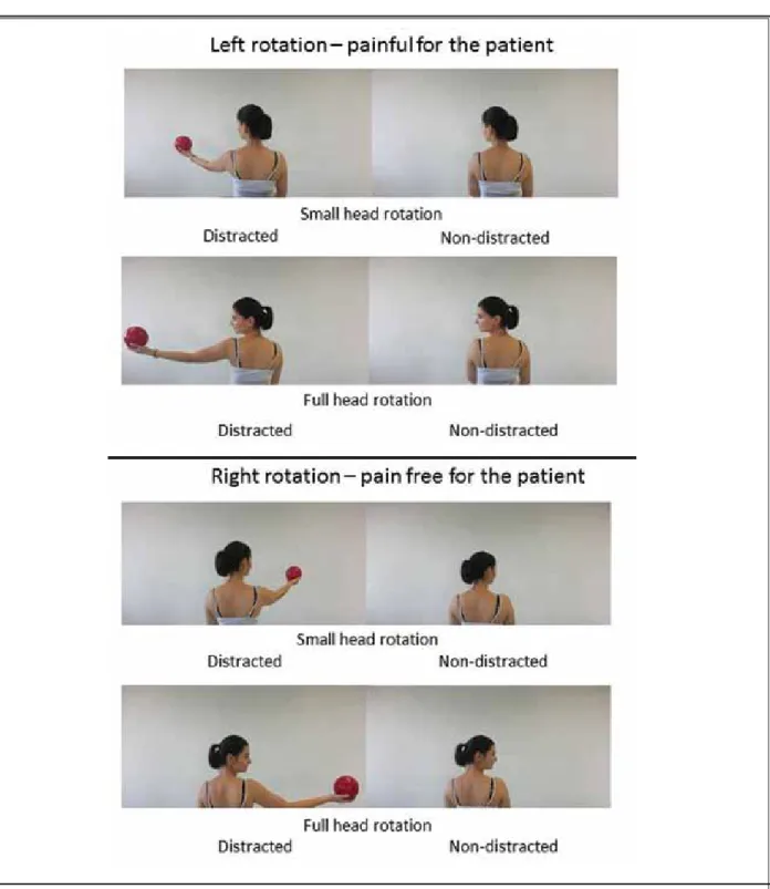
After informed consent was obtained, the patient and the control participated in an fMRI investigation in order to assess brain activity during AO+MI of 8 dif-ferent head rotation tasks (Fig. 1). In the scanner, the patient was in supine position and cushions were used to reduce head motion. Presentation of visual stimuli appeared on an LCD screen (32’’ NNL LCD Monitor,
Nor-dicNeuoLab, Bergen, Norway). The patient regarded the screen through a mirror system with a visual angle of 17° for the height and 9° for the length. The videos were presented with E-Prime 2.0 software (Psychology Software Tools, Inc., www.pstnet.com/ PA, USA) at 60 Hz. Overall, brain activity was assessed in 8 conditions [2 (“non-distracted” vs. “distracted head rotation”) x 2 (“left” vs. “right”) x 2 (“small” vs. “large amplitude”)] (Fig. 1). Thus, each video showed a certain combina-tion of amplitude (20° vs. 80°), direccombina-tion (left vs. right), and way of task execution (distracted vs. non-distract-ed). The patient observed the movements in the video and was asked to imagine at the same time being the person in the video performing the task. Thus, motor imagery (MI) was combined with action observation (AO) as this form of mental involvement (AO+MI) was previously shown to be superior to activate brain ar-eas compared to motor imagery or action observation alone (12,13). All 8 videos lasted for 16 seconds and Table 1. Results of sensorimotor and biopsychological assessments of the neck pain patient.
Inspection
Neutral head position The Patient shows a deviation of the head about 10° in lateral flexion to the right perceived as neutral head position
Range of motion
Rotation Right 80° pain-free, left only 20° due to pain Extension 70° with aberrant movement in middle cervical spine
Flexion 45°
Passive segmental motion
Lateral flexion Hypomobility in left lateral flexion at C0-C1
Extension Hypermobility at C4-C5
Hypomobility at C7-T1-T2
Sensorimotor performance
Cranio cervial flexion task 24 mmHg on the craniocervical flexion task with augmented activity of sternocleidomastoideus muscle. Balance On average, 8 seconds in tandem stance with the right leg in front and 12 seconds in tandem stance with the left leg in front could be accomplished by the patient. Cervical joint position sense Relocation of the neutral head position shows both under- and overshoots of more than 5°.
Palpation
PAIVM´s Tenderness and increased movements in the middle cervical spine on Maitland Grad II Mobilisa-tion. Stiffness and tenderness in the upper thoracic spine. Painful muscles Painful trigger points of trapezius, levator scapulae and the suboccipital extensors on the left side.
Assessments
NPAD-d FABQ CES-D
55 points interpreted as moderately restricted.
18 out of 66 points; Physical activity subscale: 6 points out of 24; Working subscale: 10 points out of 42. No noticeable fear avoidance behavior.
1 point, indicating no depressive disorder
NPAD-d = Neck Pain Disability Index – German version; FABQ = Fear avoidance beliefs questionnaire; CES-D = Center for Epidemiological Studies Depression Scale – German version; PAIVM´s = Passive accessory intervertebral movements.
Fig. 1. Conditions. Figure 1 displays the movements that were shown to the patient in the videos. The patient was asked to observe and, at the same time, imagine these tasks while undergoing an fMRI scan. Head rotations with maximal range of movement and with rotations of about 20° to the right and left side were displayed and the patient had to observe these tasks while imagining being the person in the video (AO+MI). Further, the distracted situation was displayed in videos for both sides, with full range of motion and a limited range of motion of about 20°.
were repeated twice in a randomized order. All videos were created with the same logic: each trial lasted for 2 seconds; one video consisted of 8 repetitions of one and the same trial. An auditory tone indicated the be-ginning of each movement. After each video, a resting period of 14 seconds was provided in which a white cross on a black screen was displayed. A 3T MRI scanner (Discovery MR750; GE Healthcare, Waukesha, Wisconsin USA) with a 32-channels standard head coil was used during acquisition at the Fribourg’s Hospital in Switzer-land (www.h-fr.ch/).
Data Analysis
The Statistical Parametric Mapping SPM8 software (www.fil.ion.ucl.ac.uk/spm/) working on Matlab 2012b (MathWorks, Inc., www.mathworks.com/, MA, USA) was used to analyze the functional images. Please refer to Taube et al (13) for a further detailed description. Statistical parametric maps (SPMs) were assessed by the contrast (condition > baseline) to reveal the
gen-eral pattern of activation of each experimental condi-tion (simple effects). To show differential patterns of activation between the painful and the healthy side for the patient and the control, contrasts were generated. These effects were displayed at P < 0.001 uncorrected with an extended cluster threshold of 60 contiguous voxels (P cluster < 0.05 uncorrected).
R
esults Simple EffectsNon-distracted large head rotation to the painful left side versus resting baseline
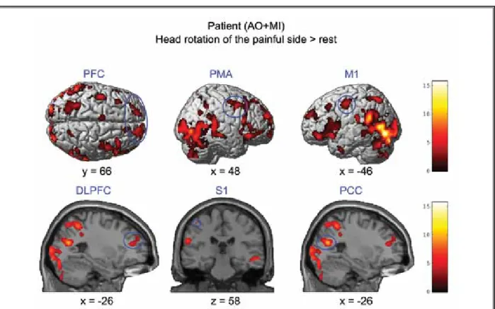
The simple effects were calculated for activation patterns of the non-distracted movement with a range of motion of 80° compared to resting baseline. Activity in areas associated with processing and expectation of pain and movement were identified and involved S1, PFC, DLPFC, and PCC. Activity in motor areas involved
Fig. 2. fMRI results – simple effects. Brain areas that were activated during combined motor imagery and action observation (AO+MI) of the pain-provoking movement to the left compared to the baseline (resting period). PFC, prefrontal cortex; PMA, premotor area; M1, primary motor cortex; DLPFC, dorsolateral prefrontal cortex; S1, primary somatosensory cortex; PCC, posterior cingulate cortex. fMRI results were displayed at P < 0.001 uncorrected with an extended cluster threshold of 60 contiguous voxels. Red-yellow colors highlighted the level of brain activation. (x, y, z) provides spatial coordinates in MNI space.
activation in M1 and PMA (Fig. 2) for the patient. The control volunteer presented activity in the visual areas (BA 17, 18, 19), superior frontal gyrus (BA 10), insula, superior temporal gyrus (BA 22, 13), and in the cerebel-lum (declive).
Non-distracted large head rotation to the pain-free right side versus resting baseline
In the patient, the activation pattern of the 80° pain-free movement to the right side during AO+MI re-vealed activity in motor areas like PMA, the supplemen-tary motor area (SMA), and the cerebellum. Further-more, the insula was significantly activated. However, no activity was found in any other brain area associ-ated with pain or the expectation of pain. Similarly, the control volunteer displayed activity in SMA, PMA, M1, caudate, thalamus, precuneus (BA 31), and superior and inferior frontal gyrus (BA 9, BA 10).
Non-distracted small head rotations to painful left side and pain-free right side versus resting baseline
The simple effects were calculated for activation patterns of the non-distracted head rotation of 20° compared to resting baseline. No activity was observed in pain-related and expectation-related brain areas for either the patient or the control irrespective of the side of rotation (left or right).
Contrasts
Non-distracted versus distracted large head rotation to the painful left side
Based on the observation that when the patient was distracted, she was able to fully rotate her head without pain, a contrast was defined to directly com-pare the non-distracted painful movement with the distracted movement (ball throwing task). AO+MI of the pain-provoking non-distracted movement revealed significantly higher activity in brain areas associated with the processing of pain such as S1, TH, and ACC, and compared to the distracted task. Furthermore, ar-eas that are associated with the expectation of pain, such as the PFC and motor areas like M1, SMA, and the cerebellum, were more strongly activated in the non-distracted condition (Fig. 3). The control did not show significant cerebral differences between distracted and non-distracted conditions.
Differences between non-distracted and distracted large head rotations to the painful left side versus
dif-ferences between non-distracted and distracted large head rotations to the pain-free right side
When comparing the differences of the pain-pro-voking non-distracted large head rotation (80°) and the distracted head rotation to the painful left side with the differences between the non-distracted pain-free movement and the distracted pain-free movement to the right side, extended activity was displayed in brain areas like ACC, PFC, and PCC. These areas are associated with the expectation of pain. Furthermore, the PMA was more strongly activated for the non-distracted painful left head rotation (Fig. 4). The control volunteer did not show significant cerebral differences between conditions.
Differences between non-distracted large and small head rotation to the pain-provoking side compared to the differences between non-distracted large and small head rotation to the pain-free side
In the patient, the comparison of differences be-tween large and small head rotations to the left side with the differences between large and small rotations to the right side revealed augmented activity in pain expectation-related brain areas like the PFC and PCC. Furthermore, the PMA showed increased activity (Fig. 5). The control did not display significant differences in cerebral activity for this interaction.
d
iscussionThe current single subject study provides evidence that maladaptive pain behavior goes along with differ-ences in activity patterns of the brain centers respon-sible for pain processing and the expectation of pain (S1, TH, In, ACC, PFC, DLPFC, PCC). First, during AO+MI of large head rotations, brain activity differed between the affected left (painful) and the non-affected right (pain-free) side, but no such differences were found for the control volunteer. Second, brain activity patterns for AO+MI of head rotations executed to the affected left side did not always provoke activity in pain-related brain areas. This means that when the head rotation was combined with a distracting motor task, in this case a ball throwing task, the patient showed signifi-cantly less activity in pain-related brain areas than in the non-distracted task execution. These results were confirmed by the behavioral data of the patient. The physical execution of the non-distracted head rotation to the pain-provoking left side was always restricted in range of motion due to pain, whereas a pain-free
Fig. 3. fMRI results comparing differences of non-distracted (painful) head rotations versus distracted head rotation (pain-free). Differences in activation patterns of brain areas during AO+MI of the pain-provoking movement (non-distracted head rotation) compared to activation patterns during AO+MI of the (non-distracted throwing task in the same direction. S1, primary somatosensory cortex; thalamus; ACC, anterior cingulate cortex; M1, primary motor cortex; SMA, supplementary motor area; PFC, prefrontal cortex; uvula and culmen as part of the cerebellum; insula. fMRI results were displayed at P < 0.001 uncorrected with an extended cluster threshold of 70 contiguous voxels. Red-yellow colors highlighted the level of brain activation. (x, y, z) provides spatial coordinates in MNI space.
rotation was possible in the presence of a distractor. Therefore, the restricted painful range of motion in the absence of a distractor can be viewed as a conditioned pain response, displayed as maladaptive pain behavior. Maladaptive pain behavior has been extensively investigated in chronic musculoskeletal pain by the fear avoidance model. Evidence supports the avoidance component as one key element in chronicity, especially in low back pain (14). However, the situation in chron-ic neck pain is less clear (15) and the present data do not point to this kind of mechanism. The patient did
not show fear, documented by a low score on the fear avoidance beliefs questionnaire (Table 1). Furthermore, at the neural level, there was no activity in the amyg-dala, which was previously demonstrated as a reliable marker for fear (16). Thus, the question of what the per-petuating factor is in this case remains speculative. As rotation of the head is a frequent everyday movement, the interrelation of pain and head rotation to the left might have been established in this patient in the early period after the accident. Furthermore, it was recently shown that even the mere intention to perform a pain-Fig. 4. Differences between non-distracted and distracted large head rotations to the painful left side versus differences between non-distracted and distracted large head rotations to the pain-free right side. (A) The contrast revealed augmented brain activity in PMA, premotor area; PFC, prefrontal cortex; PCC, posterior cingulate cortex; ACC, anterior cingulate cortex. (B) Graphics represent the contrast estimate from PCC (left plot) and ACC (right plot). Plots are based on a sphere of 10 mm around the (-6; -58; 28) voxel for PCC, and the (-12; 42; 8) voxel for ACC, red bars present the error with 90% of confidence interval. distracted head rotation and distracted head rotation of the painful side (P-nd and P-d). Non-distracted head rotation and Non-distracted head rotation of the pain-free side (Pf-nd and Pf-d). fMRI results were displayed at P < 0.001 uncorrected with an extended cluster threshold of 70 contiguous voxels. Red-yellow colors highlighted the level of brain activation. (x) provides spatial coordinates in MNI space.
ful movement can act as a covert conditioned stimulus inducing a fear response (17). Thus, this might have ad-ditionally or alternatively led to the maladaptive pain behavior. The contribution of this factor is supported by the fact that in our experiment, the mere mental simulation (AO+MI) to perform a head rotation to the left activated brain areas related to pain expectation, whereas no activation of pain related areas where found for the control.
Brain activity in pain-related areas during head rotations to the affected and non-affected side
It was recently demonstrated that not only the exe-cution, but also the observation of movements that are associated with pain, activate brain centres such as In, TH, PCC, and motor areas in patients with chronic low-er back pain (10). Thlow-erefore, the results of the present study showing differential activity of these brain areas depending on the side of head rotation are well in line
with this previous finding. In general, non-distracted large head rotations to the left (painful side) were ac-companied by activity in S1, PFC, DLPFC, and PCC. Fur-thermore, activity in motor areas involved activation in M1 and PMA, whereas rotation to the right did not dis-play significant activation of any of these areas (simple effects). When calculating contrasts, significantly larger activity was seen in ACC, PFC, PCC, and PMA when turn-ing the head to the left rather than to the right. Impor-tantly, the control did not show such side-dependent differences in activation. Our results are therefore well in line with previous reports from chronic low back pain patients (10), in whom the visualization of movements that were associated with painful experiences like car-rying heavy luggage resulted in enhanced brain activ-ity in the TH, In, SMA, PM, PCC, and cerebellum. In this previous study, it was suggested that the former experi-ence of back pain may sensitize patients so that they activate part of the pain matrix, also during observa-Fig. 5. Differences between non-distracted large and small head rotation to the pain-provoking side compared to the differences between non-distracted large and small head rotation to the pain-free side. Brain areas in which augmented brain activity was observed when taking together small and large head rotations to the pain-provoking left side compared to small and large rotations to the pain-free right side. The contrast indicated activity in PFC, prefrontal cortex (A); PCC, posterior cingulate cortex (B); PMA, premotor area (C). (D) Plot reveals the behavior of the biggest cluster, it was defined at the voxel (-18; 52; 32; PFC) of the largest cluster (cluster size: 5918), red bars present the error with 90% of confidence interval. Non-distracted large head rotation and non-distracted small head rotation of the painful side (P-and-L and P-and-S). Non-distracted large head rotation and non-distracted small head rotation of the pain-free side (Pf-and-L and Pf-and-S). fMRI results were displayed at P < 0.001 uncorrected with an extended cluster threshold of 70 contiguous voxels. Red-yellow colors highlighted the level of brain activation.
tion of potential harmful postures or movements. Our results confirm and extend these findings by showing that memory retrieval of unpleasant experiences can be highly task-specific, i.e., only the rotation to the left was associated with activation of pain-related brain ar-eas but not head rotations to the right. Furthermore, the current results demonstrate that the activation of brain areas related to the processing of pain is scaled based on the amplitude of the rotation. Large rotations caused greater activity in PFC, PCC, and PMA than small rotations. This result also fits well with the clinical pre-sentation of the patient where the execution of small rotations was associated with little or no pain.
Brain activity in pain-related areas during head rotations with and without a distracting secondary motor task
The clinical evaluation of our patient demonstrat-ed painful restrictdemonstrat-ed head rotation to the left side. However, in distracted situations, full range of motion of the cervical spine was possible without any pain. The behavioral data were complemented by brain activa-tion patterns obtained in the fMRI-scanner when con-trasting these 2 tasks (non-distracted head rotations versus distracted head rotations): While AO+MI of the non-distracted movement revealed extended activity in S1, TH, In, ACC, PFC, M1, cerebellum, and SMA, such activity was absent in the case where the same head ro-tation to the left was observed in a different, distracted context (throwing task). The selective activation of the pain matrix and the selective perception of pain indi-cate maladaptive pain behavior in our patient as the control volunteer did not show such differential activa-tion patterns. Further, the potential protective nature of pain can be refuted in this case, because within the distracted task, less activation of areas responsible for pain expectation was found. The analgesic effect of distraction is widely recognized (18). However, to our knowledge it has never been shown before that distrac-tion may serve to deactivate the pain matrix, so that movements that are usually associated with pain can suddenly be performed without discomfort.
Looking in detail at the non-distracted and dis-tracted head rotation, one may argue that although the head rotations were comparable, the 2 motor tasks clearly differ. The throwing task was undoubt-edly the more demanding task, and it is well estab-lished that increases in task difficulty enhance activity in motor centers (13,19). Thus, we would have ex-pected to see greater activity in the distracted head
rotation task than the non-distracted task, at least in motor centers. This imbalance in motor-related brain areas may also be responsible – at least in part – for the impaired ability of neck pain patients to relocate their head in predefined locations (20). However, even more importantly, the current results show that activa-tion of pain memories depend on the context. There-fore, we can conclude for this case that distracting the attention away from the pain-provoking movement prevents the perception of pain, enhances physical performance, and leads to entirely different brain ac-tivity, i.e., no activation of pain-related areas. It may therefore be argued that a certain level of attention has to be dedicated to the affected neck in order to activate the pain matrix. This may be a crucial point for the therapy of maladaptive pain memory/behavior. For neck pain patients, it was recently demonstrated that a balance training intervention proved to be ef-ficient with regard to reducing pain and improving sensorimotor function of the cervical spine (21). It may be speculated that during balancing, head movements are executed but attention is primarily allocated to keep and/or restore postural equilibrium. Thus, bal-ance exercises and the throwing task may be char-acterized as motor tasks involving the cervical spine without paying attention to the cervical motion itself. Instead, the focus is set on higher demands such as postural control or catching a ball probably prevent-ing the activation of the pain matrix.
Relevance of the Present Findings
Although the current results display only the situa-tion of one single patient with chronic, one sided neck pain so that the results cannot be generalized, they may nevertheless be important not only to improve the un-derstanding of the neural organization of maladaptive pain behavior but also to reconsider clinical evaluation and treatment strategies. From a clinical point of view, examination of chronic neck pain patients must clarify, whether symptoms and movement behaviors serve to protect tissue or whether they display maladaptive pain associated movement behavior, related to the expecta-tion of pain. For this purpose, it is important to keep in mind the differentiation of distracted versus non-distracted test-situation that we have provided by the ball throwing task (distracted) and the conscious head rotation task (non-distracted). Closely related to that, our results suggest that treatment strategies have to take into account and exploit the context in which the movement is performed.
R
efeRences1. Gatzounis R, Schrooten MG, Crombez G, Vlaeyen JW. Operant learning theory in pain and chronic pain rehabilitation.
Curr Pain Headache Rep 2012; 16:117-126.
2. Nijs J, Lluch Girbes E, Lundberg M, Mal-fliet A, Sterling M. Exercise therapy for chronic musculoskeletal pain: Innova-tion by altering pain memories. Man Ther 2014; 20:216-220.
3. Flor H. New developments in the under-standing and management of persistent pain. Curr Opin Psychiatry 2012; 25:109-113.
4. Nijs J, Meeus M, Cagnie B, Roussel NA, Dolphens M, Van Oosterwijck J, Dan-neels L. A modern neuroscience ap-proach to chronic spinal pain: Combin-ing pain neuroscience education with cognition-targeted motor control train-ing. Phys Ther 2014; 94:730-738.
5. Flor H, Braun C, Elbert T, Birbaumer N. Extensive reorganization of primary so-matosensory cortex in chronic back pain patients. Neurosci Lett 1997; 224:5-8. 6. Flor H, Nikolajsen L, Staehelin Jensen T.
Phantom limb pain: A case of maladap-tive CNS plasticity? Nat Rev Neurosci 2006; 7:873-881.
7. Swart CM, Stins JF, Beek PJ. Cortical changes in complex regional pain syn-drome (CRPS). Eur J Pain 2009; 13:902-907.
8. Apkarian AV, Sosa Y, Sonty S, Levy RM, Harden RN, Parrish TB, Gitelman DR. Chronic back pain is associated with decreased prefrontal and thalam-ic gray matter density. J Neurosci 2004; 24:10410-10415.
9. Kong J, Spaeth RB, Wey HY, Cheetham A, Cook AH, Jensen K, Tan Y, Liu H, Wang D, Loggia ML, Napadow V, Smoller JW, Wasan AD, Gollub RL. S1 is associated with chronic low back pain: A functional and structural MRI study. Mol Pain 2013; 9:43.
10. Shimo K, Ueno T, Younger J, Nishiha-ra M, Inoue S, Ikemoto T, Taniguchi S, Ushida T. Visualization of painful expe-riences believed to trigger the activation of affective and emotional brain regions in subjects with low back pain. PloS One 2011; 6:e26681.
11. Childs JD, Cleland JA, Elliott JM, Teyhen DS, Wainner RS, Whitman JM, Sopky BJ, Godges JJ, Flynn TW, American Physical Therapy A. Neck pain: Clinical practice guidelines linked to the International Classification of Functioning, Disability, and Health from the Orthopedic Section of the American Physical Therapy Associ-ation. J Orthop Sports Phys 2008; 38:A1-A34.
12. Vogt S, Rienzo FD, Collet C, Collins A, Guillot A. Multiple roles of motor imag-ery during action observation. Front Hum
Neurosci 2013; 7:807.
13. Taube W, Mouthon M, Leukel C, Hooge-woud HM, Annoni JM, Keller M. Brain activity during observation and motor imagery of different balance tasks: An fMRI study. Cortex 2014; 64C:102-114. 14. Vlaeyen JW, Crombez G. Fear of
move-ment/(re)injury, avoidance and pain dis-ability in chronic low back pain patients.
Man Ther 1999; 4:187-195.
15. Vernon H, Guerriero R, Kavanaugh S, Soave D, Puhl A. Self-rated disabili-ty, fear-avoidance beliefs, nonorganic pain behaviors are important mediators of ranges of active motion in chronic whiplash patients. Disabil Rehabil 2013; 35:1954-1960.
16. Wiech K, Tracey I. Pain, decisions, and actions: A motivational perspective.
Front Neurosci 2013; 7:46.
17. Meulders A, Vlaeyen JW. Mere inten-tion to perform painful movements elic-its fear of movement-related pain: An experimental study on fear acquisition beyond actual movements. Pain J 2013; 14:412-423.
18. Jensen KB, Berna C, Loggia ML, Wasan AD, Edwards RR, Gollub RL. The use of functional neuroimaging to evaluate psychological and other non-pharmaco-logical treatments for clinical pain.
Neu-rosci Lett 2012; 520:156-164.
19. Roosink M, Zijdewind I. Corticospinal excitability during observation and im-agery of simple and complex hand tasks: Implications for motor rehabilitation.
Behav Brain Res 2010; 213:35-41.
20. Beinert K, Keller M, Taube W. Neck mus-cle vibration can improve sensorimo-tor function in patients with neck pain.
Spine J 2015; 15:514-521.
21. Beinert K, Taube W. The effect of balance training on cervical sensorimotor func-tion and neck pain. J Mot Behav 2013; 45:271-278.
Acknowledgments
We thank Eric Dafflon for technical assistance. There are no conflicts of interests for each author.