Retinoic Acid-Binding Protein in Human Breast
Cancer and Dysplasia
1.2P R H b . . u er, 3 E Geyer,3 W. K'ung,3 A. Matter,4 . J. Torhorst,5 and U. Eppenberger 3
ABSTRACT -Seventy-five specimens of human breast tissue were checked for the presence of cellular retinoic acid-binding protein (cRABP). Fifty-two percent of the primary carcinomas and 43% of the dysplastic breast lesions (stage Mil) contained detectable amounts of cRABP, whereas no cRABP was found in normal tissue. Sucrose gradient centrifugation and electrophore-sis on agarose were used for analyelectrophore-sis of the presence of cRABP. The cRABP of human origin (normal uterus and neoplastic mammary tissue) differed in its mobility in agarose electro-phoresis from that of rat testis cRABP-J Natl Cancer Inst 61:
1375-1378, 1978.
Retinol (vitamin A alcohol) is necessary for normal growth and differentiation of epithelial cells. Recent reports show that retinol and synthetic derivatives may prevent preneoplastic changes caused by carcinogens in epithelial cells (1-4). RA exhibits similar properties (5, 6) as do synthetic analogs of RA (7, 8). Specific binding proteins have been demonstrated for both retinol and RA in various tissues (9, 10). A postulation is that both retinol and RA act through their respective binding proteins in a manner similar to that found for steroid hormones (11, 12). Ong and co-workers (13)
showed the presence of cRABP in a limited number of breast and lung tumors. but not in the surrounding noncancerous tissue. Therefore. it was of interest to determine whether this binding protein was also pres-ent in dysplastic cells.
MATERIALS AND METHODS
Chemicals.-All biochemical reagents were obtained from Sigma Chemical Co.. St. Louis. Missouri. The [I PH]RA (sp act. 1.3 Ci/mmole) was obtained from F. Hoffmann-La Roche. Ltd .. Basel. Switzerland. and was purified before each experiment. The [l PH]RA was stored in toluene at -20° C. Before use the toluene was blown off under argon and replaced by ethanol containing I mg (X-tocopherollm!. Agarose was pur-chased from Seakem-Biomedical. Rockland. Maine.
Preparation of cytosol.-Breast tissue specimens were obtained from patients during mastectomy and from biopsies. The tissue was morphologically characterized bv macroscopic examination at the time of surgery and b~' microscopic examination by the pathologist. The ti'ssue material was frozen immediately after excision and stored at -70° C. Subsequently. it was pulverized in liquid nitrogen. The tissue powder (1-1.5 g) was homogenized at pH 7.5 in ice-cold 50 mM Tris-HCI buffer in a ratio of I: 4 (wt/vol) and centrifuged at 105.000Xg for I hour. The supernatant fraction. con-taining 5-10 mg protein/mI. was recovered. Protein was determined according to the method of Lowry et al. (14).
Assay for cRABP.-Aliquots. 250 or 500 1-'1. of the supernatant fraction were incubated at 4° C for 4 hours in the dark with an equal volume of [3H]RA (10-7 M) in the assay buffer (50 mM Tris-HCl. pH 7.5). The incubation was carried out in the presence or absence of a 200-fold excess (2XIO-5 M) of unlabeled RA. The unbound ligand was removed by treatment of the solution with 250 or 500 1-'1 of a dextran-coated charcoal suspension (0.25% charcoal and 0.025% dex-tran in 50 mM Tris-HCl buffer. pH 7.5).
Sucrose gradient sedimentation.-Aliquots. 200 1-'1. of the supernatant fractions were layered on linear 5-20% sucrose gradients and centrifuged at 4° C for 16 hours at 50.000 rpm.
Agarose electrophoresis.-For the electrophoresis the method described by Wagner (15) was used. Agar was replaced by agarose because agarose has lower electro-osmosis. We prepared slab gels by heating 1 g agarose in 100 ml of 50 mM Michaelis buffer. pH 8.2. The clear solution was allowed to cool to 50° C and subsequently poured in a mold containing a glass plate (9.9X8.4 cm) to support the gel. Ten wells (50 1-'1) along the center line were punched out after the gel had formed. The glass plate containing the gel was placed on a brass plate coated with Teflon. and the temperature was lowered to 0° C by a circulating cooling mixture. We obtained electrical contacts by placing buffer-soaked filter paper at the long ends of the slabs. In the chamber was 100 mM Michaelis buffer. pH 8.2. The test solution was freed from unbound [3H]RA by the above charcoal method and introduced in the center wells (2X50 I-'lIsample). Electrophoresis was performed im-mediately to avoid diffusion and run at 120 rnA and 230 V for 90 minutes at 4° C. For analysis of the distribution of radioactivity. we sliced the gel into squares (9X3 mm). Only the anodic part of the plate was checked for specific RA binding. The gel slices were dissolved in Instagel and counted for radioactiv-ity. We identified the peaks by running [3H]RA and
ABBREVI.\TIONS I'SEO; cRABP= cellular retinoic acid-binding protein; HSA = human serum albumin; RA = retinoic acid.
I Received December 20. 1977; revised June 19. 1978; accepted June 23. 1978.
2 Supported by the F. Hoffmann-La Roche Foundation and
F. Hoffmann-La Roche. Ltd .. Basel. Switzerland.
, Hormone Laboratory and Experimental Endocrinology. Depart-ment of Gynecology. University of Basel. 4004 Basel. Switzerland.
• Pharmaceutical Research Division. F. Hoffmann-La Roche. Ltd .. Basel. Switzerland.
5 Institute of Pathology. University of Basel. Swillerland.
1376 Huber, Geyer, Kung, et al.
HSA plus [SH]RA simultaneously with tissue extracts incubated with radioactively labeled ligand. For the mixing experiment, a crude tumor extract (4 mg protein/ml) was mixed with a partially purified rat testis cRABP preparation (1.1 mg protein/ml) and analyzed as described above.
'0 .~ u '/5 .S ~ E a. u 2.4 2.0 1.6 1.2 0.8 0.4 2.4 2.0 1.6 1.2 0.8 ~ 0.4
..
2.4 2.0 1.6 1.2 0.8 0.4o
Pat. 235~
a: u Pat. 236 Pat. 237 4 8 12t
FRACTION NUMBER TOP A B C 16 20t
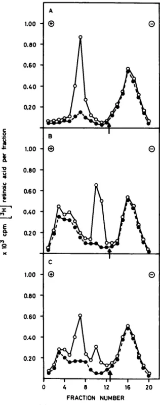
BOTTOMTEXT·FlGURE I.-Sucrose gradient sedimentation pattern of human breast tumor extracts. A: Stage Mil containing cRABP. B: Primary carcinoma containing cRABP in high quantity. C: Stage Mil with no detectable amounts of cRABP. For each expt. an average of 4-10 mg protein/ml was used. Pat. = patient.
J
NATL CANCER INSTRESULTS
Sucrose gradient sedimentation of breast tissue sam-ples incubated with [~H]RA revealed two peaks of
..
u '0 .5 "i...
e
0. u P'I 52..
1.8 1.4 1.0 0,6 0.2 0.3 0.2 0.1 Pal 333 4 TOP Pat. 333 2 4 6 8 10 12 FRACTION NUMBER 6 8, ,
e ..~
a: u "e-e -e-e_ 10 12 FRACTION NUMBER A Be
TEXT·FtGURE 2.-A: Sucrose gradient sedimentation pattern of a pri-mary tumor extract (4.6 mg protein/ml). which was centrifuged in the absence ( 0 - - 0 ) and presence (e - - - e) of 200-fold ex-cess unlabeled RA. B: Agarose electrophoresis of the same tissue sample (4.6 mg protein/ml) as in panel A. Extract was subjected to electrophoresis in the absence ( 0 - - - 0 ) and presence
(e- - - e) of 200-fold excess unlabeled RA. Arrow indicates start
of electrophoretic migration. Pat.
=
patient.Acid-Binding Protein In Human Breast Cancer and Dysplasia 1377 A 1.00 (f)
e
0.80 0.60 0.40 0.20 c .2 B~
1.00 Ef)e
!
'0 0.80 .~ u '(5 0.60 c::
..
""%'
0.40 (WI ~ E Q. 0.20 u (WI 51 >C C 1.00 (f)e
0.80 0.60 0.40 0.20o
4 8 12 16 20 FRACTION NUMBERTEXT·FlGURE 3.-Mixing expt with extracts from rat testis and hu-man breast cancer. Agarose electrophoresis was used to analyze for cRABP. A: cRABP from rat testis (1.1 mg protein/mI). B: cRABP from human breast cancer (4.6 mg protein/ml). C: Analysis of the mixed solutions from rat testis and human breast cancer. Samples run in absence ( 0 - - 0 ) and presence (e- - - e) of 200-fold excess unlabeled RA. Arrows indicate start of electrophoretic migration.
radioactivity sedimenting at 2S and 4.6S (text-fig. IA). The binding at 2S represented the cRABP inasmuch as this peak could be abolished by a 200-fold excess of unlabeled RA. This inhibition was specific for RA
VOL. 61, NO.6, DECEMBER 1978
inasmuch as a similar excess of retinol showed no inhibition. According to Sani and Hill (16), the 4.6S peak represented nonspecific binding to serum al-bumin. The pattern in text-figure IA was the one encountered in most tissue samples analyzed. Text-figure IB exhibits a profile typical of a biopsy sample with a very high concentration of cRABP, whereas the tissue sample shown in text-figure Ie contained no detectable amounts of cRABP. The criterion for the presence of a specific RA binding was the abolishment of the 2S peak by an excess of unlabeled RA.
Text-figure 2A demonstrates the sedimentation pat-tern and text-figure 2B shows the electrophoretic mi-gration of cRABP obtained from a mammary tumor. With both methods, the peak characteristic for cRABP was very prominent. The peak at fraction II (text-fig. 2B) disappeared completely with an excess of unlabeled RA. The radioactivity found in fractions 3-5 rep-resented a mixture of free and unspecifically bound ligand to HSA.
On agarose electrophoresis, cRABP of human origin did not migrate to the same position as did the rat testis cRABP.
Text-figure 3A demonstrates the electrophoretic pro-file for cRABP obtained from rat testis, and text-figure 3B shows the electrophoretic profile obtained for cRABP from human breast tumor. The maximum radioactivity in the rat testis material for cRABP was found in fraction 7 as compared to fraction II of the human sample (text-fig. 3B). The mixing experiment suggested that cRABP from the two sources was different (text-fig. 3C). Such a difference went unde-tected by sucrose gradient centrifugation of the rat testis and the human tumor extracts. The electro-phoretic migration pattern of cRABP (text-fig. 3B), obtained from a primary carcinoma, was not restricted to human breast cancer. Human uterus extracts, which contain the RA-binding protein (17), incubated with PH]RA and subjected to agarose electrophoresis, showed the ligand specifically bound also at fraction II (results not shown). Furthermore, the electropho-retic profile of extracts from human breast tumors in stage MIl (proliferation of ductules in breast tissue plus intraductal epithelial proliferation) was the same as in the primary carcinomas (results not shown).
Table I shows the distribution of cRABP in breast dysplasias, with different proliferative activities, and in primary carcinomas. In 52% of the carcinomas, cRABP
TABLE l.-Distribution of cRABP in dysplastic and neoplastic human mammary tissue
Type of No. of No. of tissue
extracts con- Percent tissue, stage" biopsies
taining cRABP
MI-F 16 0 0
MI 16 0 0
MIl 14 6 43
CA 29 15 52
• Classified according to the criteria in (18); MI-F, simple dysplasia with extensive fibrosis; MI, simple dysplasia; MIl, proliferative dysplasia; and CA, primary carcinoma.
1378 Huber, Geyer, Kung, et 81.
actIVity was detected. Also, 43% of the specimens with pronounced epithelial proliferation (stage MIl) con-tained cRABP. Neither of the 16 samples graded MI-F (fibrosis and atrophy of the breast tissue) nor MI (proliferation of ductules in breast tissue) exhibited detectable amounts of cRABP.
DISCUSSION
These results add more information to our under-standing of the distribution of cRABP in the dif-ferent stages of breast tumor formation. In contrast to Dng et al. (13), we found cRABP in only 52% of the invasive carcinomas examined. Therefore, cRABP can-not necessarily be called an obligatory product of carcinoma formation. Also, we detected the binding protein in less than half of the breast lesions of proliferative dysplasia (stage MIl), which are con-sidered as being possibly precancerous. None of the tissue specimens graded MI-F, which was considered normal tissue, contained cRABP (table 1).
Retinoids obviously influence the differentiation of epithelial cells (6, 8). Attempts to elucidate their mode of action in relation to the occurrence of steroid receptors in breast carcinomas are in progress in this laboratory. These studies and those concerning the adenylyl cyclase protein kinase system as biochemical markers (19, 20) may provide a better understanding of the many factors influencing the development of hu-man breast tumors.
REFERENCES
(I) CHU EW. MALMGREN RA: An inhibitory effect of vitamin A on the induction of tumors of forestomach and cervix in the Syrian hamster by carcinogenic polycyclic hydrocarbons. Can-cer Res 25:884-895. 1965
(2) SPORN MB. DUNLOP NM. NEWTON DL: Prevention of chemical carcinogenesis by vitamin A and its analogs (retinoids). Fed Proc 35:1332-1338. 1976
(3) MOON RC. GRUBBS CJ. SPORN MB. et al: Retinyl acetate inhibits mammary carcinogenesis induced by N-methyl-N-ni-trosourea. Nature 267:620-621. 1977
J
NATL CANCER INST(4) GRUBBS CJ. MOON RC. SPORN MB. et al: Inhibition of mam-mary cancer by retinyl methyl ether. Cancer Res 37:599-602. 1977
(5) BOLLAG W: Prophylaxis of chemically induced benign and ma-lignant epithelial tumors by vitamin A acid (retinoic acid). Eur J Cancer 8:689-693. 1973
(6) - - : Effect of vitamin A acid on transplantable and chemi-cally induced tumors. Cancer Chemother Rep 55:53-58. 1971 (7) - - : Therapeutic effect of an aromatic retinoic acid analog
on chemically induced skin papillomas and carcinomas of mice. Eur J Cancer 10:731-737. 1974
(8) - - : Therapy of epithelial tumors with an aromatic retinoic acid analog. Chemotherapy 21:236-247. 1975
(9) ONG DE. CHYTIL F: Presence of cellular retinol and retinoic acid binding proteins in experimental tumors. Cancer Lett 2: 25-30. 1976
(10) - - : Changes in levels of cellular retinol and retinoic acid binding proteins of liver and lung during perinatal develop-ment of the rat. Proc Natl Acad Sci USA 73:3976-3978. 1976 (11) BASHOR MM. TOFT DO. CHYTIL F: In vitro binding of retinol to rat tissue compounds. Proc Natl Acad Sci USA 70:3433-3487. 1973
(12) SANI BP. CORBETT TH: Retinoic acid binding protein in nor-mal tissue and experil!lental tumors. Cancer Res 37:209-213. 1977
(13) ONG DE. PAGE DL. CHYTIL F: Retinoic acid binding protein: Occurrence in human tumors. Science 190:60-61. 1975
(/4) LOWRY OH. ROSEBROUGH NJ. FARR AL. et al: Protein measure-ment with the Folin phenol reagent. J BioI Chem 193:265-275. 1951
(15) WAGNER RK: Characterization and assay of steroid hormone re-ceptors and steroid binding serum proteins by agar gel elec-trophoresis at low temperature. Hoppe Seylers Z Physiol Chem 353:1235-1245. 1972
(16) SANI BP. HILL DL: Retinoic acid: A binding protein in chick embryo metatarsal skin. Biochem Biophys Res Commun 61: 1276-1282. 1974
(17) CHYTIL F. PAGE DL. ONG DE: Presence of cellular retinol and retinoic acid binding protein in human uterus. Int J Vitam Nutr Res 45:293-298. 1975
(18) SCARFF RW. TORLONI H: Histological Typing of Breast Tu-mors. Geneva: World' Health Organization. 1968
(19) EpPENBERGER U. TALMADGE K. KONG W. et al: Adenosine 3'.5'-monophosphate dependent protein kinase and cyclic-AMP-binding in human mammary tumors. FEBS Lett 80:229-234. 1977
(20) KONG W. BECHTEL E. GEYER E. et al: Altered levels of cyclic nu-cleotides. cyclic AMP phosphodiesterase. and adenylyl cyclase activities in normal. dysplastic. and neoplastic human mam-mary tissue. FEBS Lett 82:102-106. 1977