HAL Id: tel-01400312
https://tel.archives-ouvertes.fr/tel-01400312
Submitted on 21 Nov 2016
HAL is a multi-disciplinary open access
archive for the deposit and dissemination of sci-entific research documents, whether they are pub-lished or not. The documents may come from teaching and research institutions in France or abroad, or from public or private research centers.
L’archive ouverte pluridisciplinaire HAL, est destinée au dépôt et à la diffusion de documents scientifiques de niveau recherche, publiés ou non, émanant des établissements d’enseignement et de recherche français ou étrangers, des laboratoires publics ou privés.
Shifting eating creates a misalignment of peripheral and
central circadian clocks, which leads to a metabolic
syndrome
Ahmad Kobiita
To cite this version:
Ahmad Kobiita. Shifting eating creates a misalignment of peripheral and central circadian clocks, which leads to a metabolic syndrome. Biochemistry, Molecular Biology. Université de Strasbourg, 2016. English. �NNT : 2016STRAJ003�. �tel-01400312�
UNIVERSITE DE STRASBOURG
Ecole de doctorale des Sciences de la Vie et de la santé
Thèse de doctorat
Présentée par
Ahmad KOBIITA
Soutenue le : 12 Février 2016
En vue de l’obtention du grade de
Docteur de l’Université de Strasbourg I
Discipline : Sciences du vivant
Spécialité : Aspects Moléculaires et Cellulaires de la Biologie
“Shifting eating creates a misalignment of peripheral and central circadian
clocks, which leads to a metabolic syndrome”
“
Un décalage de l’alimentation déclenche une asynchronie entre l’horloge
circadienne centrale et les horloges périphériques et engendre un syndrome
métabolique ’’
Thèse dirigée par: Pr. Pierre CHAMBON, IGBMC, Illkirch, France
Rapporteur externe: Pr. Walter WAHLI, Lee Kong Chian School of Medicine, Singapore Rapporteur externe: Pr. Filippo RIJLI, Institute Friedrich Miescher, Basel, Switzerland Examinateur: Pr. Romeo RICCI, IGBMC, Illkirch, France
“Early to bed and early to rise, makes a man healthy, wealthy and wise”
Table of contents……….1
Acknowledgments ...2 Résumé en Français ...3 Abbreviations ..9I- Introduction………11
1- Circadian rhythms .112- The molecular mechanism of circadian clocks in mammals ..12
3- Central and peripheral circadian clocks in mammals .15
a- Suprachiasmatic nucleus of the hypothalamus (SCN): the master circadian clock synchronizer 15
b- Peripheral circadian clocks ..17
4- Phenotypic effects of circadian clock mutations 18
5- External cues entrain circadian clocks ...20
a- Light-entrainment of the master SCN circadian clock .20
b- Feeding-entrainment of circadian clocks of peripheral cell/tissues ..21
6- Nuclear receptors and circadian clocks ..22
7- Circadian clocks and metabolic factors are tightly linked ..23
8- Microbiota and circadian clocks 25
References………..27
II-
Aim of the thesis………....32
III- Results and Discussion………33
1- Publication 1: Shifting the feeding of mice to the rest phase creates metabolic alterations, which, on their own, shift the peripheral circadian clocks by 12 hours. Mukherji A*, Kobiita A*, Chambon P (2015). Proc Natl Acad Sci USA, 10.1073/pnas.1519735112. (*Co-First author) 2- Publication 2: Shifting eating to the circadian rest phase misaligns the peripheral clocks with the master SCN clock and leads to a metabolic syndrome. Mukherji A, Kobiita A, Damara M, Misra N, Meziane H, Champy MF, and Chambon P (2015). ProcNatl Acad Sci USA, 10.1073/pnas.1519807112. 3- Publication 3: Homeostasis in intestinal epithelium is orchestrated by the circadian clock and microbiota cues transduced by TLRs. Mukherji A, Kobiita A, Ye T, and Chambon P. (2013). Cell 153, 812–827.
IV- General Discussion and Prospects………..99
Acknowledgments
First of all, I would like to express my sincere gratitude to Professor Pierre CHAMBON to give me the opportunity to work and learn under his supervision and for his valuable guidance, discussions and encouragement during my PhD.
I also would like to thank Dr Daniel METZGER for his support during my PhD.
I would like to thank Pr Walter WAHLI and Pr Filippo RIJLI for accepting to be the external referees and Pr Romeo RICCI for accepting to be the internal referee of my thesis work. Big thanks to all members of CHAMBON/METZGER lab especially to Atish, Laetitia, Manohar, Nisha, and Jacky for their useful discussion and their support during my PhD. I also would like to thank Valérie for her assistance.
Big thanks to the personnel of IGBMC-ICS care facilities.
I am especially grateful to my parents, my two sisters and my friends for fully supporting me in all these years.
Résumé en Français
Depuis sa création, la vie sur terre est rythmée par le cycle circadien qui en 24 heures correspond à lalternance du jour et de la nuit. La rotation de la terre autour de son axe soumet la majeure partie de la surface terrestre à une alternance de clarté et dobscurité. Le comportement et la physiologie de la plupart des êtres vivants sur la terre sont affectés en conséquence. Chez les mammifères, chaque cellule possède une horloge moléculaire circadienne qui assure lalternance régulière de périodes dactivité et de repos denviron 12 heures. En effet, la plupart des animaux chassent au moment où ils sont actifs et leur aptitude à apercevoir leur proie la plus grande. Par contre dautres, comme les souris, cherchent à être à labri et à éviter dêtre aperçus par les prédateurs. Lapport de nourriture étant circadien, de nombreux aspects de la physiologie, la glycémie et lhoméostasie sont également liés à la régulation circadienne. Une propriété importante des rythmes circadiens est quils ne dépendent pas directement de lalternance du jour et de la nuit, mais sont gouvernés par une horloge endogène qui est capable de conserver une période proche de 24 heures.
En 1971, Konopka et Benzer ont été les premiers à identifier des gènes jouant un rôle dans lhorloge circadienne chez la drosophile. Après cette découverte, une dizaine de gènes impliqués dans la genèse de lhorloge circadienne furent identifiés. Les produits de ces gènes forment des boucles dauto-activation et dauto-inhibition transcriptionelles et traductionnelles permettant ainsi la formation dune période rythmique denviron 24h. Ces gènes sont exprimés au sein de lhorloge centrale située dans les neurones du noyau suprachiasmatique (NSC) et dans toutes les cellules des tissues de la périphérie. Au niveau moléculaire, les éléments clé du système de lhorloge sont les protéines Clock (Circadian Locomotor Output Cycles Kaput) et Bmal1 (Brain and Muscle Arnt-like Protein 1). Ces dernières agissent comme des facteurs de transcription en se fixant sur la boite régulatrice E (E-Box) de la région promotrice des gènes horloges Period (Per1, Per2) et Cryptochrome (Cry1, Cry2). Lorsque lexpression des protéines Per et Cry atteint un certain niveau dans le cytoplasme, ces deux protéines se dimérisent et se transloquent dans le noyau en empêchant la fixation du dimère Clock/Bmal1, et en conséquence inhibent leur propre expression. Par ailleurs, lexpression du gène Bmal1 est régulée par lintermédiaire des facteurs de transcription appartenant à la famille des récepteurs nucléaires orphelins. Il sagit de RORs ( , ! et ") et de RevErbs ( et !), qui se fixent sur les séquences ROREs (Retinoic acid-related Orphan receptor Response Elements) présentes sur le
promoteur du gène Bmal1. Le récepteur nucléaire RevErb est un nouveau venu dans loscillateur moléculaire qui réprime à son tour lexpression de Bmal1 et par conséquence sa propre expression.
En fait, cette horloge tourne dans des conditions très déterminées, tout en restant synchronisée avec son environnement. Une question très importante est de comprendre comment cette synchronisation est établie entre lindividu et le monde extérieur ?? Cela suggère lexistence dune voie de signalisation dentrée par laquelle les stimuli externes (lumière ou autres) ajustent la période de lhorloge interne afin que sa phase reste synchronisée avec son environnement extérieur et dune autre voie de sortie (vers les organes périphériques) par laquelle cette horloge est capable de générer une réponse optimale de lensemble des fonctions physiologiques de lorganisme.
Deux propriétés fondamentales caractérisent lactivité de lhorloge circadienne: La rythmicité de son activité est endogène et proche de 24 heures.
Elle doit être synchronisée par des signaux externes (par exemple la lumière)
Chez les mammifères, dans les conditions normales, la lumière est le puissant synchronisateur de lhorloge. En effet une brève exposition à la lumière durant la nuit est capable dinduire lexpression immédiate des gènes de lhorloge tels Per1 et Per2, ce qui suggère la présence dun lien direct par lequel la lumière contrôle loscillateur de lhorloge centrale NSC.
Des travaux réalisés au cours ces dernières années ont mis en évidence dautres synchroniseurs que la lumière qui agissent sur lhorloge circadienne. De fait la nourriture agit comme un nouveau et un puissant facteur de contrôle des horloges périphériques. Léquipe dU.Schibler à Genève a démontré que la nourriture est capable dentraîner les rythmes du système circadien périphérique chez la souris. En déplaçant l'horaire de l'alimentation chez celle-ci, linversion la prise de nourriture de la phase active à la phase de repos (Restricted Feeding : RF) entraîne lhorloge périphérique et sans avoir deffet sur lhorloge centrale (NSC), ce qui conduit à un découplage entre les deux horloges. Ainsi, les horloges périphériques suivent le rythme imposé par les apports de nourriture, alors que les NSC suivent le rythme imposé par la lumière. Ces études de découplage entre les horloges centrales et périphériques indiquent que les deux synchronisateurs, la lumière et la nourriture, peuvent agir indépendamment lun de lautre. Dans
de telles conditions, les horloges périphériques peuvent être en permanence décalées par rapport à lhorloge centrale, ce qui pourra perturber lhoméostasie des fonctions corporelles et déclencher à long terme des perturbations du cycle vielle-sommeil, du métabolisme (syndrome métabolique, obésité, diabète de type II) et du comportement. Ainsi un certain nombre de données ont montré quun défaut de synchronisation de lhorloge circadienne peut être induit observé chez les personnes engagées dans une activité décalée ou nocturne et dont le régime alimentaire est décalé.
Au cours de mon travail de thèse, jai adressé plusieurs questions concernant au niveau moléculaire la compréhension de ces perturbations métaboliques consécutives à un régime alimentaire décalé dans le cadre dun travail posté.
En effet, un décalage de phase pour laccès à la nourriture (RF), de la phase active à la phase de repos chez la souris, décalait les horloges périphériques de 12 heures par rapport à lhorloge centrale. Toutefois le mécanisme moléculaire dinversion de lhorloge circadienne par lapport décalé de nourriture dans les organes périphériques restait inconnu.
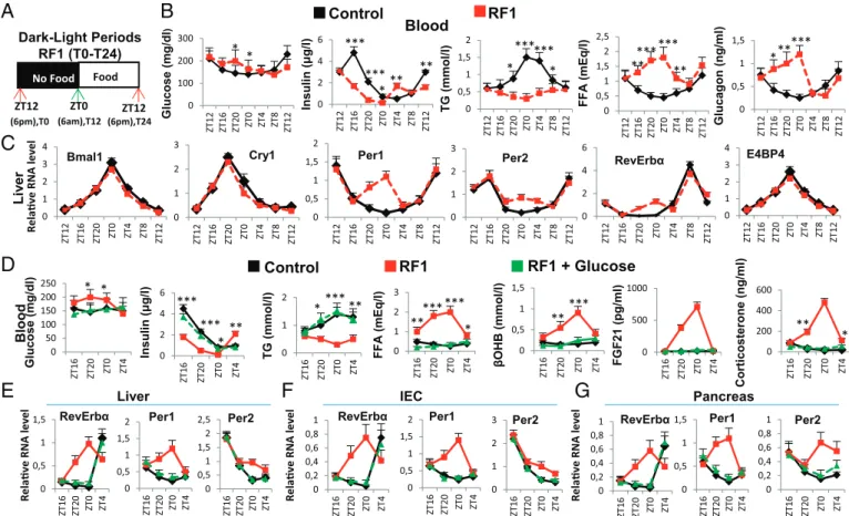
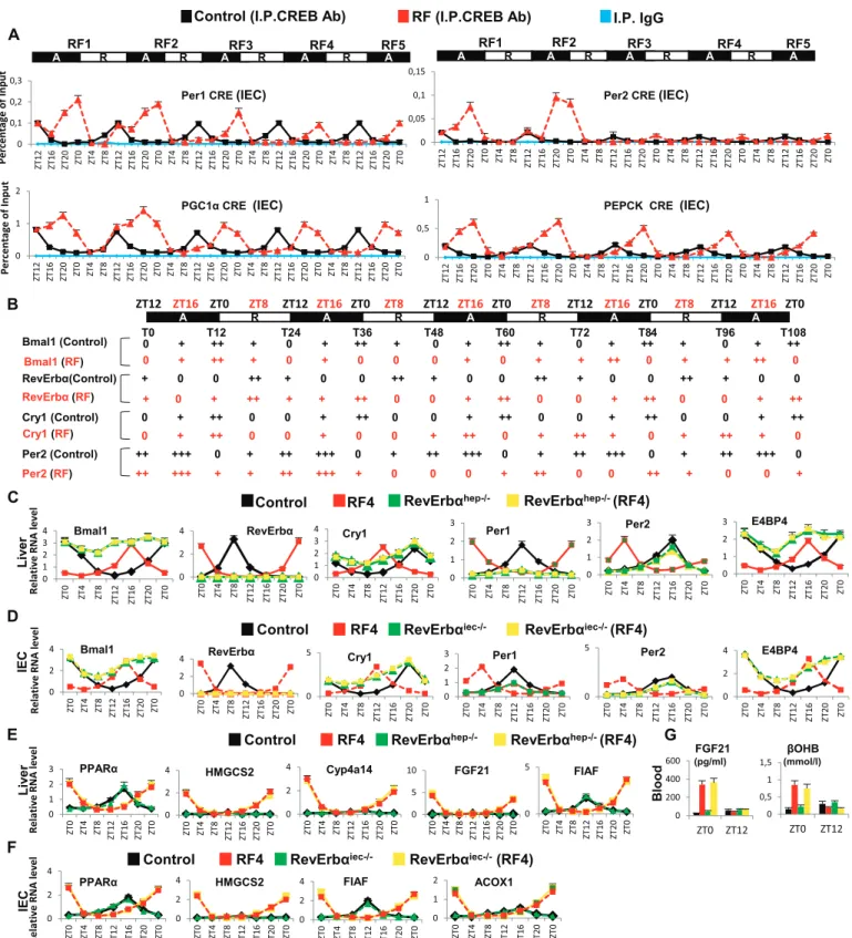
Durant ma thèse, jai montré au niveau moléculaire que les altérations métaboliques induites par linversion de phase pour lalimentation exercent une grande influence sur l'expression des éléments essentiels des horloges périphériques. En effet déplacer l'heure daccès à l'alimentation de la phase active (ZT(Zeitgeber) 12-ZT0) à la phase de repos (ZT0-ZT12) crée de fait un état de « jeûne » de 12 heures chez la souris. Nous avons donc étudié les paramètres métaboliques pendant cette période. Nous avons constaté que le passage de lhorloge circadienne initiale à la nouvelle horloge circadienne sous le régime RF comporte deux phases distinctes: (i) la rupture de lhorloge circadienne initiale, et (ii) la génération et le maintien dune nouvelle horloge circadienne décalée de 12 heures.
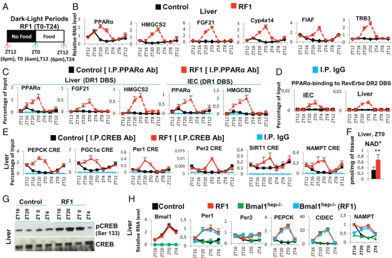
Pendant les premières heures du régime alimentaire décalé, nous avons remarqué une diminution du taux de glucose sanguin et une rapide chute au niveau de la production dinsuline. La poursuite de la baisse dinsuline permet lactivation de la lipolyse. Suite à cette activation, une augmentation accrue au niveau des acides gras libres (Free fatty-acid : FFA) survient dans le sang. Cette augmentation aberrante des acides gras libres (FFA) conduit à une surexpression pendant la phase de repos du récepteur nucléaire PPAR dont ils sont des ligands naturels. De plus, des souris portant des mutations sélectives de PPAR dans le foie (hépatocytes) et lintestin (cellules épithéliales) ont montré que laugmentation précoce de lactivité de RevErb
durant la phase de repos est dépendante de celle de PPAR . Des analyses « ChIP » poussées ont montré que la fixation de PPAR sur un élément de liaison DR2 présent dans le promoteur de RevErb était en fait à l'origine de son expression précoce pendant la phase de repos. Lensemble de ces résultats suggèrent un rôle essentiel de PPAR durant le régime alimentaire décalé qui pourrait, dans les organes périphériques étudiés, déclencher lexpression précoce de RevErb , ce qui en décalant lhorloge circadienne, entraînait un décalage complet des horloges périphériques par rapport à lhorloge centrale. En outre, lhypoinsulinémie conduit à une augmentation de lactivité du glycogène synthase kinase 3! (GSK3ß) qui, en stabilisant la protéine RevErb , augmente son niveau pendant la phase active.
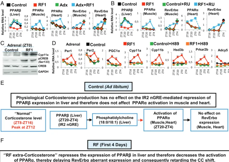
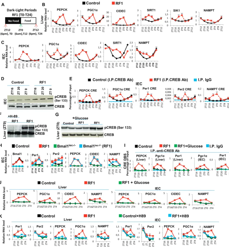
Des études avaient montré que le régime alimentaire décalé (RF), provoquait lapparition dun nouveau pic additionnel de corticostérone pendant la période de repos (ZT0), de manière non contrôlée par lhorloge centrale (NSC) et dont le mécanisme dapparition était inconnu. La biosynthèse de cette hormone se fait en plusieurs étapes. La CRH (Corticotropin-Releasing Hormone), synthétisée dans les noyaux paraventriculaires (NPV) de lhypothalamus, est libérée dans le système porte hypothalamo-hypophysaire, Ce qui induit la synthèse de lhormone adrénocorticotrope ACTH (Adrenocorticotrophin hormone), puis sa sécrétion dans la circulation générale. LACTH stimule ensuite la synthèse de glucocorticoïdes par le cortex surrénalien.
Laxe de signalisation Adénosine monophosphate cyclique (cAMP) - protéine kinase A (PKA)-Response Element-binding protein (CREB) (AMPc/PKA/CREB) joue un rôle essentiel dans la production physiologique de corticostérone au début de la phase active de la souris (ZT12) au travers de la transactivation de nombreux gènes impliqués dans la synthèse de la corticostérone. Jai donc étudié le rôle de cet axe dans la production du nouveau pic de cortisone chez la souris. Jai trouvé quune augmentation de lactivation de CREB en pCREB dans les glandes surrénales durant le régime alimentaire décalé RF, activait lexpression de différents gènes-cibles de CREB, y compris ceux qui sont impliqués dans la synthèse de la corticostérone (Cyp11a, Cyp11b, Hsd3b, Prkar2b, and Adcy5).
Dans un deuxième temps, nous avons confirmé que lhorloge centrale circadienne nest pas affectée pendant le régime alimentaire RF, même après un régime de longue durée (RF90/90 jours). En fait, nous navons détecté, ni le récepteur nucléaire PPAR , ni le récepteur du glucagon dans le NCS, récepteurs qui jouent un rôle déterminant dans la chaîne d'événements
métaboliques conduisant au décalage de lhorloge circadienne périphérique. Nous avons donc suggéré que cest labsence de ces deux récepteurs qui immunise lhorloge centrale contre le décalage provoqué dans les organes périphériques par un régime alimentaire décalé.
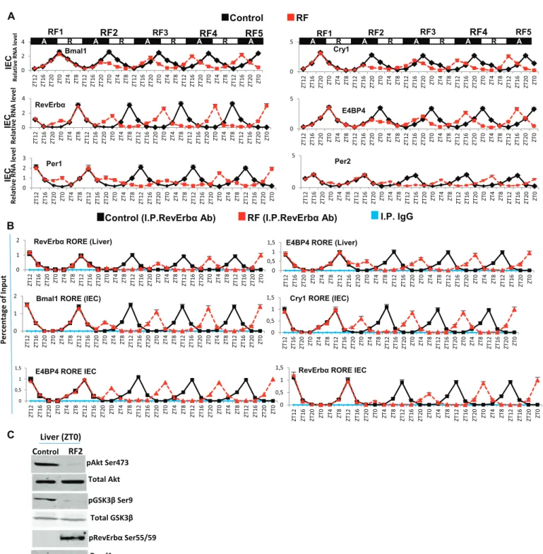
Au cours dun régime alimentaire décalé de longue durée, nous avons constaté que l'expression de certain "gènes contrôlés par les horloges périphériques" (clock-controlled genes: CCGs) impliqués dans lhoméostasie et dans le métabolisme, est décalée de 12 heures chez les souris RF par rapport aux phases de repos et dactivité contrôlées par horloge centrale (NSC), de telle sorte que lexpression périphérique de gènes normalement exprimés durant la « phase dactivité » seffectue au cours de la « phase de repos » et vice-versa, créant ainsi lapparition progressive dun syndrome métabolique pendant un régime alimentaire décalé (RF) de longue durée.
Pendant les 18 premiers mois de mon travail de thèse, nous avons exploré au niveau moléculaire, limpact du microbiote intestinal sur le fonctionnement de lhorloge circadienne et lhoméostasie des cellules de lépithélium intestinal.
Une seule couche de Cellules Epithéliales Intestinales (CEI) forme la barrière physique qui sépare le reste du corps des quelque 100.000 milliards de micro-organismes qui constituent le microbiote intestinal essentiellement composé de bactéries normalement non pathogènes, dites commensales, qui vivent en étroite symbiose avec leur hôte. De nombreuses études portant sur des souris élevées stérilement (Germ-free mice) ou dépourvues de microbiote (AIMD : antibiotic-induced microbiota-depleted) par traitement antibiotique ont révélé que la colonisation de lintestin par les bactéries est bénéfique pour léquilibre physiologique (homéostasie) de leur hôte en contrôlant de nombreux processus au niveau de lépithélium intestinal (absorption intestinale, immunité innée, prolifération cellulaire, activités métaboliques, etc,).
Des travaux réalisés au cours des 15 dernières années avaient révélé la nature du dialogue moléculaire qui sous-tend la symbiose hôte-microbiote, en démontrant quil nécessitait linteraction entre des composés provenant du microbiote et des récepteurs appartenant au groupe des « Toll-like receptors » (TLRs) localisés à la surface des CEI. La question posée était : comment ces interactions entre TLRs et composés provenant de bactéries commensales pouvaient-elles contrôler, au niveau moléculaire, de nombreuses fonctions physiologiques des CEI ? Jai démontré que lexpression des gènes TLRs est sélectivement déclenchée par un
rouage de lhorloge circadienne, le gène ROR , qui est exprimé au début de la phase circadienne dactivité. Ainsi, grâce à lactivation rythmique du rouage ROR , les signaux arythmiques émis par le microbiote sont « convertis » en une signalisation rythmique par les TLRs, qui stimulent sélectivement lexpression des gènes caractéristiques de la période dactivité. Ceci jusquau moment où lhorloge circadienne atteint la phase de repos, le rouage RervErb , de lhorloge réprime la synthèse des TLRs et autorise lexpression sélective des gènes contrôlant lhoméostasie pendant la période de repos, ainsi que la production de glucocorticoïdes par les cellules de la partie terminale de lintestin grêle, liléon.
En effet, le bon fonctionnement de cette horloge dans les cellules de lépithélium intestinal nécessite la présence de microorganismes non pathogènes qui vivent dans le tube digestif en étroite symbiose avec leur hôte. En labsence de microbiote, la désorganisation de l « axe » microbiote-TLRs-horloge circadienne se traduit dans les CEI par laltération de lexpression des gènes codant pour les protéines qui constituent les rouages de lhorloge circadienne, et aussi par laltération de lexpression des gènes qui présentent normalement un profil dexpression circadien. Il y a augmentation de lexpression des gènes qui sont exprimés durant la phase de repos au détriment de ceux exprimés pendant la phase dactivité. De plus la surproduction de glucocorticoïdes par liléon entraine lapparition dun syndrome de type pré-diabétique. En conclusion, notre étude a révélé des mécanismes moléculaires jusqualors inconnus qui sous-tendent lextraordinaire symbiose entre le microbiote et lépithélium intestinal, en démontrant comment des signaux du microbiote agissent en se liant aux TLRs des CEI pour contrôler le bon fonctionnement de leur horloge circadienne qui, elle-même, joue un rôle crucial dans le maintien de lhoméostasie intestinale. Enfin la découverte dune liaison directe entre le microbiote intestinal, le cycle circadien et léquilibre physiologique de lépithélium intestinal ouvre de nouvelles perspectives concernant lorigine et la physiopathologie des affections intestinales et (ou) systémiques qui sont liées à des altérations du microbiote et (ou) du cycle circadien, notamment chez les personnes travaillant en horaires décalées.
Abbreviations
AMPK: Adenosine monophosphate-activated protein kinase AVP: Arginine vasopressin
Bmal1: Brain and muscle aryl hydrocarbon receptor nuclear translocator (ARNT)-like cAMP: Cyclic adenosine monophosphate
CC: Circadian clock CKI: Caseine kinase I
Clock: Circadian locomotor output cycles kaput CRE: cAMP responsive element
CREB:cAMP Response Element-binding protein Cry: Cryptochrome
DBS: DNA binding sequence
DMH: Dorsomedial nucleus of the hypothalamus DBP: D site of albumin promoter
DR2: Direct repeat 2
Elovl6: Elovl fatty acid elongase 6 FAA: Food-antipatory activity FFA: Free fatty acids
FAS: Fatty acid synthase
G6pc: Glucose-6-phosphatase, catalytic subunit GABA: Gamma aminobutyric acid
GK: Glucokinase
Glut2: Glucose transporter 2
GSK3 : Glycogen synthase kinase 3 beta HFD: High-fat diet
HLF: Hepatic leukemia factor
HPA: Hypothalamicpituitaryadrenal
INS: Insulin
LDL: Low-density lipoprotein mRNA: messenger ribonucleic acid NAD: Nicotinamide adenine dinucleotide NAMPT: Nicotinamide phosphoryl-transferase Pepck: Phosphoenolpyruvate carboxykinase 2 Per: Period
PGC1: Peroxisome proliferator-activated receptor gamma coactivator1 PPAR!: Peroxisome proliferator-activated receptor alpha
PPAR": Peroxisome proliferator-activated receptor gamma RF: Restricted Feeding
ROR: Retinoic acid-related orphan receptor RORE: RevErb/ROR Response element
SCN: Suprachiasmatic nucleus of the hypothalamus SIRT: Sirtuin
Srebp1c: Sterol regulatory element binding transcription factor 1
TEF:Thyrotroph embryonic factor
TG: Triglycerides
VIP: Vasoactive intestinal peptide
WT: Wild type ZT: Zeitgeber time
Introduction
1-
Circadian rhythms
By definition, a rhythm is a change that is repeated with similar pattern during a certain period. Finding food and avoiding predators and inclement weather are the daily functions of wild animals. Some animals hunt their prey when they have the greatest ability to see (day), but others in contrary, as mice, need to avoid predators and thus are active mainly during the night. In any case, these adaptations lead to a strong circadian behavioral rhythmicity. Most processes in our body oscillate in a daily rhythmic fashion. In humans, sleep-wake cycles are the most obvious circadian manifestations of behavior (Achermann and Borbély, 2003, Borbély, 1982). These include cerebral activity, metabolism, energy homeostasis, heart rate, blood pressure, body temperature, and hormone secretion. These daily variations are connected to changes in the environment, such as regular and predictable day/night cycles, due to the fact that the Earth rotates on itself. Life has evolved and diversified through the process of natural selection to be adapted to almost every environment cues. The existence of daily rhythms in plants and animals is known for a long time, and Androsthenes Thasius described the concept of living rhythm early in the 4th century BC. He observed that leaves of certain plants (especially Tamarindus
Indicus) did not occupy the same day and night position.
The first scientific publication related to biological clocks appeared in from 1729, when Jean-Jacques d'Ortous Mairan observed the opening and closure of Mimosa Pudica leaves during the day and the night, even when the plant was kept in constant darkness. Thereafter, the observations of physiological or behavioral rhythms have accumulated during centuries. Clock genetics began in 1971 with the discovery by Ron Konopka and Seymour Benzer of the period (per) locus in Drosophila (Konopka and Benzer, 1971). In the early 20th century, circadian oscillations have been demonstrated in animals. When mice or rats are placed in constant light conditions (continuous light or darkness), the pace of locomotor activity was retained (Pittendrigh CS et al., 1976).
However, Jurgen Aschoff observations of human subjects without any external timing cue (light) for weeks revealed an unexpected finding. All individuals displayed body temperature and urine production rhythms of nearly 24 hours, and when living isolated from all
environmental cues continue to have a functioning rhythm with a period greater than 24 hours (25,9h), thereby indicating the existence of an internal biological clock (Aschoff, 1965).
2-
The molecular mechanism of circadian clocks in mammals
The molecular core clock in mammals, expressed both in brain and peripheral tissues, comprises a series of transcriptiontranslation feedback loops that result in cascades of gene expression. The mechanism of the core clock in mammals is the heterodimeric transcription factor complex of Clock and Bmal1, which activates transcription of the Period (Per) genes and Cryptochrome (Cry) genes via E-box DBS in their promoters. The Per-Cry repressive complex translocates into the nucleus to inhibit Clock-Bmal1 transactivation activity, resulting in the repression of the Per and Cry genes (Joseph Bass, 2012).
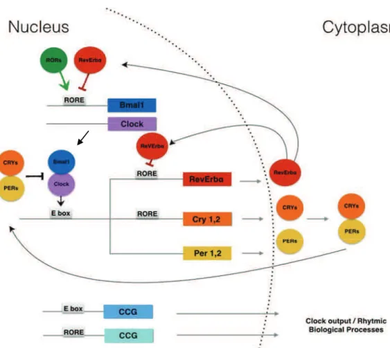
An additional feedback loop, involves the nuclear receptors RevErb and retinoic acid-related orphan receptor ROR . Clock-Bmal1 activates RevErb transcription, resulting in daily fluctuations of RevErb , which, in turn, represses Bmal1. ROR , another nuclear receptor and component of the clock machinery, competes with RevErb for the binding to the Bmal1 promoter RORE DBS, and activates its transcription. Therefore, the two nuclear receptors, RevErb and ROR , act as a new regulatory loop. They play a crucial role for a proper timing of the core clock machinery (Figure 1) (Joseph Bass, 2012).
There is a large number of genes including key regulators for cell cycle and metabolism are controlled by the components of circadian clock [the so called clock-controlled genes (CCGs)] as the heterodimer Bmal1-Clock, RevErb , ROR that suggests to contribute to the transcriptional output of the circadian clock, which are important to maintain normal cellular functions in peripheral tissues (Robles and Mann, 2013, Asher and Sassone-Corsi, 2015).
Figure 1: Transcriptional-translational feedback loops of the mammalian circadian clock. The positive elements of the loop (Bmal1/Clock) dimerize to active the rhythmic transcription of RevErb!, Crys (Cry1 and Cry2) and Pers (Per1 and Per2) genes through E-box enhancer elements. Cry-Per proteins interact with Bmal/Clock to negatively regulate Clock/Bmal1-mediated transcription. RevErb protein accumulation leads to periodic repression of Bmal1 and of its own gene expression (CCGs: clock-controlled genes).
The auto regulatory feedback loops described above take approximately 24 hours to complete a cycle and constitute a circadian molecular clock. Furthermore, clock proteins are modified in a post-translational manner by phosphorylation, acetylation, SUMOylation and ubiquitination, resulting in multiple layers of regulation to the core clock machinery. During the active phase, Per-Cry is degraded through the ubiquitylation of Cry by the FBXL3 complex. For example, phosphorylation of Per1 and Per2 by casein kinase 1 (CK1# or CKl$) can modulate the period of circadian gene expression,and RevErb has been shown to be stabilized when phosphorylated by the glycogen synthase kinase 3 (GSK3!) (Yin L et al., 2006).
Further circuits are generated through additional clock-controlled transcription factors such as DBP, TEF and HLF proteins, whose expression is E-box dependent, and E4BP4, whose
expression is RORE-dependent. Finally, E-box, RORE, and D-box can explain variable rhythms of CCGs, which control tissue-specific functions (Figure 2) (Ripperger and schibler,
2006) (Le Martelot et al., 2009) (Asher and Schibler, 2011).
Figure 2: The generation of different phases by the moleclar clock. The molecular clock in peripheral organs consists of two interlocked feedback loops. The major and essential loop involves the transcriptional activation of Period (Per1 and Per2) and Cryptochrome (Cry1, Cry2) genes by the transcription factors Clock and Bmal1, and the autorepression of Per and Cry genes by their own protein products. Another feedback loop is established through negatively and positively acting nuclear orphan receptors: the activators RORs compete with the repressors Rev-Erbs for the binding on the RORE DBS of the Bmal1 gene. Further phases are generated through additional clock-controlled transcription factors such as DBP TEF HLF whose expression is E box dependent, and E4BP4, whose expression is RORE dependent, (output regulators). The major and accessory feedback loops can therefore regulate widely different phases of genes encoding metabolic enzymes and modulators (output genes) involved in rhythmic metabolism.
3-
Central and peripheral circadian clocks in mammals
a- The Suprachiasmatic nucleus of the hypothalamus (SCN): the master circadian clock synchronizer
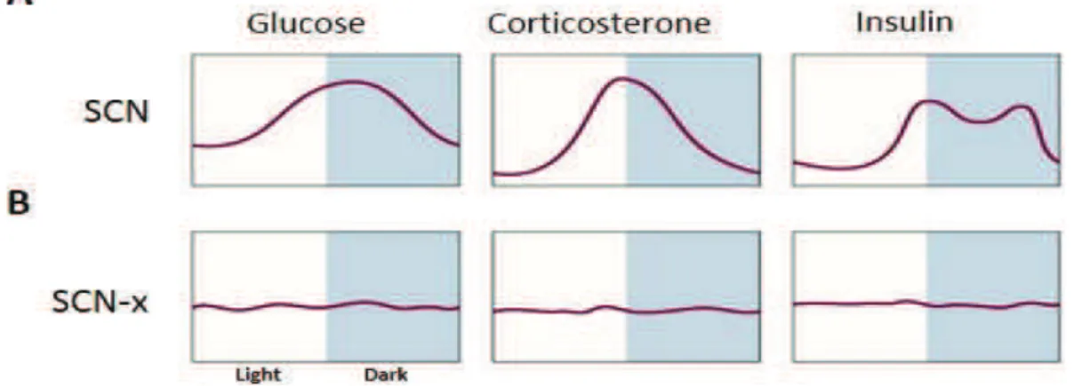
The suprachiasmatic nucleus of the hypothalamus (SCN) is a small, but extremely complex structure in the hypothalamus. Various studies of evidence show that the SCN regulates circadian rhythms, physiological and behavioral rhythms (Rusak and Zucker, 1979). Since the 1970s, lesion experiments in mammals have demonstrated its importance as a central circadian clock (Richter 1965, Moore and Eichler, 1972, Rusak, 1977). The definitive proof thats SCN
is the master synchronizer of behavior came when Ralph and colleagues have demonstrated that the behavioral rhythms of an SCN lesioned animal can be restored by transplantation of a donor SCN and exhibit the same period length of the donor animal (Ralph et al., 1990; Ralph and Menaker, 1988). In addition, in absence of SCN, different metabolites and hormones patterns lost their rhythmicity in blood. Plasma glucose, corticosterone, insulin and glucagon became completely arrhythmic by an SCN lesion in intact rats under ad libitum feeding conditions
(Figure 3) (Kalsbeek et al., 2010).
Figure 3: Daily rhythms in basal plasma glucose, corticosterone and insulin concentrations in presence (A) and absence of SCN (B) under 12 hour light/dark cycle and Ad libitum feeding condition (SCN-x: SCN lesion) (Adapted from Kalsbeek et al., 2010).
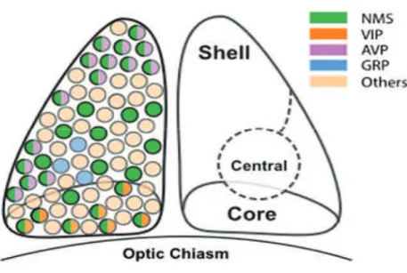
Following these important studies, it was established that in many species of mammals, the SCN control and coordinate many physiological and behavioral circadian functions (Hastings and Herzog, 2004). In mice, the SCN contains approximately ~20,000 neurons, and about
into two main anatomic-functional subparts; these designations were originally defined according to the expression of neuropeptides:
1- A ventromedial core consists of two types of neurons expressing the neuropeptides VIP (Vasoactive intestinal peptide) and GRP (Gastrin-releasing peptide).
2- A dorsomedial shell consists of neurons expressing the neuropeptide AVP (vasopressin).
Figure 4: The neuropeptide distribution in the SCN neurons. The suprachiasmatic nucleus (SCN) in the hypothalamus is divided into two major anatomical and functional subdivisions: a ventrolateral core and a dorsomedial shell. (NMS: neuropeptide neuromedin S, VIP: vasoactive intestinal peptide, AVP: arginine vasopressin, GRP: Gastrin Releasing peptide) (Adapted from Lee et al., 2015).
Several studies have indicated the presence of a large number of neurotransmitters in the SCN, which play an important role for the entrainment of the clock and for the control of different rhythms (Figure 4). VIP signaling in particular seems key to maintaining synchrony among
SCN neurons. Mice lacking VIP or its receptor display erratic free-running behavior and the rhythms of individual neurons within a single SCN are no longer held in uniform phase (Harmar et al., 2002; Colwell et al., 2003). More recently, Joseph Takahashi lab has found that the NMS (neuropeptide neuromedin S) neurons define a subset of SCN that dictate and generate SCN network synchrony and in vivo circadian rhythms through intercellular synaptic transmission (Lee et al., 2015).
b- Peripheral circadian clocks
Since the late 1990s, several teams have shown that in mammals most brain structures outside of the SCN, and even isolated cells express the molecular clock. Daily oscillation of clock genes have been observed in vitro in rat fibroblast cultures (Balsalobre et al., 1998), but in vivo, in peripheral tissues such as liver, muscle, kidneys, lungs (Abe et al., 2002; Yamazaki et al., 2000; Zylka et al., 1998). Such studies have suggested that most cells have molecular circadian clocks. The molecular mechanism of peripheral clocks was studied and based on the same molecular mechanism as in neurons of SCN (Yagita et al., 2001). Many studies on the expression of clock gene oscillations in peripheral cells have used the fibroblast culture as a model (Balsalobre et al., 1998; Welsh et al., 2004). They showed that each cell has its own molecular circadian clock. However, fibroblasts can be resynchronized by adding various factors such as a change of the culture medium, adding serum to the medium (Balsalobre et al., 1998) or the glucocorticoid dexamethasone (Balsalobre et al., 2000). An ex vivo study in 2004 has shown that the oscillations of a number of peripheral tissues such as liver, kidney and lungs persisted as long as those of the SCN (Figure 5) (Yoo et al., 2004).
Figure 5: The expression of the core circadian clock genes reflects the presence of circadian oscillators in almost every tissue in the body. Representative records of bioluminescence showing circadian profiles of mPer2 expression from various tissues from mPer2 Luc knocking animals. Tissues were explanted just before lights off (arrow). Light output (in counts per min) is plotted against previous light onset(Adapted from Yoo et al., 2004).
Lung Pituitary Kidney Days Cornea Tail Retrochiasmatic Area Liver SCN
4-
Phenotypic effects of circadian clock mutations
The molecular mechanism underlying the mammalian clockwork has been mostly studied in the mouse. The internal circadian system could be altered by a mutation of one or more clock genes, which lead to a disruption of the animal rhythms, and even in some cases to a total arrhythmia, associated to various physiological abnormalities (TABLE 1) (Ko CH and
Takahashi JS, 2006). Gene Circadian phenotype References Associated physiological abnormality References Clock#19 Semi-dominant, 4 hours longer period followed by loss-of-circadian activity rhythm in constant darkness (DD) Vitaterna et al. 1994 Hyperphagic & Obese Abnormal gluconeogenesis Hypersensitive to chemotherapeutic agent Enhanced response to cocaine Mania phenotype Decreased duration of sleep time Gorbacheva et al. 2005 ;Rudic et al 2004 ;McClung et al 2005 ;Turek et al 2005 Clock Null mutant 0.5 hour shorter period DeBruyne et al 2006 None Determined _ Per1 Null mutant 0-0.5 hour shorter period/ some animals lose circadian activity
rhythm in DD Zheng et al 2001;Cermaki an et al 2001 Lack of sensitization to cocaine Abarca et al 2002 Per2 Per2tm1Brd Null mutant 1.5 hour shorter period and tendency
for loss of circadian rhythm Bae et al 2001;Zheng et al 1999 Increased tumor development following genotoxic stress Hyper-sensitization to cocaine Improper alcohol intake Early onset of sleep Abarca et al 2002;Fu et al 2002;Toh et al 2001
Per1 & Per2 Double null mutant Complete loss of circadian activity rhythm in DD Zheng et al 2001 ; Bae et al 2001 None Determined _ Cry1 Null mutant
1 hour shorter period Van der Horst et al 2000 ; Vitaterna et al 1999
Gene Circadian phenotype References Associated physiological abnormality References Cry2 Null mutant
1 hour longer period Thersher et al 1998
None Determined _
Cry1 & Cry2 Double null
mutant
Complete loss of circadian activity rhythm in DD
Van der Horst et al 2000 ; Vitaterna et al 1999 Delayed hepatocyte re-generation Resistant to chemotherapeutic agents toxicity I Gorbacheva et al 2005;Fu et al 2002;Matsuo et al 2000 RevErb! (Nr1d1) Null mutant 0.5 hour shorter period/ Altered photic
entrainment
Preitner et al 2002
None Determined _
RORa mutant 0.5 hour shorter period Sato et al 2004 Cerebellar ataxia Abnormal bone metabolism Meyer et al 2000 ;Steinmayr et al 1998 VIP Null mutant Abnormal entrainment to light cycles Dissociated circadian wheel-running rhythms in DD Reduced amplitude in behavioral rhythms in DD Aton et al 2005; Colwell et al 2003 Impaired temporal regulation of metabolism and feeding Bechtold et al 2008 VIP Receptor2 Null mutant Abnormal entrainment to light cycles Dissociated circadian wheel-running rhythms in DD Reduced amplitude in behavioral rhythms in DD Impaired responses to light Aton et al 2005; Harmar et al 2002 Impaired temporal regulation of metabolism and feeding Bechtold et al 2008
Table1: Mouse circadian mutants and observed circadian and physiological phenotypes
5-
External cues entrain circadian clocks
a- Light-entrainment of the master SCN circadian clock
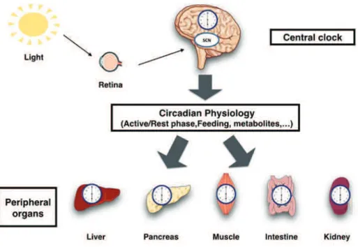
The different body oscillators have to be periodically synchronized to the environment. The light-entrained master central circadian clock (CC) in the SCN not only controls the diurnal alternance of the active phase and the rest phase, but also synchronizes the ubiquitous peripheral CCs with these phases to maintain homeostasis (Figure 6).
Figure 6: Light entrains the master clock in the suprachiasmatic nucleus (SCN), which in turn synchronizes extra-SCN and peripheral clocks.
Light enters the eye and activates different types of photoreceptors in retina. Of note, for more than 150 years, rods and cones have been considered to be the only photoreceptor cells in the retina. More recently, in 2002, intrinsically photosensitive retinal ganglion cells (ipRGCs) have been discovered melanopsin-expressing ganglion cells (non-vision pathway). The discovery of a group of blind humans who were unable to form images (absence of rods and cones), but were capable of detecting light for regulation of melatonin secretion, provided an evidence of an additional photoreceptive system in the retina through melanospsin (Berson et al., 2002, Hattar et al., 2002). Interestingly, Melanopsin knock-out mice only displayed mild phase-shifting phenotypes and the SCN clock can be photoentrained (Ruby et al., 2002, Panda et al., 2002). In addition, when a melanopsin knock-out allele was bred into Rd mice (lack of rods and cones),
the resulting mutants completely lost their circadian photoentrainement and the activity in the presence of a light-dark cycle (Panda et al., 2003, Hattar et al., 2003). This clearly demonstrated that the circadian photoperception is a property generated by the activity of rods and cones cells, in combination with melanopsin-expressing ganglion cells. Photic signals transmitted from the retina to SCN neurons lead to an influx of Ca2+ in postsynaptic neurons, which through
activation of protein kinase A (PKA) stimulates the activity of the cAMP response element binding protein (CREB) (De Cesare D et al., 2000), which is a transcription factor that induces the transcription of the Per1 and Per2 clock genes. Moreover, it has been shown that a pulse of light during the second half of the night, can immediately induce the expression of components of SCN CC as Per1 and Per2, proteins accumulate precociously, which results in a phase advance (Shearman et al., 1997, Albrecht et al., 1997).
Thus, the light-induced CREB-dependent increase in expression of Per genes, provides an explanation of how a light input can influence the function of neurons of the SCN CC.
b- Feeding-entrainment of circadian clocks of peripheral cell /tissues
Feeding and peripheral circadian clocks (PCCs) are tightly interconnected, as the feeding time can act as a potent entraining timing signal (cue) or Zeitgeber for PCC. It has been reported that PPC gene expression tissues such as liver, pancreas, intestine, kidney and heart, can be shifted by 12 hours when the food is exclusively provided during the day time (the rest phase in mice) and prevented during the night time (the active phase in mice) [the so-called Restricted Feeding (RF)], whereas the SCN central circadian clock remaining unchanged (Damiola et al., 2000). Importantly, a 12-hour RF-regime can entrain a rhythmic circadian clock in peripheral tissues of a SCN-ablated animals which are arrhythmic (Krieger et al., 1977). What are then the signals, which arise from feeding and entrain peripheral clocks? They could be the food itself, food-induced metabolites, or hormones whose secretion is controlled by feeding or its absence? A possibility is that feeding rhythms drive variations in glucose levels. The glucose metabolism is indeed well known to participate in all major biochemical functions in all cell types. Following carbohydrate-rich meals, glucose is converted into glycogen, which is stored in the liver to serve as a rapidly available fuel to mobilize glucose for brain and other tissues after feeding. Glycogenolysis is the primary mechanism by which glucose is made available. Hepatic gluconeogenesis and glycogenolysis represent the main pathway by which nutrient homeostasis is maintained over diurnal periods of feeding and fasting. Circadian variations of glucagon and insulin hormones control these biochemical processes. Interestingly
enough, the molecular mechanism through by which glucose may affect the function of circadian clock components has not been investigated.
Interestingly, not only the time of feeding could control the circadian peripheral clock, but the meal composition may also plays a crucial role in this control. In mice, a High fat diet (HFD) regime affects not only the activity, but also the cycling of clock genes and of their clock controlled targets. Recently, it has been reported that consumption of a HFD produces widespread alterations in circadian transcriptional and metabolic profiles in peripheral tissues (Kohsaka et al., 2007) (Eckel-Mahan et al., 2013).
6-
Nuclear receptors and circadian clocks
Nuclear receptors (NRs) represent a large superfamily of genes encoding receptors composed of 48 members in human and 49 in mice, which play diverse aspects involved in different biological pathways, such as energy metabolism, immunology and tissue homeostasis (Yang et al., 2006). A large number of these receptors, including both isoform ! and ! of RevErbs, RORs ( , !, ") and all three PPARs ( , !, ") are expressed in mice in a circadian manner at least at the transcript level (Yang et al., 2006). They represent a complex array of extracellular signals into transcriptional responses, many of which specifically regulate the expression of target genes. Their primary function is to mediate in target cells the transcriptional response to hormones such as the sex steroids, adrenal steroids (glucocorticoids) and retinoid ligands, as well as a variety of additional metabolic ligands. Glucocorticoids (GC) hormones might play a role in the synchronization of circadian peripheral clocks (Le Minh et al., 2001). Indeed, the level of GC displays a robust circadian rhythm. GCs are belong to a group of steroid hormones released by adrenal gland cortex that are involved in multiple functions, including inflammation, metabolism, and neurological functions. These hormones activate the glucocorticoid receptor (GR), which in glucocorticoid-responsive elements +GRE within promoter of Per genes. Although a treatment of cultured cells in vitro with dexamethasone (a synthetic GR agonist) is able to synchronize their circadian clocks, mice bearing a hepatocyte-specific knockout of the GR gene tuned their circadian liver clocks more rapidly to inverted feeding cycles than wild-type mice upon RF regime (Le Minh et al., 2001). Of note, it has been shown that GR interacts with and repressed by Cry 1 and 2 (Lamia et al., 2011).
PPARs (Peroxisome Proliferator-Activated Receptors) play a crucial role at the gene level in the regulation of mammalian metabolism, including Fatty acid oxidation and lipogenesis (Kersten et al., 2000). Three major isoforms of PPAR encoded by different genes have been
identified: PPAR , PPAR! and PPAR". The activation of target gene transcription depends directly on the binding of the free Fatty acid (FFA) to the receptor. PPAR is predominantly expressed in adipose tissue, liver, intestine, kidney, heart and skeletal muscle. During fasting and starvation period, when the carbohydrates stock has been used, the body switches to mobilizing its fat stores as a source of energy. At this point, the role of PPAR is crucial in the hepatic response to fasting by regulating genes with direct roles in ketogenesis, as shown by using gene knockout mice models and from transcriptomic analyses.More recently, it has been demonstrated that PPAR could be connected with the circadian clock machinery through a direct binding on the DR2 DBS of RevErb gene, which acts on the core circadian clocks (Duez et al., 2008).
7-
Circadian clocks and metabolic factors are tightly linked
The circadian clock machinery is controlled by transcriptional feedback loops in which clock components act through binding directly to DNA response elements. Interestingly, the same elements are present throughout the genome and regulate the expression of clock-controlled genes (CCGs) (Ueda et al., 2005). Recent genome wide transcriptome analyses in the SCN, liver and adrenal gland of mice, revealed the expression of 10% of the cell transcripts is regulated in a circadian fashion. Among these transcripts, key regulators of glucose and lipid metabolism are present (Ueda et al., 2002; Lowrey and Takahashi 2004; Hughes et al., 2009). The availability of genetic models of circadian alteration provided new opportunities to dissect the relationship between circadian clocks and metabolic systems.One of the first alteration to be studied was that of the Clock gene. It has been shown that a Clock mutation induced changes in glucose homeostasis including impairments in glucose tolerance, insulin secretion, PEPCK and Glycogen Synthase 2 (E-box) mRNA expression, as well as and increased insulin sensitivity (Kennaway et al., 2007) (Doi et al., 2010). Homozygous Clock mutant mice exhibited a dampened feeding pattern with increase food intake during the rest phase, which contributed to gain weight by fat excess. Clock mutant mice displayed multiple metabolic alterations including hyperglycemia, hypertriglyceridemia and hepatic steatosis. Moreover, a Clock mutation altered the diurnal rhythms of CC genes and of some of metabolic genes in mice brain (Turek et al., 2005). Gluconeogenesis was suppressed by a deletion of Bmal1 and mice became more to sensitive exogenous insulin (Rudic et al., 2004). In addition, a liver deletion of Bmal1 in mice induced a hypoglycemia during the rest phase, and normal total body fat as compared to the Wt mice (Lamia et al., 2008).
Metabolic sensors and circadian clock components
Most cells in mammalian body use glucose as their major source of energy. Glucose molecules are broken down within cells in order to produce adenosine triphosphate (ATP) molecules, the energy molecules that power numerous cellular processes. The maintenance of constant plasma glucose levels is therefore crucial to prevent from many metabolic disorders. The connection between the circadian clocks and metabolism is tightly dependent on the role of metabolic sensors that regulate the clock machinery.
AMPK and circadian clocks
AMP-activated protein kinase (AMPK) is one of the metabolic sensors that have been reported to transmit energy-dependent signals to the circadian clock. In the liver, it is activated in response to prolonged fasting. AMPK has been found to directly phosphorylate CKI$, resulting in increased CKI$ activity and degradation of mPER2. In addition it has been shown that AMPK phosphorylates and destabilizes mCRY1, leading to altered circadian rhythms (Lamia et al., 2009) (Feng and Lazar, 2012).
SIRT1 and circadian clocks
SIRT1 (Sirtiun 1) has emerged as a key metabolic sensor that directly links nutrient signals to metabolic homeostasis. SIRT1 is a NAD+-dependent protein deacetylase that governs many physiological processes, and has recently been found to play a key role in the control of the circadian rhythms in which circadian control is coupled to metabolism (Asher et al. 2008; Nakahata et al., 2009; Ramsey et al., 2009). In 2008, two studies from the laboratories of Ueli Schibler (Asher et al., 2008) and Paolo Sassone-Corsi (Nakahata et al., 2008) have demonstrated that SIRT1 activity is intimately linked with the hepatic circadian clock. They have shown that the circadian SIRT1 activity modulates the expression of circadian clock-controlled genes by deacetylating Per2 and Bmal1. Deacetylation of Bmal1 affects its transcriptional activity (Nakahata et al., 2008) and that of Per2 to alter its stability (Asher et al. 2008). Furthermore, SIRT1 ablation resulted in an increase in DBP and Per2 mRNA (Storch et al., 2002). Nicotinamide phosphoribosyltransferase (Nampt) is the rate-limiting enzyme in NAD+ biosynthesis and Clock and Bmal1 directly activate the Nampt promoter, leading to circadian activity of NAMPT and of cellular NAD+ levels (Nakahata et al., 2009; Ramsey et al., 2009).
It has also been shown that SIRT1 in the brain governs the central SCN circadian clock by activating transcription of the two major circadian clock components, Bmal1 and Clock (Chang et al., 2013). The discovery of circadian-directed sirtuin activity suggests that metabolites such as NAD+ could be themselves playing an important role in the cellular link between metabolism
and the circadian clocks (Paolo Sassone-Corsi, 2014).
8-
Microbiota and circadian clocks
Bacteria is a word that gives us a wrong impression of representing something dirty, contaminated or infected. We however live in a world filled with bacteria; they are everywhere and colonize our human body from birth. Bacteria reside on the skin, in the mouth and gut level; they constitute what is called the skin, oral and the intestinal flora. In intestine, it is also name the intestinal microbiota. It comprises 1014 microbial cells, around 100 times more cells than the human body itself. The intestinal epithelium has the delicate function to absorb nutrients, while being simultaneously a potential line of defense against the environment. A single layer of intestinal epithelial cells (IEC) provides a physical barrier that separates the commensal bacteria in the intestinal lumen from the underlying lamina propria and the rest of the body. IECs also express Toll like Receptors (TLRs) and are thus a part of the innate immune system, both as a physical barrier and as a first-line pathogen recognition system (Abreu, 2010). TLRs play an important role in host defense against microbial infection. Stimulation of TLRs by their cognate pathogen-associated microbial patterns (PAMPs) initiates signaling cascades leading to the activation of transcription factors, such as AP-1 and NF-%B (Hooper et al., 2001). Significant progress has been made over the past years in the understanding of TLR function (Rakoff-Nahoum et al., 2004). The contribution of the microbiota in regulation of physiology is vast and complex associated with intestinal disorders as well as with diseases such as obesity (Ley et al., 2005; Turnbaugh et al., 2006). Studies on Germ-Free (GF) and antibiotic-induced microbiota-depleted (AIMD) mice have revealed that gut colonization by the microbiota is beneficial in many respects for mammalian host homeostasis unveiled that commensal bacteria are recognized by IEC Toll-like receptors (TLRs), thus providing a new perspective on host-commensal symbiosis and raising the question of how the interaction of host-commensal bacterial products (PAMPs) with TLR members of the pattern recognition receptors (PRRs) could control at the molecular level many homeostatic processes in IECs (Abreu, 2010). Several intestinal functions (e.g., nutrient absorption, cell proliferation, motility, metabolic activities)
are known to be rhythmically regulated in a circadian manner (Hussain and Pan, 2009), and it has been shown that core components of the circadian clock could be functional in mouse small intestine in which the expression of some TLR genes may also vary around the circadian day (Froy and Chapnik, 2007). Moreover, it has been recently shown that the composition of the microbiota undergoes diurnal oscillations in both mice and humans (Thaiss et al., 2014), and that these oscillations are disturbed in mice by a genetic mutation in the clock system, as well as in jet-lag experiments (Thaiss et al., 2014) (Asher and Sassone-Corsi P, 2015). The question, which remained to be investigated, was how the gut clock interplays with metabolic pathways, and is the microbiota controlling the gut clock or vice versa?
References
1. Abe M, Herzog ED, Yamazaki S, Straume M, Tei H, Sakaki Y, Menaker M, Block GD. Circadian rhythms in isolated brain regions. J Neurosci. 2002;22:350356
2. Abreu MT (2010) Toll-like receptor signalling in the intestinal epithelium: how bacterial recognition shapes intestinal function. Nat Rev Immunol 10: 131144. doi: 10.1038/nri2707
3. Achermann P and Borbély AA (2003) Mathematical models of sleep regulation. Frontiers in Bioscience 8: 683-693.
4. Albrecht, U., Sun, Z. S., Eichele, G., Lee, C. C. A differential response to two putative mammalian circadian regulators, mper1 and mper2, to light. Cell 91: 1055-1064, 1997. 5. AschoffJ (1965) Circadian rhythms in man. Science 148:14271432
6. Asher G, Gatfield D, Stratmann M, Reinke H, Dibner C, Kreppel F, Mostoslavsky R, Alt FW, Schibler U. (2008). SIRT1 regulates circadian clock gene expression through PER2 deacetylation. Cell 134, 317328.
7. Asher, G., and Schibler, U. (2011). Crosstalk between components of circadian and metabolic cycles in mammals. Cell Metab. 13, 125137.
8. Asher, G., and Sassone-Corsi, P. (2015). Time for food: the intimate interplay between nutrition, metabolism, and the circadian clock. Cell 161, 8492.
9. Balsalobre A, Damiola F & Schibler U (1998). A serum shock induces circadian gene expression in mammalian tissue culture cells. Cell 93 929937.
10. Balsalobre A, Brown SA, Marcacci L, Tronche F, Kellendonk C, Reichardt HM, Schutz G & Schibler U (2000). Resetting of circadian time in peripheral tissues by glucocorticoid signaling. Science 289 23442347.
11. Bass J Circadian topology of metabolism (2012) .Nature 491 (7424), 348-356
12. Berson DM, Dunn FA, Takao M. Phototransduction by retinal ganglion cells that set the circadian clock. Science. 2002;295:10703.
13. Borbély AA (1982) A two process model of sleep regulation. Human Neurobiolgy 1:195-204
14. Chang H. C., Guarente L. (2013). SIRT1 mediates central circadian control in the SCN
by a mechanism that decays with aging. Cell 153, 14481460.
10.1016/j.cell.2013.05.027.
15. Colwell C, Michel S, Itri J, Rodriguez W, Tam J, Lelievre V, Hu Z, Liu X, Waschek J. Disrupted circadian rhythms in VIP- and PHI-deficient mice. Am J Physiol Regul Integr Comp Physiol.2003;285:R939R949.
16. Damiola F., Le Minh,N., Preitner,N., Kornmann,B., Fleury-Olela,F. and Schibler,U. (2000) Restricted feeding uncouples circadian oscillators in peripheral tissues from the central pacemaker in the suprachiasmatic nucleus. Genes Dev., 14, 29502961.
17. De Cesare D, Sassone-corsi P. Transcriptional regulation by cyclic AMP-responsive factors. Prog Nucleic Acid Res Mol Biol. 2000;64:343-69.
18. Doi M, Takahashi Y, Komatsu R, Yamazaki F, Yamada H, Haraguchi S, Emoto N,Ok uno Y, Tsujimoto G, Kanematsu A, et al. 2010. Salt-sensitive hypertension in circadian
clock-deficient Cry-null mice involves dysregulated adrenal Hsd3b6. Nat Med 16: 67 74.
19. Duez H, Staels B (2008) Rev-erb gives a time cue to metabolism. FEBS Lett 582(1): 1925.
20. Eckel-Mahan KL, Patel VR, de Mateo S, Ceglia NJ, Sahar SS, Dilag S, Dyar KA, Orozco-Solis R, Baldi P, and Sassone-Corsi P. Reprogramming of the Circadian Clock by Nutritional Challenge Cell, 2013 December 19,155(7), 1464-1478
21. Feng D, Lazar MA (2012) Clocks, metabolism, and the epigenome. Mol Cell 47(2):158167.
22. Froy, O., and Chapnik, N. (2007). Circadian oscillation of innate immunity components in mouse small intestine. Mol. Immunol. 44, 19541960.
23. Hattar S, Liao HW, Takao M, Berson DM, Yau KW. Melanopsin-containing retinal
ganglion cells: architecture, projections, and intrinsic
photosensitivity. Science. 2002;295:106570.
24. Hattar S, Lucas RJ, Mrosovsky N, Thompson S, Douglas RH, Hankins MW, Lem J, Biel M, Hofmann F, Foster RG, Yau KW. Melanopsin and rod-cone photoreceptive systems account for all major accessory visual functions in mice. Nature. 2003;424:76 81.
25. Harmar A, Marston H, Shen S, Spratt C, West K, Sheward W, Morrison C, Dorin J, Piggins H, Reubi J, et al. The VPAC(2) receptor is essential for circadian function in the mouse suprachiasmatic nuclei. Cell. 2002;109:497508.
26. Hastings MH & Herzog ED 2004 Clock genes, oscillators, and cellular networks in the suprachiasmatic nuclei. Journal of Biological Rhythms 19 400413.
27. Hooper, L.V., Wong, M.H., Thelin, A., Hansson, L., Falk, P.G., and Gordon, J.I. (2001). Molecular analysis of commensal host-microbial relationships in the intestine. Science
291, 881884.
28. Hughes ME, DiTacchio L, Hayes KR, Vollmers C, Pulivarthy S, et al. Harmonics of circadian gene transcription in mammals. PLoS Genet. 2009;5:e1000442.
29. Hussain, M.M., and Pan, X. (2009). Clock genes, intestinal transport and plasma lipid homeostasis. Trends Endocrinol. Metab. 20, 177185.
30. Kalsbeek A, Bruinstroop E, Yi CX, Klieverik LP, La Fleur SE, Fliers E. Hypothalamic control of energy metabolism via the autonomic nervous system. Ann. N. Y. Acad. Sci. 2010;1212:114129.
31. Kennaway DJ, Owens JA, Voultsios A, Boden MJ, Varcoe TJ. Metabolic homeostasis in mice with disrupted Clock gene expression in peripheral tissues. Am. J. Physiol. Regul. Integr. Comp. Physiol.2007;293:R1528R1537.
32. Kersten S, Desvergne B, Wahli W (2000) Roles of PPARs in health and disease. Nature 405(6785):421424.
33. Ko, C.H. & Takahashi, J.S. (2006) Molecular components of the mammalian circadian clock. Human Molecular Genetics, 15, R271-277.
34. Kohsaka A, Laposky AD, Ramsey KM, Estrada C, Joshu C, Kobayashi Y, Turek FW, Bass J. High-fat diet disrupts behavioral and molecular circadian rhythms in mice. Cell Metab. 2007; 6:414421.
35. Konopka RJ and Benzer S (1971) Clock mutants of Drosophila melanogaster. Proc Natl Acad Sci U S A 68:2112-2116.
36. Krieger DT, Hauser H, Krey LC. Suprachiasmatic nuclear lesions do not abolish food-shifted circadian adrenal and temperature rhythmicity. Science. 1977;197:3989. 37. Lamia KA, Storch KF, Weitz CJ (2008) Physiological significance of a peripheral tissue
circadian clock. Proc Natl Acad Sci USA 105:1517215177.
38. Lamia KA, Sachdeva UM, DiTacchio L, Williams EC, Alvarez JG, Egan DF, Vasquez DS, Juguilon H, Panda S, Shaw RJ, Thompson CB, Evans RM. (2009) AMPK regulates the circadian clock by cryptochrome phosphorylation and degradation. Science 326:437440. doi: 10.1126/science.1172156
39. Lamia KA, Papp SJ, Yu RT, Barish GD, Uhlenhaut NH, Jonker JW, Downes M, Evans RM (2011). Cryptochromes mediate rhythmic repression of the glucocorticoid receptor. Nature 2011;480:552556. doi: 10.1038/nature10700.
40. Lee I. T., Chang A. S., Manandhar M., Shan Y., Fan J., Izumo M., et al. . (2015). Neuromedin s-producing neurons act as essential pacemakers in the suprachiasmatic nucleus to couple clock neurons and dictate circadian rhythms. Neuron 85, 10861102. 10.1016/j.neuron.2015.02.006.
41. Le Martelot G, Claudel T, Gatfield D, Schaad O, Kornmann B, Lo Sasso G, Moschetta A, Schibler U. 2009. REV-ERBalpha participates in circadian SREBP signaling and bile acid homeostasis. PLOS Biology 7:e1000181. doi: 10.1371/journal.pbio.1000181. 42. Le Minh N, Damiola F, Tronche F, Schütz G, Schibler U (2001) Glucocorticoid
hormones inhibit food-induced phase-shifting of peripheral circadian oscillators. EMBO J 20(24):71287136.
43. Ley R., Backhed F., Turnbaugh P., Lozupone C., Knight R., Gordon J. (2005) Obesity alters gut microbial ecology. Proc Natl Acad Sci U S A 102: 1107011075.
44. Lowrey PL, Takahashi JS. Mammalian circadian biology: elucidating genome-wide levels of temporal organization. Annu. Rev. Genom. Hum. Genet. 2004;5:407441. 45. Moore, R. Y., and Eichler, V. B., 1972, Loss of circadian adrenal corticosterone rhythm
following suprachiasmatic lesions in the rat, Brain Res. 42: 201206.
46. M.R. Ralph, M. Menaker. A mutation of the circadian system in golden hamsters Science, 241 (1988), pp. 12251227
47. M.R. Ralph, R.G. Foster, F.C. Davis, M. Menaker.Transplanted suprachiasmatic nucleus determines circadian period. Science, 247 (1990), pp. 975978.
48. Nakahata Y, Kaluzova M, Grimaldi B, Sahar S, Hirayama J, Chen D, Guarente LP, Sassone-Corsi P. (2008). The NAD+-dependent deacetylase SIRT1 modulates CLOCK-mediated chromatin remodeling and circadian control. Cell 134, 329340. 49. Nakahata Y, Sahar S, Astarita G, Kaluzova M, Sassone-Corsi P. (2009). Circadian
50. Panda S, Sato TK, Castrucci AM, Rollag MD, DeGrip WJ, et al. Melanopsin (Opn4)
requirement for normal light-induced circadian phase
shifting. Science. 2002;298:22136.
51. Panda S, Provencio I, Tu DC, Pires SS, Rollag MD, Castrucci AM, Pletcher MT, Sato TK, Wiltshire T, Andahazy M, Kay SA, Van Gelder RN, Hogenesch JB. Melanopsin is
required for non-image-forming photic responses in blind
mice. Science. 2003;301:525527.
52. Pittendrigh, C.S., Daan, S.: A functional analysis of circadian pacemakers in nocturnal rodents. I. The stability and lability of spontaneous frequency. J. comp. Physiol.106, 223252 (1976)
53. Rakoff-Nahoum, S., Paglino, J., Eslami-Varzaneh, F., Edberg, S., and Medzhi- tov, R. (2004). Recognition of commensal microflora by toll-like receptors is required for intestinal homeostasis. Cell 118, 229241.
54. Ramsey K, Yoshino J, Brace CS, Abrassart D, Kobayashi Y, Marcheva B, Hong HK, Chong JL, Buhr ED, Lee C, et al. (2009). Circadian clock feedback cycle through NAMPT-mediated NAD+ biosynthesis. Science 324, 651654.
55. Richter, C. P., 1965, Biological Clocks in Medicine and Psychiatry, Charles C. Thomas, Publisher, Springfield, Ill.
56. Ripperger, U. Schibler. Rhythmic CLOCK-BMAL1 binding to multiple E-box motifs drives circadian Dbp transcription and chromatin transitions. Nat. Genet., 38 (2006), pp. 369374
57. Robles, MS & Mann, M. (2013) Proteomic approaches in circadian biology. in Handbook of experimental pharmacology: Circadian Clocks. pp. 389-407. Handbook of Experimental Pharmacology.
58. Ruby NF, Brennan TJ, Xie X, Cao V, Franken P, et al. Role of melanopsin in circadian responses to light. Science. 2002;298:22113.
59. Rudic RD, et al. (2004) BMAL1 and CLOCK, two essential components of the circadian clock, are involved in glucose homeostasis. PLoS Biol 2:e377.
60. Rutter J, Reick M, Wu LC, McKnight SL (2001) Regulation of Clock and NPAS2 DNA binding by the redox state of NAD cofactors. Science 293: 510514.
61. Rusak, B., 1977. The role of the suprachiasmatic nuclei in the generation of Circadian rhythm in the golden hamster, Mesocricetus auratus, J. Comp. Physiol. 118: 145164. 62. Rusak B, Zucker I (1979) Neural regulation of circadian rhythms. Physiol Rev 59:449
526.
63. Takahashi JS, Hong HK, Ko CH, McDearmon EL. The genetics of mammalian circadian order and disorder: implications for physiology and disease. Nat. Rev. Genet. 2008;9:764775.
64. Thaiss CA, et al. (2014). Transkingdom control of microbiota diurnal oscillations promotes metabolic homeostasis. Cell 159(3):514529.
65. Turek F.W, C. Joshu, A. Kohsaka, E. Lin, G. Ivanova, E. McDearmon, A. Laposky, S. Losee-Olson, A. Easton, D.R. Jensen, et al. Obesity and metabolic syndrome in circadian Clock mutant mice. Science, 308 (2005), pp. 10431045.
66. Turnbaugh P., Ley R., Mahowald M., Magrini V., Mardis E., Gordon J. (2006) An obesity-associated gut microbiome with increased capacity for energy harvest. Nature 444: 10271031.
67. Sassone-Corsi P. The Time of Your Life. Cerebrum. 2014 Jul-Aug; 2014: 11.
68. Shearman, L. P., Zylka, M. J., Weaver, D. R., Kolakowski, L. F., Jr., Reppert, S. M. Two period homologs: circadian expression and photic regulation in the suprachiasmatic nuclei. Neuron 19: 1261-1269, 1997.
69. Storch, K. F., Lipan, O., Leykin, I., Viswanathan, N., Davis, F. C., Wong, W. H., et al. (2002). Extensive and divergent circadian gene expression in liver and heart. Nature, 417, 7883.
70. Ueda HR, Chen W, Adachi A, Wakamatsu H, Hayashi S, Takasugi T, Nagano M, Nakahama K, Suzuki Y, Sugano S, et al.: A transcription factor response element for gene expression during circadian night. Nature 2002, 418:534-539.
71. Ueda HR, Hayashi S, Chen W, Sano M, Machida M, Shigeyoshi Y, Iino M, Hashimoto S: System-level identification of transcriptional circuits underlying mammalian circadian clocks. Nat Genet 2005, 37:187-192.
72. Welsh DK, Yoo SH, Liu AC, Takahashi JS & Kay SA 2004 Bioluminescence imaging of individual fibroblasts reveals persistent, independently phased circadian rhythms of clock gene expression. Current Biology 14 22892295.
73. Yamazaki S, Numano R, Abe M, Hida A, Takahashi R, Ueda M, Block GD, Sakaki Y, Menaker M & Tei H 2000 Resetting central and peripheral circadian oscillators in transgenic rats. Science 288 682685.
74. Yang X, Downes M, Yu RT, Bookout AL, He W, Straume M, Mangelsdorf DJ, Evans RM. 2006. Nuclear receptor expression links the circadian clock to metabolism. Cell 126: 801810.
75. Yagita K, Tamanini F, van Der Horst GT & Okamura H 2001 Molecular mechanisms of the biological clock in cultured fibroblasts. Science 292 278281.
76. Yin L., Wang J., Klein P. S., Lazar M. A. Nuclear receptor Rev-Erb alpha is a critical lithium-sensitive component of the circadian clock. Science. 2006;311:10025.
77. Yoo SH, Yamazaki S, Lowrey PL, Shimomura K, Ko CH, Buhr ED, Siepka SM, Hong HK, Oh WJ, Yoo OJ et al. 2004 PERIOD2::LUCIFERASE real-time reporting of circadian dynamics reveals persistent circadian oscillations in mouse peripheral tissues. PNAS 101 53395346.
78. Zylka MJ, Shearman LP, Weaver DR, Reppert SM. Three period homologs in mammals: differential light responses in the suprachiasmatic circadian clock and oscillating transcripts outside of brain.Neuron. 1998;20:11031110.