Publisher’s version / Version de l'éditeur:
Vous avez des questions? Nous pouvons vous aider. Pour communiquer directement avec un auteur, consultez la
première page de la revue dans laquelle son article a été publié afin de trouver ses coordonnées. Si vous n’arrivez pas à les repérer, communiquez avec nous à [email protected].
Questions? Contact the NRC Publications Archive team at
[email protected]. If you wish to email the authors directly, please see the first page of the publication for their contact information.
https://publications-cnrc.canada.ca/fra/droits
L’accès à ce site Web et l’utilisation de son contenu sont assujettis aux conditions présentées dans le site LISEZ CES CONDITIONS ATTENTIVEMENT AVANT D’UTILISER CE SITE WEB.
mAbs, 10, 6, pp. 815-826, 2018-08-15
READ THESE TERMS AND CONDITIONS CAREFULLY BEFORE USING THIS WEBSITE. https://nrc-publications.canada.ca/eng/copyright
NRC Publications Archive Record / Notice des Archives des publications du CNRC :
https://nrc-publications.canada.ca/eng/view/object/?id=3eaf8791-ec35-44a9-8e2a-b76d2804004b
https://publications-cnrc.canada.ca/fra/voir/objet/?id=3eaf8791-ec35-44a9-8e2a-b76d2804004b
NRC Publications Archive
Archives des publications du CNRC
This publication could be one of several versions: author’s original, accepted manuscript or the publisher’s version. / La version de cette publication peut être l’une des suivantes : la version prépublication de l’auteur, la version acceptée du manuscrit ou la version de l’éditeur.
For the publisher’s version, please access the DOI link below./ Pour consulter la version de l’éditeur, utilisez le lien DOI ci-dessous.
https://doi.org/10.1080/19420862.2018.1489633
Access and use of this website and the material on it are subject to the Terms and Conditions set forth at
Antigen recognition by single-domain antibodies: structural latitudes
and constraints
REVIEW
Antigen recognition by single-domain antibodies: structural latitudes and
constraints
Kevin A. Henry
aand C. Roger MacKenzie
a,baHuman Health Therapeutics Research Centre, National Research Council Canada, Ottawa, Ontario, Canada;bSchool of Environmental Sciences, University of Guelph, Guelph, Ontario, Canada
ABSTRACT
Single-domain antibodies (sdAbs), the autonomous variable domains of heavy chain-only antibodies produced naturally by camelid ungulates and cartilaginous fishes, have evolved to bind antigen using only three complementarity-determining region (CDR) loops rather than the six present in conventional VH:VLantibodies. It has been suggested, based on limited evidence, that sdAbs may adopt paratope
structures that predispose them to preferential recognition of recessed protein epitopes, but poor or non-recognition of protuberant epitopes and small molecules. Here, we comprehensively surveyed the evidence in support of this hypothesis. We found some support for a global structural difference in the paratope shapes of sdAbs compared with those of conventional antibodies: sdAb paratopes have smaller molecular surface areas and diameters, more commonly have non-canonical CDR1 and CDR2 structures, and have elongated CDR3 length distributions, but have similar amino acid compositions and are no more extended (interatomic distance measured from CDR base to tip) than conventional anti-body paratopes. Comparison of X-ray crystal structures of sdAbs and conventional antibodies in complex with cognate antigens showed that sdAbs and conventional antibodies bury similar solvent-exposed surface areas on proteins and form similar types of non-covalent interactions, although these are more concentrated in the compact sdAb paratope. Thus, sdAbs likely have privileged access to distinct antigenic regions on proteins, but only owing to their small molecular size and not to general differences in molecular recognition mechanism. The evidence surrounding the purported inability of sdAbs to bind small molecules was less clear. The available data provide a structural framework for understanding the evolutionary emergence and function of autonomous heavy chain-only antibodies.
KEYWORDS single-domain antibody; VHH; VNAR; molecular recognition; antibody: antigen interaction; paratope; epitope
Introduction
Single-domain antibodies (sdAbs) are the monomeric binding
domains of heavy chain-only antibodies that have arisen
through convergent evolution at least three times (twice in
Chondrichthyes and once in Camelidae, roughly 220 and 25
million year ago, respectively
1). The concept of autonomous,
antigen binding-competent sdAbs was first described by Ward
et al. in 1989,
2and several years later, naturally-occurring
antibodies lacking light chains were discovered in dromedary
camels
3and nurse sharks.
4The ~12–15 kDa variable domains
of these antibodies (V
HHs and V
NARs, respectively;
Figure 1
)
can be produced recombinantly and can recognize antigen in
the absence of the remainder of the antibody heavy chain. The
modular nature of V
HHs and V
NARs has been widely and
productively exploited in the development of antibody-based
drugs (reviewed in Ref.
5).
Structural studies of the first V
HHs and V
NARs isolated
6,7provided an early indication that these molecules might interact
with antigens using mechanisms distinct from those of
conven-tional antibodies. With hindsight, the notion that sdAbs might
preferentially target particular types of antigenic structures may
not seem totally unexpected, given their recombination from
distinct repertoires of V, D and J genes (see
Box 1
),
8their potential
ontogeny from separate B-cell precursors,
9and for camelid V
HHs,
their specialized constant regions bearing very long hinge
regions.
10However, the specific mechanisms of sdAb antigen
recognition (e.g., the tertiary structures and physicochemical
properties of sdAb:antigen interfaces, which may differ
funda-mentally from those of conventional antibody:antigen interfaces)
remain unclear, although several studies have suggested protein
cleft recognition as a general function for both V
HHs
11and
V
NARs.
12Over time, the idea that sdAbs can target ‘cryptic’
epitopes (so-called because they are inaccessible to conventional
antibodies, either for steric reasons or due to their fundamental
antigenic properties) has become entrenched, and although
sev-eral case studies have supported it, its gensev-erality and implications
are questionable. Several excellent recent reviews and opinion
pieces have alluded to the nature of sdAb paratopes and their
interactions with antigens, but have either not been rigorous in
their approach or have incompletely addressed the topic,
analyz-ing the properties of sdAb paratopes only, with no comparison to
those of conventional antibodies.
13–17Thus, the aim of this review
was to comprehensively investigate whether and how
sdAb:anti-gen interactions differ from conventional antibody:antisdAb:anti-gen
CONTACTKevin A. Henry Ph.D. [email protected] Human Health Therapeutics Research Centre, National Research Council Canada, 100 Sussex Drive, Ottawa, ON K1A 0R6, Canada
Color versions of one or more of the figures in the article can be found online atwww.tandfonline.com/kmab. 2018, VOL. 10, NO. 6, 815–826
https://doi.org/10.1080/19420862.2018.1489633
© 2018 The Author(s). Published with license by Taylor & Francis Group
This is an Open Access article distributed under the terms of the Creative Commons Attribution-NonCommercial-NoDerivatives License (http://creativecommons.org/licenses/by-nc-nd/4.0/), which permits non-commercial re-use, distribution, and reproduction in any medium, provided the original work is properly cited, and is not altered, transformed, or built upon in any way.
interactions, and to assess whether V
HHs and V
NARs share any
similarities in this respect despite their evolutionary divergence.
The answer to this question has direct relevance for the
‘drug-gable’ target space available to sdAbs vs. conventional antibodies.
Single-domain antibodies directed against folded
proteins
As with conventional antibodies, the bulk of sdAbs studied
have been directed against folded proteins. Certain regions
and epitopes on folded proteins are inherently more
immu-nogenic than others, a concept known as immunodominance.
The immunological mechanisms underlying B-cell
dominance are poorly understood, and patterns of
immuno-dominance probably are not completely conserved across
species.
29The first indication that sdAbs might preferentially target
different sets of epitopes compared with conventional antibodies
came from studies of anti-enzyme sdAbs (
Table 1
).
Conventional antibodies can act as enzyme inhibitors, most
commonly by inducing allosteric conformational changes or by
sterically blocking substrate access to the active site.
65It was
recognized from early structural studies of anti-lysozyme
V
HHs
6and V
NARs
7that these molecules interacted with the
enzyme in unusual fashion, probing deeply into its active site
using extended complementarity-determining region (CDR)3
loops. These results were later replicated independently using
additional V
Hs,
51V
HHs
11,47,48and V
NARs
12directed against the
active site of lysozyme, as well as with active site-binding V
HHs
against α-amylase,
31,32carbonic anhydrase,
32and urokinase.
62,63Inhibition of α-amylase was achieved by one V
HH through
penetration of the active site cleft with its CDR2 loop,
313
Conventional IgG
CH2 CH3 CH1 VH VL CLCamelid heavy
chain-only IgG
VHHShark immunoglobulin
new antigen receptor
CH2 CH3 C4 C5 CH2 CH3 C2 C3 C1 VNAR CH2 CH
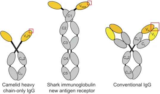
Figure 1.Domain structures of camelid heavy chain-only IgG, shark immunoglobulin new antigen receptor (IgNAR) and conventional vertebrate tetrameric IgG. The variable domain(s) of each antibody molecule are shown in yellow and the antigen-combining site is indicated by a red box.
Box 1.Immunogenetics of sdAbs
VHHs, the variable domains of camelid heavy chain-only antibodies, are recombined during B-cell development from a unique set of germline V genes and
common D and J genes (shared with the VHdomains of conventional tetrameric antibodies) located within the igh locus on chromosome 4.8Most camelid
VHH and VHgenes18are homologous to human IGHV3-family genes (~75–90% identity) and encode distinctive solubilizing residues in FR2 (Phe/Tyr42, Glu49,
Arg50 and Gly 52 using IMGT numbering; these positions map to the VH:VLinterface in conventional antibodies), although functional VHHs lacking this
consensus have been isolated.19,20Some camelid V genes may ‘promiscuously’ recombine with both heavy chain-only and conventional antibody constant
region genes.19V
HH domains bear unusually long CDR3 loops in comparison with human and murine conventional antibodies,21,22probably reflecting
increased non-templated nucleotide addition, although this may be a feature of only a subset of VHHs;21in some VHHs, the long CDR3 loop serves a dual
purpose, folding over the former VLinterface as well as interacting with cognate antigen. The rearranged VHH exon is thought to undergo elevated rates of
somatic hypermutation of both CDRs and FRs (e.g., FR1-encoding sequences immediately flanking CDR1;23–25FR2-encoding sequences which may play a role
in structuring the CDR3 loop;20,25FR3-encoding sequences that form a β-turn which can make contact with antigen, sometimes called CDR424). V HHs may
also acquire somatic insertions and deletions at higher rates than conventional antibodies,24and may under some circumstances undergo secondary
rearrangement events using a cryptic recombination signal sequence in FR3.24Some V
HH genes encode non-canonical disulfide linkages formed between
cysteine residue pairs (CDR1-CDR3, FR2-CDR3, CDR2-CDR3 or CDR3-CDR3; seeBox 2).
VNARs, the variable domains of cartilaginous fish Ig new antigen receptors, share sequence homology with T-cell receptor and Ig light chain genes4and may be
descended from Ig-superfamily cell-surface receptors.26Compared with Ig V
Hdomains, VNARs lack two β strands (C’and C’’) and consequently CDR2 is absent,
although loops connecting the C-D and D-E strands (HV2 and HV4, respectively) can make contact with antigen. During B-cell development, VNARdomains
are rearranged from a small number of loci (perhaps only three) distinct from those encoding other types of Ig molecules detectable in serum (IgM, IgW). Each locus contains one V gene, two or three D genes and one J gene and thus primary repertoire diversity is almost entirely CDR3-based:4since V
NARCDR3
loops are formed through either three or four independent rearrangement events, these tend to be long.27Unlike the V
Hdomains of IgMs and IgWs, VNARs
demonstrating that CDR3-centric binding is not the only
mechanism of competitive enzyme inhibition by sdAbs.
Competitive inhibition of these enzymes by conventional
anti-bodies targeting their active sites has not been described despite
intensive study, especially of murine antibodies against
lyso-zyme. Naturally-occurring competitive inhibitors of protease
enzymes are convex, and this appears to be a difficult geometry
for the paratopes of conventional antibodies to achieve (see
below): even in cases of near-true competitive inhibition,
con-ventional antibodies use a flat or concave V
H/V
Linterface to
bind protruding regions on enzymes and partially insert one or
more CDRs into the active site cleft in a non-substrate-like
manner.
66,67This hypothesis is supported by experiments
using purified polyclonal immunoglobulin (Ig)Gs from
enzyme-immunized dromedaries showing that competitive
inhibition was a feature of heavy chain-only IgGs, but not of
conventional IgGs.
11,32It remains unclear why immunization
with some enzymes yields mostly sdAbs with planar paratopes
and bind outside the active site, achieving allosteric or no
inhibi-tion, although tolerance mechanisms may play a role.
A second line of evidence clearly supporting distinct
specifi-cities of sdAbs vs. conventional antibodies can be found in studies
of sdAbs against pathogenic microorganisms. Stijlemans et al.
68hypothesized that the ability of a dromedary V
HH, cAb-An33, to
target a cryptic glycopeptide epitope conserved across all variant
surface glycoprotein classes of Trypanosoma brucei was due to the
V
HH’s small size as well as, potentially, the nature of this epitope.
This hypothesis was supported by the inability of rabbit and
dromedary polyclonal conventional antibodies as well as a
~90-kDa lectin to access this site. Henderson et al.
69suggested that
recognition of a conserved hydrophobic cleft on Plasmodium
AMA1 by a V
NAR(12Y-2 and its affinity-matured variants)
reflected a novel binding mode; although the epitope of a murine
conventional antibody (1F9) substantially overlapped that of V
NAR12Y-2, 1F9 binding depended to a greater degree on polymorphic
loop residues surrounding the hydrophobic trough. Likewise,
Ditlev et al.
70attributed the binding of a panel of alpaca V
HHs to
multiple domains of the malarial VAR2CSA protein to an inherent
ability of V
HHs to recognize subdominant epitopes, although
limited understanding of the human conventional antibody
response against VAR2CSA as well as irreproducibility of these
reactivity patterns by llama V
HHs
71complicated this assessment.
Probably the clearest examples of epitopes that are more
favorable for binding by sdAbs than conventional antibodies
can be found in the envelope glycoprotein trimer of HIV-1:
heterologous cross-strain neutralization is extraordinarily
dif-ficult to achieve by conventional antibodies, requiring months
of chronic infection and multiple rounds of somatic mutation
and selection, yet cross-neutralizing camelid heavy chain-only
antibodies directed against the CD4-binding site
72-75and
CD4-induced sites
76-78can be easily elicited by routine
immu-nizations with recombinant protein antigens. Similar
exam-ples can be found for other viral pathogens. Serotype
cross-neutralizing antibodies targeting the CD155-binding ‘canyon’
of the poliovirus capsid are rarely produced by the murine or
human humoral immune systems,
79,80but are apparently
common in llama heavy-chain only responses.
81Likewise,
V
HHs targeting the HBGA-binding pocket of norovirus VP1
neutralized a broad range of genotypes,
82while larger
con-ventional antibodies also made contact with antigenically
variable residues surrounding the HBGA pocket and were
thus strain-specific.
83Finally, compared with conventional antibodies, sdAbs
have been implied to have privileged access to recessed sites
on membrane proteins,
84such as ion channels and G
protein-coupled receptors (GPCRs). While this is an intriguing
hypothesis, it has yet to be substantiated by any data.
Camelid V
HHs generated against the Kv1.3 ion channel
tar-geted extracellular loops, not the channel cavity,
85and the
epitopes of V
HHs against the P2X7 ion channel were not
defined.
86Similarly, camelid V
HHs developed as potential
therapeutics against the chemokine receptors CXCR4,
84CXCR7
87and ChemR23,
88as well as V
HHs used as
crystal-lization chaperones for several GPCRs, channels and
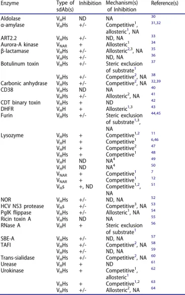
Table 1.Single-domain antibodies as enzyme inhibitors. Enzyme Type of
sdAb(s) Inhibition Mechanism(s)of Inhibition Reference(s) Aldolase VHH ND NA 30
α-amylase VHHs +/- Competitive1,
allosteric1, NA 31,32
ART2.2 VHHs +/- ND, NA 33
Aurora-A kinase VNAR + Allosteric1 34
β-lactamase VHHs +/- Allosteric2,3, NA 35
VHHs +/- ND, NA 36
Botulinum toxin VHHs +/- Steric exclusion
of substrate1 37
VHHs +/- Competitive2, NA 38
Carbonic anhydrase VHHs +/- Competitive2, NA 32,39
CD38 VHHs ND NA 40
VHHs +/- Allosteric2, NA 41
CDT binary toxin VHHs + ND 42
DHFR VHH + Allosteric1,3 43
Furin VHHs +/- Steric exclusion
of substrate1,3, NA 44,45 Lysozyme VHHs + Competitive1,2 11 VHH + Competitive1 6,46 VHH + Competitive2 47 VHHs + Competitive1 48 VHH ND NA4 49 VHH ND NA4 50 VNAR + Competitive1 7 VNAR + Competitive1 12 VHs +, ND Competitive1,2, NA 51 NOR VHHs +/- ND, NA 52 HCV NS3 protease VHs +/- Competitive3, NA 53 PglK flippase VHHs +/- Allosteric1, NA 54 Ricin toxin A VHHs ND NA 55
RNase A VHH + Steric exclusion
of substrate1 56 SBE-A VHHs +/- ND, NA 57 TAFI VHHs +/- Competitive2, NA 58 VHHs +/- ND, NA 59 Trans-sialidase VHHs +/- Competitive2, NA 60 Urease VHH + ND 61 Urokinase VHHs + Competitive1, allosteric1 62 VHHs + Competitive1,2 63 VHHs +/- Allosteric2, NA 64 1Mechanism inferred from antibody:enzyme X-ray co-crystal structures. 2Specificity for active site or non-active site regions demonstrated through
epitope mapping experiments.
3Mechanism inferred from studies of enzyme kinetics.
4Inhibition was not assessed, but structural studies showed that the antibody did
not target the active site.
Abbreviations used: ART2.2, ecto-ADP-ribosyltransferase 2.2; CDT, Clostridium difficiletransferase; DHFR, dihydrofolate reductase; HCV NS3, hepatitis C virus non-structural protein 3; NA, not applicable; ND, not determined; NOR, nitric oxide reductase; SBE-A, starch branching enzyme A.
transporters,
89–95all appear to bind solvent-exposed
extracel-lular or intracelextracel-lular loops of these receptors in a manner
similar to conventional antibodies and their fragments. By
contrast, a synthetic CXCR4-binding “i-body” engineered
from an Ig-like NCAM domain was found to penetrate deep
into the receptor’s ligand-binding pocket to occupy a truly
cryptic, partially transmembrane epitope.
96Thus, there is at
least some reason to believe that the small size of sdAbs may
grant them access to recessed regions on pores and channels,
although experimental evidence is still lacking.
Overall, the evidence is compelling that camelid V
HHs, at least,
can interact with recessed epitopes on proteins that are poorly
available for binding by conventional antibodies. Additional
examples of binding to recessed epitopes on proteins (clefts,
cav-ities, crevices or grooves) can be found for sdAbs against
lactococ-cal siphophage,
97Plasmodium falciparum MTIP,
98epidermal
growth factor receptor,
99and respiratory syncytial virus fusion
protein,
100although in these cases it is less clear that these sites
are inaccessible to conventional antibodies. While it is possible that
V
NARs may share similar cleft-binding proclivities, such claims are
based on very limited published data (three structures
7,12,69).
Moreover, it should be noted that there are many examples (not
covered in this review) of partial or complete overlap between the
epitopes of sdAbs and conventional antibodies, and thus the
degree to which sdAbs bind cryptic epitopes vs. conventional
antibody-accessible epitopes, as well as whether the magnitude of
this difference exceeds more general species-to-species reactivity
differences of conventional antibodies, remain unknown.
Single-domain antibodies direct against linear
protein epitopes
It is generally recognized that the majority of conventional
antibodies raised against folded proteins are directed against
conformational epitopes (≥90%
101), although this may depend
to some extent on the nature of the antigen. Several authors
have suggested that V
HHs, at least, are even less likely than
conventional antibodies to bind linear peptides with high
affinity.
102,103Although this is a plausible hypothesis based
on the typical structures of sdAb paratopes (see below), it has
not yet been substantiated by any data. Moreover, the
rela-tively large number of studies reporting sdAb reactivity by
western blotting suggests that sdAbs directed against
contin-uous epitopes are probably not vanishingly rare.
Single-domain antibody paratope structures
The paratopes of conventional antibodies directed against
folded proteins tend to be flat or concave;
104convex binding
sites are difficult to achieve, at least by murine and human
conventional antibodies, although synthetic conventional
antibodies can be engineered to adopt such geometries.
105By contrast, sdAb paratopes can clearly adopt both flat
106,107and convex
11topologies, although possibly only inefficiently
adopt concave ones. The CDR1 and CDR2 loops of V
HHs
depart from the typical canonical structures of conventional
antibodies (
Figure 2A
), potentially through somatic mutation
since germline human V
Hand camelid V
HH repertoires
appear to have similar canonical structures.
18Only a handful
of V
NARs have been crystallized, and several showed a
struc-tural class of CDR1 (H1-13–9) that is more common in V
HHs
than in conventional antibodies, although others had CDR1
canonical structures closer to those of V
Ldomains. The CDR3
length distributions of both V
HHs and V
NARs (
Figure 2B
) are
broader than those of conventional antibodies and biased
towards longer lengths; the long CDR loops of sdAbs may
be structurally constrained by non-canonical disulfide
lin-kages (see
Box 2
). Despite potentially elevated somatic
muta-tion rates (at least of V
HHs), the paratopes of V
HHs, V
NARs
and conventional antibodies have similar amino acid
con-tents, all being enriched for Gly, Ser and Tyr, and their
CDR sequences bear no obvious patterns of sequence
homol-ogy (
Figure 2C
,
D
). Both V
HH and V
NARparatopes have
smaller molecular surface areas and smaller diameters than
conventional antibodies (
Figure 2E
,
F
). However, sdAb
para-topes as a group are not more globally extended than those of
conventional antibodies, as reflected by the maximum
intera-tomic distance between the tips and the bases of any CDR
loop (
Figure 2G
).
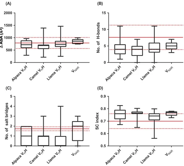
Single-domain antibody:antigen interactions
The footprints of sdAbs on antigens are smaller than those of
conventional antibodies, given that the paratopes of the
for-mer molecules are roughly half the size of the latter ones.
Using only three CDR loops (two CDR loops and potentially
two HV loops for V
NARs), sdAbs can bury similar
solvent-accessible surface areas on proteins compared with
conven-tional antibodies (
Figure 3A
). This is made possible by a
number of molecular contacts (hydrogen bonds, salt bridges)
that is slightly lower for sdAbs than in conventional
antibo-dies, but higher on a per-chain basis (
Figure 3B
,
3C
).
Moreover, the surface complementarity of sdAb:protein
inter-faces is on the high end for antibody:antigen interactions
(
Figure 3D
). Thus, sdAbs and conventional antibodies bind
protein antigens through similar types of non-covalent
inter-actions, but these are more concentrated in the smaller
para-topes of sdAbs.
Single-domain antibodies directed against small
molecules
The dominant mechanism by which conventional antibodies
interact with haptens, small-molecule lipids and
oligosacchar-ides is by forming a binding pocket at the interface between
the V
Hand V
Ldomains, typically involving the bases of the
CDR-H3 and CDR-L3 loops.
113–115Similarly, conventional
antibodies tend to accommodate short linear peptides and
nucleic acid polymers within grooves formed from both
heavy- and light-chain CDRs.
104Four studies have reported
structures of camelid V
HHs in complex with haptens and
peptides (
Table 2
); the recognition mechanism of all but one
(a methotrexate-specific V
HH with a non-canonical binding
site involving framework region (FR)3 residues located below
CDR1
113) was basically similar to that of conventional
anti-bodies, with the hapten-binding pocket formed from two or
more CDRs and extending in some instances into the former
V
Linterface. Notably, three of these V
HHs have
non-Fig 2.Properties of sdAb vs. conventional antibody paratopes. (A) Structural classification of CDR1 and CDR2 according to PyIgClassify.108 (B)CDR3 length
distributions. (C) Amino acid compositions of conventional antibody (VHdomain) and sdAb paratopes. For VHHs, sequences of CDR1, CDR2 and CDR3
(Honegger-Plückthun numbering) were used and for VNARs, sequences of CDR1 and CDR3 only were used. (D) Relatedness of conventional antibody (VHdomain) and sdAb CDR3
sequences. The phylogenetic tree was produced using neighbor-joining methods in ClustalW2 and the cladogram was visualized using iTOL109with CDR3s colored
according to species origin as in part B. (E) Molecular surface areas of conventional antibody (VH:VL) and sdAb paratopes. Areas were calculated for merged CDR
sequences (Honegger-Plückthun numbering) using PyMol. (F) Diameters of conventional antibody (VH:VL) and sdAb paratopes. Diameters were calculated as the
maximum interatomic distance between any two FR-CDR boundary residues (Honegger-Plückthun numbering). (G) Extension of conventional antibody (VH:VL) and
sdAb paratopes. Extension was calculated as the maximum interatomic distance between the CDR base (first or last residue according to Honegger-Plückthun numbering) and the CDR tip. The CDR(H)3 loop is shown in blue. In parts (E) – (G), boxplot lines represent medians, the box boundaries represent quartiles and the box whiskers represent ranges. Red dots indicate sdAbs targeting cryptic epitopes discussed in the main text. Data are representative of all complete antibody structures available in the Protein Data Bank and indexed in PyIgClassify as of January 2018.
canonical structures of either CDR1 or CDR2 that have not
been observed in structures of other V
HHs and may not be
germline-encoded.
Multiple studies have reported the isolation of
hapten-specific V
HHs without investigating their structures,
119although several also reported weaker and inconsistent
serum heavy chain-only IgG titers compared with
conven-tional IgG titers against the hapten. No studies have reported
hapten-specific V
NARs, and only one study has described a
carbohydrate-specific V
HH directed against Neisseira
menin-gitidis lipopolysaccharide;
120at least two camelid V
HHs have
been described that bind to glycopeptide epitopes.
68,76No
sdAbs of any type have been described that convincingly
bind lipids or nucleic acids. Together, the consensus of the
data is that it is probably difficult, but not impossible, for
sdAb paratopes to accommodate haptens and that three CDRs
are sufficient to form the binding pockets and grooves
required for such interactions, although potential involvement
of solubility-enhancing FR2 residues at the former V
Linter-face in pocket formation may impose restrictions on
hapten-binding specificities.
Synthetic single-domain antibodies and
non-antibody scaffolds
Fully synthetic sdAbs, derived from V
HHs, V
NARs or from rare
human and murine V
Hand V
Ldomains that remain stable and
soluble outside the context of the natural V
H:V
Lpairing, can be
engineered to bind antigens using in vitro methods (e.g., phage
display). More recently, technologies have been developed for
generating semi-synthetic sdAbs using engineered cell lines
capable of inducible V(D)J recombination
121and transgenic
mice bearing either hybrid llama-human or fully human igh
loci;
122in both cases, a limited set of V
H, D and J
Hgenes (some
of which are in non-germline configurations to promote
auton-omous folding) are rearranged in a foreign cellular or in vivo
system. Limited numbers of synthetic sdAbs have been
described and fewer still have been studied structurally in
complex with antigens. Nevertheless, the available data suggests
that some synthetic sdAbs have cleft-binding properties akin to
those of V
HHs and V
NARs
51while others employ unusual
mechanisms to interact with planar protein epitopes (e.g.,
dra-matic CDR3 restructuring of a MDM4-specific V
Hdomain to
accommodate
packing
against
a
hydrophobic
helix;
123significant involvement of FRs in binding of V
Hs to vascular
endothelial growth factor
124,125and CD40
126using distinct
mechanisms). Even less is known regarding the paratope
struc-tures and binding modes of non-Ig-based antibodies such as
variable lymphocyte receptors
127and non-antibody scaffolds
(based on monomeric non-Ig domains such as fibronectin
type III and SRC homology 3 domains), and their synthetic
origin may imply that they follow no general patterns. If so,
restrictions on the binding specificities of naturally-occurring
sdAbs may not equally affect synthetic sdAbs and non-antibody
scaffolds, although fundamental structural constraints on the
amino acid sequences that can be tolerated by stable Ig folds
would still apply.
Conclusions and perspectives
Recent work on unusual antibodies produced by
unortho-dox model organisms (e.g., cows, chickens) has spurred
renewed interest in the comparative immunology of
anti-body responses. Some ‘cryptic’ regions on proteins (e.g.,
enzyme active sites, recessed regions of viral
glycopro-teins) are clearly more accessible to sdAbs than to
con-ventional antibodies. More generally, we surmise that the
major advantage of sdAb recognition is the ability to
target conserved cleft and pocket regions (typically
bind-ing sites) on hypervariable pathogens without makbind-ing
ancillary contact with the easily mutable perimeters of
these sites. Why and how pathogen selection produced
two evolutionarily-unrelated sdAb systems in sharks and
camelids, but not in other organisms, remains to be
clar-ified. In the case of sdAbs, privileged access is conferred
by their compact paratope diameters (in the absence of a
paired V
Ldomain) rather than any global difference in
paratope shape or structure. Similar non-covalent
interac-tions mediate the binding of conventional antibodies and
sdAbs, although these are more efficiently concentrated in
the compact paratopes of sdAbs to produce high-affinity
interactions. Although it is likely that sdAb paratopes have
difficulty adopting concave geometries and recognizing
small molecules, it remains unclear whether such paratope
restrictions disfavor interaction with certain types of
pro-tein epitopes as well.
Future studies will need to rigorously assess the degree
of separation and overlap in the protein epitope space
Box 2.Non-canonical disulfide linkages of sdAbs
Some but not all camelid VHHs bear paired cysteine residues, resulting in formation of a second intradomain disulfide linkage in addition to the conserved
Cys23-Cys104 linkage (IMGT numbering) present in all Ig domains. Non-canonical disulfide linkages most commonly bridge Cys residues in CDR1 and CDR3,20,21,24but can also link FR2 and CDR3,21,22CDR2 and CDR3,23or two positions within the CDR3 loop.19The Cys residues in CDR1 are encoded by
germline VHH genes that are frequently used in the repertoires of dromedary camels, and B cells using these genes presumably acquire a partner Cys during
receptor rearrangement. Two hypotheses have been invoked to explain the presence of non-canonical disulfide linkages in VHH domains: they may impart
greater stability to the VHH fold and/or restrict the conformational flexibility of long CDR3 loops, potentially minimizing entropic penalties for antigen
binding. However, mutagenesis studies have showed that Cys residues forming non-canonical disulfide linkages can be replaced with a spectrum of other residues with only modest impairment of antigen binding affinity and thermal stability.110
Most cartilaginous fish VNARs bear an additional non-canonical disulfide linkage spanning either FR2-CDR3 (type I) or CDR1-CDR3 (types II and III16). In addition,
type I VNARs also bear a CDR3-FR4 disulfide linkage and, sometimes, an intra-CDR3 disulfide linkage (three or four intradomain disulfide linkages in total27). A
minority of VNARs (type IV) bear only the single canonical disulfide linkage. As for VHHs, most VNARCys residues in CDR1, FR2 and FR4 are probably encoded in
the germline and non-canonical disulfide linkages are formed during primary repertoire development4,27,28
Although the precise roles of non-canonical disulfide linkages in sdAb structure and function remain unclear, these linkages very likely influence sdAb paratope structure, since patterns of antigen-driven somatic hypermutation appear to vary depending on their presence and location.27
targeted by sdAbs vs. conventional antibodies, and to
explore whether sdAb-accessible (and inaccessible)
epi-topes can be predicted in silico. Basic studies of the
immu-nological functions of conventional vs. heavy chain-only
antibodies in host defense (e.g., neutralization;
opsoniza-tion; antibody-dependent cell-mediated cytotoxicity and
complement-dependent cytotoxicity) would also be highly
valuable. Given the apparent sufficiency of sdAb paratopes
to mediate high-affinity interactions with proteins, both
the evolutionary forces responsible for shaping the more
complex paired V
H:V
Lantibody system in vertebrates, as
well as the overall functions of light chains, are open
questions.
Disclosure statement
No potential conflict of interest was reported by the authors.
Funding
Funding for this work was provided by the National Research Council Canada.
Abbreviations
CDR complementarity-determining region FR framework region GPCR G protein-coupled receptor Alp aca VHH Came l VH H Lla ma VHH VNAR 0 500 1000 1500 2000 Alp aca VHH Came l VH H Lla ma VHH VNAR 0 5 10 15 No. o f H -bonds Alp aca VHH Came l VH H Lla ma VHH VNAR 0 1 2 3 4 5 No. o f s al t b ri d g es Alp aca VHH Came l VH H Lla ma VHH VNAR 0.5 0.6 0.7 0.8 0.9 SC in de x (A) (B) (C) (D)Figure 3.Properties of sdAb:antigen and conventional antibody:antigen interfaces. (A) Change in solvent-accessible surface area on proteins upon binding by conventional antibodies or sdAbs. (B) Number of hydrogen bonds and (C) number of salt bridges in conventional antibody:antigen and sdAb:antigen interfaces. (D) Shape complementarity index of conventional antibody:protein and sdAb:protein interfaces. Results in parts (A) – (C) were calculated using the PISA server, and in part (D) using the SC algorithm implemented in CCP4. Boxplot lines represent medians, the box boundaries represent quartiles and the box whiskers represent ranges. Red lines indicate the means and standard deviations for conventional antibodies.111,112Data are representative of all complete antibody:antigen co-crystal
structures available in the Protein Data Bank and indexed in PyIgClassify as of January 2018.
Table 2.Structural features of anti-hapten sdAb paratopes. Antigen sdAb Type CDR3
Length (aa)
CDR1/2 Canonical Structures
Paratope
MSA (Å2) Diameter (Å)Paratope CDR Loop Extension (Å) Reference
Reactive Red 6 VHH 17 H1-13–1, H2-15–1 4380 14.7 17.8 (CDR1), 22.8 (CDR2), 22.3 (CDR3) 116
Reactive Red 1 VHH 18 H1-16–1, H2-10–2 4242 15.0 21.8 (CDR1), 22.3 (CDR2), 22.9 (CDR3) 117
Methotrexate VHH 17 H1-13–11, H2-10–2 3866 14.7 22.4 (CDR1), 17.6 (CDR2), 21.0 (CDR3) 113
HV hypervariable Ig immunoglobulin sdAb single-domain antibody
VH variable heavy chain domain of conventional antibody
VHH variable heavy chain domain of camelid heavy chain-only
antibody
VL variable light chain domain of conventional antibody
VNAR variable domain of shark immunoglobulin new antigen
receptor.
References
1. Flajnik MF, Deschacht N, Muyldermans S. A case of convergence: why did a simple alternative to canonical antibodies arise in sharks and camels? PLoS Biol.2011;9:e1001120. doi:10.1371/jour-nal.pbio.1001120.
2. Ward ES, Gussow D, Griffiths AD, Jones PT, Winter G. Binding activities of a repertoire of single immunoglobulin variable domains secreted from Escherichia coli. Nature. 1989;341:544– 546. doi:10.1038/341544a0.
3. Hamers-Casterman C, Atarhouch T, Muyldermans S, Robinson G, Hamers C, Songa EB, Bendahman N, Hamers R. Naturally occurring antibodies devoid of light chains. Nature. 1993;363:446–448. doi:10.1038/363446a0.
4. Greenberg AS, Avila D, Hughes M, Hughes A, McKinney EC, Flajnik MF. A new antigen receptor gene family that undergoes rearrangement and extensive somatic diversification in sharks. Nature.1995;374:168– 173. doi:10.1038/374168a0.
5. Holliger P, Hudson PJ. Engineered antibody fragments and the rise of single domains. Nat Biotechnol. 2005;23:1126–1136. doi:10.1038/nbt1142.
6. Desmyter A, Transue TR, Ghahroudi MA, Thi MH, Poortmans F, Hamers R, Muyldermans S, Wyns L. Crystal structure of a camel single-domain VHantibody fragment in complex with lysozyme. Nat
Struct Biol.1996;3:803–811.
7. Stanfield RL, Dooley H, Flajnik MF, Wilson IA. Crystal structure of a shark single-domain antibody V region in complex with lysozyme. Science.2004;305:1770–1773. doi:10.1126/science.1101148.
8. Achour I, Cavelier P, Tichit M, Bouchier C, Lafaye P, Rougeon F. Tetrameric and homodimeric camelid IgGs originate from the same igh locus. J Immunol.2008;181:2001–2009.
9. Eason DD, Litman RT, Luer CA, Kerr W, Litman GW. Expression of individual immunoglobulin genes occurs in an unusual system consisting of multiple independent loci. Eur J Immunol. 2004;34:2551–2558. doi:10.1002/eji.200425224.
10. Griffin LM, Snowden JR, Lawson AD, Wernery U, Kinne J, Baker TS. Analysis of heavy and light chain sequences of conventional camelid antibodies from Camelus dromedarius and Camelus bac-trianus species. J Immunol Methods.2014;405:35–46. doi:10.1016/ j.jim.2014.01.003.
11. De Genst E, Silence K, Decanniere K, Conrath K, Loris R, Kinne J, Muyldermans S, Wyns L. Molecular basis for the preferential cleft recognition by dromedary heavy-chain antibodies. Proc Natl Acad Sci U S A.2006;103:4586–4591. doi:10.1073/pnas.0505379103. 12. Stanfield RL, Dooley H, Verdino P, Flajnik MF, Wilson IA.
Maturation of shark single-domain (IgNAR) antibodies: evidence for induced-fit binding. J Mol Biol. 2007;367:358–372. doi:10.1016/j.jmb.2006.12.045.
13. Al Qaraghuli MM, Ferro VA. Analysis of the binding loops con-figuration and surface adaptation of different crystallized single-domain antibodies in response to various antigens. J Mol Recognit.2017;30:e2592. doi:10.1002/jmr.2592.
14. Konning D, Zielonka S, Grzeschik J, Empting M, Valldorf B, Krah S, Schroter C, Sellmann C, Hock B, Kolmar H. Camelid and shark single domain antibodies: structural features and therapeutic potential. Curr Opin Struct Biol. 2017;45:10–16. doi:10.1016/j. sbi.2016.10.019.
15. Muyldermans S, Smider VV. Distinct antibody species: struc-tural differences creating therapeutic opportunities. Curr Opin Immunol. 2016;40:7–13. doi:10.1016/j.coi.2016.02.003.
16. Zielonka S, Empting M, Grzeschik J, Konning D, Barelle CJ, Kolmar H. Structural insights and biomedical potential of IgNAR scaffolds from sharks. MAbs.2015;7:15–25. doi:10.4161/ 19420862.2015.989032.
17. Desmyter A, Spinelli S, Roussel A, Cambillau C. Camelid nano-bodies: killing two birds with one stone. Curr Opin Struct Biol. 2015;32:1–8. doi:10.1016/j.sbi.2015.01.001.
18. Klarenbeek A, El Mazouari K, Desmyter A, Blanchetot C, Hultberg A, De Jonge N, Roovers RC, Cambillau C, Spinelli S, Del-Favero J, et al. Camelid Ig V genes reveal significant human homology not seen in therapeutic target genes, provid-ing for a powerful therapeutic antibody platform. MAbs. 2015;7:693–706. doi:10.1080/19420862.2015.1046648.
19. Deschacht N, De Groeve K, Vincke C, Raes G, De Baetselier P, Muyldermans S. A novel promiscuous class of camelid single-domain antibody contributes to the antigen-binding repertoire. J Immunol.2010;184:5696–5704. doi:10.4049/jimmunol.0903722. 20. Li X, Duan X, Yang K, Zhang W, Zhang C, Fu L, Ren Z, Wang C, Wu J,
Lu R, et al. Comparative analysis of immune repertoires between bactrian camel’s conventional and heavy-chain antibodies. PLoS One.2016;11:e0161801. doi:10.1371/journal.pone.0161801.
21. Harmsen MM, Ruuls RC, Nijman IJ, Niewold TA, Frenken LG, De Geus B. Llama heavy-chain V regions consist of at least four distinct subfamilies revealing novel sequence features. Mol Immunol.2000;37:579–590.
22. Muyldermans S, Atarhouch T, Saldanha J, Barbosa JA, Hamers R. Sequence and structure of VHdomain from naturally occurring
camel heavy chain immunoglobulins lacking light chains. Protein Eng.1994;7:1129–1135.
23. Vu KB, Ghahroudi MA, Wyns L, Muyldermans S. Comparison of llama VHsequences from conventional and heavy chain
antibo-dies. Mol Immunol.1997;34:1121–1131.
24. Nguyen VK, Hamers R, Wyns L, Muyldermans S. Camel heavy-chain antibodies: diverse germline VHH and specific mechanisms
enlarge the antigen-binding repertoire. EMBO J.2000;19:921–930. doi:10.1093/emboj/19.5.921.
25. Nguyen VK, Su C, Muyldermans S. van der Loo W. Heavy-chain antibodies in Camelidae; a case of evolutionary innovation. Immunogenetics.2002;54:39–47. doi:10.1007/s00251-002-0433-0. 26. Streltsov VA, Varghese JN, Carmichael JA, Irving RA, Hudson PJ,
Nuttall SD. Structural evidence for evolution of shark Ig new antigen receptor variable domain antibodies from a cell-surface receptor. Proc Natl Acad Sci U S A. 2004;101:12444–12449. doi:10.1073/pnas.0403509101.
27. Diaz M, Stanfield RL, Greenberg AS, Flajnik MF. Structural analysis, selection, and ontogeny of the shark new antigen receptor (IgNAR): identification of a new locus preferentially expressed in early develop-ment. Immunogenetics. 2002;54:501–512. doi: 10.1007/s00251-002-0479-z.
28. Diaz M, Greenberg AS, Flajnik MF. Somatic hypermutation of the new antigen receptor gene (NAR) in the nurse shark does not generate the repertoire: possible role in antigen-driven reactions in the absence of germinal centers. Proc Natl Acad Sci U S A. 1998;95:14343–14348.
29. Abdiche YN, Harriman R, Deng X, Yeung YA, Miles A, Morishige W, Boustany L, Zhu L, Izquierdo SM, Harriman W. Assessing kinetic and epitopic diversity across orthogonal monoclonal anti-body generation platforms. MAbs. 2016;8:264–277. doi:10.1080/ 19420862.2015.1118596.
30. Pinto J, Odongo S, Lee F, Gaspariunaite V, Muyldermans S, Magez S, Sterckx YG. Structural basis for the high specificity of a Trypanosoma congolense immunoassay targeting glycosomal aldolase. PLoS Negl Trop Dis.2017;11:e0005932. doi:10.1371/journal.pntd.0005932. 31. Desmyter A, Spinelli S, Payan F, Lauwereys M, Wyns L,
Muyldermans S, Cambillau C. Three camelid VHH domains in
versatility of binding topology. J Biol Chem. 2002;277:23645– 23650. doi:10.1074/jbc.M202327200.
32. Lauwereys M, Arbabi Ghahroudi M, Desmyter A, Kinne J, Holzer W, De Genst E, Wyns L, Muyldermans S. Potent enzyme inhibi-tors derived from dromedary heavy-chain antibodies. EMBO J. 1998;17:3512–3520. doi:10.1093/emboj/17.13.3512.
33. Koch-Nolte F, Reyelt J, Schossow B, Schwarz N, Scheuplein F, Rothenburg S, Haag F, Alzogaray V, Cauerhff A, Goldbaum FA. Single domain antibodies from llama effectively and specifically block T cell ecto-ADP-ribosyltransferase ART2.2 in vivo. FASEB J.2007;21:3490–3498. doi:10.1096/fj.07-8661com.
34. Burgess SG, Oleksy A, Cavazza T, Richards MW, Vernos I, Matthews D, Bayliss R. Allosteric inhibition of Aurora-A kinase by a synthetic VNAR domain. Open Biol. 2016;6:160089.
doi:10.1098/rsob.160089.
35. Sohier JS, Laurent C, Chevigne A, Pardon E, Srinivasan V, Wernery U, Lassaux P, Steyaert J, Galleni M. Allosteric inhibition of VIM metallo-β-lactamases by a camelid nanobody. Biochem J. 2013;450:477–486. doi:10.1042/BJ20121305.
36. Conrath KE, Lauwereys M, Galleni M, Matagne A, Frere JM, Kinne J, Wyns L, Muyldermans S. β-lactamase inhibitors derived from single-domain antibody fragments elicited in the Camelidae. Antimicrob Agents Chemother. 2001;45:2807–2812. doi:10.1128/ AAC.45.10.2807-2812.2001.
37. Dong J, Thompson AA, Fan Y, Lou J, Conrad F, Ho M, Pires-Alves M, Wilson BA, Stevens RC, Marks JD. A single-domain llama antibody potently inhibits the enzymatic activity of botuli-num neurotoxin by binding to the non-catalytic alpha-exosite binding region. J Mol Biol. 2010;397:1106–1118. doi:10.1016/j. jmb.2010.01.070.
38. Thanongsaksrikul J, Srimanote P, Maneewatch S, Choowongkomon K, Tapchaisri P, Makino S, Kurazono H, Chaicumpa W. A VHH that neutralizes the zinc metalloproteinase
activity of botulinum neurotoxin type A. J Biol Chem. 2010;285:9657–9666. doi:10.1074/jbc.M109.073163.
39. Desmyter A, Decanniere K, Muyldermans S, Wyns L. Antigen specificity and high affinity binding provided by one single loop of a camel single-domain antibody. J Biol Chem.2001;276:26285– 26290. doi:10.1074/jbc.M102107200.
40. Li T, Qi S, Unger M, Hou YN, Deng QW, Liu J, Lam CM, Wang XW, Xin D, Zhang P, et al. Immuno-targeting the multifunctional CD38 using nanobody. Sci Rep. 2016;6:27055. doi:10.1038/ srep27055.
41. Fumey W, Koenigsdorf J, Kunick V, Menzel S, Schutze K, Unger M, Schriewer L, Haag F, Adam G, Oberle A, et al. Nanobodies effectively modulate the enzymatic activity of CD38 and allow specific imaging of CD38+tumors in mouse models in vivo. Sci
Rep.2017;7:14289. doi:10.1038/s41598-017-14112-6.
42. Unger M, Eichhoff AM, Schumacher L, Strysio M, Menzel S, Schwan C, Alzogaray V, Zylberman V, Seman M, Brandner J, et al. Selection of nanobodies that block the enzymatic and cytotoxic activities of the binary Clostridium difficile toxin CDT. Sci Rep. 2015;5:7850. doi:10.1038/srep07850.
43. Oyen D, Srinivasan V, Steyaert J, Barlow JN. Constraining enzyme conformational change by an antibody leads to hyperbolic inhibi-tion. J Mol Biol.2011;407:138–148. doi:10.1016/j.jmb.2011.01.017. 44. Zhu J, Declercq J, Roucourt B, Ghassabeh GH, Meulemans S, Kinne J, David G, Vermorken AJ, Van de Ven WJ, Lindberg I, et al. Generation and characterization of non-competitive furin-inhibiting nanobodies. Biochem J. 2012;448:73–82. doi:10.1042/ BJ20120537.
45. Dahms SO, Creemers JW, Schaub Y, Bourenkov GP, Zogg T, Brandstetter H, Than ME. The structure of a furin-antibody complex explains non-competitive inhibition by steric exclusion of substrate conformers. Sci Rep. 2016;6:34303. doi:10.1038/ srep34303.
46. Transue TR, De Genst E, Ghahroudi MA, Wyns L, Muyldermans S. Camel single-domain antibody inhibits enzyme by mimicking carbohydrate substrate. Proteins.1998;32:515–522.
47. Chan PH, Pardon E, Menzer L, De Genst E, Kumita JR, Christodoulou J, Saerens D, Brans A, Bouillenne F, Archer DB, et al. Engineering a camelid antibody fragment that binds to the active site of human lysozyme and inhibits its conversion into amyloid fibrils. Biochemistry.2008;47:11041–11054. doi:10.1021/ bi8005797.
48. De Genst E, Silence K, Ghahroudi MA, Decanniere K, Loris R, Kinne J, Wyns L, Muyldermans S. Strong in vivo maturation compensates for structurally restricted H3 loops in antibody repertoires. J Biol Chem.2005;280:14114–14121. doi:10.1074/jbc. M413011200.
49. De Genst E, Chan PH, Pardon E, Hsu SD, Kumita JR, Christodoulou J, Menzer L, Chirgadze DY, Robinson CV, Muyldermans S, et al. A nanobody binding to non-amyloidogenic regions of the protein human lysozyme enhances partial unfolding but inhibits amyloid fibril formation. J Phys Chem B. 2013;117:13245–13258. doi:10.1021/jp403425z.
50. Dumoulin M, Last AM, Desmyter A, Decanniere K, Canet D, Larsson G, Spencer A, Archer DB, Sasse J, Muyldermans S, et al. A camelid antibody fragment inhibits the formation of amyloid fibrils by human lysozyme. Nature.2003;424:783–788. doi:10.1038/nature01870. 51. Rouet R, Dudgeon K, Christie M, Langley D, Christ D. Fully
human VHsingle domains that rival the stability and cleft
recog-nition of camelid antibodies. J Biol Chem.2015;290:11905–11917. doi:10.1074/jbc.M114.614842.
52. Conrath K, Pereira AS, Martins CE, Timoteo CG, Tavares P, Spinelli S, Kinne J, Flaudrops C, Cambillau C, Muyldermans S, et al. Camelid nanobodies raised against an integral membrane enzyme, nitric oxide reductase. Protein Sci. 2009;18:619–628. doi:10.1002/pro.69.
53. Martin F, Volpari C, Steinkuhler C, Dimasi N, Brunetti M, Biasiol G, Altamura S, Cortese R, De Francesco R, Sollazzo M. Affinity selection of a camelized VHdomain antibody inhibitor of hepatitis
C virus NS3 protease. Protein Eng.1997;10:607–614.
54. Perez C, Kohler M, Janser D, Pardon E, Steyaert J, Zenobi R, Locher KP. Structural basis of inhibition of lipid-linked oligosac-charide flippase PglK by a conformational nanobody. Sci Rep. 2017;7:46641. doi:10.1038/srep46641.
55. Rudolph MJ, Vance DJ, Cheung J, Franklin MC, Burshteyn F, Cassidy MS, Gary EN, Herrera C, Shoemaker CB, Mantis NJ. Crystal structures of ricin toxin’s enzymatic subunit (RTA) in complex with neutralizing and non-neutralizing single-chain antibodies. J Mol Biol. 2014;426:3057–3068. doi:10.1016/j.jmb.2014.05.026.
56. Decanniere K, Desmyter A, Lauwereys M, Ghahroudi MA, Muyldermans S, Wyns L. A single-domain antibody fragment in complex with RNase A: non-canonical loop structures and nano-molar affinity using two CDR loops. Structure.1999;7:361–370. 57. Jobling SA, Jarman C, Teh MM, Holmberg N, Blake C, Verhoeyen
ME. Immunomodulation of enzyme function in plants by single-domain antibody fragments. Nat Biotechnol. 2003;21:77–80. doi:10.1038/nbt772.
58. Hendrickx ML, A DEW, Buelens K, Compernolle G, Hassanzadeh-Ghassabeh G, Muyldermans S, Gils A, Declerck PJ. TAFIa inhibiting nanobodies as profibrinolytic tools and discov-ery of a new TAFIa conformation. J Thromb Haemost. 2011;9:2268–2277. doi:10.1111/j.1538-7836.2011.04495.x. 59. Buelens K, Hassanzadeh-Ghassabeh G, Muyldermans S, Gils A,
Declerck PJ. Generation and characterization of inhibitory nanobo-dies towards thrombin activatable fibrinolysis inhibitor. J Thromb Haemost.2010;8:1302–1312. doi:10.1111/j.1538-7836.2010.03816.x. 60. Ratier L, Urrutia M, Paris G, Zarebski L, Frasch AC, Goldbaum
FA. Relevance of the diversity among members of the Trypanosoma cruzi trans-sialidase family analyzed with camelids single-domain antibodies. PLoS One.2008;3:e3524. doi:10.1371/ journal.pone.0003524.
61. Ardekani LS, Gargari SL, Rasooli I, Bazl MR, Mohammadi M, Ebrahimizadeh W, Bakherad H, Zare H. A novel nanobody against urease activity of Helicobacter pylori. Int J Infect Dis. 2013;17:e723–8. doi:10.1016/j.ijid.2013.02.015.
62. Kromann-Hansen T, Louise Lange E, Peter Sorensen H, Hassanzadeh-Ghassabeh G, Huang M, Jensen JK, Muyldermans S, Declerck PJ, Komives EA, Andreasen PA. Discovery of a novel conformational equilibrium in urokinase-type plasminogen acti-vator. Sci Rep.2017;7:3385. doi:10.1038/s41598-017-03457-7. 63. Chan PH, Pardon E, Menzer L, De Genst E, Kumita JR,
Christodoulou J, Saerens D, Brans A, Bouillenne F, Archer DB. A camelid-derived antibody fragment targeting the active site of a serine protease balances between inhibitor and substrate behavior. J Biol Chem. 2016;291:15156–15168. doi:10.1074/jbc. M116.732503.
64. Kaczmarek JZ, Skottrup PD. Selection and characterization of came-lid nanobodies towards urokinase-type plasminogen activator. Mol Immunol.2015;65:384–390. doi:10.1016/j.molimm.2015.02.011. 65. Arnon R. Enzyme inhibition by antibodies. Acta Endocrinol Suppl
(Copenh).1975;194:133–153.
66. Farady CJ, Egea PF, Schneider EL, Darragh MR, Craik CS. Structure of an Fab-protease complex reveals a highly specific non-canonical mechanism of inhibition. J Mol Biol. 2008;380:351–360. doi:10.1016/j.jmb.2008.05.009.
67. Wu Y, Eigenbrot C, Liang WC, Stawicki S, Shia S, Fan B, Ganesan R, Lipari MT, Kirchhofer D. Structural insight into distinct mechanisms of protease inhibition by antibodies. Proc Natl Acad Sci U S A. 2007;104:19784–19789. doi:10.1073/ pnas.0708251104.
68. Stijlemans B, Conrath K, Cortez-Retamozo V, Van Xong H, Wyns L, Senter P, Revets H, De Baetselier P, Muyldermans S, Magez S. Efficient targeting of conserved cryptic epitopes of infectious agents by single domain antibodies: African trypanosomes as paradigm. J Biol Chem. 2004;279:1256–1261. doi:10.1074/jbc. M307341200.
69. Henderson KA, Streltsov VA, Coley AM, Dolezal O, Hudson PJ, Batchelor AH, Gupta A, Bai T, Murphy VJ, Anders RF, et al. Structure of an IgNAR-AMA1 complex: targeting a conserved hydrophobic cleft broadens malarial strain recognition. Structure.2007;15:1452–1466. doi:10.1016/j.str.2007.09.011. 70. Ditlev SB, Florea R, Nielsen MA, Theander TG, Magez S, Boeuf P,
Salanti A.. Utilizing nanobody technology to target non-immuno-dominant domains of VAR2CSA. PLoS One. 2014;9:e84981. doi:10.1371/journal.pone.0084981.
71. Nunes-Silva S, Gangnard S, Vidal M, Vuchelen A, Dechavanne S, Chan S, Pardon E, Steyaert J, Ramboarina S, Chene A, et al. Llama immunization with full-length VAR2CSA generates cross-reactive and inhibitory single-domain antibodies against the DBL1X domain. Sci Rep.2014;4:7373. doi:10.1038/srep07373.
72. Forsman A, Beirnaert E, Aasa-Chapman MM, Hoorelbeke B, Hijazi K, Koh W, Tack V, Szynol A, Kelly C, McKnight A, et al. Llama antibody fragments with cross-subtype human immunode-ficiency virus type 1 (HIV-1)-neutralizing properties and high affinity for HIV-1 gp120. J Virol.2008;82:12069–12081. 73. McCoy LE, Quigley AF, Strokappe NM, Bulmer-Thomas B,
Seaman MS, Mortier D, Rutten L, Chander N, Edwards CJ, Ketteler R, et al. Potent and broad neutralization of HIV-1 by a llama antibody elicited by immunization. J Exp Med. 2012;209:1091–1103. doi:10.1084/jem.20112655.
74. Strokappe N, Szynol A, Aasa-Chapman M, Gorlani A, Forsman Quigley A, Hulsik DL, Chen L, Weiss R, de Haard H, Verrips T. Llama antibody fragments recognizing various epitopes of the CD4bs neutralize a broad range of HIV-1 subtypes A, B and C. PLoS One.2012;7:e33298. doi:10.1371/journal.pone.0033298. 75. McCoy LE, Rutten L, Frampton D, Anderson I, Granger L,
Bashford-Rogers R, Dekkers G, Strokappe NM, Seaman MS, Koh W, et al. Molecular evolution of broadly neutralizing Llama antibodies to the CD4-binding site of HIV-1. PLoS Pathog. 2014;10:e1004552. doi:10.1371/journal.ppat.1004552.
76. Acharya P, Luongo TS, Georgiev IS, Matz J, Schmidt SD, Louder MK, Kessler P, Yang Y, McKee K, O'Dell S, et al. Heavy chain-only IgG2b llama antibody effects near-pan HIV-1 neutralization
by recognizing a CD4-induced epitope that includes elements of coreceptor- and CD4-binding sites. J Virol.2013;87:10173–10181. doi:10.1128/JVI.01332-13.
77. Matz J, Kessler P, Bouchet J, Combes O, Ramos OH, Barin F, Baty D, Martin L, Benichou S, Chames P. Straightforward selection of broadly neutralizing single-domain antibodies targeting the con-served CD4 and coreceptor binding sites of HIV-1 gp120. J Virol. 2013;87:1137–1149. doi:10.1128/JVI.00461-12.
78. Chen W, Zhu Z, Feng Y, Dimitrov DS. Human domain antibodies to conserved sterically restricted regions on gp120 as exceptionally potent cross-reactive HIV-1 neutralizers. Proc Natl Acad Sci U S A.2008;105:17121–17126. doi:10.1073/pnas.0805297105. 79. Chen Z, Fischer ER, Kouiavskaia D, Hansen BT, Ludtke SJ,
Bidzhieva B, Makiya M, Agulto L, Purcell RH, Chumakov K. Cross-neutralizing human anti-poliovirus antibodies bind the recog-nition site for cellular receptor. Proc Natl Acad Sci U S A. 2013;110:20242–20247. doi:10.1073/pnas.1320041110.
80. Puligedda RD, Kouiavskaia D, Adekar SP, Sharma R, Devi Kattala C, Rezapkin G, Bidzhieva B, Dessain SK, Chumakov K. Human monoclonal antibodies that neutralize vaccine and wild-type poliovirus strains. Antiviral Res. 2014;108:36–43. doi:10.1016/j. antiviral.2014.05.005.
81. Strauss M, Schotte L, Thys B, Filman DJ, Hogle JM. Five of five VHHs neutralizing poliovirus bind the receptor-binding site. J
Virol.2016;90:3496–3505. doi:10.1128/JVI.03017-15.
82. Koromyslova AD, Hansman GS. Nanobodies targeting norovirus capsid reveal functional epitopes and potential mechanisms of neutralization. PLoS Pathog.2017;13:e1006636. doi:10.1371/jour-nal.ppat.1006636.
83. Shanker S, Czako R, Sapparapu G, Alvarado G, Viskovska M, Sankaran B, Atmar RL, Crowe JE, Jr., Estes MK, Prasad BV. Structural basis for norovirus neutralization by an HBGA block-ing human IgA antibody. Proc Natl Acad Sci U S A.2016;113: E5830–E7. doi:10.1073/pnas.1609990113.
84. Jahnichen S, Blanchetot C, Maussang D, Gonzalez-Pajuelo M, Chow KY, Bosch L, De Vrieze S, Serruys B, Ulrichts H, Vandevelde W, et al. CXCR4 nanobodies (VHH-based single
variable domains) potently inhibit chemotaxis and HIV-1 replica-tion and mobilize stem cells. Proc Natl Acad Sci U S A. 2010;107:20565–20570. doi:10.1073/pnas.1012865107.
85. Delanote V, Janssen D, Van Hoorick D. Characterization of anti-Kv1.3 Nanobodies® and activity in inflammatory model systems. Poster presented at 12thAnnual Ion Channel Retreat. Vancouver,
British Columbia, Canada,2014.
86. Danquah W, Meyer-Schwesinger C, Rissiek B, Pinto C, Serracant-Prat A, Amadi M, Iacenda D, Knop JH, Hammel A, Bergmann P, et al. Nanobodies that block gating of the P2X7 ion channel ameliorate inflammation. Sci Transl Med. 2016;8:366ra162. doi:10.1126/scitranslmed.aaf0746.
87. Maussang D, Mujic-Delic A, Descamps FJ, Stortelers C, Vanlandschoot P, Stigter-van Walsum M, Vischer HF, van Roy M, Vosjan M, Gonzalez-Pajuelo M, et al. Llama-derived single variable domains (nanobodies) directed against chemokine receptor CXCR7 reduce head and neck cancer cell growth in vivo. J Biol Chem. 2013;288:29562–29572. doi:10.1074/jbc.M113.498436.
88. Peyrassol X, Laeremans T, Gouwy M, Lahura V, Debulpaep M, Van Damme J, Steyaert J, Parmentier M, Langer I. Development by genetic immunization of monovalent antibodies (nanobodies) behaving as antagonists of the human ChemR23 receptor. J Immunol.2016;196:2893–2901. doi:10.4049/jimmunol.1500888. 89. Burg JS, Ingram JR, Venkatakrishnan AJ, Jude KM, Dukkipati A,
Feinberg EN, Angelini A, Waghray D, Dror RO, Ploegh HL, et al. Structural biology. Structural basis for chemokine recognition and activation of a viral G protein-coupled receptor. Science. 2015;347:1113–1117. doi:10.1126/science.aaa5026.
90. Huang W, Manglik A, Venkatakrishnan AJ, Laeremans T, Feinberg EN, Sanborn AL, Kato HE, Livingston KE, Thorsen TS, Kling RC, et al. Structural insights into µ-opioid
receptor activation. Nature. 2015;524:315–321. doi:10.1038/ nature14886.
91. Hassaine G, Deluz C, Grasso L, Wyss R, Tol MB, Hovius R, Graff A, Stahlberg H, Tomizaki T, Desmyter A, et al. X-ray structure of the mouse serotonin 5-HT3 receptor. Nature. 2014;512:276–281. doi:10.1038/nature13552.
92. Ehrnstorfer IA, Geertsma ER, Pardon E, Steyaert J, Dutzler R. Crystal structure of a SLC11 (NRAMP) transporter reveals the basis for transition-metal ion transport. Nat Struct Mol Biol. 2014;21:990–996. doi:10.1038/nsmb.2904.
93. Kruse AC, Ring AM, Manglik A, Hu J, Hu K, Eitel K, Hubner H, Pardon E, Valant C, Sexton PM, et al. Activation and allosteric modulation of a muscarinic acetylcholine receptor. Nature. 2013;504:101–106. doi:10.1038/nature12735.
94. Rasmussen SG, DeVree BT, Zou Y, Kruse AC, Chung KY, Kobilka TS, Thian FS, Chae PS, Pardon E, Calinski D, et al. Crystal structure of the β2adrenergic receptor-Gs protein complex. Nature.2011;477:549–
555. doi:10.1038/nature10361.
95. Rasmussen SG, Choi HJ, Fung JJ, Pardon E, Casarosa P, Chae PS, Devree BT, Rosenbaum DM, Thian FS, Kobilka TS, et al. Structure of a nanobody-stabilized active state of the β2 adrenoceptor. Nature.
2011;469:175–180. doi:10.1038/nature09648.
96. Griffiths K, Dolezal O, Cao B, Nilsson SK, See HB, Pfleger KD, Roche M, Gorry PR, Pow A, Viduka K, et al. i-bodies, human single domain antibodies that antagonize chemokine receptor CXCR4. J Biol Chem. 2016;291:12641–12657. doi:10.1074/jbc. M116.721050.
97. Desmyter A, Farenc C, Mahony J, Spinelli S, Bebeacua C, Blangy S, Veesler D, van Sinderen D, Cambillau C. Viral infection modulation and neutralization by camelid nanobo-dies. Proc Natl Acad Sci U S A. 2013;110:E1371–9. doi:10.1073/pnas.1301336110.
98. Khamrui S, Turley S, Pardon E, Steyaert J, Fan E, Verlinde CL, Bergman LW, Hol WG. The structure of the D3 domain of Plasmodium falciparum myosin tail interacting protein MTIP in complex with a nanobody. Mol Biochem Parasitol.2013;190:87– 91. doi:10.1016/j.molbiopara.2013.06.003.
99. Schmitz KR, Bagchi A, Roovers RC, Van Bergen En Henegouwen PM, Km F. Structural evaluation of EGFR inhibition mechanisms for nanobodies/VHH domains. Structure. 2013;21:1214–1224.
doi:10.1016/j.str.2013.05.008.
100. Rossey I, Gilman MS, Kabeche SC, Sedeyn K, Wrapp D, Kanekiyo M, Chen M, Mas V, Spitaels J, Melero JA, et al. Potent single-domain antibodies that arrest respiratory syncytial virus fusion protein in its prefusion state. Nat Commun. 2017;8:14158. doi:10.1038/ncomms14158.
101. Rubinstein ND, Mayrose I, Halperin D, Yekutieli D, Gershoni JM, Pupko T. Computational characterization of B-cell epitopes. Mol Immunol.2008;45:3477–3489. doi:10.1016/j.molimm.2007.10.016. 102. Dmitriev OY, Lutsenko S, Muyldermans S. Nanobodies as Probes for Protein Dynamics in Vitro and in Cells. J Biol Chem. 2016;291:3767–3775. doi:10.1074/jbc.R115.679811.
103. Pardon E, Laeremans T, Triest S, Rasmussen SG, Wohlkonig A, Ruf A, Muyldermans S, Hol WG, Kobilka BK, Steyaert J. A general protocol for the generation of nanobodies for structural biology. Nat Protoc.2014;9:674–693. doi:10.1038/nprot.2014.039.
104. MacCallum RM, Martin AC, Thornton JM. Antibody-antigen interactions: contact analysis and binding site topography. J Mol Biol.1996;262:732–745. doi:10.1006/jmbi.1996.0548.
105. Nam DH, Rodriguez C, Remacle AG, Strongin AY, Ge X. Active-site MMP-selective antibody inhibitors discovered from convex paratope synthetic libraries. Proc Natl Acad Sci U S A. 2016;113:14970–14975. doi:10.1073/pnas.1609375114.
106. Kirchhofer A, Helma J, Schmidthals K, Frauer C, Cui S, Karcher A, Pellis M, Muyldermans S, Casas-Delucchi CS, Cardoso MC, et al. Modulation of protein properties in living cells using nanobodies. Nat Struct Mol Biol. 2010;17:133–138. doi:10.1038/nsmb.1727.
107. Chaikuad A, Keates T, Vincke C, Kaufholz M, Zenn M, Zimmermann B, Gutierrez C, Zhang RG, Hatzos-Skintges C, Joachimiak A, et al. Structure of cyclin G-associated kinase (GAK) trapped in different conformations using nanobodies. Biochem J.2014;459:59–69. doi:10.1042/BJ20131399.
108. Adolf-Bryfogle J, Xu Q, North B, Lehmann A, Dunbrack RL Jr. PyIgClassify: a database of antibody CDR structural classifications. Nucleic Acids Res.2015;43:D432–8. doi:10.1093/nar/gku1106. 109. Letunic I, Bork P. Interactive Tree Of Life (iTOL): an online tool
for phylogenetic tree display and annotation. Bioinformatics. 2007;23:127–128. doi:10.1093/bioinformatics/btl529.
110. Govaert J, Pellis M, Deschacht N, Vincke C, Conrath K, Muyldermans S, Saerens D. Dual beneficial effect of interloop disulfide bond for single domain antibody fragments. J Biol Chem.2012;287:1970–1979. doi:10.1074/jbc.M111.242818. 111. MacRaild CA, Richards JS, Anders RF, Norton RS. Antibody
recognition of disordered antigens. Structure. 2016;24:148–157. doi:10.1016/j.str.2015.10.028.
112. Kuroda D, Gray JJ. Shape complementarity and hydrogen bond preferences in protein-protein interfaces: implications for anti-body modeling and protein-protein docking. Bioinformatics. 2016;32:2451–2456. doi:10.1093/bioinformatics/btw197.
113. Fanning SW, Horn JR. An anti-hapten camelid antibody reveals a cryptic binding site with significant energetic contributions from a nonhypervariable loop. Protein Sci. 2011;20:1196–1207. doi:10.1002/pro.648.
114. Haji-Ghassemi O, Blackler RJ, Martin Young N, Evans SV. Antibody recognition of carbohydrate epitopes. Glycobiology. 2015;25:920–952. doi:10.1093/glycob/cwv037.
115. Fleming JK, Wojciak JM, Campbell MA, Huxford T. Biochemical and structural characterization of lysophosphatidic acid binding by a humanized monoclonal antibody. J Mol Biol.2011;408:462– 476. doi:10.1016/j.jmb.2011.02.061.
116. Spinelli S, Frenken LG, Hermans P, Verrips T, Brown K, Tegoni M, Cambillau C. Camelid heavy-chain variable domains provide efficient combining sites to haptens. Biochemistry. 2000;39:1217–1222.
117. Spinelli S, Tegoni M, Frenken L, Van Vliet C, Cambillau C. Lateral recognition of a dye hapten by a llama VHH domain. J
Mol Biol.2001;311:123–129. doi:10.1006/jmbi.2001.4856. 118. De Genst EJ, Guilliams T, Wellens J, O'Day EM, Waudby CA,
Meehan S, Dumoulin M, Hsu ST, Cremades N, Verschueren KH, et al. Structure and properties of a complex of α-synuclein and a single-domain camelid antibody. J Mol Biol. 2010;402:326–343. doi:10.1016/j.jmb.2010.07.001.
119. Bever CS, Dong JX, Vasylieva N, Barnych B, Cui Y, Xu ZL, Hammock BD, Gee SJ. VHH antibodies: emerging reagents for
the analysis of environmental chemicals. Anal Bioanal Chem. 2016;408:5985–6002. doi:10.1007/s00216-016-9585-x.
120. El Khattabi M, Adams H, Heezius E, Hermans P, Detmers F, Maassen B, van der Ley P, Tommassen J, Verrips T, Stam J. Llama single-chain antibody that blocks lipopolysaccharide binding and signaling: prospects for therapeutic applications. Clin Vaccine Immunol. 2006;13:1079–1086. doi:10.1128/ CVI.00107-06.
121. Gallo M, Kang JS, Pigott CR, Inventors; Innovative Targeting Solutions, Inc., Assignee. Sequence diversity generation in immu-noglobulins.2011. United States Patent US 8012714 B2. 122. Drabek D, Janssens R, de Boer E, Rademaker R, Kloess J, Skehel J,
Grosveld F. Expression cloning and production of human heavy-chain-only antibodies from murine transgenic plasma cells. Front Immunol.2016;7:619. doi:10.3389/fimmu.2016.00619.
123. Yu GW, Vaysburd M, Allen MD, Settanni G, Fersht AR. Structure of human MDM4 N-terminal domain bound to a single-domain antibody. J Mol Biol. 2009;385:1578–1589. doi:10.1016/j. jmb.2008.11.043.
124. Ma X, Barthelemy PA, Rouge L, Wiesmann C, Sidhu SS. Design of synthetic autonomous VHdomain libraries and structural analysis
of a VH domain bound to vascular endothelial growth factor. J
Mol Biol.2013;425:2247–2259. doi:10.1016/j.jmb.2013.03.020. 125. Walker A, Chung CW, Neu M, Burman M, Batuwangala T, Jones
G, Tang CM, Steward M, Mullin M, Tournier N, et al. Novel interaction mechanism of a domain antibody-based inhibitor of human vascular endothelial growth factor with greater potency than ranibizumab and bevacizumab and improved capacity over aflibercept. J Biol Chem. 2016;291:5500–5511. doi:10.1074/jbc. M115.691162.
126. Yamniuk AP, Suri A, Krystek SR, Tamura J, Ramamurthy V, Kuhn R, Carroll K, Fleener C, Ryseck R, Cheng L, et al. Functional antagonism of human CD40 achieved by targeting a unique species-specific epitope. J Mol Biol.2016;428:2860–2879. doi:10.1016/j.jmb.2016.05.014.
127. Gunn RJ, Herrin BR, Acharya S, Cooper MD, Wilson IA. VLR recognition of TLR5 expands the molecular characterization of protein antigen binding by non-Ig-based antibodies. J Mol Biol.2018;430:1350–1367. doi:10.1016/j.jmb.2018.03.016.