The CASTOR Proteins Are Arginine
Sensors for the mTORC1 Pathway
The MIT Faculty has made this article openly available.
Please share
how this access benefits you. Your story matters.
Citation
Chantranupong, Lynne; Scaria, Sonia M.; Saxton, Robert A.; Gygi,
Melanie P.; Shen, Kuang; Wyant, Gregory A.; Wang, Tim; Harper, J.
Wade; Gygi, Steven P. and Sabatini, David M. “The CASTOR Proteins
Are Arginine Sensors for the mTORC1 Pathway.” Cell 165, no. 1
(March 2016): 153–164. © 2016 Elsevier Inc
As Published
http://dx.doi.org/10.1016/j.cell.2016.02.035
Publisher
Elsevier
Version
Author's final manuscript
Citable link
http://hdl.handle.net/1721.1/108635
Terms of Use
Creative Commons Attribution-NonCommercial-NoDerivs License
The CASTOR proteins are arginine sensors for the mTORC1
pathway
Lynne Chantranupong1,2,3,4, Sonia M. Scaria1,2,3,4, Robert A. Saxton1,2,3,4, Melanie P. Gygi5, Kuang Shen1,2,3,4, Gregory A. Wyant1,2,3,4, Tim Wang1,2,3,4, J. Wade Harper5, Steven
P. Gygi5, and David M. Sabatini1,2,3,4 1 2 3 4 5
Abstract
Amino acids signal to the mTOR complex I (mTORC1) growth pathway through the Rag
GTPases. Multiple distinct complexes regulate the Rags, including GATOR1, a GTPase activating protein (GAP), and GATOR2, a positive regulator of unknown molecular function. Arginine stimulation of cells activates mTORC1, but how it is sensed is not well understood. Recently, SLC38A9 was identified as a putative lysosomal arginine sensor required for arginine to activate mTORC1 but how arginine deprivation represses mTORC1 is unknown. Here, we show that CASTOR1, a previously uncharacterized protein, interacts with GATOR2 and is required for arginine deprivation to inhibit mTORC1. CASTOR1 homodimerizes and can also heterodimerize with the related protein, CASTOR2. Arginine disrupts the CASTOR1-GATOR2 complex by binding to CASTOR1 with a dissociation constant of ~30 μM, and its arginine-binding capacity is required for arginine to activate mTORC1 in cells. Collectively, these results establish CASTOR1 as an arginine sensor for the mTORC1 pathway.
INTRODUCTION
Arginine is a conditionally essential amino acid with many metabolic and regulatory roles, serving as a proteogenic amino acid as well as a precursor for critical molecules such as nitric oxide, creatine, and glutamate (Wu and Morris, 1998). Arginine regulates key aspects of mammalian physiology, including insulin release, intestinal stem cell migration, and neonatal growth (Ban et al., 2004; Floyd et al., 1966; Rhoads et al., 2006; Yao et al., 2008). These effects stem at least in part from the ability of arginine to activate mTORC1, a master
Correspondence should be addressed to D.M.S., Tel: 617-258-6407; Fax: 617-452-3566; sabatini@wi.mit.edu.
HHS Public Access
Author manuscript
Cell. Author manuscript; available in PMC 2016 April 24.
Published in final edited form as:
Cell. 2016 March 24; 165(1): 153–164. doi:10.1016/j.cell.2016.02.035.
Author Manuscript
Author Manuscript
Author Manuscript
growth controller that integrates diverse environmental inputs to coordinate many anabolic and catabolic processes in cells (Ban et al., 2004; Dibble and Manning, 2013; Efeyan et al., 2012; Hara, 1998).
The lysosome is a critical organelle for mTORC1 activation, and amino acids promote the translocation of mTORC1 to its surface where its kinase activator Rheb, a small GTPase, resides (Buerger et al., 2006; Dibble et al., 2012; Menon et al., 2014; Saito et al., 2005; Sancak et al., 2008). Necessary for this recruitment are the Rag GTPases, which form heterodimeric complexes comprised of RagA or RagB bound to RagC or RagD (Hirose et al., 1998; Sancak et al., 2008; Schürmann et al., 1995; Sekiguchi et al., 2001). Amino acid availability controls the nucleotide state of the Rags, and this regulation depends on a complex interplay between multiple distinct factors, including Ragulator, which serves as a lysosomal scaffold for RagA/B (Bar-Peled et al., 2012; Sancak et al., 2010); FLCN/FNIP2, a GAP for RagC/D (Petit et al., 2013; Tsun et al., 2013); and GATOR1, a GAP for RagA/B and a critical negative regulator of the mTORC1 pathway (Bar-Peled et al., 2013). The GATOR2 complex, which has five subunits (mios, WDR24, WDR59, sec13, seh1L), acts upstream or parallel to GATOR1 and is a key positive regulator of the mTORC1 pathway, although its molecular function is unknown (Bar-Peled et al., 2013).
The proteins that sense amino acids and signal to the Rag GTPases were elusive until recently. We identified Sestrin2 as a cytosolic leucine sensor and SLC38A9 as a putative lysosomal arginine sensor for the mTORC1 pathway (Rebsamen et al., 2015; Saxton et al., 2015; Wang et al., 2015; Wolfson et al., 2015). While Sestrin2 interacts with GATOR2 to inhibit mTORC1 signaling in the absence of leucine, SLC38A9 forms a supercomplex with Ragulator and is necessary for transmitting arginine, but not leucine, sufficiency to
mTORC1 (Chantranupong et al., 2014; Jung et al., 2015; Lynch et al., 2000; Rebsamen et al., 2015; Saxton et al., 2015; Wang et al., 2015; Wolfson et al., 2015; Zoncu et al., 2011). Despite these advances, in human cells lacking SLC38A9 arginine starvation still inhibits mTORC1 (Wang et al., 2015), suggesting that our understanding of how arginine is sensed is incomplete.
Here, we demonstrate that CASTOR1, a previously uncharacterized protein, functions in parallel with SLC38A9 to regulate mTORC1 in response to arginine. CASTOR1 forms a homodimer and heterodimerizes with CASTOR2, also a previously unstudied protein, and both complexes interact with GATOR2 to negatively regulate mTORC1 activity. Arginine, but not other amino acids, disrupts this interaction by binding directly to CASTOR1. Importantly, activation of the mTORC1 pathway by arginine requires the arginine-binding capacity of CASTOR1. Thus, CASTOR1 is an arginine sensor for the mTORC1 pathway.
RESULTS
CASTOR1 and CASTOR2 are ACT domain-containing proteins that interact with GATOR2 Given its central role as a positive regulator of the mTORC1 pathway, GATOR2 is likely to integrate multiple amino acid inputs to mTORC1, and therefore other sensors in addition to Sestrin2 may interact with it. To identify potential GATOR2-binding partners, we
interrogated BioPlex, a database of human protein-protein interactions generated from
Author Manuscript
Author Manuscript
Author Manuscript
immunoprecipitation followed by mass spectrometry of 2,594 proteins stably expressed in HEK-293T cells (Huttlin et al., 2015). In this dataset, three core components of GATOR2 – WDR24, WDR59 and mios – were found to interact with the protein encoded by the GATS protein-like 3 (GATSL3) gene (Figure 1A). In addition, proteins encoded by the GATSL2 and FAM164A genes were also present in GATSL3 immunoprecipitates. No prior work exists for GATSL3, GATSL2, or FAM164A. For reasons that are described later, we have renamed GATSL3 as CASTOR1 (Cellular Arginine Sensor for mTORC1) and GATSL2 as CASTOR2.
In humans, CASTOR1 and CASTOR2 reside on chromosome 22 and 7, respectively, and are similar, sharing 63% protein sequence identity. Both genes are lowly expressed across most tissues, with higher expression in some, such as the muscle for CASTOR2 (Figure S1A). All human genome assemblies except the most recent (hg38) annotate on chromosome 7 an adjacent duplication of CASTOR2 termed GATSL1. GATSL1 encodes a protein that is nearly identical to CASTOR2, having only an N17K change; however, this change is not conserved across species nor does it lead to functional differences between GATSL1 and CASTOR2 (data not shown). Whether or not GATSL1 exists in the human genome is unclear, but if it does, GATSL1 is unlikely to encode for a protein that is functionally distinct from CASTOR2. Therefore, we do not consider GATSL1 further.
Orthologs of both CASTOR proteins are readily detectable in vertebrates, including zebrafish (Figure S1B and C) but are absent in other established model organisms such as
S. cerevisiae, S. pombe, C. elegans, and D. melanogaster. Database searches also revealed
the presence of potential CASTOR homologs in invertebrates such as sea urchins and sea anemones (Figure S1D). In contrast to vertebrates that encode two CASTOR proteins, these organisms encode only one CASTOR-like protein, suggesting that the duplication of an ancestral CASTOR gene yielded CASTOR1 and CASTOR2 in vertebrates.
Intriguingly, both CASTOR1 and CASTOR2 contain two tandem ACT domains of 70 - 80 residues each (Figure 1B). ACTs are evolutionarily ancient domains that function as small molecule binding modules for diverse ligands, including amino acids and nucleotides (Aravind and Koonin, 1999; Chipman, 2001; Grant, 2006; Lang et al., 2014). These domains confer complex allosteric regulation to varied proteins, predominantly bacterial enzymes involved in purine and amino acid metabolism. To date, the aromatic amino acid
hydroxylases are the only ACT-containing proteins identified and characterized in mammals (Aravind and Koonin, 1999; Carluccio et al., 2013; Grant, 2006; Kobe et al., 1999; Lang et al., 2014; Siltberg-Liberles and Martinez, 2009). A BLAST search of the NCBI protein database with each individual CASTOR ACT domain revealed that they are most similar to the ACT domains of budding yeast aspartate kinase, which binds lysine (Dumas et al., 2012), as well as several putative amino acid binding proteins in bacteria (Figure 1C). We first sought to validate the identification of CASTOR1 as a GATOR2-interacting protein. When expressed as an HA-tagged protein in HEK-293T cells, CASTOR1, but not the metap2 control protein, co-immunoprecipitated endogenous mios, an established GATOR2 component (Figure 1D). Given the sequence similarity of CASTOR1 and CASTOR2, we asked whether recombinant CASTOR2 could also interact with GATOR2.
Author Manuscript
Author Manuscript
Author Manuscript
Indeed, CASTOR2 co-immunoprecipitated an even greater amount of mios than CASTOR1 (Figure 1D).
To define which GATOR2 components bind the CASTOR proteins, we compared the ability of different GATOR2 subunits to co-immunoprecipitate CASTOR1. Together, WDR24, mios, and seh1L form a minimal unit that was sufficient to interact with CASTOR1, although it did not recapitulate the degree of binding observed with the complete GATOR2 complex (Figure S1E). Given that GATOR2 binds not only to the CASTORs but also to Sestrin1, Sestrin2, and Sestrin3 (Chantranupong et al., 2014; Kim et al., 2015; Parmigiani et al., 2014), we asked whether these proteins occupy unique sites on GATOR2. CASTOR2, but not Sestrin2, effectively displaced CASTOR1 from GATOR2, indicating that the CASTORs bind to the same site on the GATOR2 complex, and that it is distinct from that for the Sestrins (Figure S1F). Collectively, these findings establish CASTOR1 and CASTOR2 as ACT domain-containing proteins that interact with GATOR2. CASTOR1 and CASTOR2 form homo- and heterodimeric complexes
Previous structural studies show that the ACT domains of proteins can oligomerize to form multi-protein complexes (Lang et al., 2014). Consistent with this possibility, we noted a potential interaction between CASTOR1 and CASTOR2 in BioPlex (Figure 1A). Indeed, when overexpressed in cells, CASTOR1 and CASTOR2 robustly interacted with themselves and each other to form homo- and heterooligomers (Figure 2A). Gratifyingly, endogenous CASTOR2 and CASTOR1 can also participate in heterooligomeric complexes as they copurified with recombinant CASTOR1 and CASTOR2, respectively (Figure 2B and C,
Figure S2A and B). In addition to CASTOR2, we also identified FAM164A, a zinc
finger-containing protein, as a potential interacting partner of CASTOR1 (Figure 1A). However, we do not consider FAM164A further as we could not detect an interaction with CASTOR1 or GATOR2 (Figure S2C and D).
SDS-PAGE followed by Coomassie blue staining showed that purifications of recombinant CASTOR1 and CASTOR2 associate in a 1:1 ratio within the heterooligomers (Figure 2D). More definitive methods are needed to determine the exact number of proteins in the complexes, but for simplicity we refer to them as dimers. Altogether, these data support the existence of three CASTOR complexes: the CASTOR1 and CASTOR2 homodimers and the CASTOR1-CASTOR2 heterodimer.
Arginine regulates the interaction of the CASTOR1-homodimer and CASTOR1-CASTOR2 heterodimer with GATOR2
We wondered if the three CASTOR complexes we defined bind differentially to GATOR2. Indeed, when overexpressed in HEK-293T cells, the CASTOR2 homodimer interacted more strongly with endogenous GATOR2 than the CASTOR1 homodimer, while the CASTOR1-CASTOR2 heterodimer bound to GATOR2 at an intermediate level (Figure 3A). Because endogenous CASTOR2 is present in these cells, it is possible that the GATOR2 interaction we observe with the CASTOR1 homodimer is partly due to endogenous CASTOR2 that heterodimerizes with overexpressed CASTOR1. However, this is an unlikely possibility
Author Manuscript
Author Manuscript
Author Manuscript
because the RNAi-mediated depletion of CASTOR2 did not alter the level of GATOR2 that copurified with CASTOR1 (Figure S3A).
Because CASTOR1 and CASTOR2 contain ACT domains that have the potential to bind small molecules, we hypothesized that amino acids regulate the CASTOR-GATOR2 interaction in a manner analogous to how leucine controls the Sestrin2-GATOR2 association (Wolfson et al., 2015). Consistent with this prediction, in cells, amino acid withdrawal from the culture medium strengthened the interaction of recombinant CASTOR1-containing dimers with GATOR2 and re-addition of amino acids rapidly disrupted it (Figure 3A). In contrast, amino acids did not affect the interaction between the CASTOR proteins themselves whether in homo- or heterodimeric complexes (Figure 3A). Similar to
recombinant CASTOR1, endogenous CASTOR1 associated in a highly amino acid-sensitive manner with endogenous GATOR2 isolated from HEK-293T cells with an antibody directed against WDR24, as well as from a HEK-293T cell line expressing endogenously FLAG-tagged WDR59 (Figure 3B and Figure S3B).
Unlike CASTOR1, amino acids only very weakly regulated the interaction of the CASTOR2 homodimer with GATOR2 (Figure 3A and B, Figure S3B). We suspected that the slight amino acid sensitivity of this complex might stem from the small fraction of CASTOR2 that binds to endogenous CASTOR1, thus forming an amino acid-responsive heterodimer. To test this possibility, we immunoprecipitated CASTOR2-GATOR2 complexes from cells depleted of CASTOR1 by stable coexpression of Cas9 and a guide RNA (sgRNA) targeting the CASTOR1 locus. Confirming our suspicions, CASTOR1 depletion eliminated the weak amino acid sensitivity of the CASTOR2-GATOR2 interaction (Figure S3C).
Notably, these findings also suggest that the weak interaction we initially detected between GATOR2 and CASTOR1 (Figure 1D) resulted from isolating these complexes from cells growing in DMEM media, which contains high levels of amino acids that would have disrupted most of the CASTOR1-GATOR2 complexes. In contrast, the CASTOR2-GATOR2 complexes were readily detectable as they are amino acid insensitive.
To determine whether a particular amino acid modulates the interaction of CASTOR1 with GATOR2, we focused on leucine and arginine, which have long been known to regulate mTORC1 signaling (Ban et al., 2004; Blommaart et al., 1995; Fox et al., 1998; Hara, 1998; Lynch et al., 2000). In HEK-293T cells, removal of leucine or arginine from the cell medium inhibited mTORC1 signaling to a comparable degree as that of all amino acids, as detected by phosphorylated S6K1, an established mTORC1 substrate (Figure 3C). Despite similar effects on mTORC1 signaling, only arginine removal recapitulated the ability of total amino acid starvation to promote the binding of GATOR2 to CASTOR1-containing
complexes. Re-stimulation with arginine completely reversed the interaction (Figure 3C and
Figure S3D). Arginine does not appear to regulate the subcellular distribution of CASTOR1
in HEK-293T cells, as it was present in both the cytosolic and organellar subcellular fractions upon the removal and re-addition of arginine to cells (Figure S3F). CASTOR2 was in the cytosolic fraction in cells starved for or stimulated with arginine. These results are consistent with the notion that both proteins are likely cytosolic, as they lack
transmembrane domains and obvious localization signals.
Author Manuscript
Author Manuscript
Author Manuscript
Because CASTOR1 contains ACT domains, we considered the possibility that arginine might act directly on CASTOR1 to perturb its interaction with GATOR2. First, we assessed whether arginine could disrupt the interaction between CASTOR1 and GATOR2
immunopurified from amino acid starved cells. Indeed, the addition of 400 μM arginine to these purified complexes was sufficient to dissociate GATOR2 from both the CASTOR1 homodimer and CASTOR1-CASTOR2 heterodimer, with half-maximal disruption occurring at an arginine concentration of 20-40 μM (Figure 3D and 3E and Figure S3E). Arginine does so with remarkable specificity as none of the other 15 amino acids tested had the same effect.
In light of previous structural studies that reveal ligand-induced association of ACT domains (Cross et al., 2013; Cross et al., 2011; Lang et al., 2014; Tan et al., 2008), we tested whether arginine might mediate its effects on CASTOR1 by regulating the interaction between its ACT domains. We divided the CASTOR proteins in half to generate two ACT domain-containing fragments denoted as ACT1 and ACT2, and performed co-immunoprecipitation analyses in arginine-starved and -replete cells. Intriguingly, the CASTOR1 ACT domains interact with each other only when arginine is present, with arginine withdrawal from the cell medium leading to rapid dissociation of the two CASTOR1 halves. In contrast, the CASTOR2 ACT domains bound constitutively to each other, irrespective of arginine (Figure 3F). The ability of the CASTOR2 ACT domains to interact without a ligand is at odds with the fact that ligand binding induces ACT domain association, and suggests that separating the ACT domains may enable them to exhibit novel interactions that are not possible in the context of intact CASTOR2. Overall, these data are consistent with the notion that differences between the CASTOR1 and CASTOR2 ACT domains underlie the ability of arginine to regulate the interaction of GATOR2 with CASTOR1, but not CASTOR2. Taken together these data reveal a role for arginine as a regulator of the CASTOR1-GATOR2 interaction.
The CASTOR1 homodimer and CASTOR1-CASTOR2 heterodimer bind arginine with a dissociation constant of around 30 μM
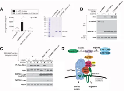
Because arginine specifically disrupts the purified CASTOR1-GATOR2 complex and modulates the interaction between the CASTOR1 ACT domains, we tested the possibility that arginine directly binds to CASTOR1. We used an equilibrium binding assay to assess whether immunopurified CASTORs from HEK-293T cells bind radiolabelled arginine. Indeed, tritiated arginine bound the CASTOR1 homodimer, but not the CASTOR2 homodimer or a control protein Rap2A, in a manner that was competed by excess nonradiolabelled arginine (Figure 4A and B). The CASTOR1-CASTOR2 heterodimer bound roughly half as much arginine as the CASTOR1 homodimer, reflecting the fact that within this complex only CASTOR1 can bind arginine (Figure 4B). Furthermore, neither radiolabelled leucine nor lysine bound to CASTOR1, consistent with the previously observed specificity for arginine for disrupting the CASTOR1-GATOR2 complex (Figure
4A).
It remained a formal possibility that arginine binds to an unidentified protein in the mammalian preparations of the CASTOR1 homodimer and CASTOR1-CASTOR2
Author Manuscript
Author Manuscript
Author Manuscript
heterodimer. To provide orthogonal evidence that CASTOR1 binds arginine, we purified the CASTOR complexes from E. coli, which do not encode a CASTOR homolog. The
CASTOR1 homodimer and heterodimer, but not Sestrin2, bound arginine to a comparable degree as the complexes prepared from human cells, demonstrating that arginine binds directly to CASTOR1 and not a co-purifying contaminating protein (Figure 4C).
A competition binding assay with increasing amounts of cold arginine revealed that the Kd
of arginine for CASTOR1 in the homodimer is 34.8 ± 5.9 μM, which is similar to its Kd in
the heterodimer of 24.2 ± 4.1 μM (Figure 4D and E). These affinities correlate well with the half maximal concentration of arginine that disrupts the interaction of GATOR2 with CASTOR1-containing complexes in vitro (Figure 3E) and activates mTORC1 in cells (Figure 4F and S4A). In combination, these data strongly support the notion that arginine binds directly to CASTOR1 to regulate its interaction with GATOR2.
CASTOR1 functions in parallel with SLC38A9 to regulate arginine sensing by mTORC1 Given the ability of arginine to bind to CASTOR1 and to modulate its interaction with GATOR2, we reasoned that CASTOR1 can affect the capacity of the mTORC1 pathway to respond to arginine. Indeed, transient overexpression of CASTOR1 driven by the strong CMV promoter inhibited mTORC1 activation by amino acids to a similar extent as expression of the dominant negative Rag GTPases mutants (Figure 5A).
Conversely, in HEK-293T and HEC59 cells, CASTOR1-depletion mediated by expression of either shRNAs or Cas9 with sgRNAs made the mTORC1 pathway substantially
insensitive to deprivation of arginine (Figure 5B and C, Figure S5B), but not to deprivation of leucine or all amino acids (Figure S5D and E). To determine if the RNAi-mediated effects are on target, we stably expressed an RNAi-resistant CASTOR1 cDNA in the CASTOR1 knockdown cells. To avoid inhibition of the mTORC1 pathway, we used the Sestrin2 promoter to express CASTOR1 at levels lower than those obtained with the CMV promoter. At this reduced level of expression, reintroduction of CASTOR1 into the
CASTOR1 knockdown cells restored the arginine responsiveness of the mTORC1 pathway, demonstrating that the RNAi effects are on target (Figure S5A). Despite the use of a weaker promoter, the level of recombinant CASTOR1 still greatly exceeded that of the endogenous protein (Figure S5A). However, this fact does not alter our conclusions because the overexpressed CASTOR1 restored, not inhibited, the arginine responsiveness of the mTORC1 pathway. Overall, these findings indicate that CASTOR1 is a negative regulator of the mTORC1 pathway.
Unlike CASTOR1, CASTOR2 constitutively associates with GATOR2 and does not bind arginine, and thus appears to be an arginine-insensitive version of CASTOR1. RNAi-mediated depletion of CASTOR2 slightly increased mTORC1 activity in the arginine-replete conditions (Figure 5D), and the knockdown of CASTOR2 together with that of CASTOR1 had a similar effect (Figure S5C). CASTOR2 levels are also partially reduced by expression of shCASTOR1_2 in HEK-293T cells, which may contribute to the slight increase in mTORC1 activation observed with this hairpin under arginine-replete conditions (Figure 5B). These results support the notion that CASTOR2, due to its inability to bind to
Author Manuscript
Author Manuscript
Author Manuscript
arginine, dampens mTORC1 activity when arginine is present. However, under arginine withdrawal, loss of CASTOR2 does not affect mTORC1 activity because CASTOR1 is still present to inhibit the mTORC1 pathway. Further corroborating an inhibitory role for CASTOR2, its transient overexpression abrogated mTORC1 activation by amino acids even more potently than overexpression of CASTOR1 (Figure 5A). Thus, both CASTOR1 and CASTOR2 are negative regulators of arginine signaling to mTORC1.
Consistent with the differential sensitivities of CASTOR1 and CASTOR2 to arginine, mild overexpression of each in HEK-293T cells had distinct effects on the response of the mTORC1 pathway to arginine. Overexpression of CASTOR2 blunted the maximal level of arginine-induced mTORC1 activity, while that of CASTOR1 reduced the sensitivity of the pathway to arginine but did not affect its maximal activity (Figure S5F). Like is the case for the Sestrins (Budanov et al., 2004; Chen et al., 2010; Lee et al., 2010; Ouyang et al., 2012), changes in CASTOR protein levels may be mediated by stress responsive transcription factors such as p53 and FOXO1. Consistent with this possibility, analysis of publically available ChIP-seq data indicates that FOXO1 binds upstream of and may regulate the expression of CASTOR2 in addition to SESN3 (Ouyang et al., 2012).
Finally, we probed the relationship between CASTOR1 and SLC38A9, a putative lysosomal arginine sensor that is required to signal the presence of arginine to mTORC1 (Wang et al., 2015). Consistent with the established role of SLC38A9, arginine-induced activation of mTORC1 signaling was severely blunted in HEK-293T cells lacking SLC38A9 (Figure
5E). RNAi-mediated depletion of CASTOR1 in SLC38A9-null cells renders the mTORC1
pathway insensitive to arginine: cells neither activated mTORC1 when arginine was present nor inactivated mTORC1 when arginine was withdrawn (Figure 5E). While other models are possible, it is likely that CASTOR1 and SLC38A9 function in parallel to enable arginine to regulate mTORC1, and in their absence, arginine signaling is almost fully defective. The binding of arginine to CASTOR1 is necessary for it to activate mTORC1
To test whether the activation of mTORC1 by arginine requires the arginine-binding capacity of CASTOR1, we used alanine scanning mutagenesis of the CASTOR1 ACT domains to identify CASTOR1 mutants that no longer bind arginine. These efforts led to the identification of I280A, a mutation within the second ACT domain that fully abrogated the ability of CASTOR1 to bind arginine in vitro (Figure 6A).
Consistent with the notion that the binding of arginine to CASTOR1 leads to the disruption of the CASTOR1-GATOR2 complex, the arginine-binding mutant of CASTOR1
constitutively interacted with GATOR2 in cells, irrespective of arginine levels (Figure 6B). Notably, this mutant bound more strongly to GATOR2 than its wild type counterpart, confirming that arginine modulates the CASTOR1-GATOR2 interaction. In addition, while the isolated ACT domains of CASTOR1 associated only in the presence of arginine, an I280A change in ACT2 fully abrogated this regulation, and provides further support for an important role for this residue in the binding of arginine to CASTOR1 (Figure S6A). Further reflecting the importance of this residue in CASTOR1 function, I280 is highly conserved in orthologs of CASTOR1 and is present in nearly all bacterial and fungal ACT
Author Manuscript
Author Manuscript
Author Manuscript
domains that share sequence homology with the CASTOR1 ACT domain (Figure 1C and
Figure S1B, C and D).
Finally, if CASTOR1 is a bona fide arginine sensor for the mTORC1 pathway, abolishing its ability to bind arginine should in turn abolish the ability of arginine to activate mTORC1 in cells. To test this hypothesis, we compared the arginine sensitivity of the mTORC1 pathway in CASTOR1 knockdown cells that stably expressed either wild type CASTOR1 or the arginine-binding mutant of CASTOR1. Unlike reintroduction of wild type CASTOR1, which restores the ability of arginine to signal to mTORC1, expression of the CASTOR1 arginine-binding mutant rendered the mTORC1 pathway inactive and insensitive to the presence of arginine (Figure 6C). In combination, these findings establish that arginine must bind to CASTOR1 in order for the mTORC1 pathway to respond to arginine.
DISCUSSION
We establish the CASTOR1 homodimer and CASTOR1-CASTOR2 heterodimer as arginine sensors for the mTORC1 pathway. First, arginine binds to both complexes at affinities that are consistent with those that activate mTORC1 in cells. Second, CASTOR1 loss leads to insensitivity of the mTORC1 pathway to arginine deprivation. Third, expression in cells of an arginine-binding mutant of CASTOR1 prevents the mTORC1 pathway from sensing the presence of arginine.
The identification of CASTOR1 and Sestrin1 and 2 as sensors for the mTORC1 pathway reveal that GATOR2 is a critical hub of amino acid sensing, where leucine and arginine signals converge upstream of the Rag GTPases to regulate mTORC1 activity (Figure 6D). Leucine and arginine have long been appreciated to be important for mTORC1 activation (Ban et al., 2004; Blommaart et al., 1995; Fox et al., 1998; Hara, 1998; Lynch et al., 2000), and our findings highlight differences in how these two amino acids are sensed. The cytosolic Sestrin proteins are likely the primary leucine sensors because their loss confers complete insensitivity of the mTORC1 pathway to leucine deprivation (Saxton et al., 2015; Wolfson et al., 2015). In contrast, arginine sensing appears to be more complex and may have inputs from two distinct cellular compartments. Loss of function experiments suggests that CASTOR1 signals the absence of arginine to inhibit mTORC1. Because CASTOR1 lacks transmembrane domains and signal sequences it is likely a soluble protein that senses free arginine in the cytosol. In contrast, SLC38A9 is needed to signal the presence of arginine, presumably lysosomal, to mTORC1. Together, both proteins appear to form parallel sensing branches that relay arginine availability to mTORC1. In the absence of both CASTOR1 and SLC38A9, arginine no longer regulates the activity of the mTORC1 pathway.
Despite these insights, several key questions remain. While CASTOR1 and Sestrin2 both bind to and likely inhibit GATOR2, whether they operate through distinct mechanisms can only be determined once the function of GATOR2 is elucidated. Furthermore, structural studies will provide insight into how the binding pocket of CASTOR1 achieves its
remarkable specificity for arginine. Importantly, these structural studies may also reveal why arginine binds to CASTOR1, but not CASTOR2. Although I280 is critical for arginine
Author Manuscript
Author Manuscript
Author Manuscript
binding to CASTOR1, this residue is also present in CASTOR2, suggesting that other unidentified residues must dictate the difference in arginine binding between these two proteins.
In addition, in vivo characterization of mice lacking the CASTOR genes will be needed to reveal how arginine sensing varies across tissues and during development. Because arginine differentially regulates each CASTOR complex, altering the expression of CASTOR1 versus CASTOR2 could serve as a means to modulate mTORC1 activity. CASTOR2 appears analogous to Sestrin3, as both are defective in amino acid binding and constitutively associate with GATOR2 to inhibit mTORC1 signaling. Thus, increased levels of CASTOR2 should blunt maximal mTORC1 signaling while those of CASTOR1 may alter the
sensitivity of the pathway to arginine, as we observed in HEK-293T cells (Figure S5F). It is likely that additional amino acid sensors exist to signal the presence of other critical amino acids for mTORC1 activity, such as glutamine (Jewell et al., 2015), as well sensors that mediate the amino acid sensitive events upstream of additional mTORC1 regulators, such as Folliculin/FNIP (Petit et al., 2013; Tsun et al., 2013). Characterizing the
evolutionary conservation of the amino acid sensors of the mTORC1/TORC1 pathway will provide insight into how varied are the amino acid inputs that drive mTORC1/TORC1 signaling in diverse organisms. For instance, budding yeast encodes a homolog of GATOR2, but not of the Sestrins or CASTORs, hinting at a divergence in the regulation of the
upstream components of the nutrient sensing pathway. This divergence may be expected given that yeast, unlike mammals, can synthesize all amino acids and thus must sense the quality and abundance of nitrogen sources rather than the identity and availability of particular amino acids. Further identification and characterization of the amino acid sensors upstream of the Rag GTPases will guide us towards a comprehensive understanding of how nutrients regulate the mTORC1 pathway.
EXPERIMENTAL PROCEDURES
MaterialsReagents were obtained from the following sources: HRP-labeled anti-mouse and anti-rabbit secondary antibodies, lamp and cathepsin antibodies from Santa Cruz Biotechnology; antibodies to phospho-T389 S6K1, S6K1, Sestrin2, Histone H3, VDAC and mios from Cell Signaling Technology; antibody to the HA epitope from Bethyl laboratories; antibody to raptor from Millipore; FLAG M2 antibody, FLAG M2 affinity gel, ATP, and amino acids from Sigma Aldrich; HA magnetic beads and RPMI without leucine, arginine, or lysine from Pierce; DMEM from SAFC Biosciences; XtremeGene9 and Complete Protease Cocktail from Roche; Inactivated Fetal Calf Serum (IFS) from Invitrogen; amino acid-free RPMI from US Biologicals; [3H]-labeled amino acids from American Radiolabeled Chemicals. The WDR24, Mios, CASTOR1, and CASTOR2 antibodies were generously provided by Jianxin Xie (Cell Signaling Technology).
Author Manuscript
Author Manuscript
Author Manuscript
Cell lines and tissue culture
All cell lines were cultured in Dulbecco’s modified Eagle’s medium (DMEM) with 10% inactivated fetal calf serum supplemented with 2 mM glutamine. All cell lines were maintained at 37°C and 5% CO2.
Preparation of cell lysates and immunoprecipitates
Cell lysate preparation, cell lysis, and immunoprecipitations were performed as described in Supplemental Experimental Procedures.
Where indicated, for transient cotransfection experiments, 2 million HEK-293T cells were plated in 10 cm dishes and transfected 24 hrs later using the polyethylenimine method (Boussif et al., 1995) with the indicated pRK5-based expression vectors: 300 ng HA-metap2, 40 ng CASTOR1-HA, 80 ng CASTOR1-FLAG, 40 ng CASTOR2-HA, 80 ng CASTOR2-FLAG; 100 ng WDR24-FLAG, 100 ng WDR59, 100 ng mios, 150 ng sec13, 150 ng seh1L, 10 ng or 40 ng or 100 ng or 600 ng of myc-Sestrin2 or myc-CASTOR2; 2 ng FLAG-S6K1, 15 ng or 60 ng HA-CASTOR2, 75 ng and 175 ng of CASTOR1-HA. The total amount of plasmid DNA in each transfection was normalized to 5 μg with empty pRK5. Thirty-six hours after transfection, cells were harvested as described above.
For experiments that required leucine, arginine or amino acid starvation or restimulation, cells were treated as previously described (Wolfson et al., 2015). Briefly, cells were incubated in leucine, arginine, or amino acid free RPMI for 50 minutes and then restimulated with the indicated amino acid(s) for 10 minutes.
Arginine binding assay
Four million HEK-293T cells were plated in a 15 cm plate four days prior to the experiment. Forty-eight hours after plating, the cells were transfected via the polyethylenimine method (Boussif et al., 1995) with the pRK5-based cDNA expression plasmids indicated in the figures in the following amounts: 15 μg FLAG-Rap2A, 400 ng CASTOR1-FLAG or CASTOR2-FLAG with 1200 ng CASTOR1-HA or 1200 ng CASTOR2-HA, 400 ng CASTOR1 I280A-FLAG with 1200 ng CASTOR1 I280A-HA. The total amount of plasmid DNA in each transfection was normalized to 15 μg with empty pRK5. Forty-eight hours after transfection cells were lysed and binding assays performed and analyzed as previously described (Wolfson et al., 2015).
In vitro CASTOR-GATOR2 dissociation assay
HEK-293T cells cotransfected with 40 ng HA and either 80 ng CASTOR1-FLAG (CASTOR1 homodimer) or 80 ng CASTOR2-CASTOR1-FLAG (CASTOR1-CASTOR2 heterodimer) were starved for all amino acids for 50 minutes, lysed and subjected to anti-HA immunoprecipitation as described previously. The CASTOR-GATOR2 complexes
immobilized on agarose beads were washed once in Triton wash buffer, three times in Triton wash buffer supplemented with 500 mM NaCl, and then incubated for 20 minutes in 1 mL of ice-cold Triton wash buffer supplemented with 500 mM NaCl and the indicated concentrations of individual amino acids. The amount of GATOR2 that remained bound to
Author Manuscript
Author Manuscript
Author Manuscript
CASTOR complexes was assayed by SDS-PAGE and immunoblotting as previously described.
Statistical analysis
Two-tailed t tests were used for comparison between two groups. All comparisons were two-sided, and P values of less than 0.001 were considered to indicate statistical significance.
Supplementary Material
Refer to Web version on PubMed Central for supplementary material.
ACKNOWLEDGEMENTS
We thank all members of the Sabatini lab for helpful suggestions, in particular Monther Abu-Remaileh and Walter W. Chen for experimental insight and advice. We are grateful to Jianxin Xie of Cell Signaling Technologies for generously providing many antibodies, in particular the CASTOR1 and CASTOR2 anti-sera. This work has been supported by grants from the US NIH (R01CA103866 and AI47389) the Department of Defense
(W81XWH-07-0448) to D.M.S. Fellowship support was provided by the John Reed UROP Fund to S.M.S.; the National Defense Science & Engineering Graduate Fellowship (NDSEG) to G.A.W.; and by the NIH to L.C. (F31 CA180271); to T.W. (F31 CA189437); U41 HG006673 to S.P.G and J.W.H., and GM095567 to J.W.H. K.S. is a Pfizer Fellow of the Life Sciences Research Foundation. D.M.S is an investigator of the Howard Hughes Medical Institute.
REFERENCES
Aravind L, Koonin EV. Gleaning non-trivial structural, functional and evolutionary information about proteins by iterative database searches. Journal of Molecular Biology. 1999; 287:1023–1040. [PubMed: 10222208]
Ban H, Shigemitsu K, Yamatsuji T, Haisa M, Nakajo T, Takaoka M, Nobuhisa T, Gunduz M, Tanaka N, Naomoto Y. Arginine and Leucine regulate p70 S6 kinase and 4E-BP1 in intestinal epithelial cells. International journal of molecular medicine. 2004; 13:537–543. [PubMed: 15010853] Bar-Peled L, Chantranupong L, Cherniack AD, Chen WW, Ottina KA, Grabiner BC, Spear ED, Carter
SL, Meyerson M, Sabatini DM. A Tumor Suppressor Complex with GAP Activity for the Rag GTPases That Signal Amino Acid Sufficiency to mTORC1. Science. 2013; 340:1100–1106. [PubMed: 23723238]
Bar-Peled L, Schweitzer LD, Zoncu R, Sabatini DM. Ragulator Is a GEF for the Rag GTPases that Signal Amino Acid Levels to mTORC1. Cell. 2012; 150:1196–1208. [PubMed: 22980980] Blommaart EFC, Luiken JJFP, Blommaart PJE, van Woerkom GM, Meijer AJ. Phosphorylation of
Ribosomal Protein S6 Is Inhibitory for Autophagy in Isolated Rat Hepatocytes. Journal of Biological Chemistry. 1995; 270:2320–2326. [PubMed: 7836465]
Boussif O, Lezoualc'h F, Zanta MA, Mergny MD, Scherman D, Demeneix B, Behr JP. A versatile vector for gene and oligonucleotide transfer into cells in culture and in vivo:
polyethylenimine. Proceedings of the National Academy of Sciences of the United States of America. 1995; 92:7297–7301. [PubMed: 7638184]
Budanov AV, Sablina AA, Feinstein E, Koonin EV, Chumakov PM. Regeneration of peroxiredoxins by p53-regulated sestrins, homologs of bacterial AhpD. Science. 2004; 304:596–600. [PubMed: 15105503]
Buerger C, DeVries B, Stambolic V. Localization of Rheb to the endomembrane is critical for its signaling function. Biochemical and Biophysical Research Communications. 2006; 344:869–880. [PubMed: 16631613]
Carluccio C, Fraternali F, Salvatore F, Fornili A, Zagari A. Structural Features of the Regulatory ACT Domain of Phenylalanine Hydroxylase. PLOS ONE. 2013; 8:e79482. [PubMed: 24244510]
Author Manuscript
Author Manuscript
Author Manuscript
Chantranupong L, Wolfson RL, Orozco JM, Saxton RA, Scaria SM, Bar-Peled L, Spooner E, Isasa M, Gygi SP, Sabatini DM. The Sestrins Interact with GATOR2 to Negatively Regulate the Amino-Acid-Sensing Pathway Upstream of mTORC1. Cell Reports. 2014; 9:1–8. [PubMed: 25263562] Chen C-C, Jeon S-M, Bhaskar PT, Nogueira V, Sundararajan D, Tonic I, Park Y, Hay N. FoxOs
inhibit mTORC1 and activate Akt by inducing the expression of Sestrin3 and Rictor. Developmental Cell. 2010; 18:592–604. [PubMed: 20412774]
Chipman D. The ACT domain family. Current opinion in structural biology. 2001; 11:694–700. [PubMed: 11751050]
Cross PJ, Allison TM, Dobson RCJ, Jameson GB, Parker EJ. Engineering allosteric control to an unregulated enzyme by transfer of a regulatory domain. Proceedings of the National Academy of Sciences of the United States of America. 2013; 110:2111–2116. [PubMed: 23345433]
Cross PJ, Dobson RCJ, Patchett ML, Parker EJ. Tyrosine latching of a regulatory gate affords allosteric control of aromatic amino acid biosynthesis. The Journal of biological chemistry. 2011; 286:10216–10224. [PubMed: 21282100]
Dibble CC, Elis W, Menon S, Qin W, Klekota J, Asara JM, Finan PM, Kwiatkowski DJ, Murphy LO, Manning BD. TBC1D7 is a third subunit of the TSC1-TSC2 complex upstream of mTORC1. Molecular cell. 2012; 47:535–546. [PubMed: 22795129]
Dibble CC, Manning BD. Signal integration by mTORC1 coordinates nutrient input with biosynthetic output. Nature cell biology. 2013; 15:555–564. [PubMed: 23728461]
Dumas R, Cobessi D, Robin AY, Ferrer J-L, Curien G. The many faces of aspartate kinases. Archives of biochemistry and biophysics. 2012; 519:186–193. [PubMed: 22079167]
Efeyan A, Zoncu R, Sabatini DM. ScienceDirect.com - Trends in Molecular Medicine - Amino acids and mTORC1: from lysosomes to disease. Trends in Molecular Medicine. 2012
Floyd JC Jr, Fajans SS, Conn JW, Knopf RF, Rull J. Stimulation of insulin secretion by amino acids. Journal of Clinical Investigation. 1966; 45:1487. [PubMed: 5919350]
Fox HL, Pham PT, Kimball SR, Jefferson LS, Lynch CJ. Amino acid effects on translational repressor 4E-BP1 are mediated primarily by l-leucine in isolated adipocytes. American journal of
physiology Cell physiology. 1998; 275:C1232–C1238.
Grant GA. The ACT Domain: A Small Molecule Binding Domain and Its Role as a Common Regulatory Element. Journal of Biological Chemistry. 2006; 281:33825–33829. [PubMed: 16987805]
Hara K. Amino Acid Sufficiency and mTOR Regulate p70 S6 Kinase and eIF-4E BP1 through a Common Effector Mechanism. Journal of Biological Chemistry. 1998; 273:14484–14494. [PubMed: 9603962]
Hirose E, Nakashima N, Sekiguchi T, Nishimoto T. RagA is a functional homologue of S. cerevisiae Gtr1p involved in the Ran/Gsp1-GTPase pathway. Journal of cell science. 1998; 111(Pt 1):11–21. [PubMed: 9394008]
Huttlin EL, Ting L, Bruckner RJ, Gebreab F, Gygi MP, Szpyt J, Tam S, Zarraga G, Colby G, Baltier K, et al. The BioPlex Network: A Systematic Exploration of the Human Interactome. Cell. 2015; 162:425–440. [PubMed: 26186194]
Jewell JL, Kim YC, Russell RC, Yu F-X, Park HW, Plouffe SW, Tagliabracci VS, Guan K-L. Metabolism. Differential regulation of mTORC1 by leucine and glutamine. Science. 2015; 347:194–198. [PubMed: 25567907]
Jung J, Genau HM, Behrends C. Amino Acid-Dependent mTORC1 Regulation by the Lysosomal Membrane Protein SLC38A9. Molecular and cellular biology. 2015; 35:2479–2494. [PubMed: 25963655]
Kim JS, Ro S-H, Kim M, Park H-W, Semple IA, Park H, Cho U-S, Wang W, Guan K-L, Karin M, et al. Sestrin2 inhibits mTORC1 through modulation of GATOR complexes. Scientific reports. 2015; 5:9502. [PubMed: 25819761]
Kobe B, Jennings IG, House CM, Michell BJ, Goodwill KE, Santarsiero BD, Stevens RC, Cotton RGH, Kemp BE. Structural basis of autoregulation of phenylalanine hydroxylase. Nature Structural & Molecular Biology. 1999; 6:442–448.
Author Manuscript
Author Manuscript
Author Manuscript
Lang EJ, Cross PJ, Mittelstädt G, Jameson GB, Parker EJ. Allosteric ACTion: the varied ACT domains regulating enzymes of amino-acid metabolism. Current opinion in structural biology. 2014; 29:102–111. [PubMed: 25543886]
Lee JH, Budanov AV, Park EJ, Birse R, Kim TE, Perkins GA, Ocorr K, Ellisman MH, Bodmer R, Bier E, et al. Sestrin as a feedback inhibitor of TOR that prevents age-related pathologies. Science. 2010; 327:1223–1228. [PubMed: 20203043]
Lynch CJ, Fox HL, Vary TC, Jefferson LS, Kimball SR. Regulation of amino acid–sensitive TOR signaling by leucine analogues in adipocytes. Journal of cellular biochemistry. 2000; 77:234–251. [PubMed: 10723090]
Menon S, Dibble CC, Talbott G, Hoxhaj G, Valvezan AJ, Takahashi H, Cantley LC, Manning BD. Spatial Control of the TSC Complex Integrates Insulin and Nutrient Regulation of mTORC1 at the Lysosome. Cell. 2014; 156:771–785. [PubMed: 24529379]
Ouyang W, Liao W, Luo CT, Yin N, Huse M, Kim MV, Peng M, Chan P, Ma Q, Mo Y, et al. Novel Foxo1-dependent transcriptional programs control T(reg) cell function. Nature. 2012; 491:554– 559. [PubMed: 23135404]
Parmigiani A, Nourbakhsh A, Ding B, Wang W, Kim YC, Akopiants K, Guan K-L, Karin M, Budanov AV. Sestrins Inhibit mTORC1 Kinase Activation through the GATOR Complex. Cell Reports. 2014; 9:1281–1291. [PubMed: 25457612]
Petit CS, Roczniak-Ferguson A, Ferguson SM. Recruitment of folliculin to lysosomes supports the amino acid–dependent activation of Rag GTPases. The Journal of Cell Biology. 2013; 202:1107– 1122. [PubMed: 24081491]
Rebsamen M, Pochini L, Stasyk T, de Araújo MEG, Galluccio M, Kandasamy RK, Snijder B, Fauster A, Rudashevskaya EL, Bruckner M, et al. SLC38A9 is a component of the lysosomal amino acid sensing machinery that controls mTORC1. Nature. 2015
Rhoads JM, Niu X, Odle J, Graves LM. Role of mTOR signaling in intestinal cell migration. American Journal of Physiology - Gastrointestinal and Liver Physiology. 2006; 291:G510–G517. [PubMed: 16710051]
Saito K, Araki Y, Kontani K, Nishina H, Katada T. Novel role of the small GTPase Rheb: its
implication in endocytic pathway independent of the activation of mammalian target of rapamycin. Journal of Biochemistry. 2005; 137:423–430. [PubMed: 15809346]
Sancak Y, Bar-Peled L, Zoncu R, Markhard AL, Nada S, Sabatini DM. Ragulator-Rag complex targets mTORC1 to the lysosomal surface and is necessary for its activation by amino acids. Cell. 2010; 141:290–303. [PubMed: 20381137]
Sancak Y, Peterson TR, Shaul YD, Lindquist RA, Thoreen CC, Bar-Peled L, Sabatini DM. The Rag GTPases bind raptor and mediate amino acid signaling to mTORC1. Science (New York, NY). 2008; 320:1496–1501.
Saxton RA, Knockenhauer KE, Wolfson RL, Chantranupong L, Pacold ME, Wang T, Schwartz TU, Sabatini DM. Structural basis for leucine sensing by the Sestrin2-mTORC1 pathway. Science. 2015:aad2087.
Schürmann A, Brauers A, Maßmann S, Becker W, Joost H-G. Cloning of a Novel Family of Mammalian GTP-binding Proteins (RagA, RagBs, RagB1) with Remote Similarity to the Ras-related GTPases. The Journal of biological chemistry. 1995; 270:28982–28988. [PubMed: 7499430]
Sekiguchi T, Hirose E, Nakashima N, Ii M, Nishimoto T. Novel G proteins, Rag C and Rag D, interact with GTP-binding proteins, Rag A and Rag B. The Journal of biological chemistry. 2001; 276:7246–7257. [PubMed: 11073942]
Siltberg-Liberles J, Martinez A. Searching distant homologs of the regulatory ACT domain in phenylalanine hydroxylase. Amino Acids. 2009; 36:235–249. [PubMed: 18368466]
Tan K, Li H, Zhang R, Gu M, Clancy ST, Joachimiak A. Structures of open (R) and close (T) states of prephenate dehydratase (PDT)—Implication of allosteric regulation by l-phenylalanine. Journal of Structural Biology. 2008; 162:94–107. [PubMed: 18171624]
Tsun Z-Y, Bar-Peled L, Chantranupong L, Zoncu R, Wang T, Kim C, Spooner E, Sabatini DM. The Folliculin Tumor Suppressor Is a GAP for the RagC/D GTPases That Signal Amino Acid Levels to mTORC1. Molecular cell. 2013; 52:495–505. [PubMed: 24095279]
Author Manuscript
Author Manuscript
Author Manuscript
Wang S, Tsun Z-Y, Wolfson RL, Shen K, Wyant GA, Plovanich ME, Yuan ED, Jones TD, Chantranupong L, Comb W, et al. Metabolism. Lysosomal amino acid transporter SLC38A9 signals arginine sufficiency to mTORC1. Science. 2015; 347:188–194. [PubMed: 25567906] Wolfson RL, Chantranupong L, Saxton RA, Shen K, Scaria SM, Cantor JR, Sabatini DM. Sestrin2 is a
leucine sensor for the mTORC1 pathway. Science. 2015:aab2674.
Wu G, Morris SM. Arginine metabolism: nitric oxide and beyond. Biochemical Journal. 1998; 336(Pt 1):1–17. [PubMed: 9806879]
Yao K, Yin Y-L, Chu W, Liu Z, Deng D, Li T, Huang R, Zhang J, Tan B, Wang W, et al. Dietary Arginine Supplementation Increases mTOR Signaling Activity in Skeletal Muscle of Neonatal Pigs. The Journal of nutrition. 2008; 138:867–872. [PubMed: 18424593]
Zoncu R, Bar-Peled L, Efeyan A, Wang S, Sancak Y, Sabatini DM. mTORC1 Senses Lysosomal Amino Acids Through an Inside-Out Mechanism That Requires the Vacuolar H+-ATPase. Science Signaling. 2011; 334:678–683.
Author Manuscript
Author Manuscript
Author Manuscript
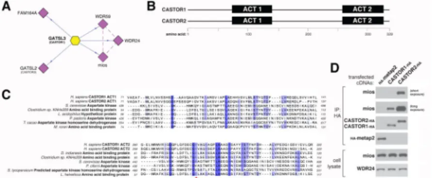
Figure 1. CASTOR1 and CASTOR2 are ACT domain-containing proteins that interact with GATOR2
(A) Endogenous GATOR2, FAM164A, and CASTOR2 co-immunoprecipitate with stably expressed CASTOR1. The schematic is adapted from the BioPlex database (Huttlin et al., 2015). Solid blue lines denote proteins that were detected by mass spectrometric analysis of CASTOR1 immunoprecipitates, and dashed purple lines indicate interactions between GATOR2 subunits that were present in Bioplex.
(B) Schematic alignment of human CASTOR1 and CASTOR2 proteins with annotated ACT domains.
(C) The ACT domains of CASTOR1 and CASTOR2 display sequence similarity with the ACT domains of fungal aspartate kinases and putative amino acid binding proteins in bacteria. Amino acid positions are colored from white to blue in order of increasing sequence identity. The red star denotes the position of the I280 residue in CASTOR1. (D) Recombinant CASTOR1 and CASTOR2 co-immunoprecipitate endogenous GATOR2, as detected by the presence of mios. Anti-HA immunoprecipitates and lysates were prepared from HEK-293T cells cotransfected with the indicated cDNAs in expression vectors. Cell lysates and immunoprecipitates were analyzed by immunoblotting for levels of indicated proteins. HA-metap2 served as a negative control.
Author Manuscript
Author Manuscript
Author Manuscript
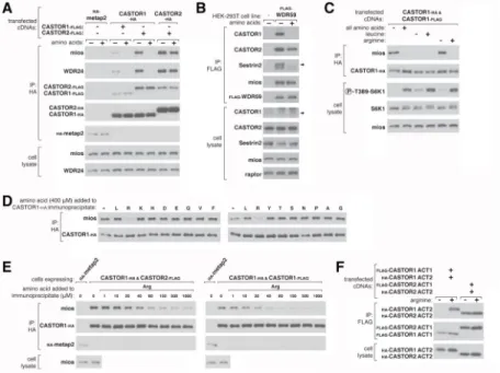
Figure 2. CASTOR1 and CASTOR2 form homo- and heterodimeric complexes
(A) Recombinant CASTOR1 and CASTOR2 coimmunoprecipitate both themselves and each other. HEK-293T cells were cotransfected with the indicated cDNAs in expression vectors and cell lysates and anti-HA immunoprecipitates were analyzed by immunoblotting for the indicated proteins as in Figure 1D.
(B) Recombinant CASTOR2 coimmunoprecipitates endogenous CASTOR1. HEK-293T cells were cotransfected with the indicated cDNAs in expression vectors and anti-HA immunoprecipitates were collected and analyzed as in Figure 1D. The arrow denotes the band corresponding to CASTOR1.
(C) Recombinant CASTOR1 coimmunoprecipitates endogenous CASTOR2. HEK-293T cells were cotransfected with the indicated cDNAs in expression vectors and anti-HA immunoprecipitates were collected and analyzed as in (A).
(D) Recombinant CASTOR1 and CASTOR2 are present in approximately equal ratios within the heterodimeric complex. SDS-polyacrylamide gel electrophoresis (PAGE), followed by Coomassie blue staining, was used to analyze the indicated protein preparations from HEK-293T cells. The asterisk denotes a common protein contaminant present in these purifications.
Author Manuscript
Author Manuscript
Author Manuscript
Figure 3. Arginine regulates the interaction of GATOR2 with CASTOR1-homodimers and CASTOR1-CASTOR2 heterodimers in cells and in vitro
(A) Amino acids differentially regulate the interaction of GATOR2 with the three CASTOR complexes. HEK-293T cells cotransfected with the indicated cDNAs were deprived of amino acids for 50 min or starved and restimulated with amino acids for 10 min. Anti-HA immunoprecipitates and cell lysates were analyzed by immunoblotting for levels of the indicated proteins.
(B) Endogenous CASTOR1, but not CASTOR2, associates with GATOR2 in an amino acid-sensitive manner. A HEK-293T cell line expressing endogenously FLAG-tagged WDR59 was treated as in (A) and anti-FLAG immunoprecipitates were analyzed by immunoblotting for the indicated proteins.
(C) Deprivation of arginine, but not leucine, promotes the interaction between the
CASTOR1 homodimer and endogenous GATOR2. Cells were deprived of leucine, arginine, or all amino acids for 50 min, and restimulated for 10 min with the respective amino acids where indicated. Anti-HA immunoprecipitates were prepared and analyzed as in (A). (D) Arginine disrupts the interaction between GATOR2 and CASTOR1-containing dimers in vitro. Anti-HA immunoprecipitates were prepared from HEK-293T cells expressing the indicated cDNAs and deprived of amino acids for 50 min. Indicated amino acids were added directly to the immunoprecipitates, which after re-washing, were analyzed as in (A).
(E) Arginine dose-dependently disrupts the interaction between GATOR2 and CASTOR1-containing dimers in vitro. The experiment was performed and analyzed as in (D), except using the indicated concentrations of arginine.
(F) Arginine regulates the interaction between the ACT domains of CASTOR1 but not CASTOR2 in cells. HEK-293T cells cotransfected with the indicated cDNAs in expression vectors were either deprived of arginine in the cell media for 50 min or starved and restimulated with arginine for 10 min. Anti-HA immunoprecipitates were prepared and analyzed as in (A).
Author Manuscript
Author Manuscript
Author Manuscript
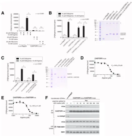
Figure 4. The CASTOR1 homodimer and CASTOR1-CASTOR2 heterodimer bind arginine with a Kd of around 30 μM
(A) Radiolabelled arginine, but not radiolabelled leucine or lysine, binds to CASTOR1 homodimers. FLAG-immunoprecipitates were prepared from HEK-293T cells cotransfected with the indicated cDNAs, and binding assays were performed with these
immunoprecipitates as described in the methods. Unlabelled amino acids were added where indicated. Values are mean ± SD of three technical replicates from one representative experiment (n.s., not significant).
(B) Arginine binds to CASTOR1-containing homo- and heterodimers, but not the CASTOR2 homodimer. FLAG immunoprecipitates of the indicated complexes were prepared from HEK-293T cells and analyzed as in (A). Equal volumes of eluants from immunoprecipitates of the denoted complexes were loaded and analyzed in SDS-PAGE, followed by Coomassie blue staining.
(C) Arginine binds to bacterially produced CASTOR1-containing complexes, but not the CASTOR2 homodimer or the control protein Sestrin2. Proteins purified from bacteria were analyzed as in (A) and (B).
(D) Arginine binds to the CASTOR1 homodimer with a dissociation constant of 34.8 μM. Binding assays were performed as in (A) with the indicated concentrations of unlabelled arginine. A representative experiment is shown, and each point represents the mean ± SD for three experiments. The Kd was calculated from four experiments.
(E) Arginine binds to the CASTOR1-CASTOR2 heterodimer with a dissociation constant of 24.2 μM. FLAG-immunoprecipitates were prepared from HEK-293T cells and analyzed as in (D).
Author Manuscript
Author Manuscript
Author Manuscript
(F) The concentration of arginine that half-maximally activates the mTORC1 pathway correlates with the concentration of arginine that disrupts half of the complexes of GATOR2 and CASTOR1 homodimers. HEK-293T cells were transfected with the indicated cDNAs and immunoprecipitates and lysates analyzed as in Figure 3C.
Author Manuscript
Author Manuscript
Author Manuscript
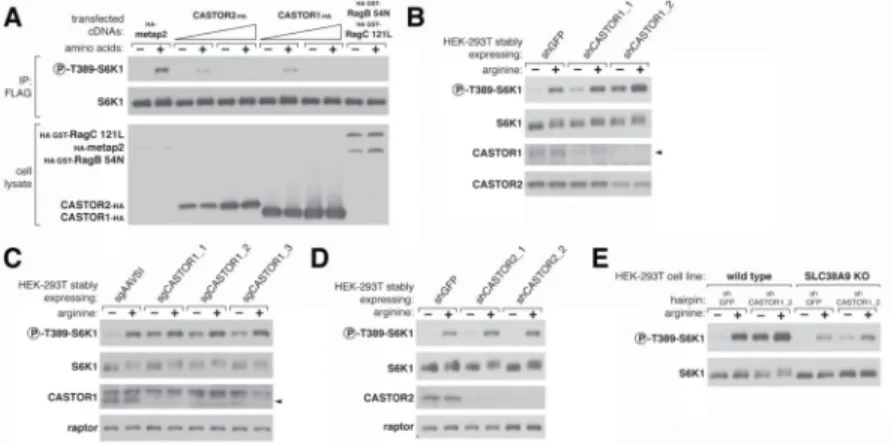
Figure 5. CASTOR1 functions in parallel with SLC38A9 to regulate arginine signaling to mTORC1
(A) Transient overexpression of recombinant CASTOR2 and CASTOR1 inhibits mTORC1 activation in response to amino acids. HEK-293T cells were cotransfected with the indicated cDNAs. Cells were treated as in Figure 3A and anti-FLAG immunoprecipitates analyzed by immunoblotting for the indicated proteins.
(B) RNAi-mediated depletion of CASTOR1 in HEK-293T cells renders the mTORC1 pathway partially insensitive to arginine deprivation. HEK-293T cells stably expressing the indicated shRNAs were starved of arginine in the cell media for 50 min or starved and restimulated with arginine for 10 min. Lysates were analyzed via immunoblotting for the indicated proteins and phosphorylation states.
(C) CRISPR/Cas9 mediated depletion of CASTOR1 in HEK-293T cells confers resistance of the mTORC1 pathway to arginine deprivation. HEK-293T cells stably coexpressing Cas9 with the indicated guide RNAs were treated as in (B) and lysates were analyzed by
immunoblotting for indicated proteins.
(D) Loss of CASTOR2 slightly increases mTORC1 activity in response to arginine. HEK-293T cells stably expressing the indicated shRNAs were treated as in (B) and lysates were analyzed by immunoblotting for indicated proteins. The normalized phosphorylated S6K1 signal under arginine stimulation for shCASTOR2_1 and shCASTOR2_2 expressing cells is 1.4 fold and 1.1 fold of shGFP expressing cells, respectively, as quantified with ImageJ.
(E) CASTOR1 and SLC38A9 likely function in parallel to signal arginine availability to the mTORC1 pathway. Wild type or SLC38A9 knockout HEK-293T cells expressing the indicated shRNAs were treated as in (B) and lysates were analyzed by immunoblotting for indicated proteins.
Author Manuscript
Author Manuscript
Author Manuscript
Figure 6. Arginine must be able to bind to CASTOR1 for it to activate mTORC1
(A) The CASTOR1 I280A mutant does not bind arginine. Binding assays were performed with FLAG immunoprecipitates of the indicated complexes as in Figure 4A.
(B) Arginine does not regulate the interaction of CASTOR1 I280A with GATOR2.
HEK-293T cells cotransfected with the indicated cDNAs in expression vectors were treated as in Figure 5B and anti-HA immunoprecipitates were analyzed by immunoblotting for levels of the indicated proteins.
(C) Reintroduction of the CASTOR1 I280A mutant into CASTOR1 knockdown cells renders the mTORC1 pathway unable to sense the presence of arginine. HEK-293T cells stably expressing the indicated shRNAs and cDNA constructs were treated as in Figure 5B and lysates analyzed by immunoblotting for indicated proteins.
(D) A model depicting how the cytosolic and lysosomal amino acid inputs impinge on CASTORs, Sestrins, and SLC38A9 to regulate mTORC1 activity.