HAL Id: hal-01663881
https://hal-amu.archives-ouvertes.fr/hal-01663881
Submitted on 14 Dec 2017HAL is a multi-disciplinary open access archive for the deposit and dissemination of sci-entific research documents, whether they are pub-lished or not. The documents may come from teaching and research institutions in France or abroad, or from public or private research centers.
L’archive ouverte pluridisciplinaire HAL, est destinée au dépôt et à la diffusion de documents scientifiques de niveau recherche, publiés ou non, émanant des établissements d’enseignement et de recherche français ou étrangers, des laboratoires publics ou privés.
Regional methylation of the 5 end CpG island of
BRCA1 is associated with reduced gene expression in
human somatic cells
Frédérique Magdinier, Lise-Marie Billard, Gaelle Wittmann, Lucien Frappart,
Mehdi Benchai, Gilbert Lenoir, Jean Francois Guerin, Robert Dante
To cite this version:
Frédérique Magdinier, Lise-Marie Billard, Gaelle Wittmann, Lucien Frappart, Mehdi Benchai, et al.. Regional methylation of the 5 end CpG island of BRCA1 is associated with reduced gene expression in human somatic cells. FASEB Journal, Federation of American Society of Experimental Biology, 2000, 14 (11), pp.1585-1594. �10.1096/fj.99-0817com�. �hal-01663881�
Regional methylation of the 5
ⴕ end CpG island of
BRCA1 is associated with reduced gene expression in
human somatic cells
FRE´ DE´RIQUE MAGDINIER, LISE-MARIE BILLARD, GAE¨LLE WITTMANN,
LUCIEN FRAPPART, MEHDI BENCHAI¨B,* GILBERT M. LENOIR, JEAN FRANC¸ OIS GUE´ RIN,* AND ROBERT DANTE1
Laboratoire de Ge´ne´tique, UMR 5641 CNRS, UCBL1, and *Laboratoire de Biologie de la Reproduction et du De´veloppement, UCBL1, 69373 Lyon cedex 08, France
ABSTRACT In mammalians, demethylation of spe-cific promoter regions often correlates with gene activation; inversely, dense methylation of CpG is-lands leads to gene silencing, probably mediated by methyl-CpG binding proteins. In cell lines and can-cers, inhibition of tissue-specific genes and tumor suppressor genes expression seems to be related to such hypermethylation. The 5ⴕ end of the breast cancer predisposition gene BRCA1 is embedded in a large CpG island of ⬃2.7 kb in length. In human sporadic breast cancers, the down-regulation of
BRCA1 does not seem to be related to BRCA1 gene
alterations. Southern blot analysis and the bisulfite sequencing method indicate that the BRCA1 CpG island is regionally methylated in all human tissues analyzed and unmethylated in the gametes, suggest-ing a role for DNA methylation in the control of gene expression. We have therefore investigated the po-tential role of methyl-CpG binding proteins in the regulation of BRCA1 gene expression. In vitro, partial methylation of constructs containing this region strongly inhibits gene expression in the presence of MeCP2 protein. Moreover, in the five human cell lines analyzed, chemically induced hypomethylation is associated with BRCA1 gene activation. These data suggest that methyl-CpG binding proteins might be associated with the control of BRCA1 gene expres-sion and that methyl-DNA binding proteins may participate in the regulation of gene expression in mammalian cells.—Magdinier, F., Billard, L.-M., Wittmann, G., Frappart, L., Benchaı¨b, M., Lenoir, G. M., Gue´rin, J. F., Dante, R. Regional methylation of the 5ⴕ end CpG island of BRCA1 is associated with reduced gene expression in human somatic cells.
FASEB J. 14, 1585–1594 (2000)
Key Words: DNA methylation䡠 oocytes 䡠 spermatozoa 䡠 breast cancer
Germ-line alterations of the BRCA1 gene confer a lifetime risk of 40% for ovarian cancers and
40 – 80% for breast cancers (1). It is likely that BRCA1 acts as a tumor suppressor gene (2). Indeed, in breast cancers linked to BRCA1, as expected for a tumor suppressor gene, allelic deletions at this locus invariably involve the wild-type allele (3, 4). BRCA1 involvement in breast cancers does not seem to be restricted to familial cancers. Despite the absence of somatic mutation in the breast tissues, a down-regulation of BRCA1 expression is associated with malignancy in human sporadic breast cancers (4 – 6). Although its biological function is still unknown, BRCA1 may have an important role in cellular differ-entiation and proliferation. In transgenic mice, ho-mozygous disruption of the Brca1 gene results in embryonic lethality (7). The progressive changes in Brca1 expression during mouse embryogenesis (8) also imply a role for Brca1 in the differentiation process. In addition, variations of Brca1 expression are observed during postnatal mammary gland de-velopment (9, 10). More recently, it had been shown that Cre-mediated invalidation of this gene affects final differentiation of the gland during gestation (11). In humans, the up-regulation of BRCA1 gene expression observed during the first stages of prena-tal development of the mammary gland also suggests a role for BRCA1 in the differentiation of the mam-mary gland (12). Taken together, these data indicate that variations of BRCA1 expression may have some physiological consequences in human breast tissue.
Analysis of the genomic region containing this gene indicates that an another gene, NBR2, lies head to head with the BRCA1 gene (13). Site-directed deletion mutagenesis experiments led to the identi-fication of a bidirectional promoter region, position ⫺258 to ⫹43 (14), and a minimal positive regulatory region has been mapped at position ⫺198 to ⫺162 (15). Structural studies have shown that a minor part
1Correspondence: Laboratoire de Ge´ne´tique, UMR 5641
CNRS, Domaine Rockefeller, UCBL1, 8 avenue Rockefeller, 69373 Lyon cedex 08, France. E-mail: dante@cismsun.univ-lyon1.fr
1585 0892-6638/00/0014-1585/$02.25 © FASEB
Vol.14, No.11 , pp:1585-1594, December, 2017
The FASEB Journal
.
139.124.156.117
to IP
www.fasebj.org
of breast cancers (⬃10%) exhibits hypermethylated sites in the region containing the putative BRCA1 promoter (16 –18). However, a down-regulation of BRCA1 expression has been observed in most spo-radic breast cancers (4 – 6), indicating that abnormal methylation of this 5⬘ end region of BRCA1 does not account for the decrease in BRCA1 expression in most cases of sporadic breast cancers. In addition, in a small series (37 cases) of sporadic breast cancers, the down-regulation of BRCA1 was not correlated with DNA hypermethylation in the vicinity of the promoter region (5).
Although transient transfection assays indicate that essential regulatory sites are not present in the upstream region (position⫺1528 to ⫺202; ref 15), it had been shown for other promoters that methyl-ation of the surrounding sequences may repress gene expression (19). The efficiency of the inhibi-tion seems to be dependent on CpG density and promoter strength (19).
We have therefore determined the methylation status of the 5⬘ end of BRCA1 in a variety of cell lines and tissues, including fetal and cancer breast tissues. This analysis led to an unexpected finding, since the 5⬘ end of BRCA1, which is embedded in a large CpG island, appears to be regionally methylated in all somatic tissues analyzed, suggesting that this region may participate in the regulation of BRCA1 gene expression.
Differences in DNA methylation are associated with differentiation and carcinogenesis, CpG meth-ylation correlating with the silencing of many genes (20, 21). Two types of mechanism could be involved in silencing genes by DNA methylation. CpG meth-ylation can down-regulate gene expression by pre-venting the binding of transcription factors to their recognition sequences or through repressor mole-cules that bind to methylated DNA (22, 23). The methyl-CpG binding proteins (MBD), a family of vertebrate proteins, bind to methylated DNA in any sequence context (22); for some members of this family it has been shown that the binding of such proteins represses gene expression at a distance (24).
Among the DNA binding proteins potentially in-volved in the negative regulation of gene expression, the methyl-CpG binding protein MeCP2 seems to play an important role (25, 26). MeCP2 is a chromo-somal protein that binds specifically to methylated CpG and represses densely methylated genes in association with a histone deacetylase complex (27, 28). Although inhibition of histone deacetylase by trichostatin A can relieve the transcriptional repres-sion mediated by MBD (27, 28), for some genes it has been shown that dense CpG island methylation might be a dominant factor in gene silencing (29).
We investigated, therefore, whether methyl-CpG
binding proteins such as MeCP2 might contribute to the control of BRCA1 expression.
MATERIALS AND METHODS
Preparation of tissues
Human tissues were snap frozen immediately after removal and stored in liquid nitrogen until use. Spermatozoa were purified from semen samples by Percoll gradient centrifuga-tion (Pharmacia, Uppsala, Sweden). One milliliter of semen was loaded at the top of a 50 –90% Percoll gradient and centrifuged at 700 g for 15 min. After centrifugation, each fraction was collected by aspiration. The spermatozoa were washed in phosphate buffer 1⫻ to remove the Percoll and resuspended in an appropriate volume of phosphate-buffered saline (PBS) 1⫻. The purity of the preparation was controlled by microscopy.
Human oocytes that had failed to fertilize 3 days after in
vitro insemination were collected from the In Vitro
Fertiliza-tion Laboratory (Hospital E. Herriot, Lyon, France). To remove the follicular cells linked to the zonae pellucida, oocytes were treated by enzymatic digestion with hyaluroni-dase (150 units, type VIII hyaluronihyaluroni-dase, Sigma, France) to discard contaminating somatic cells. Then a digestion by trypsin was performed to remove the zonae pellucida and the remaining somatic cells. Oocytes were rinsed several times in PBS 1⫻ and stored in liquid nitrogen until use. The cumulus cells were collected after hyaluronidase digestion for further studies.
Cell culture
Human breast cell lines (MCF7, BT20, and HBL 100), cervix cell line (HeLa), or kidney cell line (Bosc 23) were obtained from ATCC (Rockville, Md.) and grown in Dulbecco’s mod-ified Eagle’s medium (Sigma, L’isle d’Abeau, France) supple-mented with FCS 5% and 0.5g/ml insulin for MCF7 and BT 20 cell lines. All cells were grown at 37°C in a humidified 5% CO2atmosphere.
PCR-based methylation assay
DNA extracted from tissue samples, cell lines, and cells treated with 5azadC were digested with a fivefold excess of
RsaI or RsaI plus HpaII or CfoI and incubated overnight at
37°C in the appropriate buffer (Roche Diagnostics, Meylan, France). Control experiments were performed using the methyl-insensitive enzyme MspI. Enzymes were inactivated by heating at 65°C for 1 h and an aliquot of the reaction was used for polymerase chain reaction (PCR) amplification. The PCR amplification was performed in the following conditions: 10 mM Tris-HCl (pH 8.3), 3 mM MgCl2, 50 mM KCl, 0.1 mg/ml
gelatin, 4% DMSO, 100M of each of the four deoxyribo-nucleoside triphosphates, 0.25M of the primers (forward: 5⬘ TTG GGA GGG GGC TCG GGC AT 3⬘; reverse: 5⬘ CAG AGC TGG CAG CGG ACG GT), and 0.6 units of Taq DNA polymerase (Roche Diagnostics) after 35 cycles in an Eppen-dorf thermocycler (1 min denaturation at 94°C, 2 min annealing at 55°C, and 3 min extension at 72°C). In each experiment, the sample digested with RsaI, which does not cleave the sequence between the two primers, was amplified to verify the efficiency of the amplification. The sequence analyzed contains 9 HpaII sites and 10 CfoI sites and PCR amplification occurs only when the sites are methylated and uncut.
5-aza-2ⴕ-deoxycytidine treatments
For 5-aza-2⬘-deoxycytidine (5azadC, Sigma, France) treat-ments, cell lines were seeded at low density (3– 4⫻105cells/
100 mm dish) 16 h before treatment with a final concentra-tion of 1M 5azadC. The medium was changed 24 h after drug addition and every subsequent day. RNA and DNA were isolated after 72 h. DNAs were extracted and quantitated (Hoechst staining method) from treated cells and control cells. Different aliquots were digested by RsaI (in order to normalize the size of the DNA molecules) plus: HpaII, CfoI (methylation-sensitive enzymes), MspI (methylation-insensi-tive), and no enzyme. The 5⬘ end of the BRCA1 gene was then amplified by PCR from these samples and the signals ob-tained were quantitated by densitometry. No signal was observed after MspI digestion. The extent of demethylation was evaluated by the ratio between the signal obtained after digestion by HpaII, CfoI, or both and the signal was obtained from DNAs digested with RsaI alone. The data indicated that⬎ 90% of the DNA molecules contained unmethylated
CfoI and HpaII sites after 5azadC treatment. Exon 1, which
was found unmethylated in normal and tumoral tissues, was amplified as a control experiment of complete digestion with methylation-sensitive enzymes.
Transfections
MeCP2-HA (kindly provided by Dr. A. Bird), pCMV-gal plasmids, or pGL3-control plasmid (Promega, Lyon, France) were transfected using the calcium phosphate pre-cipitation technique (30). Cells were collected 48 h after transfection.
For the immunofluorescence assay, Bosc 23 cells were grown on Lab-tek Permanox (Nunc) 2-well chambered cov-erslips at 1⫻ 105cells per well and transfected as described
previously. After 48 h, the cells were washed twice in PBS 1⫻, fixed in paraformaldehyde 4% for 15 min at room tempera-ture, and rinsed several times with PBS 1⫻. Then cells were permeabilized in 0.1 M glycine-PBS 1⫻ buffer, followed by incubation in 0.5% Triton X-100-PBS 1⫻ buffer, and blocked in 0.2% gelatin-PBS 1⫻ buffer for 15 min at room tempera-ture. The anti-tag HA monoclonal antibody (12 CA5, Roche Diagnostics) diluted 1:40 in 0.5% Triton X-100-PBS 1⫻ buffer was incubated with the cells for 1 h. The cells were rinsed several times with PBS 1⫻ and incubated for 1 h with a 1:200 dilution of the FITC-conjugated secondary antibody (goat anti-mouse IgG, Dako, France). Fluorescence was visualized with a 25⫻ or 40⫻ immersion lens on a Leica microscope. In these experiments, MeCP2 expression was consistently ob-served in more than 50% of the cells and -galactosidase activity (30) was observed in more than 60% of the cells; five adjacent fields, at magnification 40⫻, were counted.
pGL3 constructs, methylation, and luciferase assay
A 1757 bp fragment of the BRCA1 gene containing a part of the 5⬘ CpG island and 43 bp of exon 1a was isolated from genomic DNA by PCR using primers containing KpnI site at the 5⬘ end and BglII site at the 3⬘ end (primer forward-KpnI: 5⬘ CTG GTA CCT TGG GAG GGG GCT CGG GCA A3⬘; primer reverse-BglII: 5⬘ GAA GAT CTT CCA GGA AGT CTC AGC GAG C 3⬘). The PCR products were ligated between
KpnI and BglII sites into the pGL3 basic vector (Promega) and
transformed into competent JM-109 Escherichia coli cells (Pro-mega).
In vitro methylation was performed by incubating the
p5⬘-BRCA1-Luc vector with one unit of either HpaII (CCGG sites), HhaI (GCGC sites), or SssI (CG sites) methylases per
microgram of plasmid DNA in the conditions recommended by the manufacturer (Biolabs, Beverly, Mass.); completeness of the modification was checked with the corresponding restriction enzyme. p5⬘-BRCA1-Luc constructs were trans-fected into Bosc 23 cells as described previously. Transfec-tions were optimized for 12-well plates. The cells were lysed and assayed for luciferase expression (Luciferase assay, Pro-mega) after 48 h and the light emissions were measured in a scintillation counter (Packard, Downers Grove, Ill.). A plas-mid (pGL3-control; Promega) containing a promoter and an enhancer derived from SV40 was also transfected in the same plates; values obtained for this control vector were compara-ble between each experiments.
DNA extraction
High molecular weight DNA was extracted from frozen pulverized tissue samples and cells by standard procedures (30). Briefly, samples were resuspended in 10 mM Tris-0.1M EDTA buffer and digested with proteinase K (300g/ml final concentration) in the presence of SDS. A similar method with the addition of 0.001% (V/V)-2 mercaptoethanol was used to prepare decondensed DNA from spermatozoa. When DNA was extracted from a small number of cells (6 to 10 oocytes), 2 g of pGEM-T plasmid (Promega) was added as carrier. The mixture was incubated at 37°C, phenol/chloroform extracted, and DNA was ethanol-precipitated.
RNA extraction
RNA was isolated in a single step procedure by acid-gua-nidium-thyocyanate-phenol-chloroform extraction as de-scribed previously (31). After extraction, total RNA was precipitated in isopropanol and resuspended in an appropri-ate volume of sterile wappropri-ater. The integrity and quantity of RNA were examined by gel electrophoresis. Total RNA was quan-tified by densitometry on a 1.2% agarose gel containing 0.1 g/ml ethidium bromide in comparison with serial dilutions of known amount of standard RNA (Roche Diagnostics).
Quantitative reverse transcription PCR (RT-PCR)
The RT-PCR assay was performed as described previously (32) by coamplification of 0.3 g of total RNA and a known amount of competitor RNA. Primers used were designed to amplify the cDNA fragment from exon 6 to 8 of the BRCA1 gene (C3F: 5⬘ TGT GCT TTT CAG CTT GAC ACA GG 3⬘ and C3R: 5⬘ CGT CTT TTG AGG TTG TAT CCG CTG 3⬘). Reactions were performed in 100 l containing 10 mM Tris-HCl (pH 8.3), 3 mM MgCl2, 50 mM KCl, 0.1 mg/ml
gelatin, 200M of each of the 4 deoxyribonucleoside triphos-phates, and 0.25M of the C3 primers.
After initial denaturation at 92°C for 2 min, six units of Expand Reverse Transcriptase (Roche Diagnostics) were added to the reaction mixture and incubated for 35 min at 42[de]C. Reverse transcriptase was then inactivated by heat-ing and after coolheat-ing to 0°C, PCR amplification was accom-plished by adding 0.6 units of Taq DNA polymerase (Roche Diagnostics) after 35 cycles in an Eppendorf thermocycler (1 min denaturation at 94°C, 2 min annealing at 55°C, and 3 min extension at 72°C). Aliquots were analyzed on a 2% agarose gel containing 0.1g/ml ethidium bromide and the intensity of the bands corresponding to the wild-type BRCA1 PCR products and to the competitor PCR products was determined using Image Analyzer Software (Wayne Rasband, National Institutes of Health).
1587 METHYL-CPG BINDING PROTEIN REPRESSES BRCA1
Vol.14, No.11 , pp:1585-1594, December, 2017
The FASEB Journal
.
139.124.156.117
to IP
www.fasebj.org
Southern blot analysis
Methylation patterns were determined by Southern blot experiments using methylation-sensitive restriction endonu-clease CfoI (GCGC site) and HpaII (CCGG site) or methyla-tion-insensitive isoschizomer MspI (CCGG site). In a typical experiment, 30 g of total DNA was cleaved with a 10-fold excess of TaqI or PstI for 10 to 12 h in the appropriate conditions (Roche Diagnostics). Two-thirds of the sample was then digested overnight by CfoI or HpaII or MspI. Restriction endonuclease products were separated by electrophoresis on a 1.2% agarose gel and transferred to Hybond N⫹ Nylon membrane (Amersham, France). After drying, the mem-branes were hybridized overnight at 65°C to a randomly primed32P-labeled probe (Random Primed DNA labeling kit,
Roche Diagnostics) in the hybridization solution (0.25 M sodium phosphate, 7% SDS, 1% BSA, 50g/ml yeast tRNA). Membranes were washed with increasing stringency (from 2 to 0.1⫻ SSC; 0.1% SDS) at 65°C and exposed to Hyperfilm (Amersham, France) for 1 to 7 days at ⫺70°C. Membranes could be used for rehybridization after two successive treat-ments with SDS 1% at 80°C for 30 min.
Gene probes
The probe used for Southern blot analysis of the CpG island of the BRCA1 gene was synthesized by PCR amplification of a 239 bp region (⫺1244 to ⫺1005; ref 33). The PCR fragments were cloned into a pGEM-T vector (Promega). After digestion with the appropriate enzyme, the insert was purified by agarose gel electrophoresis, followed by electroelution. Each filter was rehybridized with the exon 1 probe encompassing exon 1a of the BRCA1 gene and the bidirectional promoter (5) in order to verify that DNA cleavage with restriction endonucleases was complete.
Bisulfite modification
The sodium bisulfite modification method, followed by the sequencing of PCR products, is used to determine the CpG methylation pattern. Sodium bisulfite converts unmethylated cytosines to uracils whereas the methylated cytosines remain unmodified. In the resultant modified DNA, uracils are replicated as thymines during PCR amplification. The sodium bisulfite reaction was carried out on 4g of DNA (3 g of carrier DNA and 1 g of human genomic DNA). Alkali-denatured DNA was incubated in 3 M NaHSO3 and 5 mM
hydroquinone for 16 h at 50°C. Modified DNA was purified using the Wizard DNA Clean-up System (Promega) and eluted into 50l of sterile water. Modification was completed by 0.3 M NaOH; DNA was precipitated by 0.5 M ammonium acetate (pH 4.6) and resuspended in water.
DNA was amplified using strand-specific primers designed to amplify a 258 bp region in the CpG island of the BRCA1 gene in two separate reaction mixtures. The first round of PCR amplification was accomplished in 100 l in a buffer containing 10 mM Tris-HCl (pH 8.3), 3 mM MgCl2, 50 mM
KCl, 0.1 mg/ml gelatin, 100M of each of the four deoxyri-bonucleoside triphosphates, 0.25 M of the primers (for-ward: 5⬘ TTT TGT TTT GTG TAG GGC GGT T 3⬘; Reverse: 5⬘ CCT TAA CGT CCA TTC TAA CCG T 3⬘), and 0.6 units of
Taq DNA polymerase (Roche Diagnostics) after 35 cycles in
an Eppendorf thermocycler (1 min denaturation at 94°C, 2 min annealing at 55°C, and 3 min extension at 72°C). An aliquot of the first amplification was reamplified with internal primers (forward: 5⬘ TGA GAA TTT AAG TGG GGT GT 3⬘; reverse: 5⬘ AAC CCT TCA ACC CAC CAC TAC 3⬘) in the same conditions.
PCR products were first analyzed by digestion with restric-tion enzymes DdeI, EcoRI (Roche Diagnostics), and HphI (Biolabs) in the buffers recommended by the manufacturers. Then PCR products were cloned in a pGEM-T vector (Promega) and 10 random clones were analyzed by automatic sequencing (Eurogentec, Belgium) to determine the propor-tion of methylated (CpG) or unmethylated (TpG) sites.
RESULTS
The 5ⴕ CpG island of BRCA1 is regionally methylated in somatic tissues
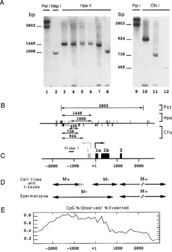
Significant variations, up to fivefold in cell lines (32) and 30-fold in sporadic breast cancers (5), in the amount of BRCA1 mRNA are not related to the methylation status of the putative promoter se-quence located at the 5⬘ end of the exon 1a (5). However, this unmethylated region (nt ⫺167 to nt ⫹484) is included in a large CpG island, spanning nucleotides⫺2200 to ⫹500 (33 and Fig. 1E), which suggests that hypermethylation of this CpG island could be associated with gene silencing. Analysis of the methylation status of the⫺1714 to ⫺1023 region using a PCR-based methylation assay indicated that this CpG island was methylated at CfoI and HpaII sites in human somatic tissues (including normal and tumoral somatic tissues and fetal tissues, 65 samples analyzed) and cell lines (data not shown). Since this assay is qualitative rather than quantitative, methylation patterns were further determined by Southern blot experiments (representative samples are shown in Fig. 1A).
DNAs were first digested with the methylation-insensitive restriction endonuclease PstI. Using probe 1 (position ⫺1244 to ⫺1005, Fig. 1C), PstI generates two bands of 3.8 kb for the BRCA1 gene and 5 kb for the BRCA1 pseudogene (Fig. 1A, lanes 1 and 9). When DNA extracted from the HBL 100 cell line is digested with the methylation-insensitive enzyme MspI, this probe reveals a 1008 bp band corresponding to the BRCA1 gene (Fig. 1A, lane 2). A faint 202 bp band corresponding to the BRCA1 pseudogene was detected at the bottom of the gel. DNAs extracted from normal tissues (Fig. 1A) were digested with the methyl-sensitive enzyme HpaII that cleaves CCGG sites when the internal cytosine at CpG dinucleotide is unmethylated. Probe 1 maps a 1449 bp band (Fig. 1A, lanes 4 –7) corresponding to the methylation of 9 CCGG sites located within the 5⬘ region of the BRCA1 gene (Fig. 1B and 1C). In spermatozoa, the CCGG sites analyzed using probe 1 are unmethylated (Fig; 1A, lane 8). DNA from the HBL 100 cell line and fetal breast tissue (Fig. 1A, lanes 10 and 11, respectively) were cleaved with the methylation-sensitive enzyme CfoI in order to map several other CpG sites. Probe 1 reveals a 924 bp band in the HBL 100 cell line and reveals a 736 bp
band in tissues, indicating that the proximal CfoI sites are unmethylated in normal tissues. In sperma-tozoa, CpG dinucleotides located within GCGC sites are unmethylated, giving rise to a 455 bp band after hybridization with probe 1.
Taken together, these data indicate that DNAs extracted from somatic tissues and cell lines were methylated at CCGG and GCGC sites within the region spanning nucleotides ⫺2000 to ⫺1000 or ⫺720 for the HBL 100 cell line, with the exception of human spermatozoa DNA, which was unmethylated at these sites (Fig. 1A).
Analysis of the methylation pattern of the body of BRCA1 gene was carried out, as described previously (5), on several fetal and adult human tissues, cell lines, and male germ cells by Southern blot after digestion by the methylation-sensitive enzymes HpaII and CfoI (Fig. 1D). The filters were successively hybridized with several probes corresponding to the different exons present in the 3⬘ region of the BRCA1 gene. The body of the gene (from the end of exon 1b to the end of exon 24) is methylated in somatic tissues (5) and mature male germ cells, as expected for non-CpG island regions. In somatic cells, the promoter region of the gene is unmethyl-ated, as described previously (5). In cell lines and tissues, the 5⬘ region is methylated at CCGG and GCGC sites in the various samples. On the other hand, this region is unmethylated in spermatozoa as expected for a CpG island.
The 5ⴕ CpG island of BRCA1 is unmethylated in human gametes
The gamete-specific pattern was further confirmed by scaling-down the bisulfite sequencing method in order to investigate CpG methylation from very small amounts of DNA, and experimental conditions were
the transcription start of both genes. The position of the first exons of the BRCA1 gene is indicated by black boxes (exons 1a, 1b, 2). The first exon of the NBR2 gene is indicated by a gray box. The scale indicates the position of the exons starting from the initiation of transcription of the BRCA1 gene (exon 1a). The black line represents the position of the 239 bp probe (probe 1). D) Methylation pattern of the BRCA1 gene in human somatic and germ cells. Data obtained from Southern blot experiments and ref 5 are summarized. The methylation status of BRCA1 DNA segments (symbolized by the arrows) is indicated: M⫹: methylated region; M⫺: un-methylated region. The gray arrow corresponds to the HBL 100 pattern. E) Distribution of the CpG sites in the 5⬘ region of the BRCA1 gene. A graph of the percentage of CpG observed divided by the expected frequency. Dinucleotide frequencies were calculated in 1000 nucleotide windows moved at 100 nucleotide intervals. The ratio (CpG-% ob-served/CpG-% expected) indicates that the 5⬘ region of the
BRCA1 gene is embedded within a CpG island beginning
upstream from the transcription start site and encompassing the first exons of the gene.
Figure 1. DNA methylation pattern of the 5⬘ region of the
BRCA1 gene in human cells and tissues. A) DNA was extracted
from the HBL 100 cell line (lanes 1–3; 9 –10); adult breast tissue (lane 4), fetal breast tissue (lane 5; 11), normal ovary (lane 6), normal kidney (lane 7), and spermatozoa purified from semen sample on Percoll gradient (lane 8; 12). South-ern blot experiments were performed as described in Mate-rials and Methods. After PstI digestion (lanes 1 and 9), probe 1 reveals two bands, the 3.8 kb band corresponding to the
BRCA1 gene and the 5 kb band to the BRCA1 pseudogene.
When cleaved with MspI (lane 2), these two bands produce a 1008 bp band for the BRCA1 gene and a 202 bp band for the pseudogene. HpaII digestion (lanes 4 –7) generates a 1449 bp band, indicating that the 9 CCGG sites at the 5⬘ end of the region analyzed are methylated. In spermatozoa (lane 8),
HpaII patterns are identical to MspI patterns, indicating that
these sites are unmethylated in these samples. The methyl-ation status of GCGC sites was investigated using CfoI diges-tion (lanes 10 –12) and probe 1. The HBL 100 cell line (lane 10) exhibits a 924 bp band and in somatic tissues (one representative sample is shown on lane 11) a 735 bp band, indicating that the proximal CfoI sites are unmethylated in normal tissues (see Fig. 1B). In spermatozoa, the CpG dinucleotides located within the GCGC sites are unmethyl-ated, giving rise to a 455 bp band. B) Position of enzymatic sites in the 5⬘ region of the BRCA1 gene. HpaII sites are indicated by upward strokes, CfoI sites by downward strokes. Arrows indicate the portion of DNA recognized by the probe and the size of the bands as revealed by autoradiography is indicated for the different couples of enzymes. C) Schematic representation of the promoter region of the BRCA1 gene. The BRCA1 gene spans 81 kb of genomic DNA and is located head to head with the NBR2 gene. The two genes are separated by a bidirectional promoter. The arrows point to
1589 METHYL-CPG BINDING PROTEIN REPRESSES BRCA1
Vol.14, No.11 , pp:1585-1594, December, 2017
The FASEB Journal
.
139.124.156.117
to IP
www.fasebj.org
chosen for obtaining a quantitative assay of the ratio methylated CpG vs. unmethylated CpG.
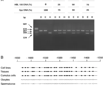
DNA extracted from purified human spermatozoa was modified by sodium bisulfite (34). This chemical treatment converts unmethylated cytosines to uracils while methylated cytosines remain unmodified. After modification, DNA was amplified by a two step PCR method, as described in Materials and Methods. The PCR product was digested by specific restriction endonucleases to determine the global methylation status of the sample. Completeness of the modifica-tion was monitored by digesmodifica-tion with DdeI, which cleaves only unconverted DNA. PCR products ob-tained from methylated molecules exhibit a new EcoRI site at position 138, whereas unmethylated molecules exhibit a new HphI site at position 165.
The sensitivity of PCR amplification after bisulfite modification was monitored by mixing different proportions of unmethylated DNA from spermato-zoa (from 25 to 100%) and methylated DNA from HBL 100 (from 0 to 75%). For each assay, an aliquot of the PCR product was cleaved with DdeI (unmodi-fied DNA), EcoRI (methylated DNA), or HphI (un-methylated DNA), loaded on a 2% agarose gel, and visualized by ethidium bromide staining. The results indicate that the amount of PCR product cleaved by enzymatic digestion is directly related to the percent-age of methylated or unmethylated DNA used in the coamplification assay (Fig. 2A). DNAs from somatic tissues and gametes were therefore modified using this method and PCR products were cloned and sequenced.
Within the region analyzed, ⫺1643 to ⫺1358, all the 24 CpG sites were unmethylated in DNA from human oocytes and spermatozoa (Fig. 2B). As ex-pected from the Southern blot experiments, all these CpG sites were methylated in all somatic tissues and cell lines, including the somatic cells of the corona radiata surrounding the oocytes (Fig. 2B). The ab-sence of DNA methylation within the CpG island in human gametes did not extend to the body of the BRCA1 gene, since control experiments indicated that two regions of the exon 11 are methylated both in somatic tissues and gametes (data not shown), suggesting that the methylation of the CpG island might play regulatory role in BRCA1 expression. Chemically induced hypomethylation elevates
BRCA1 expression
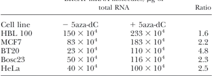
To test the potential role of DNA methylation in the control of BRCA1 expression, demethylation was induced by 5-aza-2⬘-deoxycytidine (5aza-dC) treat-ments. Human cell lines (three breast cell lines: BT20, MCF7, and HBL 100; one cervix cell line, HeLa; and one kidney cell line, Bosc 23) were grown for 72 h in the presence of 1 M of 5aza-dC. The
PCR-based methylation assay indicated that the 5⬘ end of BRCA1 was efficiently demethylated at the end of the treatment. Then the amounts of BRCA1 mRNA were determined using a competitive RT-PCR method (32).
5aza-dC treatment invariably elevated BRCA1 ex-pression in the cell lines analyzed by up to fivefold in BT20 cells and from 1.5 to 2.5 in the other cell lines (Table 1), indicating that CpG methylation is associ-ated with a low level of BRCA1 expression.
Figure 2.Methylation status of the 5⬘ region of the BRCA1
gene in human somatic tissues and germ cells. A) Sensitivity of PCR amplification after bisulfite modification. After bisulfite modification, a DNA segment of the BRCA1 CpG island (position⫺1643 to ⫺1358) was amplified (see Materials and Methods) using a two step PCR method. Ten l of PCR products obtained from DNA of spermatozoa (unmethylated) mixed with the appropriate amount (from 0 to 75%) of methylated DNA (HBL 100 cells) was digested with DdeI (lanes D), which cuts unmodified DNA; EcoRI (lanes E), which cuts at position 138 the PCR products obtained from methylated DNA; and HphI (lanes H), which cuts at position 165 the PCR products obtained from unmethylated DNA. The intensity of the corresponding bands was measured by densitometry . B) Enzymatic digestion of PCR products after bisulfite modification gives an estimation of the methylation status for only a few CpG sites (at enzymatic sites). Therefore, the methylation profile of a 258 bp region (position⫺1643 to ⫺1358 from the initiation of transcription) of the CpG island of BRCA1 containing 24 CpG sites was analyzed at the nucleotide level after cloning and sequencing the PCR prod-ucts obtained from various human tissues, cell lines, and male and female germ cells. Somatic cells of the corona radiata separated from the oocyte by enzymatic digestions were also analyzed as a control of the purity of the oocyte samples. Bisulfite sequences obtained from individual cloned frag-ments are represented as horizontal lines, with individual CpGs represented by white circles (when at least 80% of CpGs are unmethylated) or black circles (when at least 80% of the CpGs are methylated). Ten clones were analyzed for each sample. All CpG sites are methylated in somatic tissues (cumulus cells included) and human cell lines whereas the 5⬘ region of the BRCA1 gene is unmethylated in both male and female human germ cells.
In vitro methylation induces a MeCP2-dependent
repression of the BRCA1 promoter activity
The methylated domain of the BRCA1 CpG island is relatively far (⬃0.7 to 1 kb, Fig. 1C, E) from the start site of the transcription. MeCP2 is able to drive a long-range repression and might down-regulated BRCA1 expression. This hypothesis was investigated using an expression vector (p5⬘-BRCA1-Luc) contain-ing the⫺1714 to ⫹43 region of BRCA1 fused to the luciferase enzyme as a reporter gene.
Analysis of the 5⬘ end BRCA1 sequence indicates that the CCGG sequences (HpaII sites) are located outside the minimal promoter region required for full promoter activity and only one GCGC sequence (HhaI sites) is located at the 3⬘ end of this region, deletion of this latter sequence inducing only a weak inhibition of the promoter activity (15). Therefore, we take this opportunity to investigate whether the methylation of surrounding sequences might affect BRCA1 promoter activity. p5⬘-BRCA1-Luc vector was in vitro methylated using HpaII methylase or HhaI methylase and transfected in Bosc 23 cells in the presence or absence of a vector (MeCP2-Tag-HA) encoding for MeCP2. In the absence of the MeCP2-Tag-HA vector, in vitro methylation induced only a two- to threefold drop in luciferase activity; mean-while, in the presence of MeCP2, the in vitro meth-ylated vectors exhibited an almost total loss of tran-scriptional activity (Fig. 3). Control experiments using expression vectors containing the -galactosi-dase gene driven by a CMV-promoter (pCMV--gal plasmid) and a pGL3-control plasmid containing a SV40 promoter fused to the luciferase gene indi-cated that the expression of these reporters genes was not or was only minimally affected by the over-expression of the pCMV-MeCP2-HA-vector.
Therefore, cotransfection experiments in the Bosc 23 cell line indicated that partial methylation of the vector containing a part of the 5⬘ end of BRCA1, which mimics the methylation pattern observed in somatic cells, induced a MeCP2-dependent repres-sion of the transcriptional activity of this vector (Fig. 3).
DISCUSSION
In mammalian genomes, the CpG dinucleotides are underrepresented (⬃5 to 10% of their predicted fre-quency) and a high proportion (60 to 80%) is methyl-ated. However, short regions exhibit a ratio of CpG observed vs. CpG expected of at least 0.6 and a G⫹C content greater than 50%. These regions, called CpG islands, are associated with all the housekeeping genes and⬃40% of tissue-restricted genes. Most frequently, the CpG islands are located at the 5⬘ end of these genes and contain the promoter and the first exon of the associated gene (for a review, see ref 22). Although these sequences are usually unmethylated, hypermeth-ylation of CpG islands associated with gene silencing has been described for crucial regulator of growth during cancer progression (20). In addition, abnormal methylation at multiple CpG-rich sequences and gene silencing are also characteristic of human and murine cell lines (35).
In normal somatic tissues, methylated CpG islands have been found for X chromosome-inactivated genes and for imprinted genes, suggesting that the methylation of a subset of CpG islands also occurs during normal physiological process. In line with this observation, it has been shown in colon tissue that DNA methylation at CpG islands takes place during the aging process (36).
In this study we have found that the CpG island of BRCA1 is regionally methylated in all somatic human cells and tissues analyzed, suggesting that this epige-netic modification might have some consequences on BRCA1 gene expression. Since some promoters are not or are only minimally inhibited by in vitro methylation, we have investigated the methylation sensitivity of the p-5⬘-BRCA1-Luc expression vector
Figure 3. Effect of in vitro methylation on the promoter activity of BRCA1 in the presence or absence of MECP2. The 5⬘ end (nt ⫺1714 to nt ⫹43) containing the promoter region of BRCA1 was cloned upstream from the luciferase reporter gene. The constructs were methylated in vitro by M.HpaII or M.HhaI and transfected in Bosc 23 cells in the presence (ratio, 1/1) or absence of the pCMV-MeCP2-HA vector. The inhibitory effect (mean value of 4 independent experi-ments⫾ standard deviation) was determined from the ratio between the value obtained with the unmodified vector and the value obtained for the sample analyzed. PRR, positive regulatory region (15); pTR5, long terminal repeated ele-ment.
TABLE 1. Effect of 5aza-dC on BRCA1 expression
BRCA1 mRNA molecules/g of
total RNA Ratio
Cell line ⫺ 5aza-dC ⫹ 5aza-dC
HBL 100 150⫻ 104 233⫻ 104 1.6 MCF7 83⫻ 104 183⫻ 104 2.2 BT20 23⫻ 104 110⫻ 104 4.8 Bosc23 50⫻ 104 116⫻ 104 2.3 HeLa 40⫻ 104 100⫻ 104 2.5 1591 METHYL-CPG BINDING PROTEIN REPRESSES BRCA1
Vol.14, No.11 , pp:1585-1594, December, 2017
The FASEB Journal
.
139.124.156.117
to IP
www.fasebj.org
(containing the 5⬘ end of BRCA1). In vitro methyl-ation using M-HpaII generates a methylated site at position⫺167 that is not methylated in vivo, whereas the other nine sites are methylated in the cell lines and tissues analyzed.
Several papers have described a bidirectional pro-moter in this region. For example, Thakur and Croce (15) have localized the promoter region at position ⫺195 to ⫺162 and a minimal positive regulatory region at position ⫺195 to ⫺177. This short region seems to be involved in the binding of the nuclear proteins. More recently, Suen and Gross (37) also mapped the bidirectional BRCA1 promoter in this region at position⫺204 to ⫺149, and band-shift assays indicated that within this promoter the ⫺167 to ⫺149 region was the target of nuclear proteins. These data might suggest that the nucleo-tide ‘C’ at position ⫺167 is not involved in the binding of potential transcription factors.
In addition, the methylation of this site (⫺167) does not seem to be crucial for the expression of p-5⬘-BRCA1-Luc vector since the methylation of the HhaI sites (the more proximal sites are at positions ⫺564 and ⫺20) has a more pronounced inhibitory effect. Furthermore, the methylation of all CpGs induces (using SssI methylase) a very strong inhibi-tion of expression of the p-5⬘-BRCA1-luc vector, even in the absence of pCMV-MeCP2-HA vector, suggest-ing that the density of methylated sites is the main point of the inhibitory effect.
Although MeCP2 can bind DNA segments contain-ing only one methylated CpG, it had been shown that the inhibitory effect of MeCP2 is strongly depen-dent on the density of methyl-CpGs (38). Using p-5⬘-BRCA1-Luc vector cotransfected with a MeCP2 expression vector, in our experiments the inhibition seems dependent on the number of methylated CpGs, since the inhibition observed with HhaI meth-ylase (16 sites) is stronger than the inhibition ob-tained after methylation with HpaII methylase (10 sites). These data also suggest that the inhibition of the expression of the p-5⬘-BRCA1-Luc vector by DNA methylation is not the result of the methylation of a specific site, but seems to be dependent on the methylation density.
The regional methylation of the 5⬘ end BRCA1 CpG island is clearly not the consequence of a pathological process, since this pattern was observed in all normal somatic tissue samples analyzed. In contrast to the imprinted genes, this region is un-methylated in human spermatozoa and in oocytes. Furthermore, in fetal and adult tissues differential methylation between alleles was not detected. The presence, at the 5⬘ end of BRCA1, of the complete long terminal repeat element pTR5, spanning nucle-otides ⫺3123 to ⫺1273 (33), which is partially in-cluded in the BRCA1-CpG island (nt ⫺2200 to
⫹500), might account for the partial methylation of this region. In addition, the 5⬘ end of BRCA1 exhibits a complex organization since the transcription start site of BRCA1 is separated from that of the NBR2 gene by 218 bp and, therefore, its 5⬘ end region in included in the intron 1 of NBR2 gene (13). In line with this hypothesis, the promoter region of the mouse Brca1 gene, which lacks this repetitive ele-ment and complex organization (39), appears to be unmethylated in mouse somatic tissues and in the NIH 3T3 cell line (unpublished data), suggesting that insertion of the LTR element in somatic cells might have some consequences on the methylation patterns of the human BRCA1 gene.
This methylation pattern of the BRCA1 CpG island confers potential regulatory features. Our results show that BRCA1 expression is regulated at least in part by methyl-CpG binding proteins. Among this family, MeCP2 might be a good candidate since transient expression of the corresponding gene led to a methylation-dependent inhibition of BRCA1 expression. Despite the low level of MeCP2 expres-sion (unpublished data) in the cell lines analyzed, inhibition of DNA methylation resulted in an ele-vated level of BRCA1 mRNA. This chemically in-duced hypomethylation by 5aza-dC might suggest that other members of the methyl-DNA binding proteins family are also involved in this regulation.
Transient transfection of fusion proteins have identified a MeCP2 domain that is capable of long-range repression of vectors (19, 24). This protein, which binds to methylated CpG, forms a complex with histone deacetylase and the transcription re-pressor Sin3A, leading to formation of transcription-ally repressive chromatin architecture (27, 28). MBD2 and MBD3, two other methyl-DNA binding proteins, have recently been characterized and shown to participate in other histone deacetylase complexes and in gene silencing mechanisms (40, 41). The expression patterns of these methyl-DNA binding protein genes are not fully determined, but it has been shown in the rat that the level of MeCP2 expression is dependent on the tissues (42), MBD3 is associated with metastatic-associated protein 1, a protein that is overexpressed in several human can-cers (43), and MBD2 belongs to the MeCP1 deacety-lase complex, which is also present at various levels depending on the cell type and the differentiation state (40).
Although, genes possessing a 5⬘ end methylated CpG island should represent only a very small part of the mammalian genomes, differences in the amount of methyl-DNA binding proteins between tissues or cells could represent an additional mechanism for tissue-specific gene expression.
We thank Dr Adrian BIRD for the pCMV-MeCP2-HA vector. F.M. is the recipient of a fellowship from the Ligue
Nationale contre le Cancer. The present work was supported by the Ligue Nationale contre le Cancer, Comite´ de Saoˆne et Loire and the Association pour la Recherche sur le Cancer
REFERENCES
1. Ford, D., Easton, D. F., Bishop, D. T., Narod, S. A., Goldgar, D. E., Haites, N., Milner, B., Allan, L., Ponder, B. A. J., Peto, J., Smith, S., Stratton, M., Lenoir, G. M., Feunten, J., Lynch, H., Arason, A., Barkardottir, R., Ergilsson, V., Black, D. M., Kelsell, D., Spurr, N., Devilee, P., Cornelisse, C. J., Varsen, H., Birch, J. M., Skolnick, M., Santibanezkoref, M. S., Teare, D., Steel, M., Porter, D., Cohen, B. B., Carother, A., Smyth, E., Weber, B., Newbold, B., Boehnke, M., Collins, F. S., Cannon-Albright, L. A., and Goldgar, D. (1994) Risks of cancer in BRCA1-mutation carriers. Lancet 343, 692– 695
2. Smith, S. A., Easton, D. F., Evans, D. G., and Ponder, B. A. (1992) Allele losses in the region 17q12–21 in familial breast and ovarian cancer involve the wild-type allele. Nat. Genet. 2, 128 –131
3. Neuhausen, S. L., and Marshall, C. J. (1994) Loss of heterozy-gosity in familial tumors from three BRCA1-linked kindreds.
Cancer Res. 54, 6069 – 6072
4. Thompson, M. E., Jensen, R. A., Obermiller, P. S., Page, D. L., and Holt, J. T. (1995) Decreased expression of BRCA1 acceler-ates growth and is often present during sporadic breast cancer progression. Nat. Genet. 9, 444 – 450
5. Magdinier, F., Ribieras, S., Lenoir, G. M., Frappart, L., and Dante, R. (1998) Down-regulation of BRCA1 in human sporadic breast cancer; analysis of DNA methylation patterns of the putative promoter region. Oncogene 17, 3169 –3176
6. Wilson, C. A., Ramos, L., Villasenor, M. R., Anders, K. H., Press, M. F., Clarke, K., Karlan, B., Chen, J. J., Scully, R., Livingston, D., Zuch, R. H., Kanter, M. H., Cohen, S., Calzone, F. J., and Slamon, D. L. (1999) Localization of human BRCA1 and its loss in high grade, non inherited breast carcinomas. Nat. Genet. 21, 236 –240
7. Hakem, R., de la Pompa, J. L., Sirard, C., Mo, R., Woo, M., Hakem, A., Wakeham, A., Potter, J., Reitmar, A., Billia, F., Firpo, E., Hui, C. C., Roberts, J., Rossant, J., and Mak, T. W. (1996) The tumor suppressor gene Brca1 is required for embryonic cellular proliferation in the mouse. Cell 85, 1009 –1023
8. Marquis, S. T., Rajan, J. V., Wynshaw-Boris, A., Xu, J., Yin, G. Y., Abel, K. J., Weber, B. L., and Chodosh, L. A. (1995) The developmental pattern of Brca1 expression implies a role in differentiation of the breast and other tissues. Nat. Genet. 11, 17–26
9. Lane, T. F., Deng, C., Elson, A., Lyu, M. S., Kozak, C. A., and Leder, P. (1995) Expression of Brca1 is associated with terminal differentiation of ectodermally and mesodermally derived tis-sues in mice. Genes Dev. 9, 2712–2722
10. Blackshear, P. E., Goldsworthy, S. M., Foley, J. F., McAllister, K. A., Bennett, L. M., Collins, N. K., Bunch, D. O., Brown, P., Wiseman, R. W., and Davis, B. J. (1998) Brca1 and Brca2 expression patterns in mitotic and meiotic cells of mice.
Onco-gene 16, 61– 68
11. Xu, X., Wagner, K. U., Larson, L. D., Weaver, Z., Li, C., Ried, T., Hennighausen, L., Wynshaw-Boris, A., and Deng, C. X. (1999) Conditional mutant of Brca1 in mammary epithelial cells results in blunted ductal morphogenesis and tumour formation. Nat.
Genet. 22, 37– 43
12. Magdinier, F., Dalla Venezia, N., Lenoir, G. M., Frappart, L., and Dante, R. (1999) BRCA1 expression during prenatal devel-opment of the human mammary gland. Oncogene 18, 4039 – 4043 13. NBR2 Xu, C. F., Brown, M. A., Nicolai, H., Chambers, J. A., Griffiths, B. L., and Solomon, E. (1997) Isolation and charac-terisation of the NBR2 gene which lies head to head with the human BRCA1 gene. Hum. Mol Genet 6, 1057–1062
14. Xu, C. F., Chambers, J. A., and Solomon, E. (1997) Complex regulation of the BRCA1 gene. J. Biol. Chem. 34, 20994 –20997 15. Thakur, S., and Croce, C. M. (1999) Positive regulation of the
BRCA1 promoter. J. Biol. Chem. 274, 8837– 8843
16. Dobrovic, A., and Simpfendorfer, D. (1997) Methylation of the
BRCA1 gene in sporadic breast cancer. Cancer Res. 57, 3347–
3350
17. Rice, J. C., Massey-Brown, K. S., and Futscher, B. W. (1998) Aberrant methylation of the BRCA1 CpG island promoter is associated with decreased BRCA1 mRNA in sporadic breast cancer cells. Oncogene 17, 1807–1812
18. Catteau, A., Harris, W. H., Xu, C. F., and Solomon, E. (1999) Methylation of the BRCA1 promoter region in sporadic breast and ovarian cancer: correlation with disease characteristics.
Oncogene 18, 1957–1965
19. Boyes, J., and Bird, A. (1992) Repression of genes by DNA methylation depends on CpG density and promoter strength: evidence for involvement of a methyl-CpG binding protein.
EMBO J. 11, 327–333
20. Baylin, S. B., Herman, J. G., Graff, J. R., Vertino, P. M., and Issa, J. P. (1997) Alterations in DNA methylation: fundamental aspect of neoplasia. Adv. Cancer Res. 141–196
21. Jones, P. A., and Laird, P. W. (1999) Cancer epigenetics comes of age. Nat. Genet. 21, 163–167
22. Tate, P. H., and Bird, A. P. (1993) Effects of DNA methylation on DNA-binding proteins and gene expression. Curr. Biol. 3, 226 –231
23. Constaˆncia, M., Pickard, B., Kelsey, G., and Reik, W. (1998) Imprinting mechanisms. Genome Res. 9, 881–900
24. Nan, X., Campoy, F. J., and Bird, A. (1997) MeCP2 is a transcriptional repressor with abundant binding sites in genomic chromatin. Cell 88, 471– 481
25. Lewis, J. D., Meehan, R. R., Henzel, W. J., Maurer-Fogy, I., Jeppesen, P., Klein, F., and Bird, A. (1992) Purification, se-quence and cellular localization of a novel chromosomal pro-tein that binds to methylated DNA. Cell 69, 905–914
26. Nan, X., Tate, P., Li, E., and Bird, A. (1996) DNA methylation specifies chromosomal localization of MeCP2. Mol. Cell. Biol. 16, 414 – 421
27. Nan, X., Ng, H. H., Johnson, C. A., Laherty, C. D., Turner, B. M., Eisenman, R. N., and Bird, A. (1998) Transcriptional repression by the methyl-CpG-binding protein MeCP2 involves a histone deacetylase complex. Nature (London) 393, 386 –389
28. Jones, P. L., Veenstra, G. J. C., Wade, P. A., Vermaak, D., Kass, S. U., Landsberger, N., Stroboulis, J., and Wolffe, A. P. (1998) Methylated DNA and MeCP2 recruit histone deacetylase to repress transcription. Nat. Genet. 19, 187–191
29. Cameron, E. E., Bachman, K. E., Mysˇha¨nen, S., Herman, J. G., and Baylin, S. B. (199) Synergy of demethylation and histone deacetylase inhibition in the re-expression of genes silenced in cancer. Nat. Genet. 21, 103–107
30. Sambrook, J., Fritsch, E. F., and Maniatis, T. (1989): Molecular
cloning: A Laboratory Manual, 2nd Ed, Cold Spring Harbor
Laboratory, Cold Spring Harbor, N.Y.
31. Chomczynski, P., and Sacchi, N. (1987) Single-step method of RNA isolation by acid guanidinium thiocyanate-phenol-chloro-form extraction. Anal. Biochem. 162, 156 –159
32. Ribieras, S., Magdinier, F., Leclerc, D., Lenoir, G. M., Frappart, L., and Dante, R. (1997) Abundance of BRCA1 transcript in human cancer and lymphoblastoid cell lines carrying BRCA1 germ line alterations. Int. J. Cancer 73, 715–718
33. Smith, T. M., Lee, M. K., Szabo, C. I., Jerome, N., McEuen, M., Taylor, M., Hood, L., and King, M. C. (1996) Complete genomic sequence and analysis of 117 kb of human DNA containing the gene BRCA1. Genome Res. 6, 1029 –1049
34. Martin, V., Ribieras, S., Song-Wang, X. W., Lasne, Y., Frappart, L., Rio, M. C., and Dante, R. (1997) Involvement of DNA methylation in the control of the expression of an estrogen-induced breast cancer-associated protein (pS2) in human breast cancer cell lines. J. Cell. Biochem. 65, 95–106
35. Antequera, F., Boyes, J., and Bird, A. (1990) High levels of De
novo methylation and altered chromatin structure at CpG
islands in cell lines. Cell 62, 503–514
36. Toyota, M., Ahuja, N., Ohe-Toyota, M., Herman, J. G., Baylin, S. B., and Issa, J. P. CpG island methylator phenotype in colorectal cancer. (1999). Proc. Natl. Acad. Sci. USA 15, 8681– 8686
37. Suen, T. C., and Goss, P. E. (1999) Transcription of BRCA1 is dependent on the formation of a specific protein-DNA complex on the minimal BRCA1 Bi-directional promoter. J. Biol. Chem.
274,31297–31304
38. Nan, X., Campoy, F. J., and Bird, A. (1997) MeCP2 is a transcriptional repressor with abundant binding sites in genomic chromatin. Cell 88, 471– 481
1593 METHYL-CPG BINDING PROTEIN REPRESSES BRCA1
Vol.14, No.11 , pp:1585-1594, December, 2017
The FASEB Journal
.
139.124.156.117
to IP
www.fasebj.org
39. Chambers, J. A., and Solomon, E. (1996) Isolation of the murine Nbr1 gene adjacent Brca1 gene. Genomics 38, 305–313 40. Ng, H. H., Zhang, Y., Hendrich, B., Johnson, C. A., Turner,
B. M., Erdjument-Bromage, H. E., Tempest, P., Reinberg, D., and Bird, A. (1999) MBD2 is a transcriptional repressor belong-ing to the MeCP1 histone deacetylase complex. Nat. Genet. 23, 58 – 61
41. Wade, P. A., Gegonne, A., Jones, P. L., Ballestar, E., Aubry F, and Wolffe, A. (1999) Mi-2 complex couples DNA methylation to chromatin remodelling and histone deacetylation. Nat. Genet.
23,62– 66
42. Meehan, R. R., Lewis, J. D., and Bird, A. (1992) Characterization of MeCP2, a vertebrate DNA binding protein with affinity for methylated DNA. Nucleic Acids Res. 20, 5085–5092
43. Toh, Y., Pencil, S. D., and Nicolson, G. L. (1994) A novel candidate metastasis-associated gene, mta1, differentially ex-pressed in highly metastic mammary adenocarcinoma cell lines.
J. Biol. Chem. 269, 22958 –22963
Received for publication September 7, 1999. Revised for publication March 2, 2000.