Cellular and Molecular Immunotherapeutics Derived from the Bone Marrow Stroma
by
Biju Parekkadan B.S. Biomedical Engineering
Rutgers University, 2003
the Division of Health Sciences and Technology in Partial Fulfillment of the Requirements for the Degree of
Doctor of Philosophy in Chemical and Medical Engineering
at
Harvard University - Massachusetts Institute of Technology June 2008
© 2008 Massachusetts Institute of Technology. All rights reserved.
ACHIVES
Signature of Author:Biju Parekkadan H ar-MIT Division of Health Sciences and Technology
May 2, 2008
Certified by:
Shannon J. Turley Assistant Professor of Pathology Associate Director of Immunology Harvard Medical School
Thesis Supervisor
j/ ',
Certified by:
Accepted by:
Martin L. Yarmush Helen Andrus Benedict Profess f Surgery and Bioengineering Harvard-MIT Division of Health Sciences and Technology Thesis Supervisor
Martha L. Gray Edward Hood Taplin Professor of Electrica and Medical Engineering Director, Harvard-MIT Division of Health Sciences and Technology Submitted to
This thesis has been examined by a committee approved by the Harvard-MIT Division of Health Sciences and Technology,as follows:
Signature:
S('hiv Pillai, MD, PhD - Thesis ReaderT
Shiv Pillai, MD,
Associate Pr ssor of -ee, Harvard Medical School Center for C ncer se h, assachusetts General HosDital
Signature:Signature: -(avid ~Scadden,
MD - Thesis Reader) David T. Scadden, MD
Professor of Medicine, Harvard Medical School, Harvard University
Director, Center for Regenerative Medicine, Massauchesetts General Hospital Director, Harvard Stem Cell Institute, Harvard University
Signature:
(Arno W. Tilles, MD - Thesis Reader)
Arno W. Tilles, PhD
Instructor of Surgery and Bioengineering, Harvard Medical School Signature:
(Mehmet Toner, PhD - Committee Chairman) Mehmet Toner, PhD
Professor of Surgery and Bioengineering, Harvard Medical School Harvard-MIT Division of Health Sciences and Technology
Director, BioMicroElectoMe hajical Systems Resource Center, Massachusetts General
Hospital l
Signature:
(Shannon'~r-lJiy, Ph'I-- Thesis Supervisor) Shannon J. Turley, PhD
Assistant Professor of Pathology, Harvard Medical School Associate Director of Immunology, Harvay Medical School
Assistant Professpr, Department of ,aa6er Immunolo and AIDS, Dana Farber Cancer
Institute /
/
/
Signature: _
(Martin L. Yarmus l yO, PhD - Thesis Supervisor) Martin L. Yarmush, MD, PhD
V
Helen Andrus Benedict Professor of Surgery and Bioengineering , Harvard Medical School Harvard-MIT Division of Health Sciences and Technology
Director, Center for Engineering in Medicine and Surgical Services, Shriners Hospitals for Children and Massachusetts General Hospital
Cellular and Molecular Immunotherapeutics Derived from the Bone Marrow Stroma by
Biju Parekkadan
Submitted to the Harvard-MIT Division of Health Sciences and Technology on May 2, 2008 in Partial Fulfillment of the Requirements for the Degree of
Doctor of Philosophy in Chemical and Medical Engineering
ABSTRACT
The bone marrow contains a multipotent stromal cell, commonly referred to as a mesenchymal stem cell (MSC). There has been recent interest in the clinical use of MSCs for cell-based therapy because: (1) bone marrow aspiration is a routine method used in medicine thereby allowing for easy accessibility to human MSCs; (2) MSCs are easily isolated and can expand to clinical scales in a relatively short period of time; (3) MSCs can be biopreserved without loss of potency and stored for point-of-care delivery; and (4) human trials of MSCs thus far have shown no adverse reactions to allogeneic versus autologous MSC transplants suggesting that therapy can cross histocompatibility barriers.
This thesis describes the development of new modalities and indications for MSC-based treatments by leveraging the endogenous functions of these cells for therapeutic purposes. First, it is known that marrow stromal cells support hematopoiesis by secreting bioactive molecules that aid in the growth, differentiation, function and migration of hematopoietic cells within the marrow cavity. We show that these same secreted molecules derived from MSCs ex
vivo can be formulated as an intravenous drug. In a D-galactosamine model of acute liver
failure, a bolus injection of a concentrated form of MSC conditioned medium (MSC-CM) led to a significant survival benefit with a one week study endpoint. We employed in vitro and in vivo assays to demonstrate the effect of MSC-CM on leukocytes and resident liver cells. Traditional biochemical approaches were performed to identify active fractions within MSC-CM that were responsible for its therapeutic efficacy. As a corollary to an injectable drug, we developed MSC-based extracorporeal devices to serve as a dynamic source of MSC-CM in a dialysis-like setting. Liver injured rats supported by extracorporeal bioreactors seeded with MSCs had significant improvements in liver serologies and survival in the short-term, whereas a composite device containing both MSCs and hepatocytes was shown to have a long-term survival benefit after 30 days.
The second natural function of MSCs that was exploited for therapy concerns recent evidence that stromal cells can present antigens in lymphoid organs. We discovered that MSCs can express peripheral tissue antigens similar to other specialized antigen presenting cells in the thymus and lymph nodes - a process known to induce tolerance to self-reactive T cells in vivo. We show that MSC transplantation can be an effective treatment of intestinal autoimmunity in a chemically-induced model of colitis and a mouse model deficient in regulatory T cells. In addition, we demonstrate that MSC grafts increase the endogenous population of suppressor cells in vivo, which can potentially amplify and sustain the immunosuppression of the original transplant.
The proposed work is significant, as development of such therapies for acute liver failure and inflammatory bowel disease would potentially treat an estimated 100,000+ newly diagnosed patients or ones who are refractory or contraindicated to standard-of-care medical/surgical procedures. These studies may empower the future use of MSCs in other organ failure syndromes and autoimmune conditions. Finally, exploration of the therapeutic functions of MSCs is expected to enhance our understanding of the mechanisms involved in cell therapy and give further insight to the natural functions of MSCs during health and disease.
ACKNOWLEDGEMENTS
My graduate years have been a true evolution of my education and spirit, and for that I
am indebted to a number of individuals. But for the persons that may not be listed, know that you were there with me every step of the way and I won't ever forget it.
The first person to acknowledge is a man who has been much more than a mentor to me, but a friend and role model - Martin L. Yarmush. Maish saw something in me that I never knew in myself and he gave me a chance that no one had ever given me before - a chance that everyone deserves, but few are afforded. He constantly challenged me in so many ways and gave me responsibilities that matured my view of science and, ultimately the world. The simplest way to put it: Maish made me the man I am today because he taught me to believe in myself, be objective, and stay focused on the "bigger picture".
Shannon Turley has been a newfound mentor in my life and brought balance to my research. We first met in the HST Immunology course where her lecture fortified my interest in immunology. Years later I reconnected with her and she guided me in the last chapters of my graduate career, both in a literal and figurative sense. Shannon is one of the best scientists and teachers that I've interacted with because of her intelligence, charisma, and dedication to training. She is a role model for women in science and I can't think of a better figure to serve in this position.
Each committee member was chosen for a specific reason. Arno Tilles was a constant source of guidance in day-to-day activities and a confidente in difficult times. Mehmet Toner epitomized translational engineering and showed, by example, the importance of working with a team on a clinical problem. Shiv Pillai fine-tuned my work by being a wonderful source of
constructive criticism and constantly pushing me towards clear, definitive experimentation. And David Scadden provided the expertise in stem cell biology and the reality of clinical medicine that made me consider the logistics and practicality of bringing a therapeutic to the physician.
There's not a single person in the lab (CEM, BMRC) who did not play a role in this work. Frangois Berthiaume, John Levine, Zak Megeed, Monica Casali, Daniel Irimia, Koby Nahmias, Korkut Uygen and David Yarmush were excellent instructors and always knew the answer when no one else did. Without Bob Crowthier (histologist) and Don Poulsen's (illustrator) help, there probably would not be a decent figure in this work. Maria-Louisa Izamis, Suraj Patel, and Jack "Miles" Milwid were the other graduate students in the lab and I considered them my next of kin. Ilana Reis, Lynne Stubblefield, and Shelley Turok guided me through all the administrative nightmares of doctoral research performed in a large clinical center. Daan van Poll, Herman Tolboom, Kazuhiro Suganuma, and Hiroshi Yagi are the "Hands Team" - the surgical fellows that worked directly with me and together we complemented each other's skills tremendously and formed a powerful research team.
Within HST, there are many people that made me feel at home while going to school in New England. Cathy Modica, Jeanne Bruno and Patty Cunningham were my "HST moms" and
I cherished all the the discussions about life issues. Rick Mitchell, David Kuter, Carl Rosow,
Roger Mark, Valerie Pronio-Stelluto were the teachers who made me love clinical medicine and patient care. And Grace Kim, Kyle Smith, Lisa Treat, and I shared an unforgettable two months in India for our clinical preceptorship.
My friends and family are my lifeline as most know. Ryan Antonelli, Jeff Domanski, Eric
Mandelbaum, and Carolyn Halik - the four chambers of my heart. And my mother, father, sister and brothers (Tresa, Joe, Jessica, Benoy, and Benny) - the five pillars that support my soul. There are so many times that a graduate student must fail and learn from fruitless experiments. But it matters not if a person falls five times, as long as they get up six times. My friends and family not only lifted me up each and every time, but they taught me how to walk again and eventually gave me the freedom to fly. For that I dedicate this work to them.
TABLE
OF
CONTENTS
Abstract... 2 Acknowledgements... ... 3 Table of Contents... 4 List of Figures... 6 List of Tables... 7 Abbreviations ... ... 8 Section 1: Introduction... ... 9Chapter I: Mesenchymal Stem Cell Immunotherapy... 9
Section 2: A Natural Poly-Drug Based on Mesenchymal Stem Cell-Derived Molecules... 29
Chapter II: MSC-CM Reverses Fulminant Hepatic Failure... 30
Chapter III: Direct Modulation of Hepatocellular Regeneration and Death by Stromal Factors.... 45
Chapter IV: Paracrine Immunomodulation of Fibrotic Cells by Marrow Stromal Cells... 59
Chapter V: Molecular Portrait of MSC-CM... 80
Section 3: Mesenchymal Stem Cell-Laden Extracorporeal Devices... 88
Chapter VI: MSC-based Extracorporeal Bioreactor Support for Acute Settings... 89
Chapter VII: Composite Cellular Extracorporeal Device for Long-Term Survival Benefit... 97
Section 4: Cellular Transplantation of Mesenchymal Stem Cells in Autoimmune Disease... 109
Chapter VIII: Tropic Effects of Stromal Cell Transplantation in Multi-Organ Autoimmunity.. ... 110
Chapter IX: Mesenchymal Stem Cell Grafts Prevent and Ameliorate Experimental Colitis... 126
Chapter X: Peripheral Tissue Antigen Presentation by Marrow Stromal Cells... 138
Section 5: Conclusions and Future Work ... 151
LIST OF FIGURES
CHAPTER I
Figure 1.1- In Vitro Interaction of MSCs with Immune Cells... ... 15
Figure 1.2- Effects of MSC-Derived Factors on Pathogical Processes in Tissue Injury... 18
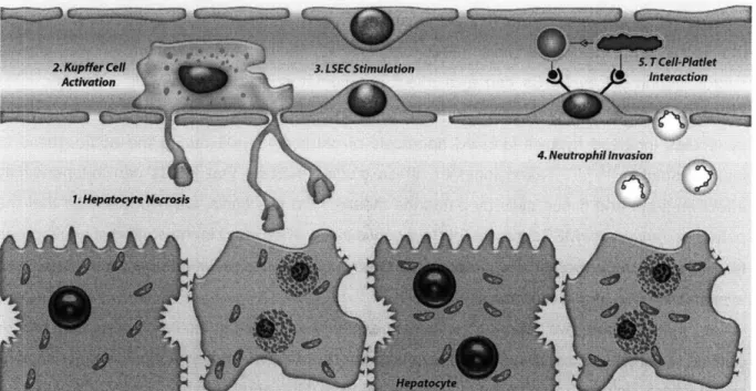
CHAPTER II Figure 2.1 - The Temporal Cellular Response to Liver Injury In Vivo... 31
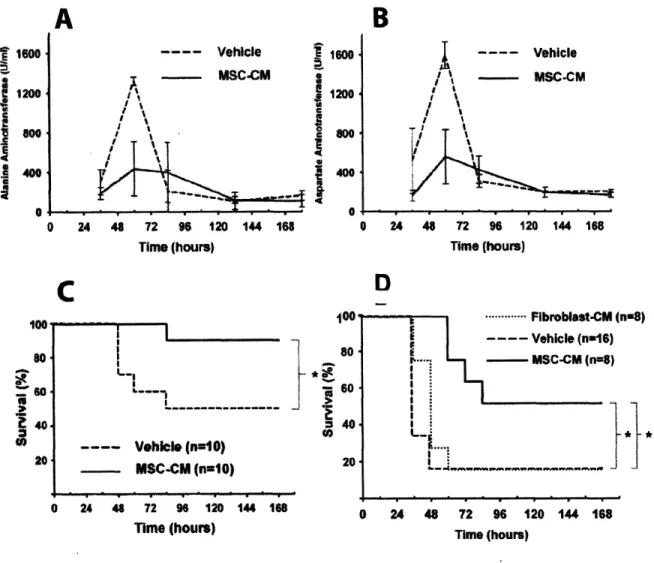
Figure 2.2 - MSC-Derived Molecules Reverse Fulminant Hepatic Failure... 36
Figure 2.3 - Infusion of MSC-CM Prevents Liver Injury and is a Cell-Specific Therapy ... 38
Figure 2.3 - MSC-CM Causes Systemic Changes of Serum Cytokines ... 40
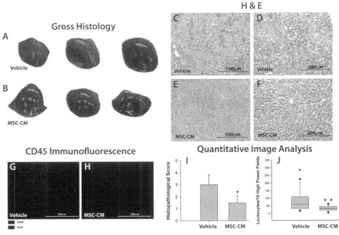
Figure 2.4 - Histopathological Analysis of Liver Tissue after MSC-CM Treatment... 41
Figure 2.5 - Alteration in Peripheral and Tissue Leukocytes in Response to MSC-CM Therapy. 43 CHAPTER III Figure 3.1 - Infusion of MSC-CM Decreases Levels of Apoptosis in Gal-N Treated Rats ... 51
Figure 3.2 - MSC-CM Inhibits Hepatocyte Apoptosis at Low Concentrations In Vitro... 52
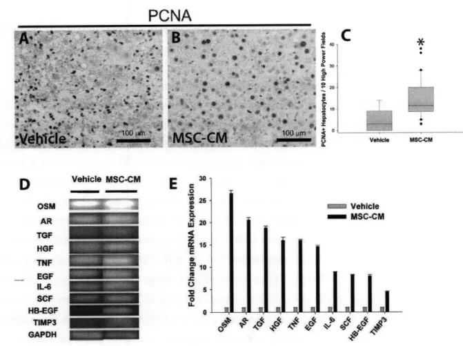
Figure 3.3 - Infusion of MSC-CM Enhances Liver Regeneration in Gal-N Treated Rats... 54
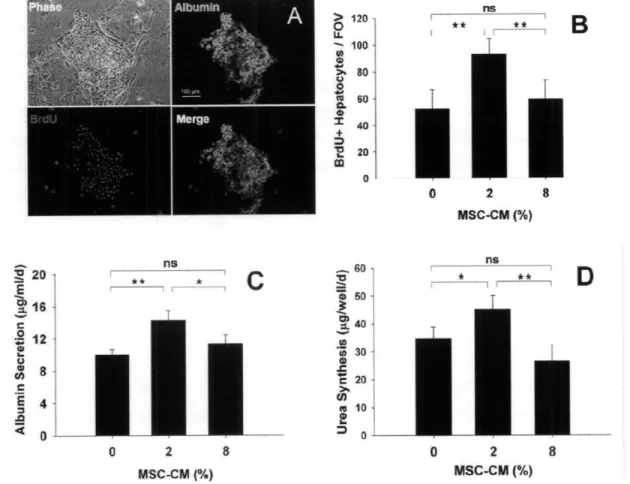
Figure 3.4 - Low Dose of MSC-CM Enhances Proliferation and Function of Hepatocytes ... 55
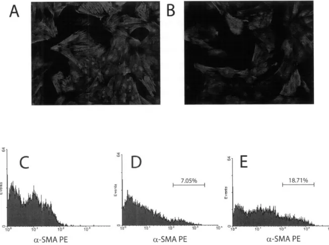
CHAPTER IV Figure 4.1 - Phenotype and Multipotency of Bone Marrow-Derived MSCs... 62
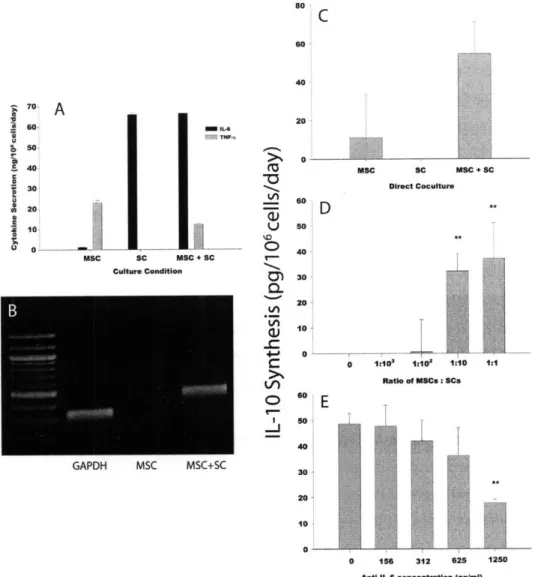
Figure 4.2 - PIP Secretion from SCs after Coculture with MSCs ... 66
Figure 4.3 - SCs do not Revert to a Quiescent Phenotype after Coculture with MSCs... 68
Figure 4.4 - BrdU Incorporation in SCs as a Function of MSC:SC Ratio ... 69
Figure 4.5 - Annexin-V Reactivity of SCs After Indirect Coculture with MSCs... 70
Figure 4.6 - IL-10 mRNA and Protein Secretion of Cytokine-Treated MSCs... 72
Figure 4.7 - Dynamic Cytokine Signaling Between MSCs and SCs ... 73
Figure 4.8 - PIP Secretion in Indirect Cocultures with Cytokine Neutralization... 75
Figure 4.9 - Decreased SC Proliferation in Response to MSC Secreted Cytokines ... 76
Figure 4.10 - MSC-Derived HGF-Mediated Apoptosis of SCs... 77
CHAPTER V Figure 5.1 - Biodistribution of Radiotraced Cell-Derived Molecules... 84
Figure 5.2 - Heparin Affinity Based Separation of MSC-CM... ... 86
Figure 5.3 - Size-Based Separation of MSC-CM ... 87
CHAPTER VI Figure 6.1 - Extracorporeal Bioreactor Circuit Design... ... 93
Figure 6.2 - MSC-EB Support Reduces Liver Injury Biomarkers and Increases Survival... 94
Figure 6.3 - Alteration of the MSC Secretome After Exposure to Liver Failure Serum... 96
CHAPTER VII Figure 7.1 - Hepatocyte Morphology During Coculture with MSCs ... 102
Figure 7.2 - Hepatocellular Function During Coculture is Dependent on MSC Dose... 104 Figure 7.3 - Hepatoprotection and Long-Term Survival Benefit After MSC+Hep-LAD Treatment 106
CHAPTER VIII
Figure 8.1 - Morphology and Immunophenotype of Infused Subpopulation of Marrow Stroma... 115
Figure 8.2 - Histological Changes in Ileal Tissue of Foxp3sf Mice after MSC Transplantation... 117 Figure 8.3 - MSC Treatment Reduces Cellularity and Activated T Cell Number... 118
Figure 8.4 - Evaluation of eGFP+ MSC Engraftment in Foxp3sf Mice... 120 Figure 8.5 - Alteration in Thymocyte Production and Serum Cytokine Levels after Cell Therapy 121 Figure 8.6 - Cotransplantation of MSCs and Tregs Increases Splenic Engrafted Tregs... 123
CHAPTER IX
Figure 9.1 - Prevention of TNBS-Induced Colitis by MSC Transplantation... 132
Figure 9.2 - Histopathological Analysis of Colitic Mice after MSC Infusion... 133
Figure 9.3 - Increased Regulatory T Cells in Gut-Associated Lymph Nodes after Cell Therapy.. 135
Figure 9.4 - Therapeutic Trial of MSC Transplantation in TNBS-Induced Colitis... 136 CHAPTER X
Figure 10.1 - Mouse and Human MSCs Express Endogenous pTAs... 143 Figure 10.2 - UEA-1+ Bone Marrow Cells Express pTAs... 145 Figure 10.3 - Immunohistochemistry of CD45-UEA-1 Localization in the Bone Marrow... 146
Figure 10.4 - Upregulation of Antigen Presentation Molecules in MSCs after IFN-y Stimulation.. 147 Figure 10.5 - Theory of Therapeutic Action Based on pTA Expression... 150
LIST OF TABLES
CHAPTER I
Table 1.1 - Immunophenotype of Human MSCs ... 10 Table 1.2 - Molecular Interactions that Facilitate MSC Homing and Engraftment to Injury Sites. 14
Table 1.3 - Examples of Clinical Trials Employing MSC Transplantation Strategies... 24
CHAPTER III
Table 3.1 - Oligonucleotide Sequences of Primers to Study Hepatocyte Regeneration ... 48
CHAPTER IX
Table 9.1 - Quantitative Histological Analysis of Colitis-Induced Mice... 131
Table 9.2 - Pathology Scoring of Microscopic Colon Sections... 131
CHAPTER X
ABBREVIATIONS
ABO - Blood Group AntigensAIRE - Autoimmune Regulator APC - Antigen Presenting Cell
BDNF - Brain-Derived Neurotrophic Factor BMP - Bone Morphogenetic Protein
CD - Chrohn's Disease CD - Cluster of Differentiation
CFU - Colony Forming Unit
CFU-F - Colony Forming Unit-Fibroblast
CNTF - Ciliary Neurotrophic Factor
CXCR - CX Chemokine Receptor DC - Dendritic Cell
EB - Extracorporeal Bioreactor
EGF - Epidermal Growth Factor
eGFP - Enhanced Green Fluorescent Protein FDA - Food and Drug Administration FGF - Fibroblast Growth Factor FHF - Fulminant Hepatic Failure GVHD - Graft Versus Host Disease HGF - Hepatocyte Growth Factor
HLA - Human Leukocyte Antigen HSC - Hematopoietic Stem Cell i.p. - intraperitoneal
IBD - Inflammatory Bowel Disease
IFN - Interferon
IGF - Insulin-like Growth Factor
IL - Interleukin
IL-1 RA - Interleukin-1 Receptor Antagonist ISEMF - Intestinal Subepithelial Myofibroblasts
LAD - Liver Assist Device LI - Liver Injured
LTC-IC - Long-Term Colony Initiating-Culture MCP - Monocyte Chemoattractant Protein MHC - Major Histocompatability Complex MLR - Mixed Lymphocyte Reaction MMP - Matrix Metalloproteinase
MSC - Mesenchymal Stem Cell
MSC-CM - MSC-Conditioned Medium mTEC - Medullary Thymic Epithelial Cell
NGF - Nerve Growth Factor NK - Natural Killer
OVA - Ovalbumin PD - Programmed Death
PDGF - Platelet-Derived Growth Factor
PE - phyocoe PG - Prostaglandin SCF - Stem Cell Factor
SCID - Severe Combined Immunodeficiency
SDF - Stromal Derived Factor Sfrp - Secreted Frizzled Protein
SSEA - Stage-Specific Embryonic Antigen TGF - Transforming Growth Factor
TNF - Tumor Necrosis Factor
Treg - Regulatory T Lymphocyte
UCB - Umbilical Cord Blood UEA - Ulex Europas Agglutinin
VEGF -Vascular Endothelial Growth Factor
CSF - Cerebrospinal Fluid
LNSC - Lymph Node Stromal Cell
AR - Amphiregulin ECM - Extracellular Matrix TIMP - Tissue Inhibitor of MMP
GI - Gastrointestinal
BMTx - Bone Marrow Transplant EKG - Electrocardiogram
LVEF - Left Ventricular Ejection Fraction PIP - Procollagen Type-I C-Peptide
FITC -Fluorescein Isothiocyanate
PCR - Polymerase Chain Reaction
WT - Wild-Type
SPECT - Single Photon Emission Computed Tomography ACTH - Adrenocoricotropic Hormone
NF-KB - Nuclear Factor-KB
PCNA - Proliferating Cell Nuclear Antigen
PCI - Percutaneous Coronary Intervention
RNA - Ribonucleic Acid
ALT - Alanine Aminotransferase
01 - Osteogenesis Imperfecta
LSEC - Liver Sinusoidal Endothelial Cell
MLN - Mesenteric Lymph Node
PET - Position Emission Tomography
GAPDH - Glyceraldehyde 3-Phosphate Dehydrogenase DAPI - 4',6-diamidino-2-phenylindole
RT-PCR - Reverse Transcription PCR
SMA - Smooth Muscle Actin
TNBS - Trinitrobenzosulfonic Acid
UC - Ulcerative Colitis
ALF- Acute Liver Failure
PBS - Phosphate Buffered Saline
TUNEL - Terminal Deoxynucloetidyl Transferase Nick End Labeling
Gal-N - D-galactosamine BrdU - Bromodeoxyuridine
ELISA - Enzyme-Linked Immunosorbent Assay
Ctx - Cell Transplant MI - Myocardial Infarction
pTA - Peripheral Tissue Antigen
SC - Stellate Cell
DNA - Deoxyribonucleic Acid
iFABP - Intestinal Fatty Acid Binding Protein
OSM - Oncostatin M H&E - Hematoxylin-Eosin
MACS - Magnetic Activated Cell Sorting
IMDM -Iscove's Modified Dulbecco's Medium PBPC - Peripheral Blood Progenitor Cell
FBS - Fetal Bovine Serum
AST - Aspartate Aminotransferase
OC - Oval Cell
ALS - Amyotrophic Lateral Sclerosis MLD - Metachromatic Leukodystrophy
AsA - Ascorbic Acid Ig - Immunoglobulin
MRI - Magnetic Resonance Imaging
CK - Cytokeratin
CHAPTER I
MESENCHYMAL STEM CELL IMMUNOTHERAPY 1.1. SIGNIFICANCE
Bone marrow-derived mesenchymal stem cells (MSCs) are an excellent candidate for cell-based therapy because: (1) bone marrow aspiration is a routine method used in medicine thereby allowing for easy accessibility to human MSCs; (2) MSCs are easily isolated and can expand to clinical scales in a relatively short period of time1'2; (3) MSCs can be biopreserved without loss of potency and stored for point-of-care delivery3'4; and (4) human trials of MSCs
thus far have shown no adverse reactions to allogeneic versus autologous MSC transplants suggesting that therapy can cross histocompatibility barriers5s7. To date, MSC transplantation
has shown encouraging results in clinical therapeutic trials of cardiovascular8'9, neurological0'o11
and immunological diseasel2"13. In this work, we have developed new immunotherapeutic
strategies for treatment of fulminant hepatic failure (FHF) and inflammatory bowel disease (IBD) by harnessing the endogenous functions of these cells. The proposed work is significant, as development of such therapies for FHF and IBD would potentially treat an estimated 100,000+
newly diagnosed patients a year as well as patients who are refractory or contraindicated to standard-of-care medical or surgical procedures.
The broader impact of the proposed studies is expected to be in multiple areas. First, the work here could advance the realization of MSCs as a practical therapy in clinical medicine, especially in organ failure and autoimmune diseases. In addition, this work can aid in the future development of MSC-based technologies and be extrapolated to other cell-based therapies to treat relevant diseases. Furthermore, exploration of the therapeutic functions of MSCs is expected to enhance our understanding of the mechanisms involved in cell therapy of these potent cells and give further insight to their natural functions during health and disease.
1.2. Origins and Developmental Biology of Mesenchymal Stem Cells
Maureen Owen14 and colleagues, based on the work of Alexander Friendenstiensi , proposed
the existence of non-hematopoietic cells in the bone marrow with the ability to give rise to mesenchymal cells. These cells were isolated from bone marrow mononuclear cells based on adherence to tissue culture plastic and initially were described as adherent, clonogenic, nonphagocytic, and fibroblastic in nature, with the ability to give rise to colony forming units-fibroblastic (CFU-F). Under certain mechanochemical stimuli, it was found that CFU-F could give rise to connective tissue cells such as osteoblasts, chondrocytes, adipocytes, and myelosupportive stroma lending credence to a stem cell hypothesis14' 16. These findings were
further popularized by Arnold Caplan in the last two decades17, who coined the term mesenchymal stem cells (MSCs) and described the process of mesengenesis. In addition, they identified the first set of MSC expressed antigens that react with SH2 (CD105) and SH3 (CD73) monoclonal antibodies. Despite lack of a consensus on the relevant cell surface markers of MSCs from other species, the current expression of a certain immunophenotype of human MSCs is listed in Table 1.118.
Table 1.1. Immunophenotype of Human MSCs
Positive Negative Inducible
CD13, CD29, CD44, CD49a, CD3, CD4, CD6, CD9, CD10, HLA-DR b, c, e, f, CD51, CD54, CD58, CD11a, CD14, CD15, CD18, CD71, CD73, CD90, CD102, CD21, CD25, CD31, CD34, CD105, CD106, CDw119, CD36, CD38, CD45, CD49d, CD120a, CD120b, CD123, CD50, CD62E, L, S, CD80, CD124, CD126, CD127, CD86, CD95, CD117, CD133, CD140a, CD166, P75, TGF- SSEA-1, ABO
blR, TGF-blIR, HLA-A, B, C, SSEA-3, SSEA-4, D7, PD-L1
Note: CD, cluster of differentiation; TGF, transforming growth factor; HLA, human leukocyte antigen; SSEA, stage-specific embryonic antigen; ABO refers to blood group antigens;
The developmental precursor of MSCs has been difficult to identify because MSCs have no distinguishing features to track in vivo. A number of studies support the concept that the typical sites of developmental hematopoiesis, including the placenta, aorta-gonad-mesonephros, and fetal liver, are also populated by embryonic MSCs19
-21. These cells are originally
independent of interactions with hematopoietic stem cells and can be found in the embryonic circulation at early stages of ontogeny22. Counter-intuitively, a novel embryological source of MSCs has been identified in the cranial neural crest. Using in situ methods with fluorescent reporting proteins, one group demonstrated a transient proliferation of Sox-1+ cells originally
from the neuroepithelium that display multipotency and that transitioned through a neural crest stage to give rise to adult MSCs23. Cells with multilineage differentiation potential and cytoskeletal elements reminiscent of adult MSCs can be isolated from the first brachial arch ectomesenchymal cells that give rise to the orofacial connective tissue24-26. These results are
consistent with the promiscuous expression of neural proteins in MSCs in their basal state27'28
1.3. Mesenchymal Stem Cell Localization and Multipotency In Vivo
The physical location, or niche, of a stem cell provides invaluable information of their role and interactions within the tissue. The MSC niche has been an elusive location and difficult to observe dynamically primarily because no unique MSC marker has been identified and the marrow cavity is difficult to probe in vivo. That stated, based on correlations between immunophenotype and ex vivo CFU-F assays, evidence supports the notion that MSCs exist in perivascular locations29,'3. Their stromal counterparts may differentiate and migrate from this
space to reside on the abluminal side of marrow sinusoids and form a three-dimensional network that invests the capillary bed. Adventitial reticular cells or pericytes that have processes projecting into the lumen of sinusoids, are likely the in vivo surrogate of CFU-Fs although clonogenicity has never been proven to date3133. These pericytes share a similar
surface and intracellular protein expression pattern with MSCs implying that the cells are related ontologically'. This location has been reproduced in artificial systems as well. Ectopic stromal cells displaying, PDGF-R, NG2, and high expression of CD146 are typically localized in perisinusoidal regions5. In addition, tissue engineered constructs juxtaposing MSCs and endothelial cells form long-lasting vascular structures with MSCs naturally displaying pericytic phenotype and function36. Such localization suggests MSCs may be intimately involved in
angiogenesis, wound healing and interactions with bloodborne entities.
In vivo transplantation of expanded MSCs has illustrated the physiological relevance of
this stem cell. Implantation of MSC grafts under the kidney capsule or in subcutaneous spaces results in the ectopic formation of bone marrow, including donor-derived bony trabeculae, myeolsupportive stroma, and adipocytes as well as host-derived hematopoietic cells that colonize and fully mature within the space'1635. If MSCs are engrafted in microencapsulated environments with pore sizes that exclude the immigration of host cells, only connective tissue cells of donor origin are observed'4. These results are all specific to MSCs, whereas experiments performed with fibroblasts or differentiated connective tissue cells fail to recapitulate the same histological image.
Though the bone marrow has been established as the primary source of MSCs, due to the invasive nature of bone marrow aspiration, efforts are underway to identify other abundant and reliable sources of MSCs for clinical purposes. The isolation of MSCs from peripheral sources such as umbilical cord blood (UCB)37'38, placental tissue19-21, and adipose tissue 39 has
been reported with cells displaying similar immunophenotypes and multipotency, though other contradictory studies report the absence of MSCs in these peripheral locations40,41.
Furthermore, it is unclear if there is a definitive relationship between these cells from various sources and it is important to be wary of interpretations of CFU-F analysis of MSCs from sites other than the bone marrow given that many adherent and clonogenic fibroblastoid cells exist in
non-hematopoietic tissues.
1.4. The Interaction Between the Bone Marrow Stroma and Hematopoiesis
MSCs exist within the bone marrow as a precursor to connective tissue components that act primarily as supportive elements to hematopoiesis. An understanding of the endogenous functions of MSCs and their precursors within the marrow can provide insight into mechanisms involved when these cells are used in a therapeutic context. The initial appreciation for the important interaction between stromal cells and hematopoietic cells was obtained from the analysis of two different spontaneous mutations in mouse colonies that led to the same anemic phenotype. Analysis of these mutant mice revealed that a stromal cell ligand known as stem cell factor (SCF) and its associated receptor, c-kit, found on hematopoietic stem cells (HSCs) was essential for the maintenance of HSCs. Other cell-cell interactions between MSC progeny, such as osteoblasts and HSCs has also proved essential for HSC self-renewal4 243. Moreover,
stromal elements secrete a number of insoluble and soluble species within the marrow space that promote the growth and differentiation of hematopoietic cell lineages44-4 7.
Experimentally, MSCs can act as a surrogate feeder layer and promote the self-renewal and differentiation of HSCs in long-term colony initiating-culture (LTC-IC) and CFU assays48 Two types of culture techniques utilizing stromal cell layers and defined chemical supplements allow for the establishment of lymphoid and myeloid cells in vitro. The Whitlock-Witte method cultures bone marrow cells on a confluent layer of irradiated stromal cells with a low-serum containing medium without corticosteroids49,50. It is a lymphoid culture system, which supports the growth of B-lymphocytes and with some modifications in culture parameters can also allow selective proliferation and differentiation of all developmental stages of pre-B-cells and
B-lymphocytes. A myelopoietic culture system, known as Dexter bone marrow cultures, maintains myeloid progenitor cells in vitro. Dexter-type culture systems differ from Whitlock-Witte cultures
by making use of high concentrations of serum and hydrocortisone and lower incubation temperatures51'5 2. Collectively, the marrow stroma can direct the differentiation of lymphoid and
myeloid cells in vitro and it is likely that the mechanisms underlying this directed differentiation will be of relevance to the immune response to MSCs in vivo.
The soluble milieu that is maintained by MSCs may also be protective in the context of immune cell development within the bone marrow. Recent evidence suggests that the bone marrow can be considered a bona fide secondary lymphoid organ that contains perisinusoidal B cell niches to partake in microbial immunity within the cavity53. Interestingly, these
perisinusoidal B cells are restricted in their ability to respond to antigen and can only perform T-independent immune reactions that may be indicative of the bone marrow being a T cell suppressed environment.
1.4. The Role of MSCs During Inflammation
MSCs are traditionally isolated from the bone marrow where these cells functionally support hematopoiesis by the secretion of cytokines, growth factors and extracellular matrix as well as by repopulating connective tissue cell types47',556. However, cells with MSC characteristics are
reportedly present in virtually every organ5 7, although more rigorous analysis of in vivo
self-renewal and potency was not determined in the study. This raises the possibility that MSCs constitute a normally quiescent pool of reparative stem cells, ready to be activated as a source of trophic factors and support in the event of injury. Mobilized and subsequently recruited MSCs may also play a role, as supported by observations that stressful events (e.g. hypoxia) result in the mobilization of MSCs into the systemic circulation58. This hypothesis is supported by the
observation that mesenchymal stem cell-like cells commonly referred to as pericytes are located almost exclusively in the perivascular space of most blood-vessels, which enhance the likelihood of their mobilization potential. In addition, molecules locally upregulated in the event of injury facilitate the recruitment of circulating MSCs59-61 (Table 1.2.). However, the physiological
role of endogenous MSCs in tissue repair remains elusive due to a lack of unique MSC markers and knockout models.
It is likely that true stem cells do not circulate peripherally, but rather produce
lineage-restricted cell types that home to tissues as a mechanism of non-parenchymal cell replenishment during injury. Fibrocytes are circulating bone marrow-derived cells (-0.1-0.5% of nonerythrocytic cells in peripheral blood) that are phenotypically a mixture of monocytes and fibroblasts expressing type I collagen and the surface markers CD1 1b, CD13+, CD34+, and CD45RO+62. In sex-mismatched bone marrow chimeras, these cells were found to be the
progeny of a radioresistant precursor from the bone marrow63. During injury, fibrocytes were rapidly and specifically found in the areas of inflammation63, fibrosis , and cancer5~8 where
they are thought to mature into tissue-resident myofibroblasts69. They express chemotactic
receptors such as CCR3, CCR5, CCR7, and CXCR4 and are absent of CCR4, CCR6, and CXCR3". Interestingly, fibrocytes express surface molecules such as MHC class II, CD80 and CD86 and were shown to present pulsed antigens to naive T cells in an efficient manner when compared to monocytes and dendritic cells, although this was not verified in vivo70.
Table 1.2. Molecular Interactions Facilitating Migration and Engraftment of MSCs to Sites of Tissue Injury
Receptor/Enzyme Tissue Ligand/Substrate Reference
c-met HGF 71,72 CXCR4 SDF-1 71,73-76 CX3CR1 Fractalkine 74 MMP2 Collagen 71,73 MMP9 Collagen 71 CD44 Hyaluronic Acid 59,77 PDGF-R PDGF-AB, BB 72,75,78 EGF-R EGF 72 FGF-R2 FGF2 72,79 IGF-R IGF-1 72,75,80 RANTES-R RANTES 75 MCP-1 R MCP-1 81,82 CCR2/3/4 CXCL8 75,81
Note: HGF, hepatocyte growth factor, SDF-1, stromal cell-derived factor-i; MMP, matrix
metalloproteinase; PDGF, platelet-derived growth factor; EGF, epidermal growth factor; FGF, fibroblast growth factor; IGF, insulin-like growth factor; MCP-1, monocyte chemoattractant protein-1
1.5. The Interaction Between MSCs and the Innate and Adaptive Immune System In Vitro MSCs have been demonstrated to modulate the immune system. MSCs were first noted to be immunopriveleged after it was observed that hMSCs could engraft without immunosuppression in a xenogenic, adult host83. The immunosuppressive ability was first exploited clinically in the
treatment of an 8-year old boy with severe, acute graft-versus host disease (GVHD), that was refractory to steroid immunosuppression84, who was successfully treated by MSC transplantation. In recent years, this immunosuppression has been found to be an active
process and the mechanisms underlying MSC immunomodulation operate at different levels of the innate and adaptive immune system. We discuss in vitro coculture experiments to exemplify the effects of MSCs on individual populations of immune cells. A schematic is shown in Figure
Ig Production IFN-y Production
K,
Proliferation ProliferationT
j
CTL formation *u IFNy on 0 , 'ýý Production Reglaor Regulatory T Cells IL-10 roduction Maturation anc expansion Production of pro-inflamitoryProliferation Cell No cell
contact contact
cytokines (IL-12,IFNy, Ilveal
Inhibitory effect
Stimulatory effect)-Figure 1.1. In Vitro Interaction of MSCs and immune cells. Schematic of MSC interactions
with distinct subsets of immune cells as adapted from Rasmusson I, Exp Cell Res (2): 198, 2006. Inhibitory effects are immune cell functions are shown by hatched arrows and stimulatory
effects shown by arrowheads. The upper right bar chart shows a representative dose response of immune cell inhibition as a function of MSC number. The lower right bar chart shows an example of results comparing the effect of MSC cell contact versus diffusible factors in immune cell inhibition. NK, B, and T refer to NK, B, and T cells, respectively. DC1: mature monocytic dendritic cells; DC2: mature plasmacytoid dendritic cells.
The majority of in vitro studies have shown that MSCs can directly inhibit CD3+, CD4+ T cell proliferation and secretion of IL-2 and IFN-y induced by mixed lymphocyte reactions (MLR), mitogens and TCR or costimulatory receptor engagement. T cells in the presence of MSCs appear to be anergized by the lack of a 'second danger signal' by MSCs, which do not express the costimulatory molecules CD80, CD86 and CD40' , however this has yet to be definitively proven. Several investigations have also shown a direct suppressive effect of MSCs on cytotoxic CD8+ T cells. MSCs prevented cytolysis of target cells by alloantigen-specific CD8+ T cells when present during the priming of cytotoxic cells86. Some investigators attribute the inhibition of cytotoxicity by MSCs to an intrinsic "veto" function or the generation of suppressor CD8+ cells after coculture87, although there exist conflicting data. Nonetheless, other reports have also observed generation of CD4+ CD25+ T cells, a cell surface marker expression pattern of both newly activated CD4+ lymphocytes and regulatory T cells (Tregs)88'89. Analysis of
Foxp3, a transcription factor required for the development and function of Tregs, was not performed in these studies to rigorously quantify the true Treg population.
Suppression, in most studies, was reproduced in the absence of cell-cell contact and in a dose-dependent manner, indicating the role of soluble factors. Furthermore, these inhibitory molecules can exert their effects across species barriers as evidenced by suppression of MLRs in xenogeneic cultures"'91. It remains highly debated as to which soluble mediators are involved in T cell suppression, although many candidates have shown promise in neutralization studies. MSC-conditioned supernatants had no anti-proliferative effect on T cells, yet did suppress the stimulation of B cells92. This suggests that MSCs can dynamically react to their
immunological environment, in the context of T cells, while also secreting immunomodulatory molecules in their quiescent, undifferentiated state, in the context of B cell development. Also, there is an approximate 1-2 order of magnitude difference between the number of MSCs needed to suppress T cell activity compared to B cell activity93'94. In contrast, other reports have shown that MSCs can stimulate antibody secretion and induce polyclonal differentiation and expansion of healthy human B cells95,96, consistent with the supportive role of stromal cells in B
lymphopoiesis. In addition, these same supportive mechanisms may advance the progression of B cell-mediated disease such as multiple myeloma and systemic lupus erythematosus questioning the use of MSCs in such disease contexts95'97. Although the principal source of
MSC immunosuppression appears to be secreted molecules, cell-cell interactions via the programmed death (PD) receptor-1 and its ligands PD-L1 and PD-L2 were shown to be
necessary for inhibition of mouse splenocytes by MSCs92. Yet, no single mechanism has yet to
MSCs also have an indirect effect on T cell function via antigen presenting cells (APCs). Dendritic cells (DCs) are the major link between innate and adaptive immunity due to their ability to present antigens with high efficiency to lymphocytes. In coculture with MSCs, monocytes failed to differentiate into DCs when cultured in lineage-specifying growth conditions98'99. In
addition, MSCs inhibited the maturation of DCs to present appropriate antigens and costimulation to T cells through CDla, CD40, CD80, CD86, and HLA-DR98,100. After coculture with MSCs, DCs were ineffective in their ability to activate lymphocytes in DC-CD4+ MLRs99.
This interaction was found to be y-secretase dependent, indicating the role of the Notch pathway in MSC-DC interactions101. Ultimately, MSCs may drive, or "license", DCs to a
suppressor phenotype which can further attenuate T cell-mediated immunity.
1.6. The Paracrine Theory of MSC Therapy in Regenerative Medicine
There has been a recent paradigm shift in what is considered to be the therapeutic promise of MSCs in diseases of vital organs. The term MSC was originally tailored for the regenerative capacity of this cell type through its ability to differentiate into mesodermal cell types including adipocytes, chondrocytes, osteocytes and stromal cells. When cultured MSCs
were also observed to adopt characteristics of cardiomyocytes'02, hepatocytes
103,104 and
several other cell types'0 5'106, many investigators put this presumed widespread regenerative
potential to the test in injury models of vital organs. Despite a lack of evidence for replaced organ function through differentiation of transplanted MSCs into functional cells, a clear therapeutic effect was consistently reported in models ranging from myocardial infarction to stroke'8"0 7 10 9.Common findings in these therapeutic trials were: (1) inhibition of inflammatory
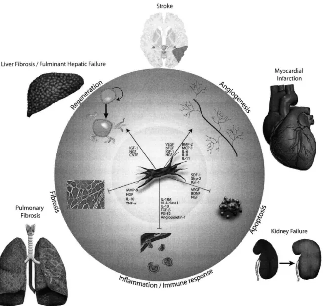
responses; (2) reduction in apoptosis; (3) prevention of fibrosis (4); stimulation of endogenous regenerative programs; and (5) neovascularization. Studies in the past years have shown that paracrine cytokines, growth factors and other secreted signaling molecules are responsible for these trophic effects. To broaden the scope of the paracrine effect of MSCs on specific cellular mechanisms, an integrated view of MSC therapy compiled from data from disease models of the heart, liver, lung, kidney and brain, is presented with emphasis on the role of specific mediators (see Figure 1.2).
1.6.1. Apoptosis
During disease, protection of injured cell mass by inhibition of programmed cell death can potentially preserve organ function. Several studies have shown that MSCs have the ability to inhibit apoptosis of various types of host cells through paracrine mechanisms. A group of
Stroke
Liver Fibr
Puli
Fi
Figure 1.2. Effects of MSC-Derived Factors on Pathogical Processes in Tissue Injury. For each growth factor, cytokine, chemokine or other mediators a summary of the observed effects and the model in which this was investigated is provided. VEGF, vascular endothelial growth factor; bFGF, basic fibroblast growth factor; IGF-1, insulin-like growth factor-i; HGF, hepatocyte growth factor; BMP-2, bone morphogenetic protein-i; MCP-1, monocyte chemoattractant protein-i; IL, interleukin; SDF-1, stromal cell-derived factor-i; Sfrp-2, secreted frizzled protein-2;
BDNF, brain-derived neurotrophic factor; NGF, nerve growth factor; IL-1RA, interleukin-1
receptor antagonist; HLA, human leukocyte antigen; TGF-P, transforming growth factor-1;
PG-E2, prostaglandin-E2; MMP, matrix metalloproteinase; TNF-a, tumor necrosis factor-a; CNTF,
ciliary neurotrophic factor.
111n11-researchers led by Dzau used a set of elegant experiments to demonstrate that the cardioprotective effect of MSC transplantation in a rat myocardial infarction model was largely ascribed to the anti-apoptotic effects of MSC molecules'1o. Local administration of MSCs into
the infarcted rat myocardium resulted in a dose-dependant normalization of cardiac function11 .
Genetic engineering of MSCs to overexpress a pro-survival gene, Akt-1, prevented massive death of transplanted cells and resulted in dramatically improved results'". These studies showed a remarkable improvement of cardiac function in a relatively short amount of time after graft administration, suggesting a role for soluble cues rather than replacement of cardiomyocytes by infused MSCs. In further experiments using conditioned medium from cultured MSCs, paracrine factors secreted by MSCs were shown to be responsible for the therapeutic effects112'113. Conditioned medium directly inhibited apoptosis of cardiomyocytes
cultured under hypoxic conditions. Moreover, injection of the conditioned medium into infarcted rat hearts reduced the number of apoptotic cardiomyocytes, reducing infarct size and preserving heart function. Silencing studies using RNA-interference identified secreted frizzled related protein 2 (Sfrp-2) as a key anti-apoptotic mediator'14
Investigations by other researchers and in other organ systems have demonstrated that several other mediators secreted by MSCs play a role in inhibition of programmed cell death. Koc et al. observed enhanced anti-apoptotic effects of MSCs modified to overexpress stromal derived factor-1 (SDF-1), identifying the SDF-1 - CX chemokine receptor-4 (CXCR4) axis as an
important mechanism of myocardial preservation via downregulation of pro-apoptotic Bax protein"" 1 16. Others demonstrated that MSC-derived insulin-like growth factor-1 (IGF-1) and
vascular endothelial growth factor (VEGF) have cardiomyocyte apoptosis-inhibiting effects using in vitro studies'17. IGF-1 was also shown to have important pro-survival effects in cultured
proximal tubular epithelial kidney cells, crucial in the development of acute kidney failure18.
Anti-apoptotic effects due to paracrine MSC factors have also been observed in injury models of the liver, kidney and brain. Increased levels of the pro-survival neurotrophins brain-derived neurotrophic factor (BDNF) and nerve growth factor (NGF) were observed in rats subjected to stroke and treated with MSC infusions, suggesting a role for these molecules in
reducing cell death in the ischemic brain" 9. In a rat model of toxin-induced fulminant hepatic failure, we demonstrated that MSC therapy using cells isolated from human bone marrow samples significantly reduced mortality and was correlated with a reduction in the number of
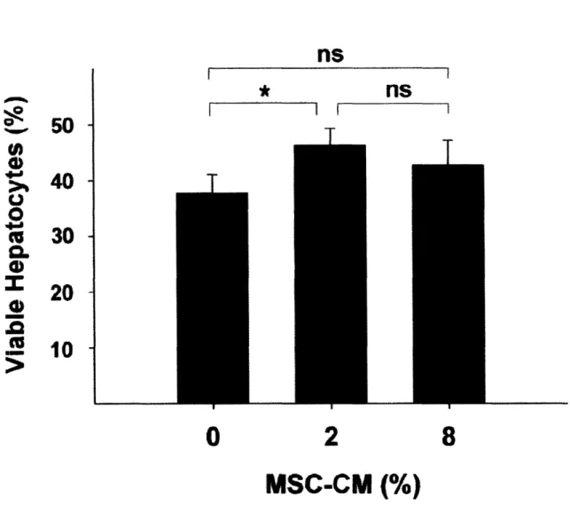
apoptotic hepatocytes (see Chapters 2 and 3)120121. In this study, several different modes of MSC therapy were assessed, including: (1) transplantation of whole cells; (2) infusion of cell lysates; (3) infusion of MSC-derived conditioned medium (MSC-CM); and (4) a MSC-based
extracorporeal device. Animal survival was found to be a function of MSC-CM dose, but above a certain threshold the conditioned medium became ineffective. These results suggest important pharmacodynamic characteristics of MSC-CM or the presence of molecules that are harmful at
high concentrations (e.g. tumor necrosis factor (TNF)-(o) 122. A separate cohort of animals was
treated using an MSC-based extracorporeal device that allowed for MSCs to dynamically supplement the animal's plasma continuously during a 10-hour treatment. The strongest effect on hepatocellular damage and survival was observed in the animals treated with this
MSC-based bioreactors (see Chapters 6 and 7). A third group of animals was unsuccessfully treated with systemically delivered MSCs, although immunorejection or increased pulmonary lodging of
large, xenogeneic cells may have been responsible for the lack of effect.
1.6.2. Immune Response
MSCs have potent inhibitory effects on immune cells. Secreted molecules inhibit lymphocyte proliferation, B-cell differentiation and the formation of natural killer cells, dendritic cells and cytotoxic T-lymphocytes89,93,99'123.Human leukocyte antigen (HLA) class I molecules, interleukin
(IL)-10, transforming growth factor (TGF)-3 and prostaglandin (PG)-E2 have been proposed as responsible mediators, but contradictory reports exist. The potency of MSC immunosuppression is illustrated by the successful treatment of severe steroid-resistant acute graft-versus host disease (GVHD) after bone marrow transplantation84. Several patients have now been
successfully treated with systemic MSC infusions, resulting in a significant survival-benefit in a randomized controlled trial13.
Infiltrating immune cells are essential in the response to tissue injury, but resolution of the inflammation is critical to prevent chronic damage and control harmful immune cell-mediated
cytotoxicityl24',125. Dampening effects on immune cells have been described in several models of vital organ injury. In a rat model of fulminant hepatic failure, we show in Chapter 2 that the number of infiltrating leukocytes was dramatically reduced after systemic administration of concentrated conditioned medium from MSC culturesl21. Fractionation of the conditioned
medium resulted in preservation of the therapeutic effects only for the heparin-binding fraction, suggesting an important role for known heparin ligands such as growth factors and chemotactic cytokines (see Chapter 5). Several groups have described a similar inhibition of accumulating immune cells in different models of lung injuryl26,127. Mei and co-workers observed enhanced
anti-inflammatory effects of genetically engineered MSCs to overexpress angiopoietin-1, suggesting a role for this molecule in the immunomodulatory effects of MSCs.
Besides the cellular immune response, the cytokine response is an important component of the inflammatory response to tissue injury. Control of this response is critical for resolution of the inflammatory infiltrate and cytotoxic effects of pro-inflammatory cytokines as described for
TNF-(a and IL-1128.MSC therapy induces local and systemic downregulation of proinflammatory
cytokines but also upregulation of anti-inflammatory cytokines including IL-10 in models of lung injury, kidney and liver failure (see Chapter 2)129-132. MSC-secreted interleukin-1 receptor antagonist (IL-1 RA) is an important factor in the inhibition of pro-inflammatory cytokine production1".
1.6.3. Fibrosis
Unresolved inflammation leads to fibrosis and scar formation. The deposition of large amounts of extracellular matrix inhibits organ function and regeneration. MSCs have the potential to inhibit scar formation and extracellular matrix accumulation in disease models of myocardial infarction, liver fibrosis, lung fibrosis and stroke. Several groups have demonstrated that administration of MSCs therapeutically inhibits fibrosis formation in rodent models of toxin-induced chronic liver injury34,135, triggering the start of a randomized controlled clinical trial
evaluating the effect of intraportally infused MSCs on patients with decompensated liver
cirrhosis136.
Transplanted MSCs were shown to express high levels of matrix metalloproteinase (MMP)-9, which directly degrades extracellular matrix'37. However, indirect immunomodulatory
effects on resident cell types likely play a role in the anti-fibrotic effects of MSCs. Using in vitro studies described in Chapter 4, we observed dynamic interactions between MSCs and activated stellate cells, the principal mediator of matrix deposition in liver fibrosis. We identified IL-10 and TNF-a as key molecules inhibiting matrix deposition by stellate cells, while MSC-derived HGF induced apoptosis in these cells'22. Such alterations in the secretome of MSCs in response to stress-signals138 is likely to have implications for the techniques required to obtain
MSC-conditioned medium preparations with an optimal therapeutic effect.
MSC therapy also inhibits matrix deposition and scar formation in models of heart infarction, lung fibrosis and stroke. Paracrine mechanisms were identified in a study effectively using a monolayer of fat-derived MSCs to prevent detrimental myocardial remodelling after infarction, which is associated with improved compliance and elasticity of the infarct zone,39' In a rat model of brain ischemia, the reduction in scar volume was accompanied by a decreased number of macrophages in the scar wall, again suggesting an indirect mechanism through immunomodulation of scar-promoting cell types 41.
1.6.4. Regeneration
Enhanced regeneration through stimulation of endogenous repair programs, either through activation of a local adult stem cell pool or via enhanced replication of differentiated cell types, has been identified as another mode of MSC therapy. Investigators implanting human MSCs into the hippocampus of severe combined immunodeficient (SCID)-mice demonstrated enhanced proliferation and differentiation of endogenous neural stem cells and suggested a direct effect of cytokines produced by the MSCs including NGF and ciliary neurotrophic factor (CNTF), both neurotrophins known to enhance neurogenesis59'119142. In coculture studies
soluble factors were shown to be responsible for increased oligodendrogenesis from neural stem cells, but a screen for candidate molecules could not identify a specific mediator'43.
Enhanced proliferation of endogenous non-stem cell types has been demonstrated in experimental liver and kidney injury131'44'45. In Chapter 3, we demonstrated that secreted
factors were sufficient to enhance liver regeneration in a model of acute liver failure in which animals were treated with the MSC-conditioned medium120. Animal studies of cisplatin-induced acute kidney failure identified IGF-1 as a crucial MSC-derived mitogenic stimulus for proximal tubular epithelial kidney cells resulting in improved organ function'18.
1.6.5. Angiogenesis
Liver regeneration' 46, recovery from ischemic heart disease'47, stroke'48 and many other forms of healing149,150 are associated with the formation of new blood vessels. Although MSCs have
the capacity to differentiate into endothelial cells51, this phenomenon is not sufficient for the formation of fully functional blood vessels, which requires many well coordinated steps. However, MSC have the capacity to provide trophic support for the entire process of angiogenesis and collateral formation. Using conditioned medium from cultured MSCs, Kinnaird and colleagues provided evidence that secreted factors are sufficient to improve new vessel formation and improve outcome in a rodent model of limb ischemia, demonstrating independent roles for both VEGF and bFGF152. Others have confirmed the importance of these two
molecules, but in vitro evidence suggests that IGF-1, hepatocyte growth factor (HGF), bone morphogenetic protein-2, monocyte chemoattractant protein (MCP)-1 and interleukins 6, 8 and
11 are also involved'17"5 3 54. MSC therapy results in a significantly increased capillary density in
models of myocardial infarction and dilated cardiomyopathy"6'51s5"516 1.7. Clinical Trials of MSC Transplantation
Physicians often do not know the exact mechanism of action of a particular therapeutic, yet this has not deterred drug usage. At this point in time, the same may be said for MSC-based cell therapies. MSCs have a number of attributes that make them a favorable candidate for the next generation of cellular medicines, in that they are: (1) readily isolated and expandable to meet clinical demands; (2) genetically stable in culture for a number of passages; and (3) are easily stored and transported for bedside use. Thus far, MSCs have been tested in a number of
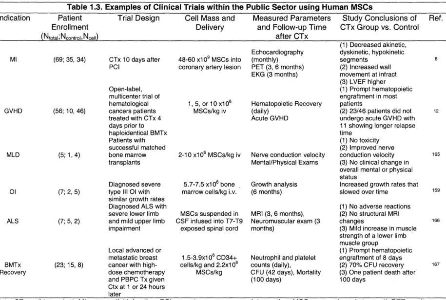
clinical scenarios and been shown to have minimal side effects, likely due to their low immunogenicity. We provide a brief review of the major clinical trials (see Table 1.3.) in progress within the field of MSC therapy with an emphasis on further understanding MSC biology. In addition, we acknowledge commercialized MSC-based products from Osiris Therapeutics, Inc. (http://www.osiristx.com/) to exemplify the development of MSC-based therapy within the biotechnology sector.
1.7.1 Orthopedic Reconstructive Grafts based on MSCs
The initial indications for MSCs were in the field of regenerative medicine for treatment of orthopedic pathologies based on the mesodermal lineage commitment of these stem cells. The first studies were seen in bone tissue engineering, whereby MSCs were seeded into biocompatible scaffolds for insertion into bone defects157. These pre-clinical test have
culminated in a Food and Drug Administration (FDA) approved product known as Osteocel® by Osiris Therapeutics, Inc. The use of MSCs for bone repair has been extended to genetic diseases of skeletal dysplasia. Osteogenesis imperfecta is an autosomal dominant disorder that results in deficiency of type I collagen, which is a critical component of the bone extracellular matrix (ECM)158. Infusion of allogeneic MSCs in childrenl59161 and in one patient in
utero1 62, showed accelerated growth velocities in all subjects compared to untreated patient
cohorts, however low engraftment was noted in all trials. Larger clinical trials are ongoing to determine the efficacy of MSC therapy as a first-line treatment for this orphan disease. These studies were further evaluated in mice models of osteogenesis imperfecta and revealed interesting dynamics of the MSC grafts in vivo. First, engraftment of osteoprogenitor cells was found to be saturated suggesting that higher doses of cells would be an ineffective strategy to improve engraftment1 63. Secondly, temporal tracking of enhanced green fluorescent (eGFP)
expressing MSCs showed that transplanted cells showed limited proliferation self-renewal capacity after engraftment, yet they could be serially passaged and repopulate another host'4.
Table 1.3. Examples of Clinical Trials within the Public Sector using Human MSCs
Indication Patient Trial Design Cell Mass and Measured Parameters Study Conclusions of Ref. Enrollment Delivery and Follow-up Time CTx Group vs. Control
(Ntotal; Ncontrol, Ncell) after CTx
MI GVHD MLD 01 ALS BMTx Recovery 48-60 x109 MSCs into coronary artery lesion
1,5, or 10 x106 MSCs/kg iv
2-10 xl 06 MSCs/kg iv
5.7-7.5 x10⁸ bone marrow cells/kg i.v. MSCs suspended in CSF infused into T7-T9
exposed spinal cord
1.5-3.9x1 06 CD34+ cells/kg and 2.2x10⁶ MSCs/kg Echocardiography (monthly) PET (3, 6 months) EKG (3 months) Hematopoietic Recovery (daily) Acute GVHD
Nerve conduction velocity Mental/Physical Exams Growth analysis (6 months) MRI (3, 6 months), Neuromuscular exam (3 months)
Neutrophil and platelet counts (daily),
CFU (42 days), Mortality (100 days) (1) Decreased akinetic, dyskinetic, hypokinetic segments (2) Increased wall movement at infract (3) LVEF higher (1) Prompt hematopoietic engraftment in most patients
(2) 23/46 patients did not
undergo acute GVHD with
11 showing longer relapse
time
(1) No toxicity (2) Improved nerve
conduction velocity (3) No clinical change in overall mental or physical status
Increased growth rates that slowed over time
(1) No adverse reactions (2) No structural MRI changes
(3) Mild increase in muscle strength of a lower limb muscle group
(1) Prompt hematopoietic
engraftment of 8 days
(2) 70% CFU recovery (3) One patient death after
100 days (69; 35, 34) (56; 10, 46) (5; 1,4) (7; 2, 5) (7; 5, 2) (23; 15, 8) CTx 10 days after PCI Open-label, multicenter trial of hematological cancers patients treated with CTx 4 days prior to haploidentical BMTx Patients with successful matched bone marrow transplants Diagnosed severe type III 01 with similar growth rates Diagnosed ALS with severe lower limb and mild upper limb impairment
Local advanced or metastatic breast cancer with high-dose chemotherapy and PBPC Tx given Ctx at 1 or 24 hours later
Note: CTx, cell transplant; MI, myocardial infarction; PCI, percutaneous coronary intervention; MSC, mesenchymal stem cell; PET, positron emission tomography; EKG, electrocardiogram; LVEF, left ventricular ejection fraction; GVHD, graft-versus-host-disease; BMTx, bone marrow
transplant; MLD, metachromatic leukodystrophy; 01, osteogenesis imperfecta; ALS, amyotrophic lateral sclerosis; CSF, cerebrospinal fluid;
1.7.2. MSC Therapy for Cardiovascular Diseases
Cardiovascular dysfunction remains atop the list of major diseases that affect patients today. In particular, myocardial infarction is still the number one killer. Death of cardiac tissue leads to contractile dysfunction and tissue remodeling that is often ineffective because of ongoing ischemia of the heart. Many investigators have examined the efficacy of intracoronary infusion of bone marrow cells or purified MSCs in the peri-infarction period. In general, bone marrow aspiration was performed after stabilization of the patients and cells were delivered by catheter-based methods to the lesion of interest. The initial excitement in using CD34+ bone marrow cell transplantation as a therapy for myocardial infarction'• was found to not have a survival benefit nor clinically ameliorate signs of disease after long term follow-up in a large cohort, multi-center trial16 9'170
. An initial trial transplanting culture amplified MSCs showed improvements in cardiac
parameters after short-term follow-up8, although no long-term data exists. In Phase I clinical
trials, ProvacelTM by Osiris Therapeutics, Inc. was found to be safe for use and enrollment of heart attack patients for larger cohort testing has begun.
1.7.3. Hematological and Immunological Use of MSCs
Indeed the natural target tissue of MSC therapy may be the hematopoietic system.
Historically, autologous MSCs were used in the setting of allogeneic bone marrow transplantation to enhance engraftment and limit co-morbidities, such as GVHD. Initial efficacy data by Lazarus et al. demonstrated no toxicity or adverse reactions to transplants, which was further elaborated by this group in a larger trial1 2'167 (Table 1.3). These studies were further extrapolated to patients undergoing severe acute GVHD and being treated with haploidentical MSCs leading to a clinical remission in a large fraction of patients relative to the standard of care 3'84. A female patient diagnosed with myelogenous leukemia given an allogeneic HSC and
MSC transplant from her haploidentical father showed rapid engraftment with a trilineage
hematological response and no signs of acute or chronic GVHD. Interestingly, several months
after treatment, chimera studies of the patient's bone marrow revealed that all MSCs were of recipient origin suggesting that the donor MSCs did not engraft in the marrow. Moreover, a patient with severe idiopathic aplastic anemia treated with MSCs led to a partial resolution of tissue hemorrhage and edema, while repopulating adipocytes and stromal cells'71. However, no functional recovery of the hematopoietic system was found with MSC transplants alone. MSCs from human marrow are being tested in a variety of immunological diseases in patients by Osiris Therapeutics, Inc. with ongoing testing in steroid refractory/acute GVHD and Crohn's disease'72 entering phase III clinical trials for their product ProchymalM.