Publisher’s version / Version de l'éditeur:
Journal of Neuroscience, 35, 3, pp. 1291-1306, 2015-01-21
READ THESE TERMS AND CONDITIONS CAREFULLY BEFORE USING THIS WEBSITE.
https://nrc-publications.canada.ca/eng/copyright
Vous avez des questions? Nous pouvons vous aider. Pour communiquer directement avec un auteur, consultez la première page de la revue dans laquelle son article a été publié afin de trouver ses coordonnées. Si vous n’arrivez pas à les repérer, communiquez avec nous à PublicationsArchive-ArchivesPublications@nrc-cnrc.gc.ca.
Questions? Contact the NRC Publications Archive team at
PublicationsArchive-ArchivesPublications@nrc-cnrc.gc.ca. If you wish to email the authors directly, please see the first page of the publication for their contact information.
NRC Publications Archive
Archives des publications du CNRC
This publication could be one of several versions: author’s original, accepted manuscript or the publisher’s version. / La version de cette publication peut être l’une des suivantes : la version prépublication de l’auteur, la version acceptée du manuscrit ou la version de l’éditeur.
For the publisher’s version, please access the DOI link below./ Pour consulter la version de l’éditeur, utilisez le lien DOI ci-dessous.
https://doi.org/10.1523/JNEUROSCI.2126-14.2015
Access and use of this website and the material on it are subject to the Terms and Conditions set forth at
Motoneurons derived from induced pluripotent stem cells develop
mature phenotypes typical of endogenous spinal motoneurons
Toma, J. S.; Shettar, B. C.; Chipman, P. H.; Pinto, D. M.; Borowska, J. P.;
Ichida, J. K.; Fawcett, J. P.; Zhang, Y.; Eggan, K.; Rafuse, V. F.
https://publications-cnrc.canada.ca/fra/droits
L’accès à ce site Web et l’utilisation de son contenu sont assujettis aux conditions présentées dans le site LISEZ CES CONDITIONS ATTENTIVEMENT AVANT D’UTILISER CE SITE WEB.
NRC Publications Record / Notice d'Archives des publications de CNRC:
https://nrc-publications.canada.ca/eng/view/object/?id=c1e21cf0-3dd1-4cc8-83b7-79f8a09cad05
https://publications-cnrc.canada.ca/fra/voir/objet/?id=c1e21cf0-3dd1-4cc8-83b7-79f8a09cad05
Development/Plasticity/Repair
Motoneurons Derived from Induced Pluripotent Stem Cells
Develop Mature Phenotypes Typical of Endogenous Spinal
Motoneurons
Jeremy S. Toma,
1X
Basavaraj C. Shettar,
1Peter H. Chipman,
1Devanand M. Pinto,
8Joanna P. Borowska,
1Justin K. Ichida,
5James P. Fawcett,
3,4Ying Zhang,
1Kevin Eggan,
5,6,7and Victor F. Rafuse
1,21Department of Medical Neuroscience,2Department of Medicine (Neurology),3Department of Pharmacology, and4Department of Surgery, Dalhousie
University, Halifax, Nova Scotia, Canada, B3H 4R2,5Howard Hughes Medical Institute,6Harvard Stem Cell Institute, Department of Stem Cell and
Regenerative Biology, and7Department of Molecular and Cellular Biology, Harvard University, Cambridge, Massachusetts 02138, and8National Research
Council, Institute for Marine Biosciences, Nova Scotia, Canada B3H 3Z1
Induced pluripotent cell-derived motoneurons (iPSCMNs) are sought for use in cell replacement therapies and treatment strategies for
motoneuron diseases such as amyotrophic lateral sclerosis (ALS). However, much remains unknown about the physiological properties
of iPSCMNs and how they compare with endogenous spinal motoneurons or embryonic stem cell-derived motoneurons (ESCMNs). In the
present study, we first used a proteomic approach and compared protein expression profiles between iPSCMNs and ESCMNs to show that
⬍4% of the proteins identified were differentially regulated. Like ESCs, we found that mouse iPSCs treated with retinoic acid and a
smoothened agonist differentiated into motoneurons expressing the LIM homeodomain protein Lhx3. When transplanted into the
neural tube of developing chick embryos, iPSCMNs selectively targeted muscles normally innervated by Lhx3 motoneurons. In vitro
studies showed that iPSCMNs form anatomically mature and functional neuromuscular junctions (NMJs) when cocultured with chick
myofibers for several weeks. Electrophysiologically, iPSCMNs developed passive membrane and firing characteristic typical of postnatal
motoneurons after several weeks in culture. Finally, iPSCMNs grafted into transected mouse tibial nerve projected axons to denervated
gastrocnemius muscle fibers, where they formed functional NMJs, restored contractile force. and attenuated denervation atrophy.
Together, iPSCMNs possess many of the same cellular and physiological characteristics as ESCMNs and endogenous spinal motoneurons.
These results further justify using iPSCMNs as a source of motoneurons for cell replacement therapies and to study motoneuron diseases
such as ALS.
Key words: electrophysiology; iPS cells; motoneuron disease; motoneurons; proteomics; stem cells
Introduction
Induced pluripotent stem cell-derived motoneurons (iPSCMNs)
are being explored as a means to study motoneuron diseases and
to develop therapeutics to treat amyotrophic lateral sclerosis
(ALS;
Dimos et al., 2008
;
Bilican et al., 2012
;
Burkhardt et al.,
2013
;
Yao et al., 2013
) and spinal muscular atrophy (
Corti et al.,
2012
). Preclinical studies are proceeding using iPSCMNs
harbor-ing genetic mutations causharbor-ing ALS to screen for small molecules
promoting motoneuron survival and function (
Egawa et al.,
2012
;
Yang et al., 2013
). Although it is hopeful that this approach
will yield novel findings, its success is highly dependent on the
supposition that iPSCMNs possess the same cellular and
physio-logical traits as their endogenous counterparts. This assumption,
however, has not been examined rigorously.
Embryonic stem cell-derived motoneurons (ESCMNs), on
the other hand, have been shown to be remarkably similar to their
endogenous counterparts (
Chipman et al., 2012
; reviewed by
Lo´pez-Gonza´lez and Velasco, 2012
). ESCMNs develop mature
electrophysiological firing properties and acquire the same
pas-sive membrane properties as spinal motoneurons (
Miles et al.,
2004
). ESCMNs project axons to muscles lining the vertebral
column when transplanted into the neural tube of chick embryos
because they express the LIM homeodomain factor conferring
medial motor column identity (
Wichterle et al., 2002
;
Soundara-rajan et al., 2006
;
Soundararajan et al., 2007
;
Soundararajan et al.,
2010
). ESCMNs form functional and remarkably mature
neuro-Received May 21, 2014; revised Nov. 25, 2014; accepted Dec. 3, 2014.
Author contributions: V.F.R., J.S.T., B.C.S., P.H.C., J.P.F., Y.Z., and K.E. designed research; V.F.R., J.S.T., B.C.S., D.M.P., and J.P.B. performed research; J.K.I. and K.E. contributed unpublished reagents/analytic tools; V.F.R., J.S.T., B.C.S., D.M.P., J.P.B., J.K.I., J.P.F., Y.Z., and K.E. analyzed data; V.F.R., J.S.T., and Y.Z. wrote the paper.
This work was supported by the Canadian Institute of Health Research (to J.P.F., V.F.R., Y.Z.), the National Sciences and Engineering Research Council of Canada (to J.P.F., V.F.R.), and ALS Canada (Bernice Ramsay Innovation Grant from ALS Canada to V.F.R.). J.S.T. was supported by a scholarship from National Sciences and Engineering Research Council of Canada. We thank Cindee Leopold for stem cell culture maintenance, Matthew Mackenzie and Caitlin Jackson-Tarlton for myotube preparation, Cheryl Rafuse for FACS analysis, Kenneth Chisholm for proteomics sample preparation, and Andrew Leslie for analysis of proteomics data. The SV2, mab35, Lhx1, and Lhx3 monoclonal antibodies were obtained from the Developmental Studies Hybridoma Bank, created by the National Institute of Child Health and Human Development of the National Institutes of Health, and maintained at The University of Iowa, Department of Biology, Iowa City, Iowa.
The authors declare no competing financial interests.
Correspondence should be addressed to Victor F. Rafuse, PhD, Department of Medical Neuroscience, Dalhousie University, 5859 College Street, P.O. Box 15000, Halifax, N.S., Canada B3H 4R2. E-mail:vrafuse@dal.ca.
DOI:10.1523/JNEUROSCI.2126-14.2015
muscular junctions (NMJs) when cocultured with muscle fibers
for several weeks (
Chipman et al., 2014
). Finally, ESCMNs
re-store contractile force to denervated muscles by forming
func-tional NMJs with denervated myofibers when transplanted into
the distal stump of a transected peripheral nerve (
Yohn et al.,
2008
) in a manner analogous to embryonic motoneurons
(
Thomas et al., 2000
). Therefore, ESCMNs develop the same
cel-lular, behavioral, and physiological characteristics as their
endog-enous counterparts and they are the same traits desired for
iPSCMNs.
In this study, we compared mouse iPSCMNs with mouse
ESCMNs to determine whether they possess the same
develop-Figure 1. Proteomic analysis of iPSCMNs and ESCMNs. A, Proteins identified in the proteomic screen from the ESCMNs and iPSCMNs were assigned a GO term using Panther classification (http://www.pantherdb.org/). The percentages of proteins matching to a GO term from either the ESCMNs or iPSCMNs were then plotted. B, The numbers of proteins identified that mapped to a defined GO term from either the ESCMNs (red) or iPSCMNs (green) or both (yellow) were compared. C, Volcano plot of all data, consisting of 3025 unique, low-variability peptides. Each data point represents a protein identified by mass spectrometry. The natural log of the ratio of expression in iPSCMNs divided by the expression in ESCMNs is plotted against the p-value.
mental and physiological characteristics. We began by comparing
global protein expression profiles between iPSCMNs and
ESCMNs and found that ⬍4% of the proteins were differentially
regulated. These results indicate that iPSCMNs and ESCMNs are
similar at the level of protein expression. We then went on to
compare iPSCMNs systematically with known traits of ESCMNs
and found that they were also very similar with respect to axon
targeting, the development of passive and active firing properties,
and their ability to form anatomically mature and functional
NMJs with cocultured muscle fibers. Finally, iPSCMNs restored
contractile force to denervated muscles to the same extent as
ESCMNs when transplanted into transected peripheral nerves of
mice. These findings support the hypothesis that iPSCMNs form
functional motoneurons that are remarkably similar to both
ESCMNs and endogenous motoneurons. These traits further
vali-date their use for studying motoneuron diseases and for developing
therapeutics aimed to treat them.
Materials and Methods
Culturing of iPSCs. Isolated mouse iPSC colonies (and ESC colonies for
experiments using ESCMNs) containing enhanced green fluorescent protein (eGFP) under the Hb9 promoter were differentiated into mo-toneurons (iPSCMNs) as described previously for mouse HBG3 ESCs (Wichterle et al., 2002;Miles et al., 2004;Soundararajan et al., 2006;
Chipman et al., 2014). For the proteomic analysis, eGFP⫹motoneurons derived from iPSCs were compared with eGFP⫹motoneurons derived from the mouse HBG3 ESC line (Wichterle et al., 2002) using the same differentiation protocol.
Culturing iPSCMNs and immunocytochemistry. iPSCMNs were
disso-ciated using TrypLE (Invitrogen) and plated on Matrigel (BD Bio-sciences)-coated coverslips (⬃350,000 cells/coverslip) and grown for 24 – 48 h in DFK10 and fixed for ⬃20 min in 3.7% formaldehyde. Cells were then immunostained for both Lhx1 [mouse monoclonal, 1:2, su-pernatant; Developmental Studies Hybridoma Bank (DSHB)] and Lhx3
(mouse monoclonal, 1:5, supernatant, DSHB) expression. Cells were incubated in primary antibody with 0.3% Triton X/PBS solution and goat serum for 1 h. Cells then underwent a 1 h incubation with the following secondary antibodies in 0.3% Triton X/PBS solution: goat anti-mouse Cy3 (Jackson Immunoresearch Laboratories, 1:500) and goat anti-rabbit Alexa Fluor 488 (Invitrogen, 1:500) for 1 h at room temper-ature. For cell counts, all Lhx3⫹/GFP⫹or Lhx1⫹/GFP⫹cells in 4 fields of view per coverslip (at 20⫻) were counted. Total cell numbers were as follows for 3 experiments: Lhx3⫹/GFP⫹cells ⫽ 844, Lhx1⫹/GFP⫹ cells ⫽ 1008 with mean cell counts of 281.3 ⫾ 131.9 (Lhx3⫹/GFP⫹, mean ⫾ 1 SD), and 336 ⫾ 63.6 (Lhx1⫹/GFP⫹). Images were acquired on a laser scanning confocal microscope (Zeiss LSM 510) and contrast and brightness adjustments were made on Adobe Photoshop CS5 software.
Proteomic analysis of iPSCMNs and ESCMNs. After
fluorescent-activated cell sorting to purify the eGFP⫹iPSCMNs and ESCMNs, cells were stored at ⫺80°C until processed. Samples were thawed and diluted in 500 l of 50 mMTEAB, pH 8 (Sigma T7408) with protease inhibitor
(Set III Calbiochem 539134). Cells were lysed cells by probe sonication, 3 ⫻ 5 s cycles at power setting 1 (Fisher sonic dismembrator model 100). An aliquot containing 50 g of protein from each sample was diluted to a final volume of 600 l in 50 mMTEAB containing 0.1% Rapigest
surfactant (Waters 186001861) samples sonicated a further 3 ⫻ 5 s cycles to ensure dissolution. Samples were reduced with 5 mMDTT (Sigma
D9163) at 60°C for 30 min, alkylated with 15 mMiodoacetamide (Sigma
I6125) for 30 min at room temp, and then digested with trypsin (Pro-mega V5113) at a 50:1 protein to trypsin ratio overnight at 37°C. Result-ing peptides acidified to pH⬍3 with TFA (Sigma 299537), filtered through 0.45 m filter to remove hydrolyzed Rapigest detergent, and desalted using SPE cartridges (Waters HLB 186000383). Samples resus-pended in 50 l water with 3% acetonitrile 0.1% formic acid. Each sam-ple was injected 6 times in random order using 1 l injection volume. Chromatographic separations were conducted using a Dionex Ultimate 3000RSLCnano system equipped with a 15 cm ⫻ 100 m Onyx mono-lithic C18 column (Phenomenex CHO-7646). The separation was per-formed using the following gradient (A: 0.1% formic acid in water, B:
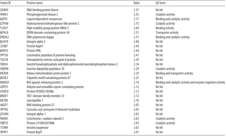
Table 1. Proteins up-regulated in iPSCMNs compared with ESCMNs (with an expression ratio >2.00)
Protein ID Protein name Ratio GO term
G5E8H3 DNA binding protein Ikaros 5.37 No hit
P09041 Phosphoglycerate kinase 2 3.35 Catalytic activity Q6ZPI3 Ligand dependent corepressor 2.77 Binding and catalytic activity Q2TPA8 Hydroxysteroid dehydrogenase like protein 2 2.75 Catalytic activity P52927 High mobility group protein HMGI-C 2.69 Binding activity Q6PAL8 DENN domain-containing protein 5A 2.51 Transporter activity Q9QUG2 DNA polymerase kappa 2.51 Binding and catalytic activity
Q62470 Integrin alpha 3 2.48 No hit
F2Z4B7 Protein Raph1 2.44 No hit
Q60953 Protein PML 2.44 No hit
E9PY58 Caseinolytic peptidase B protein homolog 2.41 No hit P70278 Stimulated by retinoic acid gene 8 protein 2.39 No hit E9Q9J4 Inositol hexakisphosphate and diphosphoinositol-pentakisphosphate kinase 2 2.34 No hit E9QN98 Inactive dipeptidyl peptidase 10 2.29 Catalytic activity
Q9CR58 Kidney mitochondrial carrier protein 1 2.29 Binding and transporter activity Q8C0E3 Tripartite motif containing protein 47 2.25 No hit
Q9QUG9 RAS guanyl-releasing protein 2 2.14 Binding and catalytic activity and enzyme regulator activity A2RT91 Ankyrin and armadillo repeat-containing protein 2.12 No hit
F6TMC0 Protein 9930021J03Rik 2.12 No hit
Q8K0F1 TBC1 domain family member 23 2.12 No hit
Q9ET80 Junctophilin 1 2.10 No hit
S4R2F7 RNA binding protein 25 2.05 No hit
E9PYH2 Cytosolic acyl coenzyme A thioester hydrolase 2.05 No hit
Q3V3R4 Integrin alpha-1 2.03 No hit
P00405 Cytochrome c oxidase subunit 2 2.03 Catalytic activity E9QP35 Protein 2310042D19Rik 2.03 Catalytic activity
F7CHR4 Inositol oxygenase 2.03 No hit
E9PXP1 Protein Brpf1 2.01 No hit
Table 2. Proteins down-regulated in iPSCMNs compared with ESCMNs (with an expression ratio <ⴚ2.00)
Protein ID Protein name Ratio GO term
E9PX29 Sptbn4 ⫺6.23 Binding and structural molecular activity
Q3UVI3 Tumor protein 63 ⫺5.53 No hit
Q9Z0R6 Intersectin 2 ⫺5.37 No hit
P23780 Beta galactosidase ⫺4.85 Catalytic activity O08644 Ephrin type B receptor 6 ⫺4.81 No hit E9QM22 WD repeat containing protein 64 ⫺4.76 No hit
P47226 Testin ⫺3.86 No hit
Q9JJ00 Phospholipid scramblase 1 ⫺3.56 No hit D3YYI5 Glyceraldehyde-3-phosphate dehydrogenase ⫺3.35 Catalytic activity B7ZCC9 Probable G-protein coupled receptor 112 ⫺3.29 Receptor activity
Q8CF02 Protein FAM25C ⫺3.25 No hit
Q9D823 60S ribosomal protein L37 ⫺3.16 Binding and structural molecular activity Q6RHR9 Membrane associated guanylate kinase WW and PDZ domain
containing protein 1
⫺3.13 Catalytic activity P97807-2 Cytoplasmic Isoform of Fumarate hydratase, mitochondrial ⫺3.03 Catalytic activity
P28658 Ataxin 10 ⫺2.77 No hit
Q920I9 WD repeat containing protein 7 ⫺2.77 Binding and structural molecular activity and enzyme regulator activity P97807 Fumarate hydratase mitochondrial ⫺2.69 Catalytic activity
P56960 Exosome component 10 ⫺2.59 Binding and catalytic activity
Q6P1J1 Crmp1 protein ⫺2.56 No hit
Q11136 Xaa-Pro dipeptidase ⫺2.53 Binding and catalytic activity and nucleic acid binding transcription factor activity E9QPI5 Sister chromatid cohesion protein PDS5 homolog A ⫺2.51 Binding
Q811D0-3 Isoform 3 of Disks large homolog 1 ⫺2.51 No hit P20060 Beta-hexosaminidase subunit beta ⫺2.51 Catalytic activity D3YU56 Inner nuclear membrane protein Man1 ⫺2.46 No hit Q9CWX2 Complex I intermediate-associated protein 30 mitochondrial ⫺2.46 No hit O70200 Allograft inflammatory factor 1 ⫺2.41 Binding
Q8BK67 Protein RCC2 ⫺2.38 Binding and catalytic and enzyme regulator activity Q8BHL5 Engulfment and cell motility protein 2 ⫺2.39 Binding
Q80SZ6 Nuclear RNA export factor 7 ⫺2.39 Binding
O35066 Kinesin-like protein KIF3C ⫺2.36 Catalytic activity and structural molecule activity P68040 Guanine nucleotide-binding protein subunit beta-2-like 1 ⫺2.34 No hit
Q9ERG0 LIM domain and actin-binding protein 1 ⫺2.34 Binding and catalytic activity and nucleic acid binding transcription factor activity and Structural molecule activity
B1AR39 Protein 2810408A11Rik ⫺2.34 No hit
E9QAF9 Protein TANC1 ⫺2.32 No hit
Q9CR98 Protein FAM136A ⫺2.32 No hit
G3UYY1 Serine hydroxymethyltransferase (Fragment) ⫺2.32 No hit
Q63918 Serum deprivation-response protein ⫺2.32 Binding and nucleic acid binding transcription factor activity
E9Q7C4 Protein Prr14l ⫺2.29 No hit
Q9R0H0-2 Isoform 2 of Peroxisomal acyl-coenzyme A oxidase 1 ⫺2.29 Catalytic activity Q684R7-3 Isoform 3 of FRAS1-related extracellular matrix protein 1 ⫺2.29 Transporter activity
Q9D7S7 60S ribosomal protein L22-like 1 ⫺2.27 Binding and structural molecule activity Q8C561 LMBR1 domain-containing protein 2 ⫺2.27 No hit
Q61526 Receptor tyrosine-protein kinase erbB-3 ⫺2.27 No hit E9PXK1 Cone-rod homeobox protein ⫺2.27 No hit
E9PW52 Protein Gm3149 ⫺2.25 No hit
Q9JIL4 Na(⫹)/H(⫹) exchange regulatory cofactor NHE_RF3 ⫺2.20 No hit Q7TPD1 F-box only protein 11 ⫺2.20 No hit
B1AVK0 Protein FAM161A ⫺2.20 No hit
Q56A10 Zinc finger protein 608 ⫺2.20 No hit
P51125 Calpastatin ⫺2.18 No hit
Q4KWH5 1-phosphatidylinositol 4 –5-bisphosphate phosphodiesterase eta-1 ⫺2.18 Binding and catalytic activity and enzyme regulator activity Q91X46 Rho guanine nucleotide exchange factor 3 ⫺2.16 Binding and catalytic activity and enzyme regulator activity
Q9D8U8 Sorting nexin-5 ⫺2.16 No hit
Q9D117 MACRO domain containing 2 ⫺2.16 No hit
P28481-2 Isoform 3 of Collagen alpha-1(II) chain ⫺2.16 Receptor activity and structural molecule activity and receptor activity P48754 Breast cancer type 1 susceptibility protein homolog ⫺2.16 Catalytic activity
Q9CWL8 Beta-catenin-like protein 1 ⫺2.16 No hit P09411 Phosphoglycerate kinase 1 ⫺2.14 Catalytic activity
Q9EQS9-3 Isoform 3 of Immunoglobulin superfamily DCC subclass member 4 ⫺2.14 Catalytic activity and receptor activity Q9D0R2 Threonine–tRNA ligase, cytoplasmic ⫺2.14 Catalytic activity
D3YU82 Probable cation-transporting ATPase 13A5 ⫺2.14 No hit
B2RXC5 Zinc finger protein 382 ⫺2.12 Binding and nucleic acid binding transcription factor activity
0.1% formic acid in acetonitrile) at 300 nl/min. The chromatographic separation gradient protocol was as follows: 3 minutes 97% A, 3% B; 5 minutes 95% A, 5% B; 90 minutes 70% A, 30% B; 95 min 3% A, 97% B; 97 minutes 3% A, 97% B; 100 min, 97% A, 3% B; 120 minutes 97% A, 3% B.
Mass spectra were acquired on a Thermo Orbitrap Velos Pro using Xcalibur software. Data were acquired using data-directed analysis in which the m/z values of tryptic peptides were measured using an MS scan in FT-MS mode followed by MS/MS scans of the 10 most intense peaks using IT-MS mode.
Data analysis was conducted using Proteome Discoverer for pro-tein identification and Sieve for chromatographic alignment, normal-ization, and peak integration. Data were then extracted from the Sieve sdb database files using a series of custom scritpts written in R. Protein ontologies were assigned using gene list analysis tools in PANTHER (www.pantherdb.org).
In ovo transplantation of iPSCMNs and immunohistochemistry. Trans-plants were grafted into male and female chick embryos as described previously (Soundararajan et al., 2006) in accordance with the guidelines of the Canadian Council on Animal Care and the Dalhousie University Committee on Laboratory Animals.
For immunohistochemical analyses, the following primary antibodies were used: rabbit anti-GFP (Millipore Bioscience Research Reagents, 1:1000), mouse anti-Tuj1 (Covance, 1:1000). Slides were incubated with primary antibodies in a solution of 0.3% Triton X/PBS with goat serum overnight. Slides were incubated with the following secondary anti-bodies in 0.3% Triton X/PBS solution: goat anti-mouse Cy3 (Jackson Immunoresearch Laboratories, 1:500) and goat anti-rabbit Alexa Fluor 488 (Invitrogen, 1:500) for 1 h at room temperature. Images were captured with a digital camera (C4742; Hamamatsu Photonics) in conjunction with digital imaging acquisition software (IPLab; Ver-sion 4.0; BD Biosciences).
Analyses of axonal projections from the chick spinal cord were per-formed as follows: using Neurolucida projections from three separate embryos, the width of a line drawn across the eGFP⫹fascicle projecting dorsally toward the epaxial muscles was compared with the width drawn across eEGFP⫹fascicles projecting ventrally toward the limb. The cross-sectional areas of the fascicles were then calculated (assuming a circular area, r2) and relative sizes compared.
iPSCMN/myofiber cocultures and immunocytochemistry. For
generat-ing myofibers, the medial head of the adductor superficialis muscles of Hamburger Hamilton (HH) St. 36 chicks were isolated and plated on 13 mm sterile thermanox plastic coverslips (Nunc) in 24-well plates as de-scribed previously before addition of iPSCMNs (Chipman et al., 2014). For immunohistochemical analysis of the cocultures, cells were incu-bated in a solution containing rabbit anti-GFP IgG (Millipore Bioscience
Research Reagents, 1:2000), mouse anti-SV2 (1:50, DSHB) primary an-tibodies and goat serum for ⬃1 h. Cells were then washed in PBS multiple times for 30 minutes before incubating in goat anti-rabbit Alexa Fluor 488 (Invitrogen, 1:500), goat anti-mouse Alex Fluor 647 (Invitrogen, 1:500), and tetramethylrhodamine-conjugated (TMR) ␣-bungarotoxin (btx; Invitrogen, 1:500). All antibodies were in a 0.3% Triton X/PBS solution. Images were acquired on a laser scanning confocal microscope (Zeiss LSM 510 or 710) or with a digital camera (C4742; Hamamatsu Photonics). Intensity of SV2 immunoreactivity and acetylcholine recep-tor area (based on TMR ␣-btx labeling) were quantified using IPLab software (version 4.0; BD Biosciences). For each measurement of inten-sity of SV2 immunoreactivity, background inteninten-sity was subtracted from the signal. Orthogonal images were rendered and edited with LSM im-aging software (Zeiss) and further contrast and brightness adjustments were performed on Adobe Photoshop version CS5.
FM4-64FX dye loading and imaging. To assess vesicular cycling at
NMJs, cocultures were incubated with 5 MFM4-64FX (hereafter
re-ferred to as FM4-64) and motor terminals of iPSCMNs were loaded by electrical stimulation. Experiments were then conducted as described previously (Chipman et al., 2014).
Intracellular recordings of iPSCMN-chick myofiber cocultures. Sharp
electrode recording techniques were used to assess synaptic function at 12- to 27-d-old cocultured NMJs as described previously (Miles et al., 2004;Soundararajan et al., 2007;Chipman et al., 2014). All cells recorded from had resting membrane potentials that varied from ⬃⫺25 to ⫺65 mV, values that were similar to those previously reported for postsynap-tic membrane potentials of in vitro rat spinal cord–myotube coculture (Robbins and Yonezawa, 1971).
Whole-cell patch-clamp recordings of iPSCMNs. iPSCMNs on
Matrigel-coated coverslips were continuously perfused in a recording chamber with oxygenated (95% O2⫹ 5% CO2) Ringer’s solution containing the following (in mM): 111 NaCl, 3.08 KCl, 11 glucose, 25 NaHCO3, 1.25 MgSO4, 2.52 CaCl2, and 1.18 mMKH2PO4, pH 7.4, at room temperature for ⬃20 minutes before recording to allow the cells to adjust to recording conditions. Perfusion continued throughout the recordings, in which a DAGE-MTI IR-1000 CCD camera connected to an Olympus BX51WI microscope was used to visualize eGFP⫹iPSCMNs. Recordings were made in current-clamp mode using a MultiClamp 700B amplifier (Mo-lecular Devices). A Digidata 1400A board (Mo(Mo-lecular Devices) controlled by pCLAMP10.3 (Molecular Devices) was used to filter analog signals at 10 kHz. Recording solution containing the following (in mM): 128
K-gluconate, 4 NaCl, 0.0001 CaCl2, 10 HEPES, 1 glucose, 5 Mg-ATP, 0.3 GTP-Li, pH 7.2, was loaded into patch-clamp recording pipettes with a resistance of 4 –7 M⍀. Next, 0.4 mg/ml lucifer yellow dilithium salt (Sigma-Aldrich) was added to the pipette solution before recording to allow visualization of recorded iPSCMNs. To ensure similar measuring
Table 2. Continued
Protein ID Protein name Ratio GO term
E9QNJ9 Fibroblast growth factor receptor 3 ⫺2.12 No hit
Q9JII6 Alcohol dehydrogenase 关NADP(⫹)兴 ⫺2.12 Catalytic activity and transporter activity
Q9CZP7 Hsp90 co-chaperone Cdc37-like 1 ⫺2.12 Binding and catalytic activity and enzyme regulator activity H3BJZ2 Protein Cdhr4 ⫺2.12 Binding and receptor activity
P53996 Cellular nucleic acid-binding protein ⫺2.10 Binding
Q80U87 Ubiquitin carboxyl-terminal hydrolase 8 ⫺2.10 Binding and catalytic activity E9Q1W0 Calcium/calmodulin-dependent protein kinase type II subunit beta ⫺2.10 No hit
J3QPC5 Protein Zfp850 ⫺2.10 No hit
P29391 Ferritin light chain 1 ⫺2.08 No hit
Q61771 Kinesin-like protein KIF3B ⫺2.08 Catalytic activity and structural molecule activity O89086 Putative RNA-binding protein 3 ⫺2.08 Binding and catalytic activity and structural molecule activity Q80US4 Actin-related protein 5 ⫺2.08 Structural molecule activity
P53996-2 Isoform 2 of Cellular nucleic acid-binding protein ⫺2.08 Binding
Q64331 Unconventional myosin-VI ⫺2.08 No hit
Q61176 Arginase-1 ⫺2.08 Catalytic activity
P97313 DNA-dependent protein kinase catalytic subunit ⫺2.03 Binding and catalytic activity Q8K3P5 CCR4-NOT transcription complex subunit 6 ⫺2.03 Binding and catalytic activity
O08677 Kininogen-1 ⫺2.03 No hit
conditions, all cells were held at ⫺60 mV with a tonic DC current. Data were obtained by Clampex 10.3 (Molecular Devices) and analyzed by AxoScope 10.2 (Molecular Devices) software. Input resistance and ca-pacitance of iPSCMNs were calculated by measuring response to repeti-tive, small negative steps of ⫺10 mV for 100 ms. For investigating firing properties of iPSCMNs, 1 s pulses of depolarizing current were delivered in increments of 5, 10, or 20 pA. Data were analyzed and statistics were generated with SigmaPlot 11 software (Systat Software).
iPSCMN implantation and force recording. All procedures performed
were approved by the Dalhousie Animal Care Committee and com-plied with the Canadian Council of Animal Care. All surgeries were
performed on male mice. The surgical technique used in this study has been described previously (Yohn et al., 2008). Immunohisto-chemistry performed on sections of nerve and muscle was conducted as described above for chick sections (1° antibodies used: TMR ␣-btx; Invitrogen), rabbit anti-GFP (Millipore Bioscience Research Re-agents). Feret’s diameter of MG muscles and muscle fibers was mea-sured using ImageJ software.
Statistical analyses. All statistics were performed using SigmaPlot 11
Software (Systat). Values are cited as mean ⫾ 1 SD. All tests used are described in the text.
Results
Protein expression is comparable between iPSCMNs
and ESCMNs
Throughout this study, we used a mouse iPSC line derived from
transgenic mice (B6.Cg-Tg(Hlxb9-EGFP)1Tmj/J) expressing
eGFP under the direction of the motoneuron-specific promoter
Hb9 (
Arber et al., 1999
;
Wichterle et al., 2002
). Approximately
20% (mean: 21.3% ⫾ 3.09) of the cells from this iPSC line
differ-entiated into GFP
⫹motoneurons when treated with retinoic acid
(RA) and a smoothened agonist (SAG). This is not significantly
( p ⫽ 0.162, n ⫽ 3) different from the percentage of motoneurons
derived from mouse HBG3 ESCs using the same differentiation
protocol (mean: 17.5% ⫾ 2.29). Therefore, the propensity for
mouse iPSCs and ESCs to differentiate into motoneurons when
treated with RA and SAG is the same.
To determine whether there are differences between
motoneu-rons derived from HBG3 ESCs (ESCMNs) and iPSC-derived
mo-toneurons (iPSCMNs), we first undertook an unbiased proteomic
approach and compared protein expression profiles between
iPSCMNs and ESCMNs. Here, ESCMNs and iPSCMNs were
sorted by fluorescence-activated cell sorting after differentiation
before proteomic analysis. After trypsin digestion of the proteins,
we performed a label-free, LC-MS/MS analysis of the resulting
peptides to compare expression profiles, as described in the
Ma-terials and Methods section. Quantification was perfomed using
peptide peak areas and peptides with good precision (relative SD
⬍30%) were retained. These peptides were then filtered to
elim-inate peptides that match to multiple proteins. The unique
pep-tides were then averaged to give overall expression levels for 3025
proteins. We next imported the dataset into Panther (
www.
pantherdb.org
) to annote known gene ontology (GO) terms to
determine whether there were overall differences in the types of
proteins identified in our screen. More than half (1531) of the
proteins in the dataset matched a GO term. The half that did not
match were likely unstudied proteins with no known function.
When the 1531 proteins were grouped into each GO term, we
found that the number (shown as a percentage) of proteins
within each group was identical (
Fig. 1
A). This indicates that the
types of proteins identified by the screen were very similar in the two
cell types. Further analysis showed that ESCMNs and iPSCMNs
ex-press the same proteins of each GO term except for the general
bind-ing category (
Fig. 1
B), in which ⬎500 general binding proteins were
the same in ESCMNs and iPSCMNs: 93 were unique to
ESCMNs and 50 to iPSCMNs (
Fig. 1
B).
To determine whether the same proteins in ESCMNs and
iPSCMNs are expressed at similar levels, we integrated the
inten-sity of each peptide and then averaged the peptides for any given
protein to provide a fold change, defined here as the protein
expression from the iPSCMNs divided by the intensity in the
ESCMNs. Based on the variability measured, a p-value was also
calculated, as shown in
Figure 1
C. We detected 28 proteins
up-regulated in iPSCMNs and 87 downup-regulated proteins (
Tables 1
,
2
, respectively). Therefore, of the original number of proteins
Figure 2. The vast majority of iPSCMNs express the LIM/homeobox protein Lhx3. Ai–Aiii, Immunolabeling showing that the majority of the eGFP⫹iPSCMNs expressed Lhx3 after 2 d in vitro. Bi–Biii, In contrast, very few eGFP⫹iPSCMNs expressed Lhx1 after the same time period. C, Percentage of Lhx3/eGFP and Lhx1/eGFP⫹cells after 2 d in vitro (n ⫽ 3 for each group). Scale bars, 10 m.
identified, ⬍4% met our criteria for being differentially regulated
(i.e., a ln expression ratio ⬎0.7 or ⬍⫺0.7 in
Fig. 1
C).
Interest-ingly, the degree of differential regulation was also modest, with
the average fold change for the upregulated and downregulated
proteins being 2.4 and ⫺2.5, respectively (
Tables 1
,
2
).
Finally, we focused on several transcription factors known to
be involved in neuron and/or motoneuron differentiation
(
Alaynick et al., 2011
;
Son et al., 2011
). Although we focused on a
number of transcription factors, including Olig2, Nkx6.1,
Nkx6.2, Ngn1, Ngn2, Lhx3, Hb9, Isl1, Isl2, Lmo4, Lhx1, FoxP1,
Ascl1, Myt1l, and Brn2, we only detected Myt1l in our dataset.
Nine unique peptides were identified for Mytl1, so it was
conclu-sively identified. Myt1l was not differentially regulated between
the two samples (ratio of 1.1, p-value ⫽ 0.5). Together, these
results indicate that ESCMNs and iPSCMNs contain similar
pro-teins at comparable levels of expression. The few propro-teins that were
differentially expressed are not directly involved in motoneuron
differentiation.
iPSCs preferentially differentiate into
Lhx3
ⴙmotoneurons when treated with
RA and SAG
Previous studies showed that mouse ESCs
treated with SAG and RA preferentially
differentiate into a specific subpopulation
of motoneurons expressing the LIM
ho-meoprotein Lhx3 (
Soundararajan et al.,
2006
;
Soundararajan et al., 2007
;
Sounda-rarajan et al., 2010
). Lhx3
⫹motoneurons
reside in the medial aspect of the medial
motor column and innervate epaxial
muscles lining the vertebral column
(
Tsuchida et al., 1994
). To determine
whether iPSCs differentiate into the same
motoneuron subset, we plated dissociated
embryoid bodies containing eGFP
⫹iP-SCMNs onto a Matrigel substrate and
cul-tured them for an additional 2 d. The cells
were then fixed and immunostained for
LIM homeodomain proteins Lhx3 or
Lhx1, the latter being a marker of
mo-toneurons innervating the dorsal muscle
mass in limbs (
Tsuchida et al., 1994
;
Fig.
2
A, B). As observed with ESCMNs
(
Soundararajan et al., 2006
;
Soundarara-jan et al., 2007
;
Soundararajan et al.,
2010
), we found that the vast majority of
the eGFP
⫹iPSCMNs were Lhx3
⫹(82 ⫾
3.7%, n ⫽ 3), whereas only 13 ⫾ 1.4%
(n ⫽ 3) expressed Lhx1 (
Fig. 2
C). These
results suggest that the process of
mo-toneurogenesis using SAG and RA is
sim-ilar between iPS and ESCs.
iPSCMNs project axons to peripheral
targets appropriate for Lhx3
ⴙmotoneurons when transplanted into
the developing chick spinal cord
In the spinal cord, Lhx3
⫹motoneurons
reside in the medial aspect of the medial
motor column (
Sharma et al., 1998
). In
the periphery, their axons selectively
proj-ect to epaxial muscles lining the vertebral
column (
Tosney and Landmesser, 1985
).
This selective guidance of axons is orchestrated, at least in part, by
intracellular signaling pathways that are activated downstream of
Lhx3 expression (
Sharma et al., 2000
;
Soundararajan et al., 2010
).
As a result, mouse Lhx3
⫹ESCMNs selectively—and correctly—
extend axons to epaxial muscles when transplanted into the
neu-ral tube of developing chick embryos at the time of
motoneurogenesis (
Soundararajan et al., 2006
;
Soundararajan et
al., 2007
;
Soundararajan et al., 2010
; i.e., HH St. 17).
To determine whether similar guidance mechanisms direct the
growth of axons from motoneurons derived from iPSCs, we
transplanted eGFP
⫹iPSCMNs into the neural tube of HH St. 17
chick embryos (n ⫽ 7). The embryos were killed 5 d later, fixed,
sectioned, and processed for Tuj1 and eGFP
immunohistochem-istry to visualize chick and transplanted neurons, respectively.
Figure 3
shows a representative pattern of axonal growth from
one transplanted embryo. eGFP
⫹axons extended out of the
chick spinal cord via the ventral root (
Fig. 3
A, D, short arrow)
around the dorsal root ganglia (DRG;
Fig. 3
A, D, arrow) to
inner-Figure 3. iPSCMNs preferentially project axons to epaxial muscles when transplanted in ovo. A–C, Cross-section through a HH St. 31 chick embryo showing eGFP⫹motor axons extended out of the spinal cord through the ventral root (A, short arrow). The majority of the eGFP⫹axons extended around the DRG (A, arrow) and into epaxial muscles (B, white arrowhead). A few eGFP⫹ axons extended ventrally into the limb bud (C, yellow arrowhead). All sections were immunolabeled with Tuj1 to visualize the endogenous chick neurons (red) and transplanted eGFP⫹iPSCMNs (yellow). D, Neurolucida reconstruction of all cross-sections from one chick embryo receiving an iPSCMN transplant. eGFP⫹iPSCMNs are shown in green. Scale bar, 100 m.
vate epaxial muscles, the appropriate
tar-get for Lhx3
⫹motoneurons (
Fig. 3
B, D,
white arrowhead). However, consistent
with the transplants containing a small
proportion of Lhx1
⫹iPSCMNs (see
above), a few eGFP
⫹axons projected into
the developing hindlimb bud (
Fig. 3
C,D,
yellow arrowhead), an appropriate target
for Lhx1
⫹motoneurons. To estimate the
relative number of eGFP
⫹axons
project-ing to the epaxial muscles and limb buds,
we created Neurolucida reconstructions
of the axonal projection patterns from
each coronal section taken from three
transplanted embryos. We then measured
the width of the projections and found
that 4.5 ⫾ 2.0 times as many eGFP
⫹axons
projected to the epaxial muscles
com-pared with the limb. This number is close
to the expected distribution of axonal
pro-jections based on the relative number of
Lhx3
⫹to Lhx1
⫹iPSCMNs in the
trans-plants (i.e., the grafts contain ⬃6.2 times as
many Lhx3
⫹motoneurons as Lhx1
⫹neu-rons;
Fig. 2
C). Therefore, the pattern of
axonal growth accurately reflects the
phenotypic identity of the transplanted
iPSCMNs and that iPSCMNs use the
same axonal guidance mechanisms as
their endogenous Lhx3
⫹and Lhx1
⫹counterparts.
iPSCMNs form functional NMJs
in vitro
Previous studies showed that ESCMNs
form functional connections with muscle
fi-bers in vitro (
Miles et al., 2004
;
Soundarara-jan et al., 2007
). To determine whether
iPSCMNs have the same capacity, we first
performed immunocytochemical analysis
of presynaptic and postsynaptic structures
associated with neurotransmission at the
NMJ using cocultures containing eGFP
⫹iPSCMNs and chick myofibers (
Miles et
al., 2004
). In the first week after plating,
numerous eGFP
⫹neurites were observed
to extend radially from spherical clusters
of iPSCMNs along individual chick
myo-fibers (
Fig. 4
A). In addition, discrete, oval,
plaque-like clusters of ␣-btx
⫹AChRs
were observed around the neurites (
Fig.
4
B–F ). This pattern of clustering closely
resembles normal in vivo development
where AChRs first cluster near, but not
always in direct contact with, innervating
motor axons (
Lupa and Hall, 1989
;
Dahm
and Landmesser, 1991
). The oval shape
and sizes of the plaques (41 ⫾ 24 m
2;
n ⫽ 60, 20 plaques measured in each of 3
separate cocultures) were similar to AChR
clusters found on developing muscles
fi-bers in ovo during the first week of
mo-toneuron innervation (i.e., E9;
Phillips
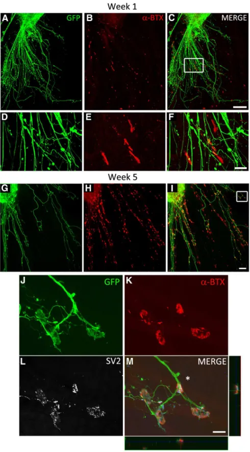
Figure 4. iPSCMNs form NMJs with cocultured chick myofibers. A–C, eGFP⫹motoneurons extend axons along myofi-bers (A) and associate with plaques of ␣-btx-labeled AChRs during the first week in culture (B, C). D–F, Boxed area in C. G–I, Contact between eGFP⫹axons and AChRs continues into week 5, when AChR clusters become much larger and maintained at myofiber regions in close proximity to eGFP⫹axons. J–L, Boxed area in I shows eGFP⫹axons (J ) contacting ␣-btx labeled AChRs with pretzel-like morphologies (K ). SV2⫹synaptic vesicles are prominent at the end-plate in the presynaptic axon (L). M, Merged image of J–L and x-z and y-z orthogonal planes of the NMJ indicated by the asterisk. Note the blue signal in the orthogonal planes and merge panel is SV2 labeling. Scale bars C, I, 100 m; F, M, 20 m.
et al., 1985
). iPSCMNs continued to maintain the eGFP
⫹neu-rites after 4 weeks in vitro (
Fig. 4
G); however, at that point, the
individual clusters of ␣-btx
⫹AChRs at the end of eGFP
⫹neurites formed structures resembling mature NMJs (
Fig.
4
H, I ). Higher magnification showed that ␣-btx
⫹AChR
clus-ters were more complex, pretzel-shaped structures (
Fig.
4
J, K ). These endplates were also significantly larger than
AChR clusters (plaques) present during the first week in
cul-ture (95 ⫾ 37 m
2, n ⫽ 60, 20 endplates measured in 3
sepa-rate cocultures, p ⬍ 0.001, Mann–Whitney rank-sum test).
This change in morphology is similar to what occurs in vivo as
the NMJs mature postnatally (
Slater, 1982
;
Balice-Gordon et
al., 1993
). By the fifth week in culture, SV2
⫹puncta were
enriched at the ␣-btx
⫹AChR rich endplates relative to the axonal
shaft (
Fig. 4
L,M). Together, this suggests that the eGFP
⫹neurites
extend from the iPSCMNs and make appropriate synaptic
connec-tions with chick myotubes.
We next investigated whether synaptic vesicles actively cycle
at the endplate to determine whether the NMJs were functional
by incubating 4-week-old iPSCMN–myofiber cocultures with
FM4-64 (
Betz and Bewick, 1992
;
Gaffield and Betz, 2006
). FM
dyes are widely used to image synaptic vesicle cycling at the NMJ
because the dye is readily taken up from the extracellular medium
into endocytosed vesicles that have fused with the plasma
mem-brane during nerve stimulation. Once the dye is trapped inside a
vesicle, it can only escape from the NMJ by subsequent exocytosis
of the vesicle (
Gaffield and Betz, 2006
). Consistent with the
for-mation of functional synapses, we found that FM4-64 was readily
taken up into cycling vesicles at presynaptic terminals using 50
Hz field electrical stimulation (1 s train of pulses, every 2 s for 5
minutes;
Fig. 5
A–D). As expected, the vast majority of the FM4-64
puncta (
Fig. 5
A,D) were adjacent to ␣-btx
⫹AChRs (
Fig. 5
C,D). We
then performed intracellular sharp electrode recordings from NMJs
labeled with a rhodamine-conjugated AChR antibody. Endplates
chosen for recordings (n ⫽ 22; in 12- to 27-d-old cocultures) had
mature morphologies (i.e., pretzel-shaped distribution of
AChRs) and were contacted by a single eGFP
⫹axon (
Fig. 6
A, B).
The amplitude of the endplate potentials
(EPPs) recorded varied between ⬃0.5 and
6 mV, which is comparable to those
re-corded from myofibers cocultured with
embryonic rate spinal cord explants (
Rob-bins and Yonezawa, 1971
), a neuronal cell
line (
Chen et al., 2001
), and ESCMNs
(
Miles et al., 2004
).
Figure 6
C, top trace,
shows 3 EPPs during a typical 5 s
record-ing in normal solution; the bottom trace
shows a similar recording in the presence
of 100
Mglutamate. The increased
fre-quency and amplitude of the EPPs in the
presence of glutamate indicates that,
like ESCMNs (
Miles et al., 2004
) and
endogenous motoneurons (
Jiang et al.,
1990
), iPSCMNs express functional
glu-tamatergic receptors. Bath application
of 25
MTTX completely blocked the
large EPPs (⬎ 1.5 mV), but not the small
EPPs (
Fig. 6
D). These results indicate
that the smaller EPPs were miniature
EPPs (mEPPs), whereas the larger EPPs
resulted from evoked neurotransmitter
release induced by action potentials in
the motoneuron. As expected, all EPPs
were blocked shortly after bath application of 100 –300
M D-tubocurarine (
Fig. 6
E; n ⫽ 3), indicating that the EPPs were
due to nicotinic neurotransmission. Together, these results
indicate that iPSCMNs form functional NMJs similar to
en-dogenous motoneurons and their ESCMN counterparts.
iPSCMNs develop appropriate passive membrane and
firing properties
Motoneurons in the spinal cord undergo significant
physiologi-cal changes during early postnatal development (
Fulton and
Wal-ton, 1986
;
Gao and Ziskind-Conhaim, 1998
;
Nakanishi and
Whelan, 2010
). For example, input resistance decreases but
whole-cell capacitance increases during the first 2 weeks of
post-natal life. These changes in passive membrane properties reflect
an overall increase in cell size and alteration in channel
proper-ties, both of which set the threshold for motoneuron excitability
(
Pinter et al., 1983
;
Fulton and Walton, 1986
;
Gao and
Ziskind-Conhaim, 1998
). Setting an appropriate threshold for activation
is an essential feature of motoneurons because it ensures proper
motoneuron recruitment and force gradation during muscle
contraction.
To ascertain whether iPSCMNs develop appropriate passive
membrane properties over time, we performed whole-cell
patch-clamp recordings on iPSCMNs cultured for 1– 6 weeks on
Matrigel-coated coverslips. We found that membrane input
re-sistance (R
m) decreased between 1–2 and 3– 4 weeks (666 ⫾ 574
to 189 ⫾ 105 M⍀;
Table 3
). This decrease in resistance correlated
well with the simultaneous increase in membrane capacitance
(C
m; 45 ⫾ 27 pF to 121 ⫾ 45;
Table 3
). These changes suggest that
the average size and/or membrane area of the iPSCMNs increased
over the first few weeks in culture, although significant size
vari-ations remained. The resting membrane potential of iPSCMNs,
however, remained similar at all time points investigated (
Table 3
).
These results are similar to those observed in cats and rats, in which
input resistance decreases overtime (
Fulton and Walton, 1986
;
Xie and
Ziskind-Conhaim, 1995
;
Gao and Ziskind-Conhaim, 1998
), whereas
the resting membrane potential does not change between late
embry-Figure 5. Active vesicular cycling occurs at NMJs formed between iPSCMNs and chick myofibers. A–C, FM4-64 puncta (A) is present in eGFP⫹axons (B) at endplates labeled with ␣-btx (C) after field electrical stimulation. D, Merged image of A–C where the blue signal is FM4-64. Scale bar, 20 m.
onic and early postnatal time periods (
Xie and
Ziskind-Conhaim, 1995
).
The probability of generating repetitive
action potentials in spinal motoneurons
in-jected with a sustained depolarizing
cur-rent increases between late embryogenesis
and early postnatal life (
Fulton and
Wal-ton, 1986
;
Gao and Ziskind-Conhaim,
1998
). This change reflects maturation in
channel properties over time. To
deter-mine whether channel properties in
iPSCMNs mature over time, we applied
stepwise injections of depolarizing current
to iPSCMNs grown in culture for 1, 2, and 5
weeks.
Figure 7
shows that iPSCMNs were
able to generate repetitive action
poten-tials at all three time points (
Fig. 7
A).
However, the amount of current required
to evoke repetitive action potentials
de-creased between 1 and 5 weeks (
Fig. 7
A).
In addition, the maximum firing rate
in-creased over time in culture (
Fig. 7
A, C)
such that, by 5 weeks, the average
fre-quency during the late phase of activity
(⬎250 ms) was 41 ⫾ 10 Hz (n ⫽ 3). This
firing rate is comparable to the firing
pat-terns of individual motor units in freely
walking rats (
Hennig and Lømo, 1985
).
Finally, the discharge rate of the evoked
action potentials decreased overtime
dur-ing the sustained injection of depolarizdur-ing
current at all three time points (
Fig. 7
A,B).
This phenomenon, known as spike-frequency
adaptation, is a typical firing pattern of spinal
motoneurons (
Granit et al., 1963
;
Kernell and
Monster, 1982
).
Action potential duration decreases in
motoneurons between late embryonic to
early postnatal life (
Gao and
Ziskind-Conhaim, 1998
). The same was true for
cul-tured iPSCMNs (
Fig. 8
A, B), in which
action potential duration (measured at
half-maximal peak amplitude in sweeps in
which a single spike was generated) was
found to decrease from 5.9 ⫾ 1.6 ms
(n ⫽ 11) at 1–2 weeks to 2.29 ⫾ 0.43 ms
(n ⫽ 10) by 5– 6 weeks. Furthermore, like
embryonic rat motoneurons (
Gao and
Ziskind-Conhaim, 1998
), only a minority
(38%, n ⫽ 13) of the recorded iPSCMNs
elicited an afterhyperpolarizing potential
(AHP) after 1 week in vitro. This
percent-age increased to 67% after 5– 6 weeks in
culture (n ⫽ 15). Together, these findings
suggest that cultured iPSCMNs mature
over time and develop passive membrane
and firing properties similar to their
en-dogenous counterparts.
Implanted iPSCMNs innervate denervated muscle fibers and
restore force after peripheral nerve injury
Embryonic rat ventral spinal cord cells (
Thomas et al., 2000
;
Liu
et al., 2013
) and mouse ESCMNs (
Yohn et al., 2008
) restore
skel-etal muscle function when transplanted into the distal stump of
transected tibial nerves. To determine whether iPSCMNs have
the same capacity, we cut the sciatic nerve and implanted
⬃10,000 iPSCMNs into the tibial nerve in mice ⬃15 mm
proxi-Figure 6. iPSCMNs form functional NMJs when cocultured with chick myofibers. A, DIC image of a chick myofiber impaled with a sharp electrode for intracellular recordings (placement of electrode indicated by white lines). B, Innervated endplates were identified as a cluster of mAb35-labeled AChRs contacted by a single eGFP⫹. Asterisks in A and B denote region impaled by the recording electrode. C, Top trace shows EPPs recorded over a 5 s period in normal solution; bottom trace shows recording from the same cell after bath application of 100 Mglutamate. Note the increase in number of EPPs. D, EPPs with an amplitude ⬎1.5 mV
were inhibited after bath application of 25 MTTX (right). Left, Recording from the same cell before TTX application. Histograms
show the amplitudes of all EPPs recorded for 90 s in the absence (left) or presence (right) of TTX. E, Intracellular recordings in the absence (left) or presence (right) of bath appliedD-tubocurarine (300 M). Note the complete absence of EPPs in the right panel.
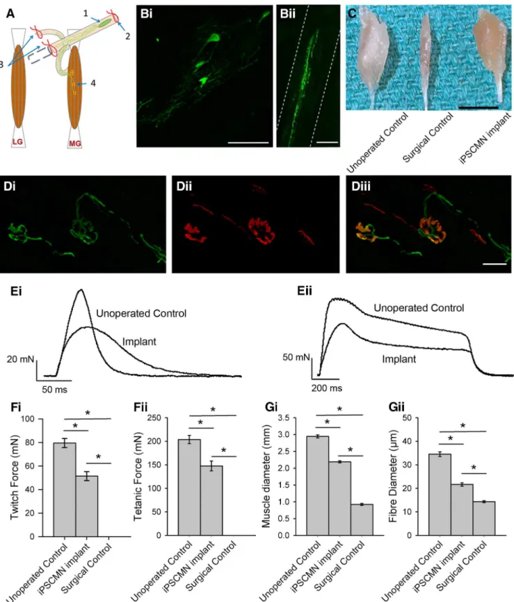
mal to the medial gastrocnemius (MG) muscle. Reinnervation of
muscles other than the MG was prevented by ligating all of the
tibial nerve branches distal to the graft site (
Fig. 9
A; see also
Yohn
et al., 2008
).
Figure 9
shows an example of a typical graft
contain-ing multiple eGFP
⫹iPSCMNs (
Fig. 9
Bi) extending neurites
along the length of the distal tibial nerve stump (
Fig. 9
Bii) 5 weeks
after transplantation. To assess synapse formation, we used
rhodamine-conjugated ␣-bungarotoxin to label AChRs at the
MG motor endplates.
Figure 9
D shows an example of two eGFP
⫹axons with typical presynaptic morphologies (
Fig. 9
Di)
innervat-ing two BTX
⫹motor endplates (
Fig. 9
Dii). These data
collec-tively demonstrate that implanted iPSCMNs survive in a host
peripheral nerve, extend axons, and form morphologically
ap-propriate connections with denervated muscle fibers.
To determine whether the contacted endplates formed
func-tional NMJs, we used an ex vivo nerve/muscle preparation to
measure contractile force of the reinnervated MG muscles.
Iso-metric twitch and tetanic contractions were elicited by applying
single and repetitive electrical pulses (50 Hz for 1 s; 20 s pulse
duration) to the distal nerve stump via a suction electrode.
Rein-nervated muscle forces were compared with both an
age-matched, unoperated control MG muscle group in which the
nerve was left intact and a surgical muscle control group in which
the sciatic nerve was cut and ligated to prevent regeneration. The
transected tibial nerve stumps of the surgical control group were
injected with media alone. As expected, electrical stimulation of the
tibial nerve stump in the control animals did not elicit muscle
con-traction (data not shown). In contrast, nerve/muscle preparations
from mice implanted with iPSCMNs 6 weeks previously produced
an average twitch force of 51.4 ⫾ 6.5 mN (n ⫽ 3) and tetanic force
of 147 ⫾ 18 mN (n ⫽ 3), respectively (
Fig. 9
Fi,Fii). These values
are ⬃2/3 that of unoperated age-matched control muscles, which
produced twitch and tetanic forces of 80 ⫾ 7 mN (n ⫽ 3) and
203 ⫾ 15 mN (n ⫽ 3), respectively. Interestingly, the contractile
forces of the muscles reinnervated by iPSCMNs, when calculated
as a percentage of unoperated control values, were greater than
those reported for rat and mouse MG muscles reinnervated by
embryonic spinal cord cells (
Thomas et al., 2000
) or ESCMNs
(
Yohn et al., 2008
). Although considerable force recovery was
observed after implantation of iPSCMNs, twitch force profiles
differed between treated and control groups (
Fig. 9
Ei). Rise times
to peak force were significantly longer in the implanted group
(76 ⫾ 12 ms) compared with control (47 ⫾ 5 ms; n ⫽ 3 for each
group, p ⫽ 0.022, t test), and relaxation times (measured as the
time taken for the force to decline to half peak force value) were
also significantly longer in the implanted group (implanted
group ⫽ 52 ⫾ 12 ms, control group ⫽ 28 ⫾ 8 ms; n ⫽ 3 for each
group, p ⫽ 0.046, t test). As expected from the force recordings,
denervation-induced muscle atrophy was attenuated
dramati-cally when the muscles were reinnervated by iPSCMNs (
Fig. 9
C).
MG muscle diameter of implanted animals was significantly
greater than those of surgical controls (control group ⫽ 2.94 ⫾
0.08 mm, implanted group ⫽ 2.19 ⫾ 0.05 mm, surgical control
group ⫽ 0.92 ⫾ 0.05 mm; n ⫽ 3 for each group, p ⬍ 0.001,
one-way ANOVA, Holm–Sidak method;
Fig. 9
Gi). Further, the
diameters of individual muscle fibers in the implanted group
were significantly greater than those of the surgical control group
(control ⫽ 34.5 ⫾ 9.4 m, implanted group ⫽ 21.6 ⫾ 6.9 m,
surgical control group ⫽ 14.3 ⫾ 3.9 m; n ⫽ 90 fibers, n ⫽ 3
animals for each group, p ⬍ 0.001, one-way ANOVA, Holm–
Sidak method;
Fig. 9
Gii). Together, these results indicate that
iPSCMNs form functional connections with denervated muscles
fibers and that this innervation limits denervation atrophy.
Discussion
The present study provides a thorough assessment of iPSCMNs
with respect to their capacity to develop into spinal
motoneu-rons. Proteomic analysis revealed that iPSCMNs and ESCMNs
have comparable protein expression profiles. The identified
pro-teins in the ESCMNS and iPSCMNs were not only similarly
grouped into known GO terms, but the proteins in each group
were the same except for 143 binding proteins that were uniquely
expressed in ESCMNs or iPSCMNs. These results indicate that
the cellular makeup of ESCMNs and iPSCMNs are remarkably
similar. In addition, when protein expression levels were
com-pared between the two cell types, we found only 28 proteins to be
upregulated and 87 proteins downregulated in iPSCMNs
com-pared with ESCMNs. None of the upregulated or downregulated
proteins is known to play a significant role in motoneuron
differ-entiation. The remarkable similarities in protein identification
and expression between ESCMNs and iPSCMNs suggest that
these two cell types are very similar in nature. It will be interesting
nonetheless to pursue whether proteins identified as being
differ-entially regulated can affect motoneuron phenotypes that are
more subtle than those used in this study. For example,
Be-taIVSigma1 spectrin (Sptbn4 in
Table 2
) is concentrated at the
nodes of Ranvier in the peripheral nervous system (
Berghs et al.,
2000
) and regulates, at least in part, its size (
Lacas-Gervais et al.,
2004
). Whether the nodes of Ranvier would differ between iPSCMNs
and ESCMNs remains to be determined.
Interestingly, even though we focused on 15 transcription
fac-tors involved in neuron and/or motoneuron differentiation, only
Myt1l was identified in our dataset. Myt1l was not differentially
expressed in ESCMNs and iPSCMNs. The reasons that we did not
detect more transcription factors involved in motoneuron
differ-entiation in unclear, but likely reflects the proteomic approach
used in this study. Some peptides are better at being ionized and
thus are more amenable to identification in a mass spectrometer.
Future studies could use a more targeted approach using multiple
reaction monitoring analysis to identify all known transcription
factors involved. Despite this limitation, the present study
indi-cates that the protein compositions of motoneurons derived
from ESCs and iPSCs are remarkably similar.
Axonal trajectories of iPSCMNs are appropriate for their LIM
homeodomain expression pattern
Similarities in LIM homeodomain protein expression between
ESCMNs and iPSCMNs suggest that they undergo similar
devel-opmental programs when cultured in the presence of SAG and
RA. The vast majority of ESCMNs (
Soundararajan et al., 2006
)
and iPSCMNs were immunopositive for the Lhx3 (
Fig. 2
), a LIM
homeodomain protein expressed by motoneurons in the medial
aspect of the medial motor column (
Sharma et al., 1998
). Like
ESCMNs (
Soundararajan et al., 2006
;
Soundararajan et al., 2007
;
Soundararajan et al., 2010
), their axonal trajectories reflected the
nature of their LIM homeodomain protein expression pattern
Table 3. Passive membrane properties of iPSCMNs
Weeks in culture n Cm(pF) Rm(M⍀) Vm(mV) 1–2 16 45.25 ⫾ 26.91** 666.38 ⫾ 573.95** ⫺45.19 ⫾ 9.47 3– 4 9 121.33 ⫾ 45.15** 189.33 ⫾ 104.37∧ ⫺47.33 ⫾ 6.19 5– 6 16 80.19 ⫾ 21.10** 195.69 ⫾ 59.54∧ ⫺50.44 ⫾ 7.84
Cm, Whole cell capacitance; Rm, membrane input resistance; Vm, resting membrane potential.
Values are mean ⫾ SD; **statistically significant difference from all other means (one-way ANOVA, p ⬍ 0.05),
when transplanted into the neural tube of developing chick
em-bryos at the time of motoneurogenesis. The majority of iPSCMNs
projected axons out of the ventral root and then dorsally around
the DRG to the epaxial muscles along nerve pathways taken by
developing chick Lhx3
⫹motoneurons (
Fig. 3
). Far fewer axons
targeted limb musculature. These results suggest that iPSCMNs
express guidance factors necessary for this targeting, such as
EphA4 (expressed by both Lhx1
⫹motoneurons that innervate
the dorsal limb muscle mass as well as Lhx3
⫹motoneurons that
innervate epaxial muscles) and FGFR1 (expressed by Lhx3
⫹mo-toneurons). Both molecules are known to be required for proper
guidance of developing Lhx3
⫹motoneurons (
Helmbacher et al.,
2000
;
Shirasaki et al., 2006
;
Soundararajan et al., 2010
).
iPSCMNs form functional NMJs in vitro
Previous studies have shown that iPSCMNs contact myotubes
formed from muscle cell lines (i.e., C2C12 cells) and extend
neu-rites in close vicinity to AChR clusters (
Hu and Zhang, 2009
;
Mitne-Neto et al., 2011
). The present study extends these
find-ings to show that iPSCMNs form stable NMJs with anatomical
and physiological features typical of mature endplates when
cocultured with chick myotubes for several weeks. Anatomically,
iPSCMNs initially formed endplates that were plaque shaped
(
Fig. 4
E) but later became characteristically pretzel shaped (
Fig.
4
K ). This transition in endplate appearance normally occurs
during the first postnatal weeks in mice and likely involves
bi-drectional signaling between motoneurons and muscle fibers
(
Balice-Gordon et al., 1993
;
Bolliger et al., 2010
). Our SV2
im-munolabeling (
Fig. 4
L) and FM dye studies (
Fig. 5
) indicated that
iPSMNCs correctly localized synaptic vesicles at endplates, where
they cycled appropriately in response to neural stimulation (
Betz
and Bewick, 1992
;
Ribchester et al., 1994
). Furthermore, we
showed that EPPs and TTX-insensitive mEPPs were present at
NMJs formed by iPSCMNs in vitro (
Fig. 6
), indicating that the
junctions were functional and capable of inducing depolarization
in postsynaptic myofibers. As expected, the largest EPPs were
Figure 7. iPSCMNs develop appropriate motoneuron firing properties. A, Current-clamp recordings of membrane potentials in response to 500 ms current injections measured from iPSCMNs after 1, 2, and 5 weeks in vitro. iPSCMNs were capable of firing repetitive action potentials at all ages, however, the amount current required decreased with age. Values of injected current (from top to bottom trace, respectively) for 1 week were 25, 70, and 110 pA; for 2 weeks, they were 30, 65, and 185 pA; and for 5 weeks, they were 20, 90, and 220 pA. B, Plots of instantaneous firing frequency versus time during a single current pulse shows spike frequency adaptation. C, Plot showing that maximum firing frequency increases with days in culture (i.e., 1, 2, and 5 weeks).