Characterization of yeast mutants lacking alkaline ceramidases YPC1 and YDC1
Natalia S. Voynova
1, Shamroop K. Mallela
1, Hector M. Vazquez
1, Vanessa Cerantola
1, M elanie Sonderegger
1, Jens Knudsen
2, Christer S. Ejsing
2& Andreas Conzelmann
11
Department of Biology, University of Fribourg, Fribourg, Switzerland; and
2Department of Biochemistry and Molecular Biology, University of Southern Denmark, Odense, Denmark
Correspondence: Andreas Conzelmann, Department of Biology, University of Fribourg, Ch. du Mus ee 10, CH-1700 Fribourg, Switzerland.
Tel.: +41 26 300 8631;
fax: +41 26 300 9735;
e-mail: [email protected]
Present addresses: Natalia S. Voynova, Temasek Life Sciences Laboratory, National University of Singapore, Singapore City, Singapore
Vanessa Cerantola, Philip Morris Products SA, 2000 Neuchatel, Switzerland
Keywords
secretion; chronological life span; vesicular traffic; synthetic genetic array; sphingolipid;
aureobasidin A.
Abstract
Humans and yeast possess alkaline ceramidases located in the early secretory pathway. Single deletions of the highly homologous yeast alkaline ceramidases YPC1 and YDC1 have very little genetic interactions or phenotypes. Here, we performed chemical-genetic screens to find deletions/conditions that would alter the growth of ypc1Δydc1Δ double mutants. These screens were essentially negative, demonstrating that ceramidase activity is not required for cell growth even under genetic stresses. A previously reported protein targeting defect of ypc1Δ could not be reproduced and reported abnormalities in sphingolipid bio- synthesis detected by metabolic labeling do not alter the mass spectrometric lipid profile of ypc1 Δ ydc1 Δ cells. Ceramides of ypc1 Δ ydc1 Δ remained normal even in presence of aureobasidin A, an inhibitor of inositolphosphorylceramide synthase. Moreover, in caloric restriction conditions Ypc1p reduces chronologi- cal life span. A novel finding is that, when working backwards as a ceramide synthase in vivo, Ypc1p prefers C24 and C26 fatty acids as substrates, whereas it prefers C16:0, when solubilized in detergent and working in vitro. Therefore, its physiological activity may not only concern the minor ceramides containing C14 and C16. Intriguingly, so far the sole discernable benefit of conserving YPC1 for yeast resides with its ability to convey relative resistance toward H
2O
2.
Introduction
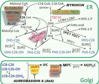
Discovery of alkaline ceramidases in yeast While mammalian cells contain acid, neutral and alkaline ceramidases residing in lysosomes, at the plasma mem- brane and in the early secretory pathway, respectively, only alkaline ceramidases have been described in yeast (Mao &
Obeid, 2008). They go by the names of Ypc1p and Ydc1p (Fig. 1), are highly homologous over their entire sequence (54% identity) and reside in the ER, where also Lag1p and Lac1p, the two redundant acyl-CoA dependent cera- mide synthases are located. Ypc1p and Ydc1p seem to be homeostatic enzymes, which cannot only hydrolyze cera- mides into long chain base (LCB) and fatty acid, but also
generate ceramides through the reverse reaction. Indeed, YPC1 was discovered as a gene enabling cells, when overexpressed, to grow on fumonisin B1, a competitive inhibitor of Lag1p and Lac1p; its homolog YDC1 was subsequently shown to have ceramidase activity also (Mao et al., 2000a, b; Fig. 1). Similarly, in lac1 Δ lag1 Δ cells, LCBs accumulate and this renders the Ypc1p- or Ydc1p-mediated ceramide synthesis thermodynamically possible (Schorling et al., 2001; Cerantola et al., 2009).
Yeast ceramidases Ypc1p and Ydc1p show slightly different substrate specificities: Ypc1p hydrolyzes ceramides con- taining phytosphingosine (PHS) or dihydrosphingosine (DHS), whereas Ydc1p is a dihydroceramidase, and this LCB specificity of Ydc1p is also observed for the reverse reaction (Mao et al., 2000b; Cerantola et al., 2009).
Published in )(06<HDVW5HVHDUFK±
which should be cited to refer to this work.
http://doc.rero.ch
Physiological expression levels of Ypc1p and Ydc1p
At their physiological expression levels, Ypc1p and Ydc1p markedly enhance growth of lac1Δlag1Δ cells lacking acyl- CoA-dependent ceramide synthesis in that lac1Δlag1Δ cells grow much better than lac1Δlag1Δypc1Δydc1Δ (Cerantola et al., 2009). Thus, even when expressed from their endogenous promoters, they may contribute to cera- mide biosynthesis if Lac1p and Lac1p are not operational.
Moreover, the reverse ceramidase activity of Ypc1p and Ydc1p in microsomal detergent extracts from wild-type (WT) cells can easily be detected and is quite substantial, as expression of GAL1 promoter driven YPC1, placed on a 2 l or a centromeric vector, increases the activity only 25 and 2.3-fold over WT levels, respectively (Mao et al., 2000a; Ramachandra & Conzelmann, 2013; therein Supporting Information, Fig. S2d).
Impact of Ypc1p and Ydc1p on sphingolipid biosynthesis
The simultaneous presence of acyl-CoA-dependent cera- mide synthases and alkaline ceramidases in the ER seems to create the potential for a futile circle. Indeed, in WT cells, the overexpression of YPC1 and YDC1 was shown to significantly increase the levels of free LCBs and LCB- phosphates and to reduce the biosynthetic flow of LCBs toward mature sphingolipids, whereas deletion of YPC1 caused a significant increase of mature sphingolipids as detected by metabolic labeling with tritiated palmitate
([
3H]C16:0) or [
3H]serine (Mao et al., 2000a, b). One also has to consider the possibility that ceramidases potentially generate biologically important alterations in the local sphingolipid composition of a membrane under certain circumstances, alterations that do not occur in ypc1 Δ ydc1 Δ (yy ΔΔ ) cells.
Aim of the study
It is likely that the Synthetic Genetic Array (SGA) studies performed in the past using ypc1 Δ or ydc1 Δ single mutants might have missed potential genetic interactions between a complete lack of alkaline ceramidase activity and other gene deletions because of potential paralog compensation between YPC1 and YDC1 (DeLuna et al., 2008). We therefore did a chemical-genetic screen to find nonessential genes, which would impact the growth rate in the yy ΔΔ background. We also followed up on a few published phenotypes of ypc1 Δ or ydc1 Δ cells and tested if they were exacerbated in yy ΔΔ double mutant.
Materials and methods
Strains and growth conditions
Saccharomyces cerevisiae strains used are listed in Table S1, plasmids in Table S2. Mutant strains were gen- erated using standard methods for crossing of single mutants, for plasmid transfection or for gene disruption using deletion cassettes generated by PCR. Cells were grown on rich medium (YPD or YPG) or synthetic com- plete media (yeast nitrogen base YNB, United States Bio- logical) containing 2% glucose (D) or galactose (G) as a carbon source. Unless indicated otherwise, synthetic com- plete medium with 2% glucose was used.
Synthetic genetic arrays
Screens were performed according to published protocols (Collins et al., 2010). The measurement of growth, the analysis and the visualization of high-throughput screen data were conducted with the help of the S
CREENM
ILLsoftware (Dittmar et al., 2010). A more detailed descrip- tion of this and the following methods is to be found in the supporting information.
Fluorescence microscopy
For fluorescent imaging, cells were collected when in exponential phase at 30 ° C unless indicated otherwise. Cells were imaged using an Olympus BX54 microscope equipped with a piezo-positioner (Olympus).
Z sections (7 – 10 each 0.5 l m apart) were projected to
Fig. 1. Major pathways of sphingolipid biosynthesis in yeast. Gene names are in italic, essential genes in red and enzyme inhibitors in bold italics. A detailed description of the biosynthetic reactions can be found in recent reviews (Dickson et al., 2006; Dickson, 2010).
http://doc.rero.ch
two-dimensional images and analyzed with the C
ELLM soft- ware (Olympus).
Mass spectrometric lipid analysis
For Fig. 4a, lipids were extracted and analyzed in negative and positive ion mode by direct infusion mass spectrome- try using an LTQ Orbitrap XL mass spectrometer equipped with the automated nanoflow ion source Triver- sa NanoMate (Advion Biosciences) as described (Ejsing et al., 2006, 2009). For Fig. 5a and b, lipids were extracted and analyzed as described before (Hanson & Lester, 1980;
Cerantola et al., 2009) and inositolphosphorylceramide (IPC) and mannosyl-IPC (MIPC) lipids were identified based on release of characteristic fragment ions. Before injection, all lipid extracts were mixed with a fixed amount of lipid extract from WT cells grown in [
13C]glucose as the only carbon source and the data from different cell lines were made comparable by normalizing the signal intensities of this internal standard as a reference.
Triton X-100 solubilization assay
Crude membranes were isolated from early logarithmic cells by breaking cells with glass beads in ice-cold TNE-I buffer (50 mM Tris-HCl, pH 7.4, 150 mM NaCl, and 5 mM EDTA) supplemented with protease inhibitors (1 mM PMSF, 4 lM leupeptin, and 2 lM pepstatin) as described (Grossmann et al., 2008). Debris was removed, and membranes were sedimented at 16 000 g for 75 min and resuspended in TNE-I buffer. For the determination of detergent resistance, aliquots corresponding to 50 lg of membrane protein in 100 lL TNE-I buffer were treated with increasing concentrations of Triton X-100 (0 – 0.8%) at room temperature for 30 min. The nonsolubilized mate- rial was sedimented by centrifugation (16 000 g at 4 ° C for 30 min) and washed with 100 l L of detergent-free buffer.
The other methods used are described in the support- ing information.
Results
Combining ceramidase deficiency with a further genetic deletion
The yyΔΔ, ypc1Δ and WT strains were robotically crossed with the 4978 individual strains of the nonessential dele- tion strain collection of S. cerevisiae in quadruplicate.
Selected triple mutants were tested either on synthetic complete selection media or the same supplemented with 0.03 l g mL
1of Aureobasidin A (AbA; Fig. 1), 25 l M PHS or 100 mM CaCl
2or on media lacking inositol. The whole screen was carried out twice. Each
ypc1Δydc1ΔxxxXΔ triple mutant was compared with the corresponding xxxXΔ single or ypc1ΔxxxXΔ double mutant to find xxxX Δ mutants, in which the deletion of YPC1 and YDC1 caused a growth phenotype. Twenty sig- nificant hits (P-value ≤ 0.05) were obtained and these were further verified by independent crosses and subse- quent random sporulation analysis, tetrad analysis and serial dilution growth tests. None of the 20 could be vali- dated through these further tests. While numerous xxxX Δ mutants were reproducibly resistant or hypersensitive to AbA, or PHS or CaCl
2, or to the absence of inositol, as reported before (Tani & Kuge, 2010, 2012; Young et al., 2010) none of these altered sensitivities was exacerbated or mitigated in the ypc1Δ or yyΔΔ backgrounds. Yet, many more deletion mutants than previously reported (BIOGRID (http://thebiogrid.org/) were found to be AbA hypersensitive (N.S. Voynova and H. M. Vazquez, unpub- lished data).
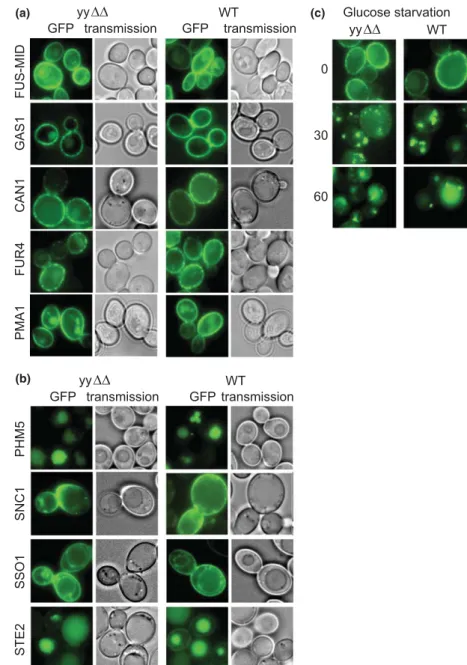
Deletion of YPC1 and YDC1 does not affect the trafficking of Fus-Mid-GFP and other plasma membrane proteins
It has been recognized for some time that sphingolipids are required for trafficking of GPI anchored proteins (e.g.
Gas1p) and many multispan membrane proteins (e.g.
Pma1p) transiting through the secretory pathway to the plasma membrane or the vacuole (Horvath et al., 1994;
Bagnat et al., 2000; Gaigg et al., 2005). It is believed that targeting of many membrane protein requires association with sphingolipid- and ergosterol-rich subdomains of the membrane, which often are referred to as ‘rafts’. Such proteins usually cannot be solubilized with nonionic detergents at 4 ° C are said to be ‘detergent resistant’, a finding that in the past has been used as a proxy for potential raft association of proteins. A deletion strain library was screened microscopically for strains, in which the detergent resistant, chimerical Fus-Mid-GFP protein was not properly targeted to the plasma membrane (Pros- zynski et al., 2005). Deletion of several enzymes metabo- lizing sphingolipids (ELO3, SUR2, YPC1; Fig. 1) or making ergosterol (ERG4, ERG6) caused Fus-Mid-GFP to accumulate in the Golgi or the vacuole. Deletion of YPC1 provoked the accumulation of Fus-Mid-GFP in the Golgi.
Expecting to see a stronger phenotype in yyΔΔ cells, we expressed the Fus-Mid-GFP construct of Proszynski et al.
in our strains. In spite of numerous efforts, we observed no mislocalization of Fus-Mid-GFP neither in ypc1Δ cells nor in yy ΔΔ cells (Fig. 2a), although we used the same genetic background and the same induction protocol as the original report (Proszynski et al., 2005).
As shown in Fig. 2a, other raft-associated proteins such as Gas1p and Pma1p also were properly localized at the
http://doc.rero.ch
cell surface in yy ΔΔ cells and evaluation of data showed no statistically differences in subcellular localization of Gas1p and Pma1p between WT and yy ΔΔ cells. This argued that in yyΔΔ cells the sphingolipid rich rafts in the ER form correctly.
The yeast plasma membrane is known to comprise several subdomains, which are enriched for specific sets of membrane proteins, namely the dotty membrane compartment of Can1p (MCC), the meshlike compart- ment of Pma1p (MCP) and the dotty compartment of TORC2 (MCT). The dotty MCC contains the proton symporters Can1p, Tat2p and Fur4p, carrying arginine, tryptophan and uracil, respectively, and several other membrane proteins; the MCC colocalizes with and is
stabilized by the underlying eisosomes (Malinska et al., 2003; Grossmann et al., 2008). Importantly, genetic and pharmacological data indicate that sphingolipids and ergosterol are important for the maintenance of the MCC and of eisosomes (Grossmann et al., 2008;
Fr€ ohlich et al., 2009). As can be seen in Fig. 2a, Can1p- GFP and Fur4p-GFP showed the typical spotty appear- ance in the plasma membrane indicating their proper integration into the MCC also in yyΔΔ. In contrast, Pma1p showed the typical homogenous distribution of the PMP. Thus, alkaline ceramidases do not seem to be required for surface transport of GPI proteins and multi- span plasma membrane proteins nor their segregation into subdomains.
FUS-MID CAN1 FUR4 PMA1 GAS1
Glucose starvation
30
60 0
yy WT
PHM5 SNC1 SSO1 STE2
transmission GFP
yy WT
transmission GFP
transmission GFP
yy WT
transmission GFP
(a)
(b)
(c)
Fig. 2. Subcellular localization and organellar distribution of GFP-tagged membrane proteins. (a) WT and yy ΔΔ cells expressing Fus- Mid-GFP behind the GAL1 promoter were grown to mid-log phase on raffinose, then switched to galactose for 3 h at 30 ° C prior to visualization by fluorescent microscopy as described (Proszynski et al., 2005). Cells expressing GFP-tagged Gas1p, Can1p, Fur4p and Pma1p proteins under constitutive promoters were grown to the exponential phase before viewing. (b) WT and yy ΔΔ cells expressing either of GFP-Phm5p, GFP-Snc1p, GFP-Sso1p or Ste2p-GFP from centromeric plasmids were grown to mid-log phase at 30 ° C. The a -factor receptor Ste2p, normally expressed only on MATa cells, is internalized in these MATa cells, because they themselves produce a -factor and because the receptor is overexpressed. (c) Stress-induced endocytosis of Can1p is normal. WT and yyΔΔ cells harboring pCAN1-GFP were grown to exponential phase and resuspended in glucose-free medium. 0, 30 or 60 min later the cells were collected and viewed.
http://doc.rero.ch
Intracellular trafficking pathways in yy ΔΔ cells are normal
As sphingolipids also regulate intracellular protein target- ing and as these processes determine the morphology of organelles, we surveyed organellar morphology by intro- ducing into the yy ΔΔ background mtGPF, GFP-Vph1p, GFP-Sec63p, RFP-Sec7p and GFP-Sed5p for life staining of mitochondria, vacuoles, ER, trans-Golgi and cis-Golgi, respectively. As shown in Fig. S1, the morphology of all these compartments was perfectly normal in yy ΔΔ cells.
We also checked intracellular trafficking routes in yy ΔΔ cells more directly by monitoring the distribution of pro- teins, which are continuously commuting between organ- elles. Observation of the Golgi v-SNARE Snc1p-GFP assesses the traffic from endosomes to the plasma mem- brane and back. In WT cells, GFP-Snc1p primarily labels the plasma membrane as well as small internal punctate structures that correspond to early endosomes and trans- Golgi structures (Lewis et al., 2000). No difference was found between yy ΔΔ and WT cells (Fig. 2b). The corre- sponding plasma membrane t-SNARE GFP-Sso1p was also distributed normally in yy ΔΔ cells (Fig. 2b).
Phm5p is a vacuolar enzyme known to transit from the Golgi to the vacuole directly via endosomes and the mul- tivesicular body (MVB) pathway (Dunn et al., 2004).
Phm5p-GFP was correctly targeted to the vacuolar lumen in yy ΔΔ cells (Fig. 2b) indicating that the MVB pathway was functioning correctly. Furthermore, Ste2p-GFP, the yeast a-mating factor receptor, which undergoes endocy- tosis and trafficking to the vacuole in a ligand-dependent fashion (Stefan & Blumer, 1999) was internalized nor- mally in yyΔΔ cells (Fig. 2b).
Constitutive and stress-induced endocytosis is normal in yy ΔΔ cells
Normal and stress-induced endocytosis of plasma mem- brane proteins can be induced and regulated by LCBs, and this explains why exogenously added PHS can inhibit the growth of auxotrophic cells requiring nutrient trans- porters at their surface (Chung et al., 2000, 2001; Zano- lari et al., 2000). As shown in Fig. S2a, the membrane seeking fluorescent dye FM4-64 entered yyΔΔ cells via en- dosomes and reached the vacuolar membrane at a normal rate, that is, within 45 min. Similarly, the water-soluble fluorescent dye Lucifer Yellow was taken up by yyΔΔ cells at a normal rate (Fig. S2b).
As shown in Fig. 2c, upon acute glucose starvation of WT cells Can1p-GFP appeared in endosomes after 30 min and in vacuoles after 60 min and endocytosis of Can1p-GFP followed the same kinetics in yy ΔΔ cells, demonstrating that stress-induced endocytosis of nutrient
transporters is unaffected in yyΔΔ cells. Other kinds of stress such as an inhibition of protein synthesis or heat shock were also tested as stimuli for Can1p endocytosis.
While all these agents could trigger rapid endocytosis of Can1p, no difference between WT and yy ΔΔ could be observed (not shown).
ISC1 encodes a hydrolase for IPC, MIPC and M(IP)
2C and isc1 Δ contain significantly increased amounts of these complex sphingolipids when metabolically labeled with [
3H]DHS (Sawai et al., 2000). Isc1 Δ have also been shown to secrete a small fraction of vacuolar enzymes such as CPY (Bonangelino et al., 2002). Although similar labeling experiments also showed a significant increase in complex sphingolipids in yyΔΔ cells (Mao et al., 2000b), yyΔΔ did not secrete the vacuolar carboxypetidase CPY (Fig. S3a).
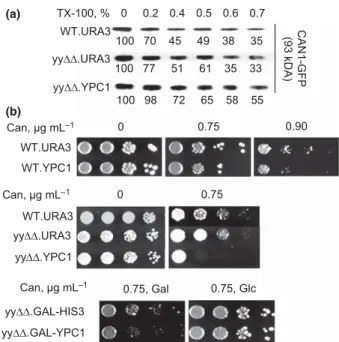
Detergent resistance of lipid raft proteins As mentioned, the bulk of Gas1p, Can1p, Fur4p and Pma1p are typically found in detergent resistant mem- branes, but become detergent extractable when sphingo- lipid biosynthesis is compromised (Bagnat et al., 2000).
To directly test if the loss of ceramidase function affects the association of these proteins with lipid rafts, cell membranes of yy ΔΔ cells were incubated with 1% of Tri- ton X-100 at 4 ° C for 30 min and then loaded at the bot- tom of a stepwise density gradient (Bagnat et al., 2000).
Solubilized proteins remain at the bottom of the tube during the following ultracentrifugation, while detergent resistant membranes float to the top. We find that Gas1p and Can1p are still floating in yyΔΔ cells, albeit somewhat less rapidly than in WT cells (Fig. S3b).
Detergent resistance of membrane proteins can also be probed simply by extracting them with different concen- trations of nonionic detergent. Indeed, Can1p resists extraction at low detergent concentrations in WT cells but not so in mutants, in which the MCC compartment is destabilized (Grossmann et al., 2008). As shown in Fig. 3a, detergent resistance of Can1p-GFP was the same in yy ΔΔ as in WT cells.
We also used a strain, in which YPC1 was overexpres- sed. In this yy ΔΔ .YPC1 strain, Can1p-GFP was more resistant to detergent extraction than in WT (Fig. 3a).
We therefore asked the question whether the increased detergent resistance of Can1p in yyΔΔ.YPC1 cells protect Can1p from being endocytosed. Increased stability of Can1p in the plasma membrane has been shown to lead to an increased sensitivity to canavanine, a toxic arginine analogue that enters the cell via Can1p, and canavanine sensitivity therefore formed the bases for genetic screens to discover regulators of Can1p-endocytosis (Lin et al., 2008). As shown in Fig. 3b, WT and yy ΔΔ cells display the same moderate sensitivity to canavanine but overex-
http://doc.rero.ch
pression of YPC1 makes cells more sensitive to canavanine.
The same was observed when YPC1 was overexpressed from a GAL1 promoter (Fig. 3b, bottom). These results are unexpected as overexpression of YPC1 would be expected to increase LCB levels, which are known to destabilize the transporters and promote their endocytosis and this ought to make cells canavanine-resistant rather than hypersensitive (Chung et al., 2000). One speculative explanation would be that local LCB and free fatty acid concentrations at the plasma membrane may allow Ypc1p to work in the reverse direction, thereby reducing LCB and increasing ceramide levels.
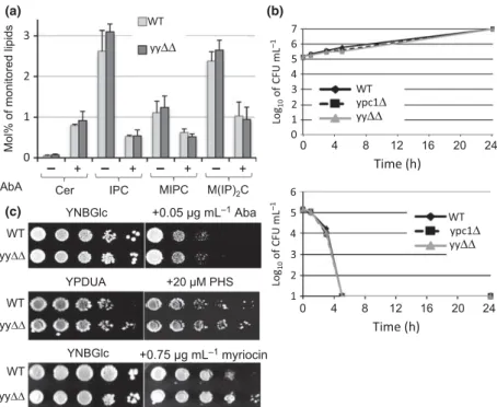
Cells lacking alkaline ceramidases have a normal sphingolipid composition
Metabolic labeling with [
14C]serine demonstrated that yy ΔΔ cells accumulated the same ceramide species as WT
cells (Fig. S4a). The same was true also after treatment with AbA, a cyclic depsipeptide antifungal antibiotic, which inhibits the IPC synthase Aur1p, thereby causing accumu- lation of ceramides (Endo et al., 1997; Nagiec et al., 1997;
Fig. S4a). AbA treatment caused a drastic reduction of labeling of IPCs, and to a lesser degree, of MIPCs and M (IP)
2Cs, as expected (Fig. S4b). When analyzed by mass spectrometry, no significant difference in any class of sphingolipid was observable between WT and yy ΔΔ cells (Fig. 4a). Treatment with AbA for 4 h again led to the expected drastic increase of ceramide levels and a marked reduction of IPC, MIPC and M(IP)
2C levels (Fig. 4a). (The reduction of IPCs under AbA is more pronounced than that of MIPC and M(IP)
2C, as not only caused by dilution of IPCs during a further cell division but mainly by the fur- ther maturation of premade IPC to MIPC and M(IP)
2C.) When analyzing the different species containing 42–46 car- bon atoms and three or four hydroxyl groups in their cera- mide moiety, the profile of yy ΔΔ cells was the same as the one of WT (not shown). These findings are apparently in contrast to the earlier observed marked increase of all com- plex sphingolipids observed in ypc1 Δ and yy ΔΔ cells labeled with [
3H]C16:0 (Mao et al., 2000a, b). The discrepancy may be explained by the fact that our mass spectrometric method fails to detect the ceramides with C14 and C16 fatty acids, as they are two or three orders of magnitude less abundant than those with C24 and C26 (Montefusco et al., 2013, 2014), and secondly by assuming that the sphingoli- pids previously observed after labeling with [
3H]C16:0 con- tained nonelongated [
3H]C16:0 and hence were derived from these minor ceramides. It also may be that there are different ceramide pools and that only ceramides made from fatty acids taken up from the medium are substrates for Ypc1p and Ydc1p. Overall, at their physiological expres- sion levels, Ypc1p and Ydc1p do not seem to be able to reduce the levels of the major C26:0-containing ceramides accumulating under AbA.
yy ΔΔ cells grow normally on aureobasidin A and high concentrations of PHS
In spite of these results, we wondered if Ypc1p and Ydc1p could mitigate the toxicity of ceramides that accumulate when cells are exposed to AbA. As shown in Fig. 4b, high concentrations of AbA killed the yy ΔΔ cells no faster than WT cells and no viable cells remained after 5 h of treat- ment. Also, on low concentrations of AbA, yy ΔΔ cells were not growing less well than WT (Fig. 4c). Overexpression of YPC1 rendered cells more resistant to high concentrations of PHS in the culture medium (Mao et al., 2000b), an effect believed to be due to an increased ceramide biosyn- thesis through reverse ceramidase activity. This suggests
yy .URA3 yy .YPC1
0.75 0
WT.URA3
yy .GAL-YPC1 yy .GAL-HIS3
0.75, Glc WT.URA3
WT.YPC1 Can, μg mL
–1Can, μg mL
–10.75, Gal
0.75 0.90
Can, μg mL
–10 TX-100, %
100 77 100 98
51 61 35 33 72 65 58 55 100 70 45 49 38 35 yy .URA3
yy .YPC1
WT.URA3 (93 kDA) CAN1-GFP
(a)
(b)
Fig. 3. (a) Association of Can1p with detergent resistant membranes in yy ΔΔ and YPC1 overexpressing cells. WT and yy ΔΔ containing empty vector (WT.URA3 or yyΔΔ.URA3 respectively) and yyΔΔ with YPC1 behind the ADH1 promoter (yy ΔΔ .YPC1) were grown to exponential phase. Membrane proteins (50 l g per sample) were treated with increasing concentrations of Triton X-100 for 30 min at room temperature exactly as described (Grossmann et al., 2008). After high speed centrifugation, the nonsolubilized proteins of the pellet were resolved by SDS-PAGE and detected by specific antibodies on Western blots. Percent of detergent resistant Can1p-GFP was calculated as compared with the nontreated sample and indicated below the bands. (b) YPC1 overexpression makes cells canavanine hypersensitive. WT and yy ΔΔ cells with YPC1 behind the ADH1 (top panels) or the GAL1 promoter (bottom panel) were grown to exponential phase and were serially diluted on the medium containing canavanine or solvent. Cells were incubated at 30 °C for 3 days.
http://doc.rero.ch
that yyΔΔ may be hypersensitive to PHS. However, yyΔΔ and WT cells grew at the same rate on medium supple- mented with PHS. On media containing myriocin, which blocks LCB synthesis (Fig. 1) and therefore may render breakdown of ceramides unnecessary, yy ΔΔ grew slightly better than WT (Fig. 4c). Overall, Ypc1p and Ydc1p, when expressed from their endogenous promoter, do not seem to have any homeostatic role when cell growth is compro- mised by artificially increased ceramide or LCB levels.
In vivo substrate specificity of the reverse ceramidase activity of Ypc1p
To characterize the ceramides that potentially could be generated locally in the ER by YPC1-dependent reverse ceramidase activity, in vivo we analyzed the sphingolipid profile of lag1Δlac1Δ cells overexpressing YPC1 (2Δ.YPC1) by LC-MS/MS. The 2 Δ .YPC1 strain is only viable as long as it harbors the multicopy plasmid carrying YPC1 and all its ceramides are made by Ypc1p (and endogenous Ydc1p). As shown in Fig. 5a, the parental YPK9 cells only make IPCs and MIPCs with 42, 44 or 46 C atoms and
mostly four or three hydroxyl groups in their ceramide moiety, quite in agreement with the literature. (DHS and PHS are counted as contributing two and three hydroxyls, respectively, the remaining hydroxyl groups residing on the fatty acid moiety.) As the most abundant yeast sphin- golipids contain PHS with 18, less frequently 20 carbon atoms, the predominance of IPC44 and IPC46 suggests that the sphingolipids of YPK9 WT cells most frequently contain C26:0 fatty acids and their mono-hydroxylated derivatives. According to the sphingolipid profile shown in Fig. 5a and b, 2Δ.YPC1 cells contain much lower amounts of IPCs and MIPCs than WT cells, but they also predominantly make IPC44 and IPC46 species, although IPC42 and IPC40 species represent a higher fraction of total IPCs than in parental YPK9 cells. In all cells, basi- cally only IPC44 and IPC46 are used for MIPC biosynthe- sis (Fig. 5b). For unknown reasons, all sphingolipid classes of 2 Δ .YPC1 contain relatively more species with three than with four hydroxyls, while the reverse is true in WT cells. In summary, the profile of sphingolipids of 2 Δ .YPC1 suggests that Ypc1p in vivo preferentially uses very long chain fatty acids.
(a)
(c)
(b)
Fig. 4. Sphingolipid levels and sensitivity to AbA are not altered in yy ΔΔ cells. (a) WT and yy ΔΔ cells growing exponentially at 30 ° C and reaching a density of OD
600= 0.4 – 0.6 were either extracted directly or further incubated in the presence of AbA (0.25 l g mL
1) for 4 h during which time they reached an OD
600of 1.0–1.6. Lipids from treated and nontreated cells were subjected to lipidomic analysis. Bars indicate standard deviations of four determinations (two biological replicates each run twice). (b) ypc1Δ , yy ΔΔ and WT cells were grown till exponential phase. 10
5cells were suspended in fresh synthetic medium containing 5.0 l g mL
1of AbA (bottom panel) or not (top panel). Cells were further incubated at 24 °C, and viability was measured at 0, 1, 3, 5 and 24 h by plating an aliquot of cells onto YPD. Duplicate preparations were examined. After culture for 3 days at 30 ° C, CFUs were counted. (c) WT and yy ΔΔ cells were grown till exponential phase and then replicated by serial 10-fold dilution on media supplemented or not with AbA, PHS or myriocin. Plates were incubated at 30 ° C for 3 days.
http://doc.rero.ch
In vitro substrate specificity of the reverse ceramidase activity of Ypc1p
The original standard assay of reverse ceramidase of Ypc1p activity utilizes [
3H]C16:0 and PHS as substrates (Mao et al., 2000b). We tried to elucidate the fatty acid specific- ity of this assay by adding different concentrations of unla- beled fatty acids of variable chain length as competitive inhibitors. This demonstrated that C16:0 was by far the best competitor, and hence, the best substrate compared with shorter and longer fatty acids (Fig. 5c). Also unsatu- rated and alpha-hydroxylated fatty acids, which are not made by yeast cells but may be taken up from the surroundings, are not better substrates for Ypc1p than C16:0 (Fig. S5a). Thus, the in vitro test does not seem to reflect the situation in vivo, as discussed below.
Previous studies have shown that lcb1 Δ mutants can take up and utilize not only the physiological
D-erythro but also
L-threo forms of LCBs, which latter are trans- formed in vivo into
D-erythro type LCBs (Watanabe et al., 2002). Neither
L- nor
D-threo forms of DHS could be utilized efficiently in the standard assay (Fig. S5b), sug- gesting that
L-threo DHS could not be transformed into
D
-erythro DHS in vitro. On the other hand,
D-sphingosine, the predominant LCB in mammalian sphingolipids, was a good substrate for reverse ceramidase activity, although a sphingosine containing ceramide was not hydrolyzed in the microsomal ceramidase assay of Ypc1p in the original report (Mao et al., 2000b).
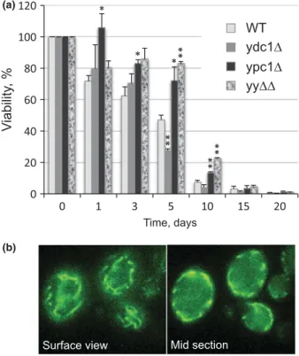
Testing mitochondrial functions of yy ΔΔ
Recent data have shed light on the role of a previously unsuspected mitochondrial metabolism of sphingolipids.
During diauxic shift Isc1p moves from the ER to the outer membrane of mitochondria (Vaena de Avalos et al., 2004), where it causes an increase of a -hydroxylated phytoceramide, which is believed to be a precondition for the postdiauxic induction of genes involved in aerobic carbon metabolism (Kitagaki et al., 2007, 2009). More- over, deletion of ISC1 increases iron levels, reactive oxygen species, oxidative stress markers and H
2O
2sensi- tivity of cells, and thereby causes premature aging, that is increased apoptosis and a drastic reduction of the chro- nological life span (CLS; Almeida et al., 2008). Similarly, ypc1 Δ cells were found to be hypersensitive to 2 – 5 mM H
2O
2(Higgins et al., 2002; Hillenmeyer et al., 2008). In view of this, we decided to test mitochondrial function in
IPC36:0-3IPC40:0-3IPC42:0-2IPC42:0-3IPC42:0-4IPC42:1-3IPC44:0-3IPC44:0-4IPC44:1-4IPC46:0-3IPC46:0-4IPC46:0-5IPC46:1-4IPC46:1-5
2 .YPC1 + AbA 2 .YPC1 – AbA
YPK9 + AbA YPK9 – AbA
2 .YPC1 + AbA 2 .YPC1 – AbA
YPK9 + AbA YPK9 – AbA 0
20 40 60 80 100 120
MIPC34:0-2MIPC34:0-3MIPC34:0-4MIPC34:0-5MIPC34:1-3MIPC34:1-4MIPC34:1-5MIPC36:0-2MIPC36:0-3MIPC36:0-4MIPC36:1-3MIPC36:1-4MIPC44:0-3MIPC44:0-4MIPC46:0-3MIPC46:0-4MIPC46:0-5 0
20 40 60 80 100