1Cellular Reprogramming and Oncogenesis Laboratory, Equipe labellisée la Ligue contre le cancer, Labex DEVweCAN, Université de Lyon, Université Claude Bernard Lyon 1, INSERM 1052, CNRS 5286, Centre Léon Bérard, Centre de recherche en cancérologie de Lyon, Lyon, France. 2Department of Genome Regulation, Max Planck Institute for Molecular Genetics, Berlin, Germany. 3Broad Institute of MIT and Harvard, Cambridge, MA, USA. 4Harvard Stem Cell Institute, Cambridge, MA, USA. 5Department of Stem Cell and Regenerative Biology, Harvard University, Cambridge, MA, USA. 6GReD, Université Clermont Auvergne, CNRS, INSERM, BP38, Clermont-Ferrand, France. 7Department of Biochemistry and Molecular Genetics, University of Illinois at Chicago, Chicago, IL, USA. 8Apoptosis, Cancer and Development Laboratory, Université de Lyon, Université Claude Bernard Lyon 1, INSERM 1052, CNRS 5286, Centre Léon Bérard, Centre de recherche en cancérologie de Lyon, Lyon, France. 9Cytometry Facility, Université de Lyon, Université Claude Bernard Lyon 1, Centre Léon Bérard, Centre de recherche en cancérologie de Lyon, INSERM 1052, CNRS 5286, Lyon, France. 10Research Pathology platform, Department of translational research and innovation, Centre Léon Bérard, Lyon, France. 11Institute for Dental Research and Oral Musculoskeletal Research, Center for Biochemistry, University of Cologne, Cologne, Germany. 12Department of Translational Research and Innovation, Centre Léon Bérard, Lyon, France. 13These authors contributed equally: Aurélia Huyghe, Giacomo Furlan, Duygu Ozmadenci. ✉e-mail: fabrice.lavial@lyon.unicancer.fr
M
ouse embryonic stem cells (mESC) are mainly regulated by four signalling cues1–3. Leukaemia inhibitory factor (lif)4, Wnt3a5 and Bmp46 sustain self-renewal, whereas fibroblast growth factor 4 (Fgf4) triggers exit from self-renewal via Erk1/2 activation7. Conventional culture conditions require Lif and serum or knockout serum replacement (hereafter serum/Lif) to maintain a self-renewing state. In this state, the Fgf–MAPK and the repressive Gsk3α/β–Tcf7l1 pathways remain active, lead-ing to a heterogeneous population of cells. These metastable mESCs exhibit fluctuating expression of Nanog and detectable levels of lineage-affiliated genes8. The suppression of MAPK sig-nalling (via Mek1/2 blockade) and activation of the Wnt pathway (via Gsk3α/β blockade) (hereafter 2i), supports self-renewal of mESCs and instructs a ground state of pluripotency8–10. The 2i mESCs display uniform Nanog expression and negligible levels of lineage-affiliated genes. This finding has led to the establishment of germline-competent ESCs from recalcitrant mouse strains and from rat11,12. However, the prolonged blockade of Mek1/2 has been shown to compromise mESC genomic stability, calling into question the use of 2i13. Thus, the identification of endogenouspathways controlling naive pluripotency is crucial not only for advancing our understanding of embryonic development but also for best developing strategies to generate stable human naive pluripotent cells.
Netrins are secreted proteins that have been identified as having a role in axon guidance during nervous system develop-ment14,15. Netrin-1 (encoded by the Ntn1 gene), which was ini-tially purified as a soluble laminin-related molecule that elicited the growth of commissural axons, is now considered as a pleiotro-pic ligand involved in development and diseases14,16,17. Most func-tions of Netrin-1 are mediated through the receptors deleted in colorectal carcinoma (Dcc), Unc5 homologues (that is, Unc5A, Unc5B, Unc5C and Unc5D) and neogenin (Neo1)18–20. The char-acterization of this repertoire led to identification of Netrin-1 as a bifunctional molecule that exerts opposing effects—attracting or repelling—neurons, endothelial or immune cells, depend-ing on the receptors it engages21–23. Our previous work showed that it constrains apoptosis during reprogramming24 but the func-tion of this pathway in self-renewal and lineage commitment remains unknown.
Netrin-1 promotes naive pluripotency through
Neo1 and Unc5b co-regulation of Wnt and
MAPK signalling
Aurélia Huyghe
1,13, Giacomo Furlan
1,13, Duygu Ozmadenci
1,13, Christina Galonska
2, Jocelyn Charlton
2,3,4,5,
Xavier Gaume
1, Noémie Combémorel
1, Christina Riemenschneider
2, Nicolas Allègre
6, Jenny Zhang
7,
Pauline Wajda
1, Nicolas Rama
8, Pauline Vieugué
8, Isabelle Durand
9, Marie Brevet
10, Nicolas Gadot
10,
Thomas Imhof
11, Bradley J. Merrill
7, Manuel Koch
11, Patrick Mehlen
8,12, Claire Chazaud
6,
Alexander Meissner
2,3,4,5and Fabrice Lavial
1✉
In mouse embryonic stem cells (mESCs), chemical blockade of Gsk3α/β and Mek1/2 (2i) instructs a self-renewing ground state
whose endogenous inducers are unknown. Here we show that the axon guidance cue Netrin-1 promotes naive pluripotency by triggering profound signalling, transcriptomic and epigenetic changes in mESCs. Furthermore, we demonstrate that Netrin-1
can substitute for blockade of Gsk3α/β and Mek1/2 to sustain self-renewal of mESCs in combination with leukaemia inhibitory
factor and regulates the formation of the mouse pluripotent blastocyst. Mechanistically, we reveal how Netrin-1 and the balance of its receptors Neo1 and Unc5B co-regulate Wnt and MAPK pathways in both mouse and human ESCs. Netrin-1 induces Fak
kinase to inactivate Gsk3α/β and stabilize β-catenin while increasing the phosphatase activity of a Ppp2r2c-containing Pp2a
complex to reduce Erk1/2 activity. Collectively, this work identifies Netrin-1 as a regulator of pluripotency and reveals that it mediates different effects in mESCs depending on its receptor dosage, opening perspectives for balancing self-renewal and lineage commitment.
Here, we reveal an early developmental function for Netrin-1 in the control of naive pluripotency. We found that Netrin-1 and its receptors Neo1 and Unc5B control Wnt and MAPK signalling in mESCs and support self-renewal in combination with Lif. In vivo, Netrin-1 regulates the formation of the pluripotent compartment of mouse preimplantation embryos. Our findings shed light on an unexpected regulator of pluripotency and reveal that a single ligand can have diverse effects in stem cells depending on its rep-ertoire of receptors.
Results
Netrin-1 is regulated by Wnt and MAPK signalling in mouse and human pluripotent stem cells. To identify regulators of naive plu-ripotency, we compared the transcriptomes of serum/Lif mESCs supplemented with inhibitors of Gsk3α/β (CHIR99021), Mek1/2 (PD0325901) or both (2i) for 48 h. Among the transcripts affected by modulation of Wnt and MAPK signalling, Ntn1 was induced by Gsk3α/β inhibition and by 2i but repressed by Mek1/2 blockade at both transcript and protein levels (Fig. 1a,b), whereas other Netrin family members (Netrin-4, Netrin-5, Netrin-G1 and Netrin-G2)25 remained unaffected (Extended Data Fig. 1a). Since Netrin-1 is expressed at basal levels in serum/Lif but is elevated following Gsk3α/β inhibition (Fig. 1a,b), we investigated whether it consti-tutes a target of Wnt. Consistently, mESCs treated with recombinant Wnt3a increased Netrin-1 levels, whereas treatment with Lif and Bmp4 had no effect (Fig. 1c). Netrin-1 induction was also observed in human induced pluripotent stem (hiPS) cells in response to Gsk3α/β inhibition or Wnt3a stimulation (Extended Data 1b). Since canonical Wnt stimulation has been shown to alleviate the repres-sive effect of Tcf7l126, we evaluated Netrin-1 expression in mESCs lacking Tcf7L1 and/or Lef1 (J.Z., B.R. Shy and B.J.M., manuscript in revision). Netrin-1 was released in the absence of Tcf7l1 but reduced if both Tcf7l1 and Lef1 were depleted, indicating that Tcf7l1 acts as a repressor of Netrin-1 activation by Lef1 (Extended Data Fig. 1c,d). Collectively, these results demonstrate that Netrin-1 is regulated by Wnt and MAPK signalling in pluripotent stem cells.
The activation of the Netrin-1–Neo1–Unc5B signalling axis sustains Nanog and mESC undifferentiated state. The distribu-tion of naive pluripotency factors such as Nanog is heterogeneous in serum/Lif and becomes homogeneous with 2i9. Using Ntn1βgeo knock-in reporter mESCs27, we found that Netrin-1 expression is confined to 8% of serum/Lif mESCs, confirming its basal expres-sion in this condition. This β-galactosidase-positive fraction increased to 26 and 23% in the presence of Gsk3α/β inhibitor
and 2i, respectively (Fig. 1d). Exploration of single-cell transcrip-tomic data28 showed similar results (Extended Data Fig. 1e) but revealed that the mean Ntn1 expression level per cell is higher with 2i (Extended Data Fig. 1f).
Due to its induction with 2i, we investigated whether Netrin-1 could actively instruct ground-state pluripotency features. In serum/Lif mESCs, mouse induced pluripotent stem cells (miPSCs) and preimplantation embryos, the Dcc receptor was not expressed but Unc5B and Neo1 were detected (Extended Data Fig. 2a,b). We therefore generated mESCs that exogenously express different haemagglutinin (HA)-tagged Netrin-1 forms from Cre-excisable transgenes (Fig. 1e), wild-type (WT) Netrin-1 or Netrin-1 mutated on residues critical for its interaction with the Dcc-related protein Neo1 (Netrin-1(Neo1-mut)) or with Unc5B (Netrin-1(Unc5B-mut))17,20 (Fig. 1f,g). We established mESC monoclones express-ing the Netrin-1 proteins at similar levels (Extended Data Fig. 2c), comparable to its level in 2i (Extended Data Fig. 2d). Fluorescence-activated cell sorting (FACS) analysis showed that the size and granulometry of Netrin-1(WT) cells became homogeneous, with a prominent contribution of Unc5B (Extended Data Fig. 2e). We next revealed that Nanog heterogeneity was reduced in Netrin-1(WT) mESCs grown in serum/Lif (Fig. 1h,i). This observation was associ-ated with increased levels of Esrrb and Sox2, whereas Oct4 levels remained constant (Fig. 1j). Both Netrin-1 mutants failed to con-fer this effect, highlighting the complementary roles of Neo1 and Unc5B (Fig. 1h–j). Transcriptomic data of single mESCs grown in serum/Lif also revealed a correlation between Ntn1 and Esrrb levels29 (Extended Data Fig. 2f).
We next assessed whether activation of Netrin-1 signalling, by sustaining Nanog, safeguards the undifferentiated state. When cells were grown without Lif for 7 d and replated for 7 d with Lif (Fig. 1k), expression of Netrin-1(WT) conferred strong resistance to differentiation, whereas both mutants failed to do so, reinforcing the involvement of both receptors in Netrin-1 function (Fig. 1l,m). As expected, excision of the Netrin-1 transgene abrogated the effect in revertant cells (Fig. 1n,o). mESCs with doxycyxline (dox)-induc-ible (doxi) Netrin-1 expression also presented enhanced resistance to differentiation upon dox addition (Extended Data Fig. 2g–i).
We next demonstrated that sustained expression of Netrin-1 severely impairs mESC differentiation. We first evaluated whether Netrin-1 maintains the expression of naive pluripotency markers in differentiation conditions. Nanog and Esrrb were still expressed in Netrin-1(WT) mESCs after 6 d of culture in N2B27 medium without Lif, but were not expressed in control cells (Fig. 1p). Immunofluorescence confirmed that some Netrin-1(WT) mESCs
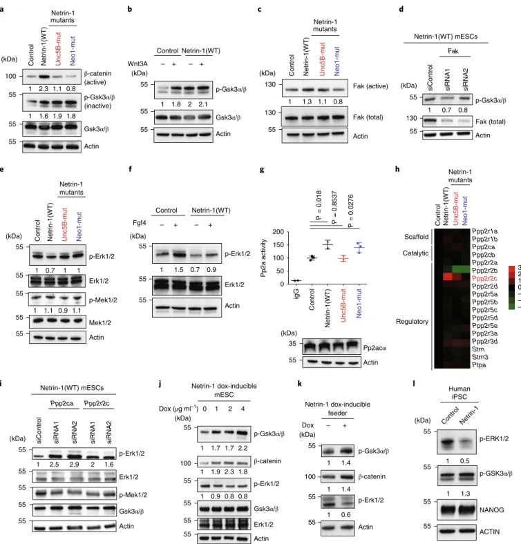
Fig. 1 | Netrin-1 signalling controls pluripotency features. a, Ntn1 transcript levels in mESCs grown as indicated. Data are log2 FPKM values normalized to serum/Lif mESCs. Inh, inhibitor. b, Western blots performed in similar settings as a. c, Western blot in mESCs cultured in serum/Lif as indicated (3 independent experiments). d, Representative bright-field images of Ntn1βgeo mESCs grown as in a. Scale bars, 250 µm. Percentages of positive cells are indicated. n is the total number of cells counted. e, Schematic of the approach. The mESCs were stably transfected with the depicted constructs. IRES, internal ribosome entry site; Pac, puromycin N-acetyltransferase; pA, polyA. f, Netrin-1 protein structure. LN, laminin-like domain; LE, EGF repeats. g, Representation of the Netrin-1 mESC mutants. Netrin-1(Neo1-mut) harbours the L111E mutation, whereas Netrin-1(Unc5B-mut) contains R348A/ R349A/R351A mutations. h, Nanog and Oct4 immunofluorescence on indicated mESCs. Scale bars, 50 µm. i, Quantification of the Nanog/Oct4 intensity ratio. n is the number of cells analysed. The centre line represents the median of the data, the box edges represent upper and lower quartiles, whiskers show highest and lowest values excluding outliers. Two-tailed Student’s t-test. j, Western blot on indicated mESCs. k, Scheme depicting assays for exit from pluripotency. l,m, Colony counts for alkaline phosphatase activity (l) and images from a representative experiment (m) from assays for exit from pluripotency. Data are mean ± s.d., n = 3 independent experiments; two-tailed Student’s t-test. n,o, Assays for exit from pluripotency. n, Images from a representative experiment (n = 3 independent experiments). o, Colony counts. Data are mean ± s.d., n = 3 independent experiments; two-tailed Student’s t-test. p, Western blot for Nanog and Esrrb after 6 d in N2B27 (–Lif) (n = 3 independent experiments). q,r, Nanog and Esrrb immunofluorescence after 6 d in N2B27 (−Lif). q, Representative images. Bars: 50 µm. r, Counts of Nanog- and Esrrb-positive cells. Data are mean ± s.d., n = 3 independent experiments; two-tailed Student’s t-test; 488 control and 416 Netrin-1(WT) mESCs. s, Western blot of embryoid bodies (n = 3 independent experiments). t, Histological analysis of teratoma. Scale bars, 250 µm. Four independent teratomas were analysed per cell line. u,v, Long-term self-renewal assays. u, Colony counts, normalized to the number of colonies formed by control cells for each passage (P) (red dotted line). v, Images from a representative experiment (n = 2 independent experiments).
sustain Nanog and Esrrb expression in differentiation-promoting conditions (Fig. 1q,r). Similarly, after 7 d in non-adherent culture conditions, embryoid bodies derived from Netrin-1(WT) mESCs failed to repress Nanog or Esrrb expression (Fig. 1s) or to induce differentiation genes (Extended Data Fig. 2j). Teratoma assays
also revealed a severe differentiation defect caused by sustained expression of Netrin-1 (Fig. 1t). Finally, when cells were grown at clonal density on laminin in N2B27 medium with Lif for five passages, Netrin-1 expression increased the self-renewal ability of mESCs (Fig. 1u,v). Collectively, these results showed that the
+ Mek1/2 in h + Gsk3 α/ β inh + 2i 1.57 1.05 0.52 0 –0.52 –1.05 –1.57 + Mek1/2 inh+ Gsk3 α/β inh Serum/Lif + 2i Netrin-1 Actin Control+ Lif + Bmp4 + Gsk3 α/β inh + Wnt3a Netrin-1 Actin Ntn1 Nanog Dnmt3b Dnmt3a Pou5f1 Sox2 Serum/Lif 2i Ntn1βgeo Gsk3α/β inh Mek1/2 inh Netrin-1
(WT) (Neo1-mut)Netrin-1 (Unc5B-mut)Netrin-1
Unc5B Neo1 L111 Unc5B interaction Dcc–Neo1 interaction R348-R349-R351
LN LE1 LE2 LE2
Nanog Oct 4 DAPI Merg e
Control (Neo1-mut)Netrin-1 (Unc5B-mut)Netrin-1
Contro l Netrin-1(Unc5B-mut) Netrin-1(Neo1-mut) Netrin-1(WT) 0.2 0.6 0.4 0.8 Nanog/oct4 1 Unc5B Neo1 Unc5B Neo1 n = 25 9 n = 12 4 P = 2.3 × 10 –16
Ntn1 IRES pacpASTOP GFPpA Promoter Intron CAG loxP loxP TAT-Cre GFPpA Nanog Esrrb Oct4 Actin Control Netrin-1(Unc5B-mut ) Netrin-1(Neo1-mut) Netrin-1(WT) Sox2 Control Netrin-1(WT) Netrin-1 (Unc5B-mut) Netrin-1 (Neo1-mut) 7 d – Lif + Lif7 d Replate 0 50 100 Colony numbe r 150 P = 0.004 Netrin-1(WT) Control 200 µm 200 µm Netrin-1(WT) Nanog Esrrb Actin Control Netrin-1(WT) EB (d) 0 7 0 7 Netrin-1 (WT) 1 1.7 1.3 1.1 1 1.4 1.3 0.6 1 1.6 1.4 1.3 8% (n = 248) 6% (n = 177) 26% (n = 196) 23% (n = 243) Nanog Esrrb Actin Day 0 Day 3 Day 6
N2B27 – Lif Contro l Netrin-1(WT) Contro l Netrin-1(WT) Contro l Netrin-1(WT) Control Revertant Contro l Netrin-1(Unc5B-mut) Netrin-1(Neo1-mut) Netrin-1(WT) Contro l Netrin-1(WT) N2B27 + Lif P3
Colony forming abilit
y P2 P3 P4 P5 0 1 2 3 4 5Netrin-1(WT) Contro l Netrin-1(WT) Nanog
Dapi Esrrb Merge
Nanog + cells (% ) 0 10 20 30 0 20 40 60 80 ESrrb + cells (% ) Control Netrin-1(WT) Control Netrin-1(WT) 200 0 50 100 Colony number 150 200 250 P = 0.001 P = 0.076 n = 322 P = 8.8 × 10 –12 n = 12 5 P = 0.17 5 P = 0.0001 48 h 48 h a b c e h m q s t u v r n p o i j k l f g d 24 h (kDa) (kDa) (kDa) (kDa) (kDa) 70 55 70 55 55 55 55 55 55 55 55 55 55 55 55 Control Netrin-1(WT) Revertant mESC
Netrin-1–Neo1–Unc5B signalling axis sustains Nanog and protects mESCs from differentiation.
The activation of the Netrin-1–Neo1–Unc5B signalling axis induces transcriptomic and epigenetic changes in mESC. To gain insight into Netrin-1 function, we compared the transcriptomes of control and 1(WT) mESCs grown in serum/Lif. Netrin-1(WT) affected expression of 434 genes (fold change (FC) > 1.5 or FC < −1.5 at adjusted P < 0.05), with strong repression of differen-tiation genes (Gata6 and Gata4) (Fig. 2a). Functional-annotation clustering of differentially expressed genes (DEGs) by gene ontol-ogy (GO) Panther analysis revealed association with ‘embryo development’, ‘endoderm development’ and ‘regulation of MAPK cascade’ (Extended Data Fig. 2k). Differences were also observed in cell proliferation, in agreement with the accelerated growth of Netrin-1(WT) mESCs (Extended Data Fig. 2l,m). Because similar GO terms are modulated with 2i8, we investigated whether Netrin-1 triggered ground-state pluripotency features. We compared the transcriptomes of control and Netrin-1(WT) mESCs grown in serum/Lif with those from published datasets30,31 (see Methods). The transcriptome of Netrin-1(WT) cells acquired limited but signifi-cant similarities with 2i mESCs (Fig. 2b). To evaluate the role of the receptors, we next compared the transcriptomic effects of Netrin-1(WT), Netrin-1(Unc5B-mut) and Netrin-1(Neo1-mut) (Fig. 2c,d). Both mutant forms failed to induce the full transcriptomic signa-ture, supporting the importance of the interaction of Netrin-1 with both Neo1 and Unc5B in regulation of mESC physiology.
We assessed whether Netrin-1 signalling instructs ground-state-related epigenomic modifications. We found that Netrin-1(WT) mESCs displayed increased activity of the naive Oct4 distal enhancer alongside Jarid2 and Prdm14 enhancers, which were also induced with 2i32 (Fig. 2e). Chromatin immunoprecipitation with sequencing (ChIP–seq) analyses revealed a global increase in both H3K4me3 and H3K27me3 histone marks in Netrin-1(WT) mESCs (Fig. 2f), which included increased H3K4me3 enrichment at pluri-potency genes Nanog and Sox2 (Fig. 2g). We also observed increases in both H3K4me3 and H3K27me3 at bivalent promoters8 (Fig. 2h,i). In 2i, reduced levels of Dnmt3A, Dnmt3B, Dnmt3L and Uhrf1 trigger genome-wide DNA hypomethylation10,13,33,34. By contrast, restricted representation bisulfite sequencing revealed that Netrin-1(WT) mESCs (grown for more than 30 passsages in the presence of the transgene) display DNA methylation resembling that of control cells (mean = 0.25 ± 0.003 and 0.24 ± 0.004 respectively) (Fig. 2j and Extended Data Fig. 2n). In addition, even if the level of Dnmt3A was slightly reduced (Extended Data Fig. 2o), Uhrf1 protein level was induced in Netrin-1(WT) mESCs (Fig. 2k). Moreover, even though 2i represses differentiation genes of the three germ layers8, ectoder-mal genes were not downregulated in Netrin-1(WT) mESCs (Fig. 2l).
Collectively, these results indicate that the Netrin-1–Neo1–Unc5B signalling axis induces transcriptomic and epigenomic changes in mESC that share limited analogies with 2i.
Netrin-1 controls Wnt and MAPK signalling by modulating Gsk3α/β and Erk1/2 in mouse and human pluripotent stem cells. We dissected how Netrin-1 instructs pluripotency features at the molecular level. With 2i, blockade of Gsk3α/β activates the Wnt pathway, whereas Mek1/2 inhibition suppresses MAPK signalling9. The Wnt pathway was strongly activated in Netrin-1(WT) mESCs, as indicated by β-catenin levels, and expression of Netrin-1 mutants showed that Neo1 and Unc5B were required for this effect (Fig. 3a). Gsk3α/β activity was also reduced in Netrin-1(WT) mESCs, as revealed by increased levels of its inactive form35 (Fig. 3a). Moreover, the level of inactive phosphorylated (p-) Gsk3α/β in Netrin-1(WT) mESCs was equivalent to that in con-trol cells treated with Wnt3a, and could not be increased further by Wnt3a addition (Fig. 3b).
Netrin-1 has been linked to various kinases36–40 that may be involved in Gsk3α/β phosphorylation. Among these, we found slightly increased levels of active Fak in Netrin-1(WT) mESCs (Fig. 3c). Because Fak was shown to phosphorylate Gsk3α/β41, we investigated whether the effect of Netrin-1 on Wnt is mediated by Fak. Depletion of Fak using short interfering RNA (siRNA) in Netrin-1(WT) mESCs (>80%; Extended Data Fig. 3a) reduced p-Gsk3α/β, thereby reducing Wnt-pathway activation (Fig. 3d).
We next showed that Netrin-1(WT) mESCs harbour reduced levels of p-Erk1/2, whereas p-Mek1/2 levels remained similar to those in the control cells (Fig. 3e). Because MAPK activation is con-trolled by Lif and Fgf, we determined whether Netrin-1 signalling modulated the sensitivity of mESCs to these cytokines. Deprivation and stimulation experiments indicated that Fgf4 responsiveness was markedly reduced in Netrin-1(WT) mESCs, as indicated by p-Erk1/2 (Fig. 3f). In similar settings, Lif-mediated p-Erk1/2 and p-Stat3 inductions were not affected (Extended Data Fig. 3b). The link between Unc5B and the phosphatase complex Pp2a36 prompted us to investigate whether this complex regulated the decrease in p-Erk1/2 in mESCs42. Immunoprecipitation of the catalytic sub-unit Pp2acα showed that Netrin-1 activation triggered a substantial increase of its phosphatase activity (Fig. 3g). The qualitative com-position of the complex has been shown to modulate its activity43, and we observed an induction of the regulatory subunit ppp2r2c in Netrin-1(WT) mESCs (Fig. 3h). To evaluate whether the Pp2a com-plex is responsible for MAPK attenuation, we attempted to rescue p-Erk1/2 levels by siRNA-mediated depletion of Ppp2ca or Ppp2r2c. We showed that knockdown (>80%; Extended Data Fig. 3c) of both subunits rescued p-Erk1/2 level, while levels of p-Mek1/2 and p-Gsk3α/β remained steady (Fig. 3i).
Fig. 2 | 1 signalling triggers transcriptomic and epigenetic changes in meSCs. a, Volcano plot comparing transcriptomes of control and Netrin-1(WT) mESCs. n = 3 independent samples. Benjamini–Hochberg adjusted P values of the comparisons were computed using the limma-voom workflow; modified two-sided t-test. b, Hierarchical clustering of transcriptomes with published datasets30,31 (see Methods for details). c,d, Comparison of the effects of Netrin-1(WT), Netrin-1(Unc5B-mut) (c) and Netrin-1(Neo1-mut) (d) on the transcriptome of mESCs. n = 3 independent samples. Benjamini–Hochberg adjusted P values of the comparisons were computed using the limma-voom workflow; modified two-sided t-test. e, Luciferase assays. Data are normalized to Renilla activity produced by a plasmid as a control of transfection efficiency and expressed as the mean ± s.d. (n = 4 independent experiments). Two-sided Student’s t-test. f–i, H3K4Me3 and H3K27Me3 distribution in control and Netrin-1(WT) mESCs. Data are representative of 3 independent experiments. f, H3K4Me3 and H3K27Me3 distribution in control and Netrin-1(WT) mESCs. Data are representative of 3 independent experiments. Modified two-sided t-test. Normalized enrichment of H3K4me3 and H3K27me3 for ENCODE-annotated embryonic stem cell peaks (ENCFF001XWU and ENCFF001XWQ, respectively) located at least 10 kb from bivalent promoters. g, Representative browser tracks of H3K4me3 enrichment (in fragments per kilobase of transcript per million (FPKM)) at Nanog and Sox2 loci. h, Normalized H3K4Me3 and H3K27Me3 enrichment at bivalent promoters in control and Netrin-1(WT) mESCs. i, Representative browser tracks of H3K27me3 enrichment (FPKM) at bivalent loci. j, Heat map of methylation levels for 1.3 million matched CpGs in control, Netrin-1(WT) mESCs grown in serum/Lif and 2i mESCs. Each horizontal line is one CpG. k, Western blot for Uhrf1 in indicated mESCs grown in serum/Lif (3 independent experiments). l, Expression of differentiation-related genes in control, 2i and Netrin-1(WT) mESCs. Dendogram presents RNA-seq data. FPKM values are normalized to control mESCs and presented as log2 values. Colour scale is provided.
mESCs with Netrin-1-doxi expression showed dose-dependent changes in Wnt and MAPK signalling following dox addition (Fig. 3j). When mESCs were plated on Netrin-1-doxi feeder cells, with or without dox treatment, we observed similar changes in signalling, indicating that Netrin-1 can act in a paracrine manner on pluripotency (Fig. 3k). Finally, human iPS cells exogenously expressing Netrin-1 showed limited but similar elevation of Wnt
and reduction of MAPK activities—but these were not sufficient to increase Nanog expression (Fig. 3l).
Collectively, these results show that activation of Netrin-1 sig-nalling promotes Wnt sigsig-nalling by activating Fak, which triggers Gsk3α/β inactivation and β-catenin stabilization. Netrin-1 also modifies Pp2a complex composition and activity to dephosphory-late Erk1/2.
8,597 genes, geometric mean
Serum-1 Serum-3 Serum-2 Serum-1 Serum-2 Netrin-1(WT)-2 Netrin-1(WT)-1
2i-1 2i-2 2i-3
2i-1 2i-2 This study Ref. 30 Ref. 31 1.57 1.05 0.52 0 –0.52 –1.05 –1.57 Ectoder m Mesoder m Endoder m Tgm3 Tcf15 Sox18 Pax6 Bmp6 Runx1Msx2 Bmp4Dab2 Sox17 Gata4 Gata6 Pdgfra Hnf1bControlNetrin-1(WT) 2i 1.0 0.8 0.6 0.4 0.2 0.0 n = 1,292,395 CpGs Control Netrin-1(WT) 2i Contro l Netrin-1(WT) Uhrf1 Actin 1 2.3 Fold change 0.5 0.2 0.1 0.05 0.02 0.5 0.2 0.1 0.05 0.02 2 5 10 20 50 100 200 0.5 0.2 0.1 0.02 0.01 2 5 20 50 200 Netrin-1(WT)/control 0.05 0.2 0.1 0.02 0.01 2 5 20 50200 Netrin-1(WT)/control Netrin-1(Unc5B-mut)/control NS (adjusted P < 0.05) Significant (Netrin-1(WT)/control) Significant (Netrin-1(Unc5B-mut)/control) Significant both Amn Gata6 Gata4 Bmp6 Zscan4f 2 5 10 20 50 100 0.01 0.005 0.002 Netrin-1(Neo1-mut)/control Amn Gata6 Gata4 Bmp6 Zscan4f NS (adjusted P > 0.05) Significant (Netrin-1(WT)/control) Significant (Netrin-1(Neo1-mut)/control) Significant both 0.1 0.01 0.5 0.2 0.01 2 5 200 NS (adjusted P > 0.05) Adjusted P value 10–3 10–4 10–5 a b c e f g h i j k l d Amn Gata6 Gata4 Fgfr2 Runx1 Bmp6 Pou5f1 Ntn1 Zscan4e Zscan4f Sox2 1 0 4 6 2 PE DE Pou5f1
Relative luciferase activity
Jarid2 Prdm14 Control Netrin-1(WT) Netrin-1(Neo1-mut) Netrin-1(Unc5B-mut) P = 0.0058 P = 0.0391 0 2.0 2.5 1.0 0.5 1.5 P = 0.426 P = 0.0005 0 –2 –4 2 4
Distance from peak center (kb)
Normalized coverage 25 20 15 10 5 0
All ES-annotated peaks (ENCODE) with bivalent promoters removed –20 –10 0 10 20 4 2 0 6 Control Netrin-1(WT) 0 1,000 0 1,000 Sox2 Nanog 0 3000 300 chr3:34,539,459–34,557,041 chr6:122,651,181–122,668,697 H3K4me3 H3K4me3 H3K27me3 0 –2 –4 2 4 0 –2 –4 2 4
Distance from peak center (kb)
Normalized coverage 30 20 10 0 30 20 10 0 H3K4me3 H3K27me3 Bivalent promoters Control Netrin-1(WT) chr15:98,614,299–98,642,915 chr15:102,743,975–102,876,952 Hoxc cluster Wnt1 Ddn 0 3000 300 0 3000 300 H3K27me3 Control Netrin-1 (WT) Control Netrin-1 (WT) Control Netrin-1 (WT) Control Netrin-1 (WT) 55 (kDa) 100 Significant in (Netrin-1(WT)/control)
β-catenin (active) Contro l Unc5B-mut Neo1-mu t Netrin-1(WT) Netrin-1 mutants a b c d e f g h i j k l p-Gsk3α/β (inactive) Gsk3α/β Actin p-Gsk3α/β Gsk3α/β Actin Control Netrin-1(WT) Wnt3A – + – + Actin p-Erk1/2 Erk1/2 p-Mek1/2 Mek1/2 p-Erk1/2 Erk1/2 Actin Control Netrin-1(WT) Fgf4 – + – + Netrin-1 mutants Human iPSC Control Netrin-1 p-GSK3α/β p-ERK1/2 ACTIN 1 0.7 1 1 1 1.1 0.9 1.1 1 1.5 0.7 0.9 1 2.3 1.1 0.8 1 1.6 1.9 1.8 1 1.8 2 2.1 Actin Erk1/2 Gsk3α/β p-Erk1/2 p-Gsk3α/β β-catenin Netrin-1 dox-inducible mESC 0 1 2 4 Dox (µg ml–1) Pp2acα Actin 0 50 100 150 200 Pp2a activit y igG p-Erk1/2 Erk1/2 Actin Ppp2ca Ppp2r2c siContro l
siRNA1 siRNA2 siRNA1 siRNA2
Netrin-1(WT) mESCs Ppp2r1a Ppp2r1b Ppp2ca Ppp2cb Ppp2r2a Ppp2r2b Ppp2r2c Ppp2r2d Ppp2r5a Ppp2r5b Ppp2r5c Ppp2r5d Ppp2r5e Ppp2r3a Ppp2r3d Strn Strn3 Ptpa Netrin-1 mutants 3 2 1 0 –1 –2 –3 Scaffold Catalytic Regulatory Netrin-1 mutants Fak (active) Fak (total) Actin p-Gsk3α/β Actin Fak
siControl siRNA1 siRNA2
Netrin-1(WT) mESCs 1 0.7 0.8 Fak (total) 1 2.5 2.9 2 1.6 1 1.3 1 0.5 1 1.7 1.7 2.2 1 1.9 2.3 1.8 1 0.9 0.8 0.8 1 1.3 1.1 0.8 Actin β-catenin p-Erk1/2 p-Gsk3α/β Netrin-1 dox-inducible feeder – + Dox 1 1.4 1 1.4 1 0.6 Contro l Unc5B-mut Neo1-mu t Netrin-1(WT) Contro l Unc5B-mut Neo1-mu t Netrin-1(WT) Contro l Unc5B-mut Neo1-mu t Netrin-1(WT) Contro l Unc5B-mut Neo1-mu t Netrin-1(WT) P = 0.01 8 P = 0.8537 P = 0.0276 Gsk3α/β p-Mek1/2 NANOG (kDa) (kDa) (kDa) (kDa) (kDa) (kDa) (kDa) (kDa) (kDa) (kDa) (kDa) 100 55 55 55 55 55 55 130 130 55 55 130 55 55 55 55 55 55 55 55 55 35 55 55 55 55 55 55 55 100 55 55 55 55 55 100 55 55 55 55 55 55
Fig. 3 | Netrin-1 regulates Gsk3α/β and erk1/2 activities in mouse and human pluripotent stem cells. a, Western blot of Wnt-pathway proteins in the indicated mESCs grown in serum/Lif. b, Effect of Netrin-1 signalling on Wnt3a sensitivity. Control and Netrin-1(WT) mESCs were serum-starved overnight and stimulated with recombinant Wnt3a for 6 h before sample collection. c, Western blot of phosphorylated (active) and total Fak levels in control and Netrin-1(WT) mESCs. d, Western blot of Netrin-1(WT) mESCs transfected with control siRNA or two independent siRNAs targeting Fak. e, Western blot of MAPK proteins in the indicated mESCs grown in serum/Lif. f, Effect of Netrin-1 on Fgf4 sensitivity. Control and Netrin-1(WT) mESCs were serum-starved overnight and stimulated with recombinant Fgf4 for 20 min before sample collection. g, Modulation of Pp2a activity by Netrin-1 signalling in mESCs. Top, phosphatase activity of the complex in Pp2acα (or control IgG) immunoprecipitates from control and Netrin-1-expressing mESCs. Data are mean ± s.d. (n = 3 independent experiments). Two-sided Student’s t-test. Bottom, western blot showing Pp2acα levels in the corresponding mESC populations. h, Dendogram showing levels of transcripts of Pp2a subunits. The raw FPKM data are normalized to control mESCs and presented as log2 values. Three independent samples. i, Western blot in Netrin-1(WT) mESCs transfected with control siRNA or two independent siRNAs targeting Ppp2ca or Ppp2r2c. j, Western blot showing Wnt and MAPK activation in Netrin-1-doxi mESCs. Dox was added at the indicated concentrations for 48 h. k, Netrin-1-expressing feeder cells trigger similar signalling changes in mESCs. The irradiated feeder was plated and untreated or treated with 2 μg ml−1 dox for 24 h. The next day, mESCs were plated on the feeders and grown for 3 d before collection. l, Western blot of Wnt and MAPK proteins in control and Netrin-1(WT) human iPS cells. Western blots in a–g and i–l are representative of 3 independent experiments.
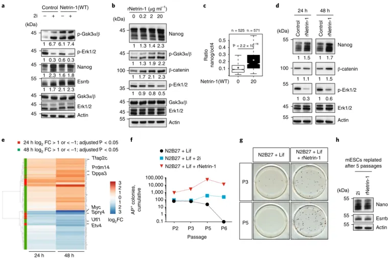
Netrin-1 supports mESC self-renewal in combination with Lif. To compare the magnitude of the changes induced by Netrin-1 and 2i, control and Netrin-1(WT) mESCs were treated with 2i for 48 h in serum/Lif (Fig. 4a). Basal levels of p-Gsk3α/β, Nanog and Esrrb in Netrin-1(WT) mESCs were similar to those in 2i-treated mESCs. However, there was less reduction in MAPK signalling in the Netrin-1(WT) cells (Fig. 4a), and these cells remained responsive to 2i treatment, confirming that Netrin-1 only partially mimics signalling changes induced by 2i.
We assessed whether recombinant Netrin-1 (rNetrin-1; see Methods) triggers similar changes to transgenes in mESCs. Using western blot and immunofluorescence, we showed that 48 h treat-ment with different doses of rNetrin-1 led to Nanog induction, dem-onstrating a paracrine effect of rNetrin-1 (Fig. 4b,c). This treatment also led to dose-dependent changes of β-catenin, p-Gsk3α/β and p-Erk1/2, as expected (Fig. 4b). We next dissected the sequence of events by analysing mESCs at 24 and 48 h of treatment. Changes in signalling appeared with different kinetics: Erk1/2 phosphorylation
was decreased at 24 h, whereas β-catenin was induced only at 48 h (Fig. 4d). Transcriptomic analyses enabled us to define clusters of early and late responders (Fig. 4e). At 24 h, there was a very lim-ited response (35 DEGs, log2 FC <1 or >1 and adjusted P < 0.05) and significant downregulation was detected for pluripotency genes (Myc and Utf1). Differentiation genes (Hand1) and Fgf tar-gets (Etv4, Spry4 and Dusp6) were also repressed, in line with the rapid changes of p-Erk1/2. At 48 h, a larger transcriptomic response was evident (193 DEGs), including upregulation of Tfap2c, Prdm14 and Dppa3. Most of the endoderm and mesoderm genes repressed in Netrin-1(WT) mESCs (Fig. 2l) were not repressed at these early time points, suggesting that Netrin-1 induces signalling changes prior to repressing differentiation.
We next investigated whether rNetrin-1 can support mESC self-renewal in combination with Lif. mESCs were grown at clonal density on laminin in N2B27 + Lif with rNetrin-1 or 2i for six passages (18 d) (Fig. 4f,g). Colony-formation assays confirmed that Lif is not sufficient to maintain self-renewal in serum-free
p-Gsk3α/β Actin 1 6.7 6.1 7.4 1 0.3 0.6 0.3 p-Erk1/2 Erk1/2 Gsk3α/β Nanog 1 2.3 1.6 1.8 Control Netrin-1(WT) 2i a e f g h b c d – + – + Esrrb 1 1.7 2.1 2.3 Nanog rNetrin-1 (µg ml–1) 0 0.2 2 20 1 1.3 1.4 2.3 0.1 0.3 0.2 0.4 Rati o nanog/oct 4 0.5 Netrin-1(WT) 0 * * 20 n = 525 n = 571 β-catenin Actin p-Erk1/2 p-Gsk3α/β 1 1.7 2.1 2.3 1 0.9 0.8 0.5 1 1.3 1.9 2.2 Gsk3α/β Erk1/2 Nanog Erk1/2 p-Erk1/2 β-catenin 24 h 48 h
Control rNetrin-1 Control rNetrin-1
Actin Nanog Esrrb Actin 2i rNetrin-1 mESCs replated after 5 passages P3 P5
N2B27 + Lif N2B27 + Lif+ rNetrin-1
N2B27 + Lif + rNetrin-1 N2B27 + Lif + 2i N2B27 + Lif P2 P3 P5 P6 Passage 100,000 10,000 0.1 10 100 1,000 1 AP + colonies, cumulative 24 h log2 FC > 1 or < –1; adjusted P < 0.05 24 h 48 h 1 1.5 1 1.7 1 1.1 1 1.5 1 0.3 1 0.6 1 1 0 2 3 2 3 log2FC Myc Spry4 Utf1 Etv4 Prdm14 Dppa3 Tfap2c 48 h log2 FC > 1 or < –1; adjusted P < 0.05 P < 2.2 × 10⁻¹⁶ (kDa) (kDa) (kDa) (kDa) 45 45 45 55 45 45 45 45 45 100 35 45 45 55 55 100 55 45 55 55 55 55
Fig. 4 | Recombinant Netrin-1 supports meSC self-renewal in combination with Lif. a, Western blot comparing signalling and pluripotency changes induced by Netrin-1 and 2i. Control and Netrin-1(WT) mESCs were grown in serum/Lif and treated with 2i for 2 d (3 independent experiments). b, Western blot of Nanog, Wnt and MAPK proteins in response to increasing doses of rNetrin-1. Cells were treated with the indicated concentrations for 48 h (3 independent experiments). c, Quantification of the Nanog/Oct4 intensity from immunofluorescence in single cells from the different populations. n corresponds to the number of cells. The centre line represents the median of the data, box edges show the upper and lower quartiles and whiskers show the highest and lowest values, excluding outliers. Two-tailed Student’s t-test. d, Western blot of pluripotency and signalling changes occurring after 24 and 48 h of treatment with 20 μg ml−1 rNetrin-1 (3 independent experiments). e, Heat map of DEGs. RNA-seq was performed on untreated or rNetrin-1-treated mESCs. n = 4 independent samples. P values from two-sided Wald test; two-sided Benjamini–Hochberg test for adjustment. f, Self-renewal assays. E14Tg2a mESCs were maintained for six passages in the indicated conditions. After splitting at P2, P3, P5 and P6, cells were counted and similar cell numbers were plated at clonal density in serum/Lif for 7 d and the number of alkaline phosphatase-positive colonies was counted to evaluate the self-renewal potential of the cells. Data from one representative experiment of two independent experiments. g, Self-renewal abilities of mESCs grown in the indicated conditions for three or five passages. Similar results were obtained from 3 independent experiments. h, Western blot of pluripotency factors. mESCs cultured in N2B27 + Lif + 2i or N2B27 + Lif + rNetrin-1 for 5 passages (15 d) were cultured in serum/Lif for a further 7 d before collection (3 independent experiments).
medium (Fig. 4f,g). However, the addition of rNetrin-1 enabled sustained self-renewal, similar to 2i (Fig. 4f,g). When replated in serum/Lif after five passages in N2B27 + Lif + rNetrin-1 or N2B27 + Lif + 2i, mESCs exhibited similar Nanog and Esrrb levels (Fig. 4h). Together, these results show that rNetrin-1 co-regulates Wnt and MAPK signalling and sustains self-renewal in combina-tion with Lif.
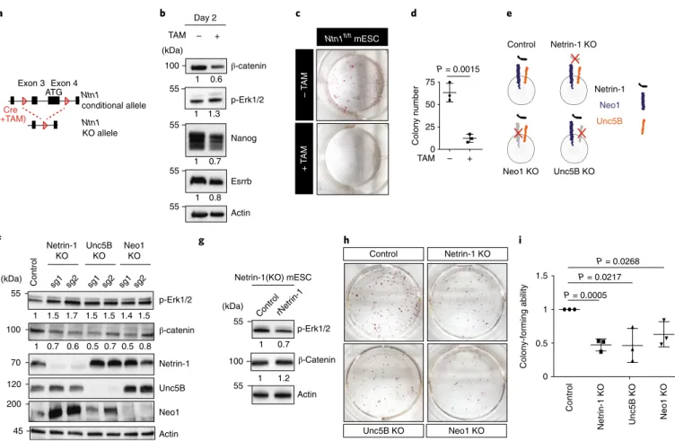
The Neo1 and Unc5b receptors are required for endogenous Netrin-1 function in mESCs. Because Netrin-1 is expressed at basal levels in serum/Lif (Fig. 1b), we assessed its endogenous func-tion in mESCs. We generated Ntn1 condifunc-tional-knockout (KO) mESCs by crossing Ntn1fl/fl (ref. 44) and Rosa26CreERT2 mice (Fig. 5a) (Extended Data Fig. 4a). Because feeder cells secrete Netrin-1,
Ntn1fl/fl mESCs were adapted on gelatin. After 48 h, Ntn1
dele-tion induced changes in signalling and pluripotency that mainly mirrored gain of function. Wnt-pathway activation was reduced, whereas MAPK activity was induced and Nanog and Esrrb levels were reduced (Fig. 5b). However, the expression of epiblast (Fgf5 and Otx2) or primitive endoderm (Gata4 and Gata6) transcripts was not significantly induced (Extended Data Fig. 4b). Netrin-1 loss also led to a significant decrease of mESC self-renewal abil-ity (Fig. 5c,d) with no significant changes in proliferation and cell death (Extended Data Fig. 4c,d). These defects, observed in
the first days following Netrin-1 deletion, were compensated and
Ntn1−/− mESCs could be maintained at high density for more than 20 passages (Extended Data Fig. 4e).
We next generated Netrin-1-, Neo1- and Unc5B-KO mESCs using clustered regularly interspaced short palindromic repeats (CRISPR)–CRISPR-associated protein 9 (Cas9) genome editing (Fig. 5e,f and Extended Data Fig. 4f). CRISPR–Cas9-mediated loss of Netrin-1 led to similar changes in p-Erk1/2 and β-catenin levels as the conditional KO (Fig. 5f), and these changes were partially rescued by treatment with rNetrin-1 (Fig. 5g). Loss of Netrin-1 trig-gered a self-renewal defect (Fig. 5h,i) that was compensated in 2i (Extended Data Fig. 4g), in agreement with the effects of Netrin-1 on Wnt and MAPK. Importantly, in serum/Lif, Unc5B and Neo1 single-KO induced signalling and clonogenicity changes that largely mimicked loss of Netrin-1 (Fig. 5f–i), indicating that a tight regula-tion of receptor dosage is required to co-regulate Wnt–MAPK and therefore self-renewal.
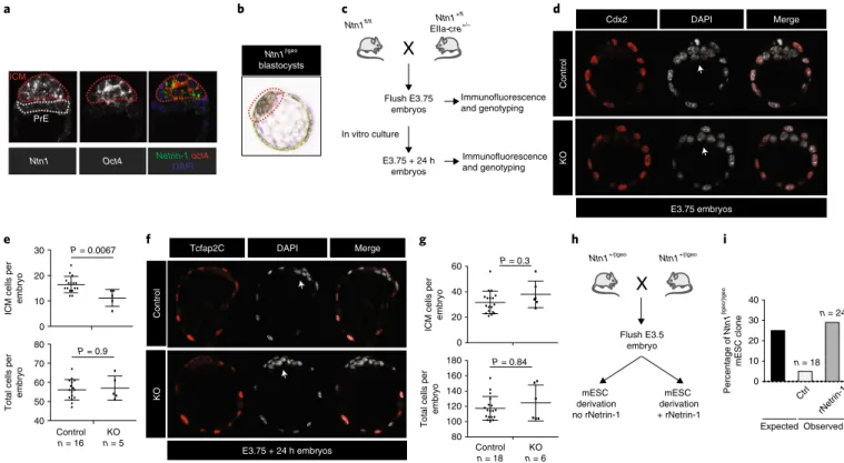
Netrin-1 regulates cell-fate allocation in preimplantation embryos. Netrin-1 depletion has been reported to cause embryonic lethality at embryonic day (E)14.5 (ref. 45), but no function has been reported in preimplantation embryos. Due to the unexpected func-tion we described in mESCs, we assessed whether Netrin-1 could be expressed and have a role at an earlier stage. In situ hybridization
Control Netrin-1 KO Unc5B KO Neo1 KO Colony-forming ability 1 1.5 0.5 0 P = 0.0005 P = 0.0217 P = 0.0268 Exon 3 a f g h i b c d e Exon 4 ATG Ntn1 conditional allele Ntn1 KO allele Cre (+TAM) Day 2 – + TAM β-catenin Nanog Esrrb p-Erk1/2 Actin 1 0.6 1 1.3 1 0.7 1 0.8 (kDa) 100 55 55 55 55 – TA M Ntn1fl/fl mESC + TA M Colony number 75 0 25 50 – + TAM P = 0.0015 Control Netrin-1 Unc5B Neo1 Netrin-1 KO Neo1 KO Unc5B KO p-Erk1/2 β-catenin Actin sg1 sg2 sg1 sg2 sg1 sg2 Control Netrin-1 KO Unc5BKO Neo1KO 1 1.5 1.7 1.5 1.5 1.4 1.5 1 0.7 0.6 0.5 0.7 0.5 0.8 55 100 45 p-Erk1/2 Actin ControlrNetrin-1 Netrin-1(KO) mESC 1 1.2 1 0.7 β-Catenin (kDa) (kDa) 55 100 55 Control Netrin-1 KO Unc5B KO Neo1 KO Netrin-1 Unc5B Neo1 70 120 200
Fig. 5 | endogenous Netrin-1 controls pluripotent features via Neo1 and Unc5b. a, Schematic of the Ntn1 conditional allele. KO, knockout. b, Western blot for Wnt and MAPK proteins in Ntn1fl/fl mESCs treated overnight or not with tamoxifen (TAM) in N2B27 + Lif for 48 h before collection (3 independent
experiments). c, Colony-formation assays. Similar results were obtained from 3 independent experiments. d, Colony counts. Data are mean ± s.d., n = 3 independent experiments; two-sided Student’s t-test. e, Scheme depicting the Netrin-1(KO), Neo1(KO) and Unc5B(KO) mESCs generated using CRISPR– Cas9. f, Effect of Netrin-1, Neo1 and Unc5B depletion on Wnt and MAPK pathways (3 independent experiments). g, Western blot for Wnt and MAPK proteins. Netrin-1(KO) mESCs were treated for 48 h with rNetrin-1 (20 μg ml−1) before collection (3 independent experiments). h,i, Effect of ligand or receptor depletion on mESC self-renewal ability. Cell lines from e were analysed by colony-formation assay. h, Bright-field images of a single experiment representative of 3 independent experiments. i, Colony counts. Data are mean ± s.d., n = 3 independent experiments; two-sided Student’s t-test.
of Ntn1 and Oct4 revealed that Ntn1 expression was confined to the naive epiblast, while no signal was detected in the adjacent primitive endoderm (Fig. 6a). Previously published single-cell transcriptomic data confirmed significantly higher Ntn1 expression in epiblas-tic cells than in primitive endoderm in E4.5 embryos46 (Extended Data Fig. 4h). The use of Ntn1βgeo embryos also showed specific βgal activity in the epiblast of blastocysts, indicating that Netrin-1 is expressed in pluripotent blastomeres (Fig. 6b).
We next analysed E3.75 and E4.5 (E3.75 grown in vitro for 24 h) embryos from intercrosses between Ntn1fl/fl males and Ntn1fl/WT; EIIa-cre+/− females (Fig. 6c). This strategy was selected because off-springs inheriting EIIa-cre maternally exhibit a widespread reporter expression47. Netrin-1 depletion led to a significant reduction of the number of cells in the inner cell mass (DAPI+Cdx2−) in E3.75 embryos, indicating a function for Netrin-1 in the homeostasis of the inner cell mass (Fig. 6d,e). This defect was compensated when embryos were grown in vitro and analysed 24 h later, in agree-ment with a lack of requireagree-ment for Netrin-1 at these embryonic stages (Fig. 6f,g).
We finally assessed whether Netrin-1 controls mESC deriva-tion. Starting from blastocysts obtained through Ntn1+/βgeo mice crosses27 (Fig. 6h), 18 expanded blastocyst outgrowths were sub-sequently amplified. Among these, a single Ntn1βgeo/βgeo mESC line was detected (Fig. 6i), a deviation from the expected 1:2:1
genotype frequency. The use of rNetrin-1 rescued this deviation (Fig. 6i), indicating that Netrin-1 controls optimal pluripotency capture. Together, these results reveal an unexpected function for Netrin-1 during preimplantation development.
Netrin-1 exerts different effects in mESCs depending on the Neo1/Unc5B stoichiometry. Netrin-1 has been shown to trigger opposite responses depending on the receptor dosage21–23. We inves-tigated whether it also exerts different functions depending on the Neo1/Unc5B stoichiometry in mESCs and differentiated deriva-tives. While Netrin-1 and both receptors are expressed in mESCs, embryoid body formation induced Netrin-1 expression and tilted the dosage toward a Neo1low/Unc5Bhigh ratio (Fig. 7a). Unc5b induc-tion was already detected in epiblast-like (EpiLC) cells48 (Extended Data Fig. 5a). To alter experimentally the ligand and receptors ratio, we engineered control and Netrin-1(WT) mESCs to express Neo1 or Unc5B on addition of dox (Fig. 7b). We found that the decrease in p-Erk1/2 triggered by Netrin-1 was more pronounced when Neo1 was exogenously expressed (Fig. 7c). By contrast, when Unc5B was induced, p-Erk1/2 levels were increased by Netrin-1, indicating that the ligand has different effects on MAPK depend-ing on the balance of its receptors (Fig. 7d). In line with a func-tion of p-Erk1/2 in lineage commitment, we found that this setting, Netrin-1highNeo1lowUnc5Bhigh, triggered downregulation of Nanog
Ntn1βgeo blastocysts a b e f g h i c d Ntn1fl/fl Ntn1 +/fl EIIa-cre+/– Flush E3.75 embryos In vitro culture Immunofluorescence and genotyping Ntn1+/βgeo Ntn1+/βgeo Flush E3.5 embryo mESC derivation no rNetrin-1 Percentage of Ntn1 βgeo/ βgeo mESC clone 20 30 10 0 40 Expected Ctrl rNetrin-1 n = 18 n = 24
Total cells per
embryo P = 0.9 ICM cells pe r embryo 10 0 20 30 P = 0.0067 PrE mESC derivation + rNetrin-1 P = 0.84 20 0 60 40 P = 0.3 ICM cells pe r embryo E3.75 embryos E3.75 + 24 h embryos Cdx2 DAPI Merge Contro l KO
Tcfap2C DAPI Merge
Contro l KO E3.75 + 24 h embryos Immunofluorescence and genotyping 60 70 50 40 80 Control n = 16 n = 5KO Control n = 18 n = 6KO
Total cells per
embryo 120 140 100 80 160 180 Observed ICM PrE
Ntn1 Oct4 Netrin-1DAPIoct4
Fig. 6 | Netrin-1 regulates cell-fate allocation in preimplantation embryos. a, In situ hybridization of Netrin-1 and Oct4 in an E4.5 embryo. Representative of 17 E4.5–E4.75 embryos from 3 independent experiments. ICM, inner cell mass; PrE, primitive endoderm. b, X-gal activity in blastocyst-stage embryos. Ntn1βgeo reporter embryos were flushed at E3.5 and grown in vitro for 24 h before fixation (9 embryos from 3 independent experiments). c, Scheme depicting the intercrosses. d, Image of control and Netrin-1(KO) E3.75 embryos. Cdx2 immunofluorescence marks trophectoderm cells. Arrows indicate ICM cells (4 independent experiments). e, Average number of ICM cells per embryo. Data are mean ± s.d. of (DAPI+ cells) − (Cdx2+ cells); 16 control and 5 Netrin-1(KO) embryos were analysed. Control, Ntn1+/+ and Ntn1+/− embryos; Netrin-1(KO), Ntn1−/−. Two-sided non-parametrical Wilcoxon test. f, Control and Netrin-1(KO) E3.75 embryos grown in vitro for a further 24 h. Immunofluorescence for Tcfap2C marks trophectoderm cells. Arrows indicate epiblast cells (3 independent experiments). g, Number of ICM cells per embryo. Data are mean ± s.d. of (DAPI+ cells) − (Tcfap2c+ cells). n = 18 (control) and 6 (Netrin-1(KO)) embryos. Two-sided non-parametrical Wilcoxon test. h, Scheme of the intercrosses. i, The percentage of Ntn1βgeo/βgeo mESC lines obtained from Ntn1+/βgeo heterozygous crosses. n represents the number of lines generated and is indicated on the graph. Embryos were flushed at E3.5, grown on feeder cells in N2B27 + Lif + 2i for 3 d and then in serum/Lif. Netrin-1 status was evaluated by western blot.
and Esrrb (Fig. 7e) and a reduction of mESC resistance to differ-entiation (Fig. 7f,g). In addition, by subjecting mESCs expressing solely Unc5B (Neo1(KO)) or Neo1 (Unc5B(KO)) to differentiation in N2B27 − Lif, we found that mESCs expressing Unc5B exhibited increased induction of differentiation genes (Fig. 7h). Collectively, these data indicate that the effect of Netrin-1 on self-renewal is tightly regulated by the balance of Netrin-1 receptors.
Netrin-1 coordinates differentiation in vitro and in vivo. Loss of a pro-self-renewal signal in mESCs leads to their accelerated dif-ferentiation, as shown for Lif4, Wnt49 and Bmp6. However, because Netrin-1 can either repress or induce p-Erk1/2, we assessed whether and how its loss affects mESC differentiation. We subjected Ntn1fl/fl mESCs, untreated or treated with TAM, to differentiation assays. In embryoid bodies, Netrin-1 deletion led to a delay in the induction p-Erk1/2 Erk1/2 – + – + Dox Control Unc5B-doxi Netrin-1 Unc5B-doxi p-Erk1/2 Erk1/2 – + – + Dox Control Neo1-doxi Netrin-1 Neo1-doxi d – Dox + Dox (Unc5B) Control Unc5B-doxi Netrin-1 Unc5B-doxi Control Unc5B-doxi Colony number Nanog Esrrb Dox – + Actin Netrin-1 Unc5B-doxi c Netrin-1 Unc5B Neo1 Control mESC + Dox Control Neo1-doxi Control Unc5B-doxi + Dox a Day 0 Day 4 Neo1 Actin Unc5B b h e f g i n o p j k l m Netrin-1 Neo1-doxi Netrin-1 Unc5B-doxi Netrin-1(WT) mESC Ntn1fl/fl mESC
*
Chimaeras (% of pups) 0 5 10 15 12,5%n = 16 0% n = 20 – + TAM Wnt3 Mixl1 Foxa2 Amn Cdh2 Cer1Relative transcript level
Control Neo1 KO
Unc5B KO
Cleaved-caspase-3
+
cells per embryo
0 2 4 6 – n = 9 + Netrin-1 Unc5b Neo1 Wnt MAPK n = 8 TAM 8 – TA M + TA M Cleaved Caspase 3 DAPI GFP Merge * * 1 1 0.8 0.5 1 1 0.7 0.9 TAM N2B27 – Lif day 2 Proliferation inde x 0 1 2 – + P = 0.0128 Netrin-1 Unc5B-doxi 30 0 10 20 Dox – + Neo1 Actin Unc5B Netrin-1 N2B27 – Lif Day 0 Day 4 Day 8 P = 0.0095 Dox – + 30 0 10 20 40 P > 0.99 Nes P = 0.015 P = 0.095 P = 0.2 P = 0.0076 0 2 4 6 8 0 2 4 6 Tubb3
Relative transcript level
TAM P = 0.018 P = 0.0092 P = 0.0026 P = 0.014 0 1 2 0 1 2
Embryoid body day 7
– + – + – + P = 0.0025 P = 0.007 P = 0.0001 (kDa) (kDa) (kDa) (kDa) (kDa) 200 125 55 55 55 55 55 55 70 200 125 55 55 55 P P P Erk1/2 Naive pluripotency features Mek1/2 Pr55γ Fak Gsk3α/β β-catenin β-catenin Axin Apc Pp2aα
of differentiation genes of the three germ layers (Fig. 7i), indicat-ing that Netrin-1 contributes to coordinated differentiation. Cells depleted of Netrin-1 generated teratomas of similar size to those generated by control cells (Extended Data Fig. 5b). To assess whether these defects were associated with differentiation, prolifer-ation and/or cell death, we performed guided neural differentiprolifer-ation, during which Netrin-1 is induced and the switch of receptors occurs (Fig. 7j). After 8 d, we observed a reduced induction of differentia-tion transcripts in the absence of Netrin-1 (Extended Data Fig. 5c). This difference was accompanied by a reduction of proliferation, which was observed as early as day two (Fig. 7k), without signifi-cant difference in cell death (Extended Data Fig. 5d), indicating that Netrin-1 controls differentiation and proliferation. When injected into blastocysts, mESCs depleted of Netrin-1 harboured a reduced ability to give rise to coat-colour chimaeras (Fig. 7l,m). To better characterize this defect, GFP-labelled Ntn1fl/fl mESCs, untreated or treated with TAM, were aggregated with moru-las. Immunofluorescence analysis of late blastocysts for GFP and cleaved Caspase-3 revealed an increase in the number of apoptotic cells in embryos injected with Netrin-1-depleted cells, suggesting that Netrin-1 promotes cell survival (Fig. 7n,o). Altogether, these results demonstrate that Netrin-1 deregulation triggers differentia-tion, proliferation and survival defects.
Discussion
In this study, we document that the neuronal guidance cue Netrin-1, which is expressed in the epiblast and in mESCs, is an autocrine and paracrine factor that promotes pluripotent features. Netrin-1 controls self-renewal of mESCs and triggers signal-ling, transcriptomic and epigenetic features that partially overlap with the ground state of pluripotency9 (Fig. 7p). Even if Netrin-1 signalling acts by reducing MAPK and promoting Wnt in a similar manner to 2i, it targets a different effector of the MAPK pathway—namely Erk1/2—via Pp2a. In line with this difference and in contrast to prolonged Mek1/2 blockade, we did not observe global DNA hypomethylation in Netrin-1(WT) mESCs13. Because the use of a Src inhibitor preserves mESCs genomic integrity, and since Src and Fak are interconnected50, it will be interesting to assess whether Src inhibition triggers a similar cascade to the one described here.
Using genetic models, we reveal that Netrin-1 influences cell-fate allocation during preimplantation development (Fig. 6). Despite this effect, mouse embryos lacking zygotic Netrin-1 expression develop normally through the epiblast stage and die at E14.5 (ref. 45). The cause of the embryonic death is currently unknown, but it coin-cides with the embryonic lethality in Unc5B-null mice23. It remains to be investigated whether, as with gp130, such a role may be accen-tuated in the context of delayed implantation51.
We show that Netrin-1 exerts different effects on MAPKs depending on the Neo1/Unc5B dosage. The repressive activity of Netrin-1 on MAPKs is converted to a stimulation by Unc5B induc-tion, but questions remain to be answered. First, the factors trig-gering Unc5B induction remain to be identified. Publicly available resources indicate that transcription factors such as Otx2 or Cdx2, MyoD and Gata3 induce Unc5B when expressed in mESCs (Gene Expression Omnibus GSE31381)52. Second, the molecular mecha-nisms responsible for the differential effects of Netrin-1 remain to be dissected. Because these effects are mediated by the Pp2a subunit Ppp2r2c, whose paralogues Ppp2r2a and Ppp2r2d were shown to affect the Nodal pathway in opposite ways43, it will be interesting to assess whether Pp2a composition is responsible for the differential effects of Netrin-1. Finally, while Netrin-1 has been shown to medi-ate differential responses in various cell types21,22, our study suggests that this ability reflects a fundamental characteristic of this protein, which manifests earlier in development than previously proposed, and which might govern other cellular responses than cell migra-tion, such as cell-fate decisions.
Collectively, our work positions Netrin-1 as a signalling pathway that participates in feedback loops with Wnt and MAPK in pluripo-tent cells, and demonstrates that a single ligand can trigger different effects in stem cells depending on the stoichiometry of its recep-tors, opening fascinating perspectives for regenerative medicine and cancer biology.
Online content
Any methods, additional references, Nature Research reporting summaries, source data, extended data, supplementary informa-tion, acknowledgements, peer review information; details of author contributions and competing interests; and statements of data and code availability are available at https://doi.org/10.1038/s41556-020-0483-2.
Received: 3 November 2017; Accepted: 13 February 2020; Published online: 30 March 2020
References
1. Evans, M. J. & Kaufman, M. H. Establishment in culture of pluripotential cells from mouse embryos. Nature 292, 154–156 (1981).
2. Martin, G. R. Isolation of a pluripotent cell line from early mouse embryos cultured in medium conditioned by teratocarcinoma stem cells. Proc. Natl Acad. Sci. USA 78, 7634–7638 (1981).
3. Dunn, S. J., Martello, G., Yordanov, B., Emmott, S. & Smith, A. G. Defining an essential transcription factor program for naive pluripotency. Science 344, 1156–1160 (2014).
4. Williams, R. L. et al. Myeloid leukaemia inhibitory factor maintains the developmental potential of embryonic stem cells. Nature 336, 684–687 (1988). 5. Sato, N., Meijer, L., Skaltsounis, L., Greengard, P. & Brivanlou, A. H.
Maintenance of pluripotency in human and mouse embryonic stem cells
Fig. 7 | Netrin-1 exerts different effects in meSCs depending on receptor balance. a, Western blot in mESCs and day 4 embryoid bodies. b, Schematic of mESC lines generation. c–e, Western blot of p-Erk1/2 levels in indicated cell lines. f,g, Assays for exit from pluripotency performed on the indicated cell lines. f, Representative images of a single experiment representative of 3 independent experiments. g, Colony counting. Data are mean ± s.d. (n = 3 independent experiments). Two-sided Student’s t-test. h, Results of quantitative PCR with reverse transcription (RT–qPCR), showing transcript levels at day 8 of differentiation in N2B27 − Lif. Data are normalized to housekeeping genes and value 1 is given to day 8 control mESCs. Data are mean ± s.d. (3 independent experiments). Two-sided Student’s t-test. i, RT–qPCR showing levels of differentiation transcripts in day 7 embryoid bodies generated with Ntn1fl/fl mESCs, untreated or treated with TAM. Data are mean ± s.d., n = 3 independent experiments; value 1 is given to untreated mESCs.
Two-sided Student’s t-test. j, Western blot showing Netrin-1, Unc5b and Neo1 expression during neural differentiation. k, Proliferation assays. Ntn1fl/fl mESCs,
untreated or treated with TAM, were grown for 2 d in N2B27 − Lif and cell number was counted. Data are mean ± s.d., 3 independent experiments; two-sided Student’s t-test. l,m, Blastocyst injections performed with Ntn1fl/fl mESCs, untreated or treated with TAM prior to injection. l, Example of a coat
colour chimaera obtained following injection of Ntn1fl/fl mESCs. m, Percentage of chimaeras obtained (n represents the number of live pups obtained and
is indicated on the graph). n, Image of representative embryos immunostained for GFP and cleaved Caspase-3. Morulas are aggregated with 5–10 Ntn1fl/fl
mESCs, untreated or treated with TAM. Blastocysts were fixed 36 h after aggregation. Asterisks mark apoptotic cells. o, Count of cells positive for cleaved Caspase-3. Data are mean ± s.d., 3 independent experiments; n represents the number of embryos analysed and is indicated on the graph. Two-sided Student’s t-test. p, Graphical summary of the results. Western blots in a,c–e,j, are representative of 3 independent experiments.
through activation of Wnt signaling by a pharmacological GSK-3-specific inhibitor. Nat. Med. 10, 55–63 (2004).
6. Ying, Q. L., Nichols, J., Chambers, I. & Smith, A. BMP induction of Id proteins suppresses differentiation and sustains embryonic stem cell self-renewal in collaboration with STAT3. Cell 115, 281–292 (2003). 7. Kunath, T. et al. FGF stimulation of the Erk1/2 signalling cascade triggers
transition of pluripotent embryonic stem cells from self-renewal to lineage commitment. Development 134, 2895–2902 (2007).
8. Marks, H. et al. The transcriptional and epigenomic foundations of ground state pluripotency. Cell 149, 590–604 (2012).
9. Ying, Q. L. et al. The ground state of embryonic stem cell self-renewal. Nature 453, 519–523 (2008).
10. Ficz, G. et al. FGF signaling inhibition in ESCs drives rapid genome-wide demethylation to the epigenetic ground state of pluripotency. Cell Stem Cell 13, 351–359 (2013).
11. Buehr, M. et al. Capture of authentic embryonic stem cells from rat blastocysts. Cell 135, 1287–1298 (2008).
12. Li, P. et al. Germline competent embryonic stem cells derived from rat blastocysts. Cell 135, 1299–1310 (2008).
13. Choi, J. et al. Prolonged Mek1/2 suppression impairs the developmental potential of embryonic stem cells. Nature 548, 219–223 (2017).
14. Serafini, T. et al. The netrins define a family of axon outgrowth-promoting proteins homologous to C. elegans UNC-6. Cell 78, 409–424 (1994). 15. Kennedy, T. E., Serafini, T., de la Torre, J. R. & Tessier-Lavigne, M. Netrins
are diffusible chemotropic factors for commissural axons in the embryonic spinal cord. Cell 78, 425–435 (1994).
16. Cirulli, V. & Yebra, M. Netrins: beyond the brain. Nat. Rev. Mol. Cell Biol. 8, 296–306 (2007).
17. Grandin, M. et al. Structural decoding of the netrin-1/UNC5 interaction and its therapeutical implications in cancers. Cancer Cell 29, 173–185 (2016). 18. Bell, C. H. et al. Structure of the repulsive guidance molecule
(RGM)-neogenin signaling hub. Science 341, 77–80 (2013).
19. Rajagopalan, S. et al. Neogenin mediates the action of repulsive guidance molecule. Nat. Cell Biol. 6, 756–762 (2004).
20. Xu, K. et al. Neural migration. Structures of netrin-1 bound to two receptors provide insight into its axon guidance mechanism. Science 344,
1275–1279 (2014).
21. Ko, S. Y., Dass, C. R. & Nurgali, K. Netrin-1 in the developing enteric nervous system and colorectal cancer. Trends Mol. Med. 18, 544–554 (2012). 22. Hong, K. et al. A ligand-gated association between cytoplasmic domains of
UNC5 and DCC family receptors converts netrin-induced growth cone attraction to repulsion. Cell 97, 927–941 (1999).
23. Lu, X. et al. The netrin receptor UNC5B mediates guidance events controlling morphogenesis of the vascular system. Nature 432, 179–186 (2004). 24. Ozmadenci, D. et al. Netrin-1 regulates somatic cell reprogramming and
pluripotency maintenance. Nat. Commun. 6, 7398 (2015).
25. Rajasekharan, S. & Kennedy, T. E. The netrin protein family. Genome Biol 10, 239 (2009).
26. Wray, J. et al. Inhibition of glycogen synthase kinase-3 alleviates Tcf3 repression of the pluripotency network and increases embryonic stem cell resistance to differentiation. Nat. Cell Biol. 13, 838–845 (2011).
27. Skarnes, W. C., Moss, J. E., Hurtley, S. M. & Beddington, R. S. Capturing genes encoding membrane and secreted proteins important for mouse development. Proc. Natl Acad. Sci. USA 92, 6592–6596 (1995). 28. Kumar, R. M. et al. Deconstructing transcriptional heterogeneity in
pluripotent stem cells. Nature 516, 56–61 (2014).
29. Guo, G. et al. Serum-based culture conditions provoke gene expression variability in mouse embryonic stem cells as revealed by single-cell analysis. Cell Rep. 14, 956–965 (2016).
30. Bulut-Karslioglu, A. et al. Inhibition of mTOR induces a paused pluripotent state. Nature 540, 119–123 (2016).
31. Boroviak, T. et al. Lineage-specific profiling delineates the emergence and progression of naive pluripotency in mammalian embryogenesis. Developmental Cell 35, 366–382 (2015).
32. Galonska, C., Ziller, M. J., Karnik, R. & Meissner, A. Ground state conditions induce rapid reorganization of core pluripotency factor binding before global epigenetic reprogramming. Cell Stem Cell 17, 462–470 (2015).
33. Habibi, E. et al. Whole-genome bisulfite sequencing of two distinct interconvertible DNA methylomes of mouse embryonic stem cells. Cell Stem Cell 13, 360–369 (2013).
34. von Meyenn, F. et al. Impairment of DNA methylation maintenance is the main cause of global demethylation in naive embryonic stem cells. Molecular Cell 62, 848–861 (2016).
35. Beurel, E., Grieco, S. F. & Jope, R. S. Glycogen synthase kinase-3 (GSK3): regulation, actions, and diseases. Pharmacol. Ther. 148, 114–131 (2015). 36. Guenebeaud, C. et al. The dependence receptor UNC5H2/B triggers apoptosis
via PP2A-mediated dephosphorylation of DAP kinase. Molecular Cell 40, 863–876 (2010).
37. Ren, X. R. et al. Focal adhesion kinase in netrin-1 signaling. Nat. Neurosci. 7, 1204–1212 (2004).
38. Liu, G. et al. Netrin requires focal adhesion kinase and Src family kinases for axon outgrowth and attraction. Nat. Neurosci. 7, 1222–1232 (2004). 39. Moore, S. W. & Kennedy, T. E. Protein kinase A regulates the sensitivity of
spinal commissural axon turning to netrin-1 but does not switch between chemoattraction and chemorepulsion. J. Neurosci. 26, 2419–2423 (2006). 40. Qu, C. et al. c-Jun N-terminal kinase 1 (JNK1) is required for coordination of
netrin signaling in axon guidance. J. Biol. Chem. 288, 1883–1895 (2013). 41. Gao, C. et al. FAK/PYK2 promotes the Wnt/β-catenin pathway and intestinal
tumorigenesis by phosphorylating GSK3β. eLife 4, e10072 (2015). 42. Sangodkar, J. et al. All roads lead to PP2A: exploiting the therapeutic
potential of this phosphatase. FEBS J. 283, 1004–1024 (2016).
43. Batut, J. et al. Two highly related regulatory subunits of PP2A exert opposite effects on TGF-β/Activin/Nodal signalling. Development 135, 2927–2937 (2008). 44. Dominici, C. et al. Floor-plate-derived netrin-1 is dispensable for commissural
axon guidance. Nature 545, 350–354 (2017).
45. Bin, J. M. et al. Complete loss of netrin-1 results in embryonic lethality and severe axon guidance defects without increased neural cell death. Cell Rep. 12, 1099–1106 (2015).
46. Nakamura, T. et al. A developmental coordinate of pluripotency among mice, monkeys and humans. Nature 537, 57–62 (2016).
47. Heffner, C. S. et al. Supporting conditional mouse mutagenesis with a comprehensive cre characterization resource. Nat. Commun. 3, 1218 (2012). 48. Hayashi, K., Ohta, H., Kurimoto, K., Aramaki, S. & Saitou, M. Reconstitution
of the mouse germ cell specification pathway in culture by pluripotent stem cells. Cell 146, 519–532 (2011).
49. ten Berge, D. et al. Embryonic stem cells require Wnt proteins to prevent differentiation to epiblast stem cells. Nat. Cell Biol. 13, 1070–1075 (2011). 50. Mitra, S. K. & Schlaepfer, D. D. Integrin-regulated FAK–Src signaling in
normal and cancer cells. Curr. Opin. Cell Biol. 18, 516–523 (2006). 51. Nichols, J., Chambers, I., Taga, T. & Smith, A. Physiological rationale for
responsiveness of mouse embryonic stem cells to gp130 cytokines. Development 128, 2333–2339 (2001).
52. Correa-Cerro, L. S. et al. Generation of mouse ES cell lines engineered for the forced induction of transcription factors. Sci. Rep. 1, 167 (2011).
Publisher’s note Springer Nature remains neutral with regard to jurisdictional claims in
published maps and institutional affiliations.