Abstract — We have developed a versatile delivery platform comprising a novel composite of two biomaterials with proven track records: apatite and poly(lactic-co-glycolic acid) (PLGA). These composites have been tested in the delivery of a model protein, bovine serum albumin (BSA), as well as a growth factor, bone morphogenetic protein-2 (BMP-2), which is a potent inducer of bone formation. The controlled release strategy is based on the use of a polymer with acidic degradation products to control the dissolution of a basic inorganic component, resulting in protein release. The release profile can be modified systematically by changing variables that affect polymer degradation and/or apatite dissolution, such as polymer molecular weight, polymer composition, apatite loading, and apatite particle size. We have found that an increase in polymer molecular weight and polymer hydrophobicity led to slower polymer degradation, and in turn, slower apatite dissolution and protein release. Protein release was enhanced by reducing apatite particle size and by lowering the apatite content in the composites. We anticipate that this delivery platform can be extended to the controlled release of other therapeutic proteins and chemicals.
Index Terms — Composite, apatite, PLGA, bone
morphogenetic protein
*To whom correspondence should be addressed
Manuscript received November 3, 2003. This work was supported by the Singapore-MIT Alliance (MEBCS Program).
T.-H. Yong is with the Department of Materials Science and Engineering, Massachusetts Institute of Technology, Cambridge, MA, 02139, USA (e-mail: say1@mit.edu).
E. A. Hager is with the Department of Materials Science and Engineering, Massachusetts Institute of Technology, Cambridge, MA, 02139, USA (e-mail: zlahager@mit.edu).
J. Y. Ying is with the Department of Chemical Engineering, Massachusetts Institute of Technology, Cambridge, MA, 02139, USA (phone: +1-617-253-2899; e-mail: jyying@mit.edu) and the Institute of Bioengineering and Nanotechnology, 51 Science Park Road #01-01, The Aries, Singapore Science Park II, Singapore 117586 (phone: +65-6874-9350; e-mail: jyying@ibn.a-star.edu.sg).
I. INTRODUCTION
one is a naturally regenerative tissue; it is able to heal from injury by recapitulating the embryonic skeletal developmental process. However, an estimated 5-10% of fractures fail to recover properly and proceed to delayed union or nonunion [1]. The repair of bone loss associated with trauma and cancer is also typically not observed. Current treatment involves the implantation of autogenous or allogeneic bone grafts, a procedure that an estimated 300,000 patients undergo in the United States each year [2]. Autografts, long considered the gold standard in bone grafting, are plagued with problems of limited availability and morbidity associated with graft harvest. The use of allografts is dampened by the risks of disease transfer and the costs of maintaining bone banks. Synthetic grafts constructed of polymers, metals, and ceramics are also used, but their mechanical incompatibility with bone tissue often leads to implant failure. The ideal solution would be to regenerate bone to fill the defects. Bone morphogenetic proteins (BMPs), a group of potent growth factors belonging to the transforming growth factor-β (TGF-β) superfamily, have the ability to elicit new bone formation. These proteins provide a promising alternative to current bone grafts.
Among the osteoinductive BMPs, BMP-2, BMP-4 and BMP-7 appear to have greater potency [3,4] and are being produced with high bioactivity and purity via recombinant DNA technology. BMP-2 and BMP-7 are currently in clinical use. In vitro administration of BMP-2 and BMP-7 to embryonic rat calvarial cells, rat osteosarcoma cells, or mouse fibroblasts resulted in enhanced osteogenic activity, as evidenced by differentiation into osteoblasts and elevated expression of bone mineralization proteins. In vivo treatment with BMP-2 or BMP-7 augmented the healing of defects in rodents [5,6], rabbits [7-11], dogs [12-14], sheep [15], and non-human primates [9,16,17]. Critical-size defects, which do not heal spontaneously, were bridged within 3 months in primates [16,17]. These animal studies validate the safety and efficacy of BMP-2 and BMP-7 in promoting orthopedic repair. However, the results from human trials have shown more variation. Geesink et al. reported that amongst six patients receiving
Apatite-Polymer Composite Particles for
Controlled Delivery of BMP-2
Tseh-Hwan Yong
1, Elizabeth A. Hager
1, and Jackie Y. Ying
2,3*1
Department of Materials Science and Engineering,
2Department of Chemical Engineering,
Massachusetts Institute of Technology, Cambridge, MA 02139-4307, USA.
3
Institute of Bioengineering and Nanotechnology, 51 Science Park Road #01-01,
The Aries, Singapore Science Park II, Singapore 117586.
BMP-7 with a collagen carrier for the treatment of high tibial osteotomy, four showed bridging by 6 weeks, one by 10 weeks, and one showed no bone formation during the course of the study [18]. When similar BMP-7 carriers were implanted in the maxillary sinuses of three patients with maxillary atrophy, only one exhibited satisfactory bone formation whereas the remaining two showed little bone formation after 6 months [3]. This suboptimal response in humans may be attributed to a smaller population of multipotent cells that are also less responsive than those in small animals.
To improve the efficacy of BMP treatment in higher mammals, optimal delivery of the therapeutic protein is crucial. The objective of our research is to develop carriers that can retain and meter out BMPs at the appropriate dose and for a sufficient duration to allow the recruitment, migration, differentiation, and proliferation of bone-forming cells. In addition to sustained release, we seek to reduce burst release to avoid the adverse effects of an overdose and to conserve the protein, making the formulation more cost-effective.
In this paper, we describe a controlled delivery system for BMP-2 constructed from composites of apatite and poly(lactic-co-glycolic acid) (PLGA). The controlled release strategy is based on the use of a polymer with acidic degradation products to control the dissolution of a basic inorganic component (apatite) on which a therapeutic agent (BMP-2) has been adsorbed. The dissolution of the inorganic substrate results in the release of the therapeutic agent. This strategy allows the systematic modulation of the release profile by changing variables that affect polymer degradation and/or apatite dissolution. These variables include polymer type, polymer molecular weight, polymer composition (including copolymers and blends), apatite loading, and apatite particle size. Controlled release of a model protein, bovine serum albumin (BSA), was first established, and then extended to BMP-2.
II. EXPERIMENTAL
A. Synthesis of Hydroxyapatite and Carbonated Apatite Hydroxyapatite (HAP) and carbonated apatite (CAP) were synthesized according to the method developed by Ahn et al. [19-21]. For the synthesis of HAP, 900 ml of 0.167 M Ca(NO3)2 (Fluka) and 900 ml of 0.100 M
(NH4)2HPO4 (Fluka) were prepared in distilled water. The
pH of the (NH4)2HPO4 solution was raised to 10.4 with
ammonium hydroxide. The Ca(NO3)2 solution was added
to the (NH4)2HPO4 solution at a rate of ~ 3 ml/min. The
resulting suspension was stirred at room temperature for 72 h. After this aging step, the white precipitate was collected by centrifugation, washed with aqueous NH4OH solutions
of decreasing pH, followed by two ethanol washes. The gel was air-dried overnight, then oven-dried at 120°C for 24 h. The dried gel was ground in a heated mortar and calcined at
550°C for 2 h (ramp rate of 10°C/min). After calcination, the HAP powder was sieved through a 45-µm mesh. CAP was synthesized by the same method but with the following modifications. (NH4)HCO3, the carbonate
source, was added to the (NH4)2HPO4 solution to a
concentration of 0.100 M. After the oven-drying step, the gel was ground and sieved. The powder was not calcined to avoid driving off the carbonate groups at elevated temperatures.
For the synthesis of submicron-sized apatite particles, modifications were made to the above protocol to reduce agglomeration. Tween 80 (Aldrich) was added as a surfactant to constitute 10 v/v% of the (NH4)2HPO4/
(NH4)HCO3 solution. Apatite was collected and washed by
ultrafiltration instead of centrifugation. The washed apatite was dried by lyophilization, which produced a fine, fluffy powder without the need for grinding or sieving. Calcination, which leads to grain growth, was not performed on these apatite powders. The hydroxyapatite and carbonated apatite thus prepared are referred to as sHAP and sCAP, respectively.
B. Characterization of Apatite
Powder X-ray diffraction (XRD) patterns of the various apatite powders were obtained with a Siemens D5000 θ-θ diffractometer (45 kV, 40 mA, Cu Kα). Grain size
analyses were performed on the <002> diffraction peaks using Scherrer’s method. The BET surface areas of the apatite powders were determined by nitrogen adsorption on a Micromeritics ASAP 2000/2010 Analyzer. Particle size distribution was evaluated using a Horiba CAPA-300 Particle Size Analyzer.
C. Preparation of Apatite-Protein Complexes
Fluorescein isothiocyanate-bovine serum albumin (FITC-BSA, Sigma), was used as a model protein. FITC-BSA was dissolved in distilled water and added to an aqueous suspension of HAP, sHAP, CAP, or sCAP. The suspension was stirred at room temperature for 16 h to allow the adsorption of the protein onto the apatite. The BSA-apatite complex was collected by centrifugation, washed with distilled water, and lyophilized. BMP-2 (R&D Systems) was adsorbed onto apatite by the same procedure, except the adsorption time was reduced to 8 h. The amount of protein adsorbed was determined by measuring the protein concentrations of the initial stock solution and the supernatant after adsorption, and taking the difference. FITC-BSA concentration was analyzed by Coomassie Plus total protein assay (Pierce). BMP-2 concentration was evaluated with an enzyme-linked immunosorbent assay (ELISA) kit (R&D Systems).
D. Preparation of Composite Microparticles
Composite microparticles were synthesized by a modification of a solid-in-oil-in-water emulsion process
previously used to encapsulate proteins in the solid state [22]. A typical synthesis involves dissolving 250 mg of PLGA (24 kd, Alkermes) in 2 ml of dichloromethane. To this polymer solution, 10 mg of protein-apatite complex was added and vortexed to create a uniform suspension. The solid-in-oil suspension was homogenized in 50 ml of 0.1 w/v% aqueous methyl cellulose solution at 8000 rpm for 2 min at room temperature. The resulting solid-in-oil-in-water suspension was heated at 30°C for 3 h to drive off dichloromethane and solidify the particles. The particles were collected by centrifugation, washed with distilled water, and lyophilized. This protocol was systemically varied one parameter at a time to investigate the effect of different processing parameters.
E. Characterization of Microparticle Morphology and Size
The morphology of the composite particles was evaluated by environmental scanning electron microscopy (ESEM, FEI/Philips XL30 FEG) at 6-7 kV. The average particle size and size distribution were determined by measuring the dimensions of a minimum of 100 particles under ESEM.
F. Evaluation of In Vitro Release
Thirty mg of protein-loaded composite microparticles were re-suspended in 1.5 ml of pH 7.4 N,N-bis(2-hydroxyethyl)-2-aminoethanesulfonic acid (BES) buffer supplemented with 0.02 w/v% sodium azide. The samples were incubated at 37°C. At pre-determined time intervals (1, 4, 7, 14, 21, 28, 42, 56… days, up to 20 weeks), the samples were centrifuged, 0.75 ml of the supernatant was withdrawn and replaced with 0.75 ml of fresh BES buffer. For samples containing FITC-BSA, the collected supernatant was filtered and assayed for fluorescence, and stored at 4°C until further analysis of protein concentration with the Coomassie Plus total protein assay. For samples containing BMP-2, the collected supernatant was filtered and stored at -20°C until evaluation by a BMP-2 ELISA kit. The protein concentration at each time point was used to construct the cumulative release profiles.
III. RESULTS AND DISCUSSION
A. Characterization of Apatite
HAP and CAP had approximately the same particle size. The calcination step in the preparation of HAP led to grain growth and lower surface area (Table 1). The use of surfactant in the synthesis of sHAP and sCAP contributed to less agglomeration and smaller particle size. The particle sizes of sHAP and sCAP were 1.0 µm and 0.7 µm, respectively.
The higher the surface area, the greater the capacity is for protein adsorption. However, the availability of surface
area for adsorption is constrained by the pore size of the material. Although the surface area of CAP was 3.5 times higher than that of HAP, the amount of BSA adsorbed was only 2.4 times higher. The mean pore size of CAP was 8.2 nm, which was equivalent to the hydrodynamic diameter of a BSA molecule. Therefore, pores at the smaller end of the distribution were inaccessible to BSA. Even in larger pores, the adsorption of one BSA molecule could have blocked off passage to the rest of the pore.
TABLE 1
PROPERTIES OF APATITE PREPARED WITH AND WITHOUT SURFACTANT
Property CAP HAP
Particle Size (µm) 6.8 5.3
Grain Size (nm) 11.6 22.9
Surface Area (m2/g) 302 86
Pore Volume (cm3/g) 1.24 0.52
Mean Pore Radius (nm) 8.2 12.2
Maximum BSA Adsorption (wt%) 31.4 15.9
B. Effect of Processing Parameters on Composite Particle Size
ESEM micrographs of two representative sets of composite microparticles are shown in Fig. 1. Both sets of particles have an average diameter of ~ 40 µm. Particles prepared from submicron-sized sCAP have smoother surfaces whereas particles containing micron-sized CAP appear rougher and more porous.
Fig. 1. ESEM micrographs of composite microparticles prepared from (a) sCAP and (b) CAP.
(a)
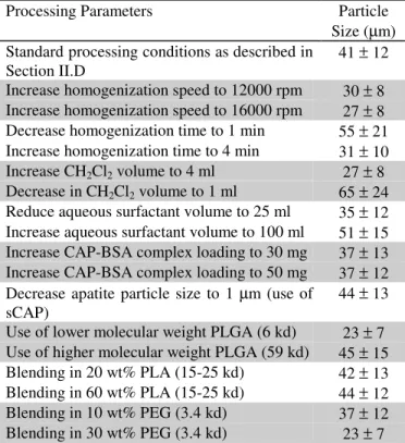
The effect of various processing parameters on the size of the composite particles is summarized in Table 2. For each parameter, low and high values around a midpoint were investigated to obtain a sense of the general trend. Variations that caused higher shear during homogenization led to the creation of smaller particles. For example, increase in homogenization speed and homogenization time directly increased the amount of shear experienced by the suspension. Decrease in polymer solution concentration and polymer molecular weight reduced the viscosity of the organic phase and also reduced particle size concomitantly.
TABLE 2
EFFECT OF PROCESSING PARAMETERS ON COMPOSITE PARTICLE SIZE
Processing Parameters Particle
Size (µm) Standard processing conditions as described in
Section II.D 41 ± 12
Increase homogenization speed to 12000 rpm 30 ± 8 Increase homogenization speed to 16000 rpm 27 ± 8 Decrease homogenization time to 1 min 55 ± 21 Increase homogenization time to 4 min 31 ± 10 Increase CH2Cl2 volume to 4 ml 27 ± 8
Decrease in CH2Cl2 volume to 1 ml 65 ± 24
Reduce aqueous surfactant volume to 25 ml 35 ± 12 Increase aqueous surfactant volume to 100 ml 51 ± 15 Increase CAP-BSA complex loading to 30 mg 37 ± 13 Increase CAP-BSA complex loading to 50 mg 37 ± 12 Decrease apatite particle size to 1 µm (use of
sCAP) 44 ± 13
Use of lower molecular weight PLGA (6 kd) 23 ± 7 Use of higher molecular weight PLGA (59 kd) 45 ± 15 Blending in 20 wt% PLA (15-25 kd) 42 ± 13 Blending in 60 wt% PLA (15-25 kd) 44 ± 12 Blending in 10 wt% PEG (3.4 kd) 37 ± 12 Blending in 30 wt% PEG (3.4 kd) 23 ± 7
C. Effect of Processing Parameters on the In Vitro Release from Composite Microparticles
1) Polymer Molecular Weight
The molecular weight of a biodegradable polymer influences its degradation rate and lifetime. As molecular weight increases, degradation becomes slower and longevity is extended. Apatite-polymer composite particles were prepared from PLGA of different molecular weights ranging from 6 kd to 59 kd. The onset of the accelerated phase of protein release was observed to occur at different times depending on the molecular weight of the polymer used (Fig. 2a). For particles fabricated from the short-chained, 6 kd and 13 kd PLGA, the rapid release phase began immediately. An upturn in the release profile was seen at ~ 4 weeks for 24 kd PLGA, whereas release remained gradual up to 8 weeks for 59 kd PLGA. Such control of release rate by polymer molecular weight may be useful in sequential release, such as to direct the debut of a protein in a wound healing process. Polymers of different
molecular weights could also be blended together to equalize the production of acid over time. Through the use of an equi-proportion blend of 6, 13, 24, 59 and 75 kd PLGA, a sustained and gradual release of BMP-2 was obtained (Fig. 2b). 0.0 0.2 0.4 0.6 0.8 1.0 1.2 0 20 40 60 80 100 Time (days) C um ul at iv e B S A R el ea se ( g/ m g ca rr ie r) 6 kd 13 kd 24 kd 59 kd 0.0 1.0 2.0 3.0 4.0 5.0 6.0 7.0 8.0 0 20 40 60 80 100 Time (days) C um ul at iv e B M P -2 R el ea se (n g/ m g ca rr ie r) 13 kd 6, 13, 24, 59, 75 kd blend 59 kd
Fig. 2. Effect of PLGA molecular weight on protein release. (a) Particles contained 15 µg of FITC-BSA per mg carrier. (b) Particles contained 64.5 ng of BMP-2 per mg carrier.
A comparison of Figs. 2a and 2b also reveals that the release profiles of BSA and BMP-2 from these composites are very similar, suggesting the same release mechanism for both proteins.
2) Polymer Hydrophobicity Blends of PLGA and PLA
Composite particles were synthesized from blends of PLGA (50:50 copolymer) and poly(lactic acid) (PLA). As the proportion of PLA in the composite particles was increased, the release rate decreased (Fig. 3). Lactic acid (LA) contains a methyl group that augments its hydrophobicity relative to glycolic acid (GA). Hence, PLA is more hydrophobic than PLGA. An increase in polymer
(b) (a)
hydrophobicity would delay water penetration and polymer degradation, and consequently, reduce protein release from composite particles. This parameter could be adjusted by using different types, blends, or compositions of polymers. For example, the LA to GA ratio in PLGA could be varied to obtain a range of hydrophobicities and degradation rates.
0.0 0.1 0.2 0.3 0.4 0.5 0.6 0 20 40 60 80 100 Time (days) C um ul at iv e B S A R el e as e ( g/ m g c ar ri er ) PLGA PLGA+PLA (4:1) PLGA+PLA (3:2)
Fig. 3. Effect of polymer hydrophobicity on protein release. Particles contained 15 µg of FITC-BSA per mg carrier. Molecular weight of PLGA was 24 kd; molecular weight of PLA was 20-30 kd.
Blends of PLGA and a Hydrophilic Polymer
Polyethylene glycol (PEG), a hydrophilic, biocompatible, and non-degradable polymer, was blended with PLGA in the fabrication of composite particles. Increase in PEG content led to faster initial release (Fig. 4). All release profiles showed an upturn at approximately 21 days, which corresponded to the degradation of 24 kd PLGA. 0.0 0.1 0.2 0.3 0.4 0.5 0.6 0 10 20 30 40 50 60 Time (days) C um ul at iv e B S A R el ea se ( g/ m g ca rr ie r) PLGA + PEG (7:3) PLGA + PEG (9:1) PLGA
Fig. 4. Effect of incorporating a hydrophilic polymer (PEG, 3.4 kd) on protein release. Particles contained 9 µg of FITC-BSA per mg carrier. Molecular weight of PLGA was 24 kd.
The hydrophilicity of PEG would draw water into the particles where the PEG chains reside. In addition, PEG might leach out of the polymer matrix, leaving behind pores [23]. The aqueous channels thus formed would facilitate water penetration and PLGA hydrolysis, as well as protein diffusion out of the particles. Therefore, the incorporation of a hydrophilic polymer would be expected to increase the rate of release.
3) Apatite Particle Size
CAP-BSA complexes prepared from sCAP (1 µm) and CAP (7 µm) were incorporated into composite particles. Reducing the apatite particle size resulted in release that more closely approximated zero-order for a longer period of time (Fig. 5). In contrast, the release plateaued after the first week of release from composite particles containing the larger, 7 µm-sized CAP particles.
0.0 0.5 1.0 1.5 2.0 2.5 3.0 3.5 0 20 40 60 80 100 Time (days) C um ul at iv e B S A R el ea se ( g/ m g ca rr ie r) 1 micron 7 microns
Fig. 5. Effect of apatite particle size on protein release. Particles were fabricated from 59 kd PLGA and contained 18 µg of FITC-BSA per mg of carrier.
For a given amount of acid produced by polymer degradation, a certain amount of apatite would be dissolved. Consequently, the protein adsorbed on that amount of apatite would be released. For small apatite particles, we surmise that most of the area available for protein adsorption is on the surface of the particles due to their higher surface/volume ratio, and their lower porosity and pore size compared to larger particles. (sCAP has porosity of 0.27 cm3/g and mean pore size of 4.2 nm
compared to porosity of 1.24 cm3/g and mean pore size of
8.2 nm for CAP.) As the surface of the apatite particles is gradually eroded by acid dissolution, the protein is released. For larger, porous apatite particles, the protein may be adsorbed on the surface as well as within the pores inside the particles. To free the protein adsorbed within the pores, a larger volume of apatite has to be dissolved. This difference in adsorption site may account for the difference in the release profiles for the 1 µm- and 7 µm-sized apatite particles.
4) Addition of Buffering Apatite
In addition to apatite-protein complexes, we loaded varying amounts of bare apatite with no adsorbed protein, referred to as “buffering apatite”, into the composite particles. As the proportion of buffering apatite was raised, the release rate decreased (Fig. 6). Protein release was dependent on the dissolution of apatite by acidic polymer degradation products. By incorporating buffering apatite into the composite particles, a portion of the acidity was neutralized by this bare apatite instead of by apatite-protein complexes. As a result, less protein was released for each unit of acid produced.
0.0 0.1 0.2 0.3 0.4 0.5 0.6 0 10 20 30 40 50 60 Time (days) C um ul at iv e B S A R el ea se ( g/ m g ca rr ie r) 30 mg CAP/BSA 30 mg CAP/BSA + 20 mg CAP 30 mg CAP/BSA + 50 mg CAP
Fig. 6. Effect of buffering apatite on protein release. Particles were fabricated from 24 kd PLGA and contained (♦) 9, ( ) 8.4 and ( ) 7.6 µg of FITC-BSA per mg of carrier.
5) Apatite-BSA Complex Loading in Particles
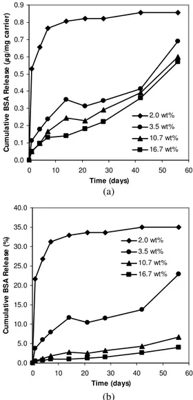
Different amounts of CAP-BSA with the same BSA content (9 wt%) were loaded into composite particles constructed of 24 kd PLGA. Increasing the CAP-BSA loading reduced the proportion of PLGA in the particles and diminished the amount of BSA released (Fig. 7a). In contrast, raising the protein loading of polymeric particles prepared by water-in-oil-in-water emulsification tends to hasten and increase protein release [24,25].
If the CAP-BSA loading in the composite particles were decreased to a very low level, the complex would be quickly depleted by acid evolution from PLGA such that at later time points, even with the continual degradation of PLGA, there would be no further protein release. On the other hand, if the proportion of CAP-BSA were increased, some of the complex might remain after complete PLGA degradation. Plotting the percent cumulative release from such particles would suggest seemingly low release (Fig. 7b). For our experiments, we have chosen to err on the side of over-loading rather than under-loading apatite-protein complexes so that protein was continually released and tracked over the course of our investigation. The complex loading can be adjusted so that for a precious therapeutic
protein, a high percentage of the protein is released before polymer degradation is complete.
0.0 0.1 0.2 0.3 0.4 0.5 0.6 0.7 0.8 0.9 0 10 20 30 40 50 60 Time (days) C um ul at iv e B S A R el ea se ( g/ m g ca rr ie r) 2.0 wt% 3.5 wt% 10.7 wt% 16.7 wt% 0.0 5.0 10.0 15.0 20.0 25.0 30.0 35.0 40.0 0 10 20 30 40 50 60 Time (days) C um ul at iv e B S A R el ea se (% ) 2.0 wt% 3.5 wt% 10.7 wt% 16.7 wt%
Fig. 7. Effect of apatite-protein complex loading on protein release, expressed as (a) cumulative mass of BSA released per mg carrier and (b) percentage cumulative release of BSA. Apatite-protein loading in composite particles was 2.0, 3.5, 10.7 and 16.7 wt%. Particles contained 2, 3, 9 and 15 µg of FITC-BSA per mg carrier, respectively. Molecular weight of PLGA used was 24 kd.
IV. CONCLUSION
We have prepared composites of a biodegradable polymer (PLGA) and an inorganic substrate (apatite) as delivery vehicles for BSA and BMP-2. These composites were formed into microparticles by a solid-in-oil-in-water suspension process. The composites were capable of sequestering proteins, preventing their premature release. Controlled release was accomplished through the interplay of acidic product formation by polymer degradation and dissolution of the basic apatite. Factors that influence polymer degradation and apatite dissolution include
(b) (a)
polymer molecular weight, polymer hydrophobicity, apatite particle size, and apatite loading. We have demonstrated the feasibility of using these factors to modulate protein release profiles. Sustained release was obtained for as short as 1 week to more than 3 months by varying PLGA molecular weight and apatite loading. Low burst, zero-order release was achieved for over 4 weeks through the use of submicron-sized apatite particles. The rate of release was systematically lowered through the addition of buffering apatite. This delivery platform can potentially be extended to other proteins. In addition, the administration of a combination of therapeutic proteins may be achieved by loading each protein into a differently formulated composite to obtain a superposition of release profiles.
REFERENCES [1] Wozney, J. M. Spine 2002, 27, S2-S8.
[2] Davies, J. E., Ed. Bone Engineering; 1st ed.; EM Squared Incorporated: Toronto, Canada, 2000.
[3] Groeneveld, E. H. J.; Burger, E. H. Eur. J. Endocrinol. 2000, 142,
9-21.
[4] Hoffman, A.; Weich, H. A.; Gross, G.; Hillmann, G. Appl.
Microbiol. Biotech. 2001, 57, 294-308.
[5] Lutolf, M. P.; Weber, F. E.; Schmoekel, H. G.; Schense, J. C.; Kohler, T.; Muller, R.; Hubbell, J. A. Nat. Biotechnol. 2003, 21,
513-518.
[6] Saito, N.; Okada, T.; Horiuchi, H.; Narumichi, M.; Takahashi, J.; Nawata, M.; Ota, H.; Nozaki, K.; Takaoka, K. Nat. Biotechnol.
2001, 19, 332-335.
[7] Koempel, J. A.; Patt, B. S.; O'Grady, K.; Wozney, J. M.; Toriumi, D. M. J. Biomed. Mater. Res. 1998, 41, 359-363.
[8] Kokubo, S.; Fujimoto, R.; Yokota, S.; Fukushima, S.; Nozaki, K.; Takahashi, K.; Miyata, K. Biomaterials 2003, 24, 1643-1651.
[9] Suh, D. Y.; Boden, S. D.; Louis-Ugbo, J.; Mayr, M.; Murakami, H.; Kim, H.-S.; Minamide, A.; Hutton, W. C. Spine 2002, 27, 353-360.
[10] Ueki, K.; Takazakura, D.; Marukawa, K.; Shimada, M.; Nakagawa, K.; Takatsuka, S.; Yamamoto, E. J. Crano-Maxillofac. Surg. 2003,
31, 107-114.
[11] Zegzula, H. D.; Buck, D. C.; Brekke, J.; Wozney, J. M.; Hollinger, J. O. J. Bone Joint Surg. [Am] 1997, 79-A, 1778-1790.
[12] Cullinane, D. M.; Lietman, S. A.; Inoue, N.; Deitz, L. W.; Chao, E. Y. S. J. Orthop. Res. 2002, 20, 1240-1245.
[13] Itoh, S.; Kikuchi, M.; Takakuda, K.; Nagaoka, K.; Koyama, Y.; Tanaka, J.; Shinomiya, K. J. Biomed. Mater. Res. 2002, 63,
507-515.
[14] Murakami, N.; Saito, N.; Takahashi, J.; Ota, H.; Horiuchi, H.; Nawata, M.; Okada, T.; Nozaki, K.; Takaoka, K. Biomaterials
2003, 24, 2153-2159.
[15] den Boer, F. C.; Wippermann, B. W.; Blokhuis, T. J.; Patka, P.; Bakker, F. C.; Haarman, H. J. T. M. J. Orthop. Res. 2003, 21,
521-528.
[16] Ripamonti, U.; Ramoshebi, L. N.; Matsaba, T.; Tasker, J.; Crooks, J.; Teare, J. J. Bone Joint Surg. [Am] 2001, 83-A, S1-116.
[17] Cook, S. D.; Wolfe, M. W.; Salkfeld, S. L.; Rueger, D. C. J. Bone
Joint Surg. [Am] 1995, 77-A, 734-750.
[18] Geesink, R. G. T.; Hoefnagels, N. H. M.; Bulstra, S. K. J. Bone
Joint Surg. [Br] 1999, 81-B, 710-718.
[19] Ahn, E. S., Nanostructured Apatites as Orthopedic Biomaterials, Ph.D. Thesis, Department of Chemical Engineering, Massachusetts Institute of Technology: Cambridge, MA, 2001
[20] Ahn, E. S.; Gleason, N. J.; Nakahira, A.; Ying, J. Y. Nano Lett.
2001, 1, 149-153.
[21] Ying, J. Y.; Ahn, E. S.; Nakahira, A., US Patent 6,013,591 (2000) [22] Castellanos, I. J.; Carrasquillo, K. G.; Lopez, J. D. J.; Alvarez, M.;
Griebenow, K. J. Pharm. Pharmacol. 2001, 53, 167-178.
[23] Cleek, R. L.; Ting, K. C.; Eskin, S. G.; Mikos, A. G. J. Control.
Release 1997, 48, 259-268.
[24] Yang, Y. Y.; Chung, T. S.; Ng, N. P. Biomaterials 2001, 22,
231-241.
[25] Sun, S. W.; Jeong, Y. I.; Jung, S. W.; Kim, S. H. J. Microencapsul.


