HAL Id: hal-00387498
https://hal.archives-ouvertes.fr/hal-00387498
Submitted on 25 May 2009
HAL is a multi-disciplinary open access archive for the deposit and dissemination of sci-entific research documents, whether they are pub-lished or not. The documents may come from teaching and research institutions in France or abroad, or from public or private research centers.
L’archive ouverte pluridisciplinaire HAL, est destinée au dépôt et à la diffusion de documents scientifiques de niveau recherche, publiés ou non, émanant des établissements d’enseignement et de recherche français ou étrangers, des laboratoires publics ou privés.
Production of oligosaccharides and cellobionic acid by
Fibrobacter succinogenes S85 growing on sugars,
cellulose and wheat straw
Régis Nouaille, Maria Matulova, V. Patoprsty, A.M. Delort, Evelyne Forano
To cite this version:
Régis Nouaille, Maria Matulova, V. Patoprsty, A.M. Delort, Evelyne Forano. Production of oligosac-charides and cellobionic acid by Fibrobacter succinogenes S85 growing on sugars, cellulose and wheat straw. Applied Microbiology and Biotechnology, Springer Verlag, 2009, 83, pp.425-433. �hal-00387498�
Production of oligosaccharides and cellobionic acid by Fibrobacter succinogenes S85 growing on sugars, cellulose and wheat straw
Régis Nouaille1, 2, Maria Matulova1, 3, Vladimír Pätoprstý3, Anne-Marie Delort1 and Evelyne Forano2
(1) Laboratoire de Synthèse et Etude de Systèmes à Intérêt Biologique, UMR 6504 Université Blaise Pascal-CNRS, 63177 Aubière cedex, France
(2) Unité de Microbiologie UR454, Centre de Recherches de Clermont-Ferrand-Theix, INRA, 63122 Saint-Genès-Champanelle, France
(3) Institute of Chemistry, Center for Glycomics, Slovak Academy of Sciences, Dubravska cesta 9, 842 38 Bratislava, Slovak Republic
Abstract : Extracellular culture fluid of Fibrobacter succinogenes S85 grown on glucose, cellobiose, cellulose or wheat straw was analysed by 2D-NMR spectroscopy. Cellodextrins did not accumulate in the culture medium of cells grown on cellulose or straw. Maltodextrins and maltodextrin-1P were identified in the culture medium of glucose, cellobiose and cellulose grown cells. New glucose derivatives were identified in the culture fluid under all the substrate conditions. In particular, a compound identified as cellobionic acid accumulated at high levels in the medium of F. succinogenes S85 cultures. The
production of cellobionic acid (and cellobionolactone also identified) was very surprising in an anaerobic bacterium. The results suggest metabolic shifts when cells were growing on solid substrate cellulose or straw compared to soluble sugars.
Keywords: Rumen, Fibrobacter succinogenes, Cellobionate, NMR, Oligosaccharides Introduction :
Cellulose is the most abundant biomass in the world, and its biological degradation is a key step in global carbon cycling as well as a promising approach for the production of bioenergy (Lynd et al. 2002). The rumen ecosystem, by exploiting cellulolytic microorganisms, is the most efficient process for cellulose transformation into useful products. The rumen cellulolytic microorganisms, which possess very active and complex hydrolytic systems, are thus potential biocatalysts for the synthesis of highly added value products. Fibrobacter succinogenes S85 is recognised as one of the most active cellulolytic rumen bacteria. Its enzymatic system has been extensively studied by molecular and biochemical approaches, and many different cellulases and hemicellulases have been identified (Krause et al. 2003). In addition, the complete genome sequence of the strain S85 of F. succinogenes revealed more than 100 predicted enzymes active against plant polysaccharide (Qi et al. 2005), suggesting a high potential hydrolytic activity of this organism. The sugar metabolism by F. succinogenes S85 was also extensively investigated (Forano et al. 2008), while metabolisation of natural substrates was much less explored.
In a previous work, we showed that resting cells of F. succinogenes S85 were able to synthesise and release oligosaccharides identified by 2D-NMR techniques as maltodextrins (MD), maltodextrin-1-phosphate (MD-1P) and another unknown phosphorylated sugar. These metabolites were produced when cells were metabolising glucose and cellobiose, but also their natural substrate cellulose (Matulova et al. 2001; Nouaille et al. 2004, 2005). The synthesis and excretion of MD and MD-1P were unexpected in bacteria specialised in cellulose degradation. In addition, we found that no cellodextrins were accumulating in extracellular media of cells whatever the substrate and particularly cellulose, contrary to what was generally admitted. These results prompted us to investigate whether growing bacteria were still producing these oligosaccharides. Extracellular culture fluids of bacteria growing on soluble sugars and also on cellulose and wheat straw were analysed by 2D-NMR spectroscopy for the presence of α(1–4) and β(1–4) glucose oligosaccharides, phosphorylated or not. Analysis of sugar metabolism and its regulation in F. succinogenes growing on natural substrate is a prerequisite for further attempts to improve lignocellulose bioconversion by this species.
Materials and methods :
Experimental procedures were mostly as described in Nouaille et al. (2004) or Matulova et al. (2005). Bacterial growth
F. succinogenes S85 (ATCC 19169) was grown at 39°C for 10 h in an anaerobic chemically defined medium (Gaudet et al. 1992) with 8 g L−1 glucose or cellobiose or for 4 days with 10 g L−1 of Sigmacell 20 cellulose or ground wheat straw: dried Triticum sp. straw was first chopped up to a length particle of 5–10 mm and then ground to particles <500 µm (Waring laboratory blender, VWR International, Fontenay-sous-Bois, France). Cellulose and wheat straw cultures (in triplicate) were harvested after 1, 2 or 4 days of growth. Extracellular media were separated from cells by centrifugation (15 min at 20, 000×g) before analysis.
NMR experiments
After centrifugation, pH of cell-free supernatants was corrected to 7.40, and the supernatants were freeze-dried two times with D2O. Samples were finally dissolved in a mixture of 470 µL 99.98% D2O, 20 µL
10 mM sodium 3-(trimethylsilyl) propionate TSP-d4 (δ 0.0) and 10 µL 50 mM 1-O-methyl-β-D
-xylopyranose (δ 4.331/104.79) used as standards. Samples were subjected to liquid state NMR measurements at 300 K on Bruker Avance DSX 300 MHz and 500 MHz spectrometers in 5 mm TXI inverse probes (1H, 13C, 15N) and Varian 600 MHz UNITY INOVA 600 NB spectrometer in 5 mm
1H{13C, 15N}PFG Triple Res IDTG600-5 probe, respectively, all with z-gradients. The following
techniques were used for the assignment of NMR signals: one-dimensional transient gradient-enhanced nuclear overhauser enhancement spectroscopy (ge-NOESY), ge-1D TOCSY (gradient-enhanced one-dimensional total-correlation spectroscopy), two-one-dimensional gradient enhanced proton-homonuclear shift correlation spectroscopy (ge-COSY), 1H–13C gradient-enhanced heteronuclear single quantum coherence spectroscopy (ge-HSQC) and 1H–13C heteronuclear multiple quantum correlation spectroscopy (HMBC). Standards were dissolved in an anaerobic buffer (50 mM potassium phosphate, 40 mM Na2CO3, 3 mM cysteine) at pH 7.4; they were freeze-dried and dissolved in D2O.
Metabolite assays
Succinate and acetate were quantified using enzymatic assays (Roche Diagnostics, Meylan, France). Cultures were done in triplicate. Standard error was lower than 12%.
Chemicals
TSP-d4 was purchased from Eurisotop (Saint-Aubin, France). Glucose, cellobiose, Sigmacell 20 cellulose
and 1-O-methyl-β-D-xylopyranose were from Sigma-Aldrich (Saint-Quentin Fallavier, France). All
enzymes and other chemicals were purchased from Sigma-Aldrich or Roche. Results
Growth of F. succinogenes S85 on different substrates
Growth of F. succinogenes S85 on glucose, cellobiose, cellulose and wheat straw was monitored by quantification of the production of succinate and acetate, the two main products of its metabolism. Identical growth was obtained on glucose and cellobiose, and the same amounts of succinate (8.4 ± 1.0 mM) and acetate (2.3 ± 0.8 mM) were produced in the extracellular medium after 10 h (stationary phase, not shown). When cells were grown on cellulose and wheat straw, respectively, 10.5 and 7 mM of succinate and 8 and 4 mM of acetate were produced after 4 days (stationary phase), on the example shown in Fig. 1. Both on cellulose and wheat straw, acetate was produced in higher amount than succinate during the first 24 h, while after this time, succinate was the major metabolite produced (Fig. 1). Cultures on glucose and cellobiose always produced succinate in higher concentration than acetate (mean ratio about 3.5). There appears thus to be a metabolic shift after the first day of culture on solid substrate.
Fig. 1 Production of succinate (squares) and acetate (circles) by F. succinogenes S85 growing on cellulose (open symbols) or wheat straw (closed symbols). Cells were cultivated for 4 days in mineral medium with 10 g L−1 Sigmacell 20 cellulose or
wheat straw. Metabolites were quantified by enzymatic assays. Standard error was less than 12%
Analysis of glucose, cellobiose and cellulose culture media
1H NMR COSY spectra of extracellular media obtained after 10 h growth on glucose or cellobiose and
1 day growth on cellulose are presented in Fig. 2 as an example. The left-hand part of Fig. 2 shows spectra of the region of the H1/H2 cross-peaks of non-substituted MD (δ 5.41/3.59 ppm, Fig. 2a, b), MD-1P (δ 5.46/3.52 and 5.41/3.59, Fig. 2c) and of the unknown compound X (glucose-MD-1P-derivative, 5.46/3.49, Fig. 2a, b and c). These metabolites were previously observed in resting cell incubations (Nouaille et al. 2005). The signals of α-glucose were also present in this part of the figure, corresponding to free glucose (α-glucose, αGlc, δ 5.24/3.54, Fig. 2a and 2b) or corresponding to residual cellobiose (α reducing end, αCB δ 5.24/3.59, Fig. 2b). In the right part of Fig. 2 are detected the signals of free glucose (β-glucose, βGlc, δ 4.65/3.24, Fig. 2a) and cellobiose (β reducing end, βCB, δ 4.68/3.30 and non-reducing glucose moiety of CB, β 4.52/3.33, Fig. 2a and b). The presence of cross-peaks at δ 4.54/3.34 suggested the synthesis of CD of dp > 2 (Fig. 2b). A huge signal of a new metabolite, named X2 (δ 4.63/3.39) was detected in Fig. 2a, b and c.
Fig. 2 Part of 1H−1H COSY NMR spectra of culture fluid on glucose, cellobiose and cellulose. F. succinogenes cells were grown for 10 h on glucose (a) or cellobiose (b) or for 1 day on cellulose Sigmacell 20 (c). Glc glucose, CB cellobiose, CD cellodextrins, MD linear maltodextrins, MD-1P maltodextrin-1-phosphate, X glucose-1P derivative, X2 unknown glucose derivative, S side bands
Identification of X2
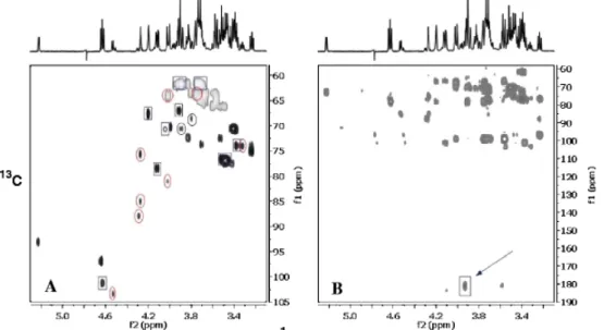
Various 1D and 2D-NMR techniques were used to analyse the cellobiose culture media in order to identify X2. The COSY spectrum revealed the presence of glucose and X2 both in a rough ratio 1:1 and a further series of non-identified signals. Starting with selective excitation of the signal at δ 4.63, series of 1D selective TOCSY, 1D selective NOESY were obtained. Two-dimensional HSQC and HMBC NMR spectra were also registered (Fig. 3). NMR data indicated the presence of disaccharide in which a non-reducing part of the molecule is β-glucose, which is linked to an acyclic carbon chain. The type of linkages between the two sugar moieties was confirmed by responses in the 1D 1H transient NOESY
spectra. After a selective excitation of H1 at δ 4.63 (glucose of non-reducing end), besides its own H3 (δ 3.52) and H5(δ 3.49), two other signals at δ 4.21 and 4.13 appeared, which were previously identified by 1D selective TOCSY as H4 and H3 signals of the acyclic moiety (Table 1). In the HMBC spectrum, a cross-peak due to H2/C1 of acyclic carbon chain was observed, confirming thus the presence of carboxyl group at C1 (Fig. 3b). These data suggest that X2 is cellobionic acid in form of cellobionate (CBA-S, cellobionic acid salt). NMR spectra of cellobionate and cellobionolactone were previously published by Higham et al. 1994. Our data (Table 1, Fig. 4) are in very good agreement with the spectrum of CBA-S shown there. Moreover, the published spectrum of cellobionolactone (CBA-L) helped us to identify further signals in our spectra and to reveal the presence of this compound in the F. succinogenes culture medium (Fig. 4). Anomeric signal due to non-reducing glucose unit of CBA-L was found at δ 4.54.
Reducing end glucose unit in the form of lactone showed a typical signal at δ 4.27 in the 1H NMR spectrum (Fig. 4). However, HSQC spectrum (Fig. 3a) indicated that it represents overlapped signals of three protons of the lactonic part of the molecule. Their assignment was done only tentatively by comparison with the data of glucose, gluconolactone and cellobiose. The ratio of signals due to cellobiose/glucose/CBA-S/CBA-L was 0.1:1:1:0.6 in the spectrum shown. The quantification in the case of CB, CBA-S and CBA-L was done by integration of H1 signal due to non-reducing end units and H1 α and β signals of reducing-end glucose. The ratio CBA-S/CBA-L varied from 1 to 1.7 depending on the experiments.
Fig. 3 Two-dimensional 1H−13C heterocorrelated HSQC (a) and HMBC (b) spectra of the extracellular medium of F.
succinogenes grown on cellobiose. Spectrum A Cross-peaks marked by squares are due to cellobionic acid in sodium salt form
(CBA-S), while those marked by circles are due to a cellobionolactone (CBA-L). Other signals present in the spectrum are due to α and β glucose (H1 signals at δ 5.23/93.1 and 4.65/96.9, respectively) formed by degradation of cellobiose. H1 signal at δ 4.51 reveals the presence of a trace amount of cellobiose still present in the mixture. Arrow in the spectrum B indicates a cross-peak due to a long range coupling constant from H2 to carboxyl group in the lactone part of the molecule in CBA-S
Table 1 1H and 13C NMR chemical shifts of cellobionate and cellobionolactone
Chemical Sugar unit shift δ/ppm
H1/C1 H2/C2 H3/C3 H4/C4 H5/C5 H6,H6´/C6 CBA-S Glc(1→4) 4.63/101.2 3.39/73.9 3.52/76.7 3.46/76.7 3.49/76.8 3.91, 3.73/61.8 COO−– (CHOH)5―CH2OH 182.2 3.92/66.9 4.13/78.3 4.21/67.6 4.05/70.1 4.02, 3.76/64.1 CBA-L* Glc(1→4) 4.54/103.3 3.34/73.9 3.52/76.9 3.41/70.6 3.50/76.8 3.95, 3.73/61.8 Glc-lactone – 4.29/85.1 4.29/75.6 4.30/87.8 4.04/81.0 3.72, 3.57/65.4 Assignment of CBA-L was done only tentatively by comparison of its chemical shifts with those due to glucose,
Fig. 4 Part of the 1H spectrum of the extracellular medium of F. succinogenes grown on cellobiose. Glc+CB red.alpha overlapped H1 signals due to α-glucose and reducing α-glucose units of cellobiose, Glc+CB red.beta overlapped H1 signals due to β-glucose and reducing β-glucose units of cellobiose, CBA nr salt H1 signal due to non-reducing unit glucose of cellobionate, CBA nr lactone H1 signal due to reducing unit glucose of cellobionolactone, CB H1 signal due to non-reducing unit glucose of cellobiose, CBA lactone* tentatively attributed overlapped H2, H3 and H4 signals of the gluconolactone part of the cellobionolactone molecule, H4 salt H4 of the acyclic part of the cellobionate molecule,
CBA-H3 salt CBA-H3 of the acyclic part of the cellobionate molecule, CBA-H5 salt H5 of the acyclic part of the cellobionate molecule
Analysis of wheat straw culture media :
We also checked for the presence of these compounds in the culture media of bacteria grown on wheat straw. Samples were analysed by 1H–1H COSY. Spectra collected after 1 and 4 days of bacterial growth
are presented in Fig. 5. Identified signals were mainly due to free xylose, arabinose and arabinoglucuronoxylan oligosaccharides, released during hemicellulose degradation in wheat straw (Matulova et al. 2005). The H1 anomeric signal due to non-reducing Glc unit of cellobionate (CBA-S, H1/H2: δ 4.63/3.38) was also detected in the COSY spectra from the beginning of bacterial growth (Fig. 5a) until the end of the culture (Fig. 5b). Its concentration, quantified from H1/H2 signal volumes in COSY spectra, varied from 1 to 1.5 mM. The presence of cellodextrin cross-peaks (δ 4.53–4.52/3.36– 3.32) was not detected unambiguously in any spectrum because a lot of H1/H2 cross-peaks were detected in the region δ 4.60–4.50/3.35–3.30. However, as shown previously in similar experiments and also complementary experiments (Matulova et al. 2005), cellodextrins are likely not present in the straw culture medium. The metabolite denoted X (H1/H2 and C1: δ 5.46/3.48 and 94.2) and assigned to a derivative of Glc-1P was detected in the early stage of the culture (after 8 h, not shown), but it was not found in the spectra registered at later stages (Fig. 5). MD and MP-1P were not identified in the wheat straw culture media.
Fig. 5 1H–1H COSY NMR spectra of culture media collected after 1 day (a) and 4 days (b) during the degradation of wheat straw by F. succinogenes S85. GlcA −Xyl glucuronic moiety in xylo-oligosaccharides, Xyl −GlcA xylose substituted by glucuronic
acid in xylo-oligosaccharides, Arap free arabinose in the pyranose form (α and β anomers), Xyl free xylose (α and β anomers),
Xyl red reducing end of xylose (α and β anomers) in xylooligosaccharides, Xyl int internal xylose in xylo-oligosaccharides, Araf −Xyl arabinosyl moiety in xylo-oligosaccharides, X2 cellobionate, ST standard, 1-O-methyl-β-D-xylopyranose
Oligosaccharide production in growing cells :
The presence of different sugars detected by NMR in the extracellular medium of the four culture conditions is summarised in Table 2. The first important result is that neither CD nor cellobiose is accumulating in the extracellular medium of cells growing on cellulose or straw. Also, no glucose could be detected when cells were grown on solid substrate. Maltose-1P was only found when cellulose was the growth substrate, while non-phosphorylated MDs were only produced when cells were growing on the soluble sugars glucose and cellobiose. The Glc-1P derivative (X) was found under all conditions of growth, while Glc-1P could not be detected on any substrate. Finally, cellobionic acid (cellobionate) was detected under the four conditions of growth; its concentration was in the millimolar range. This concentration value indicates that this compound, as well as the other ones detected in the extracellular medium, was not released from cell lysis (Wells and Russell 1996), but was actively excreted from the bacteria. The presence of all the sugars and sugar derivatives detected previously in resting cells (Nouaille et al. 2005) is also reported in Table 2 for comparison.
Table 2 Detection of different sugars in the extracellular culture fluid of F. succinogenes S85 grown on glucose, cellobiose, cellulose and wheat straw (growing cells) or in the extracellular incubation medium of cells incubated with glucose, cellobiose and cellulose (resting cells)
Substrate Experimental conditions
MD-1P MD X Glc CB CD Glc-1P CBA
Glucose Growing cells − + + + + − − +
Resting cells + + − + − − + −
Cellobiose Growing cells − + + + + + − +
Resting cells + + − + − − + −
Cellulose Growing cells + − + − − − − +
Resting cells + + + + − − + −
Straw Growing cells − − + Transient − − − − +
MD-1P maltose-1-phosphate, MD linear maltodextrins, X glucose-1P derivative, Glc glucose, CB cellobiose, CD cellodextrins
of dp > 2, CBA cellobionic acid
Discussion :
On cellulose and wheat straw, the production of organic acids by F. succinogenes S85, a good indicator of the bacterial growth (Fields et al. 2000), was very similar (Fig. 1). However, the concentration of both acetate and succinate was lower on straw, probably because of less available substrate for growth. Indeed, although F. succinogenes S85 degrades very efficiently the hemicelluloses of straw and particularly xylans, it is not able to metabolise the pentoses or xylooligosaccharides released (Matulova et al. 2005) and only uses glucose and cellobiose for growth (Matte et al. 1992; Matheron et al. 1997). On both solid substrates, a shift in the production of the metabolites acetate and succinate was observed after about 24 h, contrary to what was found on glucose or cellobiose. This reflects a modification in the fluxes in the two corresponding metabolic pathways. As the acetate pathway is an efficient means of producing ATP (Miller 1978; Matheron et al. 1997), this shift might be due to different energy requirements between the beginning and the end of the batch culture on solid substrates. For example, there could be a need of more energy in the first hours of growth on solid substrate for induction of synthesis of specific proteins involved in adhesion (Roger et al. 1990) or cellulolysis (Béra-Maillet et al. 2000). In addition, acetate and succinate production may be affected differently by several factors such as pH, as shown previously on continuous cultures of S85 (Weimer 1993) or on other bacteria (Desvaux 2001). As F. succinogenes also produces formate as a minor metabolite (Miller 1978; Matheron et al. 1997), it would have been interesting to monitor its concentration during the growth on the two substrates to see the evolution of its production with time.
The analysis of the oligosaccharides produced in the culture medium on the four substrates shows specific features.
MD or MD-1P that were produced by resting cells of F. succinogenes (Nouaille et al. 2004, 2005) were also found under more physiological conditions of cells growing on cellulose. They were not detected on straw, either because they are not produced at all or because of a too small concentration on this substrate that also provided lower growth (Fig. 1). We previously observed with resting cells (Nouaille et al. 2005) that the ratio MD-1P/MD was increasing with the DP of the substrate (glucose < cellobiose << cellulose). This phenomenon appears amplified in growing cells where MD-1P was only detected with cellulose, while MD was only detected with glucose and cellobiose (Table 2). One of the main difference between growing and resting cells concerns Glc-1P and X: Glc-1P was present in resting cells, and its concentration was increasing with the size of the substrate (Table 2, Nouaille et al. 2005), while it was absent in growing cells. On the contrary, X was not detected in resting cells metabolising glucose and cellobiose, while it was present in high quantity in cells metabolising cellulose; in growing cells, X was found under the four substrate conditions, it appears in higher quantity in extracellular medium of cells grown on cellulose (Fig. 2). These results suggest that Glc-1P might be the precursor of X. In the case of cellulose, the larger accumulation of X (both in growing and in resting cells) could be explained by a higher Glc-1P production. Growth on cellulose or straw differs from that on soluble sugars by several factors: (a) the necessity for bacteria first to adhere to the substrate and second to degrade it, (b) different growth rate, (c) different physiological state of the bacteria; the carbon flow is consequently different in cellulose and/or wheat straw grown cells. The metabolism of another anaerobic cellulolytic bacterium Clostridium cellulolyticum was studied in details in batch and continuous cultures on cellobiose or cellulose, and a modification of metabolic fluxes was also observed between the two substrates (Desvaux et al. 2000; Desvaux et al. 2001).
Accumulation of phosphorylated sugars in the extracellular medium of growing bacteria (present work) but also of resting cells (Nouaille et al. 2004, 2005) is quite surprising. Their estimated concentration in the millimolar range exclude their release by bacterial cell lysis as this would imply an extremely high intracellular concentration, far above that previously measured in F. succinogenes (Matheron et al. 1998). They could be actively excreted from the cells by a specific permease, such as that reported to occur for G6P in Escherichia coli (Van der Zee et al. 1996).
In F. succinogenes resting cells, no CB or CD was detected in the extracellular medium of cells incubated with glucose or cellulose. On the contrary, cells growing on glucose accumulated CB in the culture fluid, and cells growing on cellobiose accumulated CB and CD in the culture medium; these results are in accordance with those of Wells et al. (1995). However, in cellulose cultures, no CB or CD could be detected in the medium. The same results were observed on wheat straw cultures (present work and Matulova et al. 2005). This suggests that CD are utilised by the cells as soon as they are released during cellulose degradation. This result is in agreement with the concept that the depolymerisation of insoluble cellulose to soluble cellodextrins limits the cellulose fermentation, as proposed by Pavlostathis et al. (1988) for Ruminococcus albus. Such mechanism could avoid a feed-back inhibition of cellulases by cellodextrins. In addition, we did not find any glucose accumulation in the milieu of cells grown on cellulose, contrary to what was found previously in cellulose-fed continuous cultures of F. succinogenes and particularly at low pH (Weimer 1993). This might be explained by different growth rate in batch and continuous cultures.
Several groups showed previously that F. succinogenes could be co-cultured with non-cellulolytic bacteria on cellulose and concluded that F. succinogenes was providing cellodextrins to the other species (Scheifinger and Wolin 1973; Kudo et al. 1987; Wells et al. 1995). Altogether, the results of the present work on growing cells suggest that maltodextrins and/or maltodextrin-phosphate and/or X may be additional substrates of this inter-species cross-feeding. Indeed, two of the species used in the co-cultures, Selenomonas ruminantium and Streptococcus bovis, are amylolytic and thus able to use maltodextrins, while the third one (Treponema bryantii) is saccharolytic (Stewart et al. 1997).
A new compound, identified as cellobionic acid, was present in growing cells whatever the substrate, while it was not detected in resting cells (Table 2). The presence of cellobionic acid is very surprising. It is usually produced in aerobic lignocellulolytic fungi through the activity of a cellobiose dehydrogenase (CDH) that converts cellobiose into cellobionolactone, an unstable compound rapidly spontaneously hydrolysed into cellobionic acid (Higham et al. 1994; Zamocky et al. 2006). CDH activity was also found in cellulolytic bacteria (Li et al. 1996). Cellobionolactone was also identified in the culture media of F. succinogenes S85. Sugar dehydrogenase with a broad specificity is found in bacteria; for example, the
soluble aldose sugar dehydrogenase (sGdh) from E. coli can utilise glucose or arabinose, but displays higher activity towards oligomeric substrates such as maltose, maltotriose or cellobiose (Southall et al. 2006). Putative sGdh homologs have been identified in diverse prokaryotic phyla, including the proteobacteria and some firmicutes, and even in Archaea (Oubrie 2003). The precise role of the sugar dehydrogenases is not completely clear. In aerobic fungi, CDH might be involved in regulation of cellulase expression as it produces cellobionolactone, which is a potent inducer of cellulase genes in some of these organisms such as Trichoderma reesei (Ilmen et al. 1997). CDH was also proposed to directly participate in cellulose depolymerisation (Mansfield et al. 1997). The E. coli as well as other enteric bacterial sGdh was proposed to be part of a periplasmic electron transfer system that can serve to input sugar-derived electrons into the respiratory network (Southall et al. 2006). In F. succinogenes, there is no clear evidence for the presence of a Gdh (no highly homologous gene was found in the genome of the S85 strain, http://blast.jcvi.org/rumenomics/), and additional experiments are necessary to discover the mechanism of cellobionolactone/cellobionic acid formation as well as its role in the bacterium.
References
Béra-Maillet C, Gaudet G, Forano E (2000) Endoglucanase activity and relative expression of glycosyl-hydrolase genes of
Fibrobacter succinogenes S85 grown on different substrates. Biochim Biophys Acta 1543:77–85
Desvaux M, Guedon E, Petitdemange H (2000) Cellulose catabolism by Clostridium cellulolyticum growing in batch culture on defined medium. Appl Environ Microbiol 66:2461–2470
Desvaux M, Guedon E, Petitdemange H (2001) Metabolic flux in cellulose batch and cellulose-fed continuous cultures of
Clostridium cellulolyticum in response to acidic environment. Microbiology 147:1461–1474
Fields MW, Mallik S, Russell JB (2000) Fibrobacter succinogenes S85 ferments ball-milled cellulose as fast as cellobiose until cellulose surface area is limiting. Appl Microbiol Biotechnol 54:570–574
Forano E, Delort AM, Matulova M (2008) Carbohydrate metabolism in Fibrobacter succinogenes: what NMR tells us. Microb Ecol Heath Disease 2:94–102
Gaudet G, Forano E, Dauphin G, Delort AM (1992) Futile cycling of glycogen in Fibrobacter succinogenes as shown by in
situ 1H-NMR and 13C-NMR investigation. Eur J Biochem 207:155–162
Higham CW, Gordon-Smith D, Dempsey CE, Wood PM (1994) Direct 1H evidence for conversion of β-D-cellobiose to
cellobionolactone by cellobiose dehydrogenase from Phanerochaete chrysosporium. FEBS Lett 351:128–132
Ilmen M, Saloheimo A, Onnela ML, Penttila ME (1997) Regulation of cellulase gene expression in the filamentous fungus
Trichoderma reesei. Appl Environ Microbiol 63:1298–1306
Krause DO, Denman SE, Mackie RI, Morrison M, Rae AL, Attwood GT, McSweeney CS (2003) Opportunities to improve fiber degradation in the rumen: microbiology, ecology, and genomics. FEMS Microbiol Rev 27:663–693
Kudo H, Cheng KJ, Costerton JW (1987) Interactions between Treponema bryantii and cellulolytic bacteria in the in vitro degradation of straw cellulose. Can J Microbiol 33:244–248
Li X, Huang Y, Xu D, Xiao D, Jin F, Gao P (1996) Cellobiose-oxidizing enzyme from a newly isolated cellulolytic bacterium
Cytophaga sp LX-7. Biotechnol Lett 18:205–210
Lynd LR, Weimer PJ, van Zyl WH, Pretorius IS (2002) Microbial cellulose utilization: fundamentals and biotechnology. Microbiol Mol Biol Rev 66:506–577
Mansfield SD, Jong E, Sadler JN (1997) Cellobiose dehydrogenase, an active agent in cellulose depolymerisation. Appl Environ Microbiol 63:3804–3809
Matheron C, Delort AM, Gaudet G, Forano E (1997) Re-investigation of glucose metabolism in Fibrobacter succinogenes S85 using NMR and enzymatic assays. Evidence of pentose phosphates phosphoketolase and pyruvate-formate-lyase activities. Biochim Biophys Acta 1355:50–60
Matheron C, Delort AM, Gaudet G, Forano E (1998) In vivo 13C NMR study of glucose and cellobiose metabolism by four
cellulolytic strains of the genus Fibrobacter. Biodegradation 9:451–461
Matte A, Forsberg CW, Verinder-Gibbins AM (1992) Enzymes associated with metabolism of xylose and other pentoses by
Prevotella (Bacteroides) ruminicola strains, Selenomonas ruminantium D, and Fibrobacter succinogenes S85. Can J Microbiol
38:370–376
Matulova M, Delort AM, Nouaille R, Gaudet G, Forano E (2001) Concurrent maltodextrin and cellodextrin synthesis by
Fibrobacter succinogenes S85 as identified by 2D NMR spectroscopy. Eur J Biochem 268:3907–3915
Matulova M, Nouaille R, Capek P, Péan M, Forano E, Delort A-M (2005) Degradation of wheat straw by Fibrobacter
succinogenes S85: a liquid and solid state NMR study. Appl Environ Microbiol 71:1247–1253
Miller T (1978) The pathway of formation of acetate and succinate from pyruvate in Bacteroides succinogenes. Arch Microbiol 117:145–152
Nouaille R, Matulova M, Delort AM, Forano E (2004) Production of maltodextrin-1-phosphate by Fibrobacter succinogenes S85. FEBS Lett 576:226–230
Nouaille R, Matulova M, Delort AM, Forano E (2005) Oligosaccharide synthesis in Fibrobacter succinogenes S85 and its modulation by the substrate. FEBS J 272:2416–2427Oubrie A (2003) Structure and mechanism of soluble glucose dehydrogenase and other PQQ-dependent enzymes. Biochim Biophys Acta 1647:143–151
Pavlostathis SG, Miller TL, Wolin MJ (1988) Fermentation of insoluble cellulose by continuous cultures of Ruminococcus
Qi M, Nelson KE, Daugherty SC, Nelson WC, Hance IR, Morrison M, Forsberg CW (2005) Novel molecular features of the fibrolytic intestinal bacterium Fibrobacter intestinalis not shared with Fibrobacter succinogenes as determined by suppressive subtractive hybridization. J Bacteriol 187:3739–3751
Roger V, Fonty G, Komisarczuk-Bony S, Gouet P (1990) Effects of physicochemical factors on the adhesion to cellulose Avicel of the ruminal bacteria Ruminococcus flavefaciens and Fibrobacter succinogenes subsp. succinogenes. Appl Environ Microbiol 56:3081–3087
Scheifinger CC, Wolin MJ (1973) Propionate formation from cellulose and soluble sugars by combined cultures of Bacteroides
succinogenes and Selenomonas ruminantium. Appl Microbiol 26:789–795
Stewart CS, Flint HJ, Bryant MP (1997) The rumen bacteria. In: Hobson PN, Stewart CS (eds) The rumen microbial ecosystem, Second editionth edn. Blackie, London, pp 10–72
Southall SM, Doel JJ, Richardson DJ, Oubrie A (2006) Soluble aldose sugar dehydrogenase from Escherichia coli. A highly exposed active site conferring broad substrate specificity. J Biol Chem 281:30650–30659
Van der Zee JR, Postma PW, Hellingwerf KJ (1996) Quantitative conversion of glucose into glucose-6-phosphate by intact
Escherichia coli cells. Biotechnol Appl Biochem 24:225–230
Weimer PJ (1993) Effects of dilution rate and pH on the ruminal cellulolytic bacterium Fibrobacter succinogenes S85 in cellulose-fed continuous culture. Arch Microbiol 160:288–204
Wells JE, Russell JB, Shi Y, Weimer PJ (1995) Cellodextrin efflux by the cellulolytic ruminal bacterium Fibrobacter
succinogenes and its potential role in the growth of non-adherent bacteria. Appl Environ Microbiol 61:1757–1762
Wells JE, Russell JB (1996) The effect of growth and starvation on the lysis of the ruminal cellulolytic bacterium Fibrobacter
succinogenes. Appl Environ Microbiol 62:1342–1346
Zamocky M, Ludwig R, Peterbauer C, Hallberg BM, Divne C, Nicholls P, Haltrich D (2006) Cellobiose dehydrogenase-a flavocytochrome from wood-degrading, phytopathogenic and saprotropic fungi. Curr Protein Pept Sci 7:255–280