HAL Id: tel-02855053
https://tel.archives-ouvertes.fr/tel-02855053
Submitted on 8 Jun 2020HAL is a multi-disciplinary open access
archive for the deposit and dissemination of sci-entific research documents, whether they are pub-lished or not. The documents may come from teaching and research institutions in France or abroad, or from public or private research centers.
L’archive ouverte pluridisciplinaire HAL, est destinée au dépôt et à la diffusion de documents scientifiques de niveau recherche, publiés ou non, émanant des établissements d’enseignement et de recherche français ou étrangers, des laboratoires publics ou privés.
Role of microRNAs and their machinery in hepatocytes
and pancreatic beta cells
Gaia Fabris
To cite this version:
Gaia Fabris. Role of microRNAs and their machinery in hepatocytes and pancreatic beta cells. Cell Behavior [q-bio.CB]. COMUE Université Côte d’Azur (2015 - 2019), 2019. English. �NNT : 2019AZUR4030�. �tel-02855053�
Rôle des microARNs et de leur machinerie dans les hépatocytes et
les cellules béta pancréatiques
Role of microRNAs and their machinery in hepatocytes and pancreatic beta cells
Gaia FABRIS
IRCAN CNRS UMR 7284
– INSERM U1081
Présentée en vue de l’obtention du grade de docteur en Sciences
d’Université Côte d’Azur
Mention : interactions moléculaires et
cellulaires
Dirigée par : Giulia Chinetti
Co-encadrée par : Patricia Lebrun Soutenue le : 7 Juin 2019
Devant le jury, composé de :
Francesco Beguinot, Professeur d’Endocrinologie, Università di Napoli Federico II
Giulia Chinetti, Professeure de Biochimie, UCA Nice
Magalie Ravier, CRCN, Inserm, IGF Montpellier
Emmanuel Van Obberghen, Professeur de Biochimie, UCA Nice
Rôle des microARNs et de leur machinerie dans les
hépatocytes
et les cellules béta pancréatiques
Jury :
Président de jury
Emmanuel Van Obberghen, Professeur de Biochimie, UCA Nice
Rapporteurs
F a es o Begui ot, P ofesseu d’Endocrinologie, Département de
Médecine translationnelle Université de Naples Federico II, Italie
Magalie Ravier, CRCN, Inserm, Institut de Génomique Fonctionnelle
Montpellier, France
Résumé
Da s le foie o al de l’adulte les h pato tes so t da s u tat de uies e e, sauf lo s d’u e situatio d’ag essio ph si ue ou to ique. Dans ces cas-là, afin de pouvoir ai te i leu ôle da s l’ho ostasie de l’o ga is e les h pato tes e do ag s ou sauvegardés vont proliférer. Un des facteurs limitants pour la prolifération est la p se e d’a ides a i s, ui so t essai es pour la synthèse des protéines au cours de la p olif atio et di isio ellulai e, et pou l’a ti atio de la oie TOR, ui joue un rôle clef dans la régulation de la prolifération cellulaire.
Le ut de o p ojet de th se tait d’ tudie da s des h pato tes adultes le ôle des a ides a i s da s la gulatio de la t a sitio e t e l’ tat de uies e e et la croissance cellulaire. Pour cela nous avons utilisé comme modèle de foie partiellement reséqué des hépatocytes de rats adultes en culture primaire non- o flue te. J’ai d ou e t ue l’a se e d’a ides a i s i duit l’e t e des h pato tes da s u tat de quiescence couplée à une augmentation de la protéine Drosha; cet accroissement étant indépendant de la voie mTOR. Inversement, la réduction de Drosha permet aux hépatocytes de proliférer, et cet effet semble indépendant des microARNs, qui sont les pa te ai es lassi ues de D osha. L’e se le de os sultats le t da s les hépatocytes adultes l’e iste e d’effets o -classiques de Drosha dans le contrôle de la régulation de la croissance suite un stress nutritionnel. Un tel mécanisme pourrait
t e i l pou la p e tio et/ou le t aite e t de l’i suffisa e h pati ue.
Le second projet de ma thèse concerne le rôle du microARN-375 dans la régulation de la sécrétion de l’i suli e e po se au glu ose da s les îlots de La ge ha s de ats et d’o igi e hu ai e. Le ta olis e gl ol ti ue et o datif so t nécessaires pour la réponse sécrétoire au glucose. Alors que plusieurs microARNs sont connus pour odule l’ho ostasie des ellules ta, le microARN-375 se démarque par sa forte expression dans les cellules béta dans lesquelles il régule le fonctionnement, la prolifération et la différentiation. Sachant que le métabolisme du glucose est au centre de toutes les fonctions de la cellule béta, nous avons étudié son impact sur le métabolisme dans des îlots humains et murins. Notre approche a fait appel soit à la surexpression du microARN-375, soit à sa réduction dans de îlots humains ou murins. E suite ous a o s a al s la s tio de l’i suli e et le ta olis e. De plus, ous
a o s ega d l’e p essio du microARN-375 et la réponse sécrétoire au glucose dans des îlots de rats à différents stades de leur développement. Nous avons trouvé que la surexpression du microARN-375 dans les îlots murins et humains diminuait la sécrétion de l’i suli e e po se au glu ose, ais pas elle au α-cétoisocaproate ou au KCl. De plus, le microARN-375 duit la o so atio d’O2 engendrée par la glycolyse et le métabolisme du py u ate, ais pas elle e po se au α-cétoisocaproate. En même temps, la production de lactate est augmentée suggérant que le pyruvate est détourné des mitochondries. Enfin, la réduction du niveau du microARN-375 est associée à la atu atio des ellules ta de at foetal et l’appa itio d’u e po se s toi e au glucose. Pris dans leur ensemble nos résultants identifient le microARN-375 comme étant un régulateu e t al du ta olis e du glu ose et de la s tio de l’i suli e, et ainsi il pourrait être un rouage déterminant pour la maturation fonctionnelle de cellules béta.
Mots clés : hépatocytes, prolifération cellulaire, acides aminés, Drosha; diabète, métabolisme du glucose, s tio de l’i suli e, îlots pancréatiques, microARNs.
Abstract
In an adult healthy liver, hepatocytes are in a quiescent stage unless a physical injury, such as ablation, or a toxic attack occurs. However, to maintain their important organismal homeostatic role, the damaged or remaining hepatocytes will start proliferating to restore their functional mass. One of the limiting conditions for cell proliferation is amino acid (aa) availability, necessary both for the synthesis of proteins important for cell growth and division, and for the activation of the mTOR pathway, known for its considerable role in the regulation of cell proliferation. The aim of my main PhD project was to investigate the role of amino acids in the regulation of the switch between quiescence and growth of adult hepatocytes. To do so I used non-confluent primary adult rat hepatocytes as a model of partially ablated liver. I discovered that the absence of aa induces in primary rat hepatocytes the entrance in a quiescence state together with an increase in Drosha protein, which does not involve the mTOR pathway. Conversely, knockdown of Drosha allows the hepatocytes to proliferate and this effect appears to be independent of miRNAs, the canonical downstream partners of Drosha. Taken together, these observations reveal an intriguing non-canonical effect of Drosha in the control of growth regulation of adult hepatocytes responding to a nutritional strain, and they may help to design novel preventive and/or therapeutic approaches for hepatic failure.
A side project of my PhD involved the role of microRNA-375 in the regulation of glucose-induced insulin secretion in rat and human pancreatic islets. Enhanced β-cell glycolytic and oxidative metabolism are necessary for glucose-induced insulin secretion. While several microRNAs modulate β-cell homeostasis, microRNA-375 stands out as it is highly expressed in β-cells where it regulates β-cell function, proliferation and differentiation. As glucose metabolism is central in all aspects of β-cell functioning, we investigated the role of microRNA-375 in this process using human and rat islets; the latter being an appropriate model for in depth investigation. We used forced expression and repression of microRNA-375 in rat and human primary islet cells followed by analysis of insulin secretion and metabolism. Additionally, microRNA-375 expression and glucose-induced insulin secretion were compared in islets from rats at different developmental ages. We found that overexpressing of microRNA-375
in rat and human islet cells blunted insulin secretion in response to glucose, but not to α-ketoisocaproate or KCl. Further, microRNA-375 reduced O2 consumption related to gl ol sis a d p u ate eta olis , ut ot i espo se to α-ketoisocaproate. Concomitantly, lactate production was augmented suggesting that glucose-derived pyruvate is shifted away from mitochondria. Finally, reduced microRNA-375 expression as asso iated ith atu atio of fetal at β-cells and acquisition of glucose-induced insulin secretion function. Altogether our findings identify microRNA-375 as an effi a ious egulato of β-cell glucose metabolism and of insulin secretion and could be dete i a t to fu tio al β-cell developmental maturation.
Keywords: hepatocytes, cell proliferation, amino acids, Drosha; diabetes, glucose metabolism, insulin secretion, pancreatic islets, microRNAs.
“ While
the heart resounds and attracts the music of the mandolin, there, inside, you filter and distribute, you separate and divide, you multiply and lubricate, you raise and gather
the threads and the grams of life, the final
distillate,
the intimate essences. Submerged viscus, measurer of the blood, you live full of hands and full of eyes,
measuring and transferring in your hidden
alchemical chamber. “
Ode to the liver – Pablo Neruda
« Mientras el corazón suena y atrae la partitura de la mandolina, allí adentro tú filtras y repartes, separas y divides, multiplicas y engrasas, subes y recoges los hilos y los gramos de la vida, los últimos licores, las íntimas esencias. Víscera submarina, medidor de la sangre, vives lleno de manos y de ojos, midiendo y trasvasando en tu escondida cámara de alquimista. » Oda al hígado – Pablo Neruda
Contents
List of figures ... 6 List of abbreviations ... 8 Introduction ... 13 Liver ... 14 Anatomy ... 14From the macroscopic to the microscopic structure ... 15
o An overview on hepatic cell populations ... 15
Hepatocytes... 18
Hepatocyte: a deeper look inside the cell ... 21
Liver regeneration ... 23
Liver regeneration: the pathways involved ... 24
o The priming phase ... 25
o The proliferation phase ... 26
o The termination phase ... 27
Liver-related diseases ... 28
Liver metabolism ... 31
Liver glucose metabolism ... 31
Gluconeogenesis ... 33
Glycogen metabolism: glycogenesis and glycogenolysis ... 34
Glycolysis ... 35
Liver fatty acid metabolism: from lipogenesis to fatty acid oxidation ... 36
Liver amino acid metabolism ... 38
Amino acid regulatory mechanisms ... 42
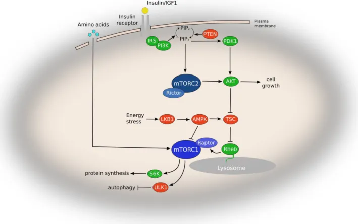
Intracellular amino acid sensing: the mTOR pathway ... 43
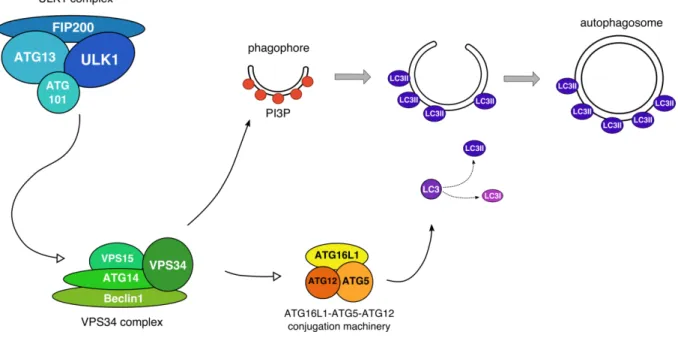
Intracellular amino acid sensing: autophagy ... 46
Metabolism in proliferative and quiescent cells ... 48
Pancreas ... 49
Both an exocrine and an endocrine organ ... 49
The insulin secretion signaling pathway ... 51
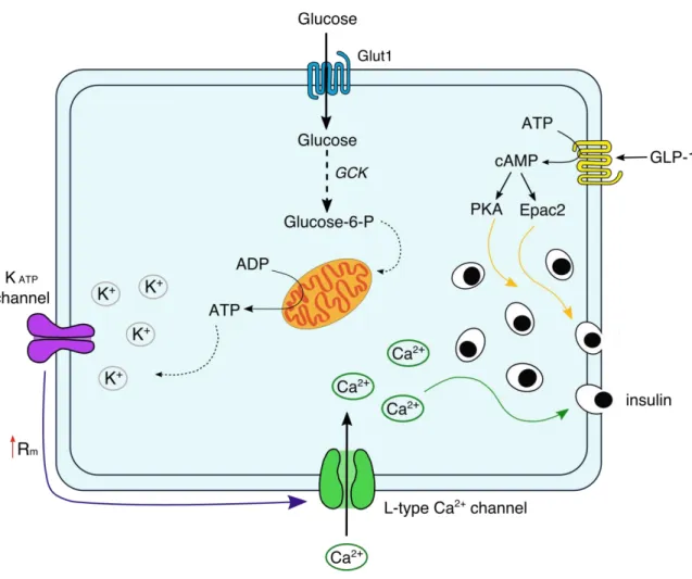
The importance of amino acid in insulin secretion ... 53
The liver-pancreatic islets axis ... 55
MicroRNAs ... 56
MiRNAs in the liver ... 56
MiRNAs and hepatocyte metabolism ... 59
MiRNAs and pancreatic beta cells ... 60
i‘NAs a d β-cell differentiation/de-differentiation ... 60
MiRNAs and β-cell identity ... 61
Mi‘NAs a d β-cell function ... 64
Origins of miRNAs: hidden in the genome ... 65
MicroRNA biogenesis: the canonical pathway ... 66
MicroRNA biogenesis: the non-canonical pathways ... 70
Results ... 72
Amino acid-induced regulation of hepatocyte growth: possible role of Drosha... 74
Abstract ... 75
Introduction ... 76
Results ... 78
Amino acid deprivation blocks primary rat hepatocyte proliferation and induces a metabolic rest without affecting the mitochondria ... 78
Amino acid-deprived hepatocytes are able to exit growth arrest after the addition of
essential amino acids ... 81
In aa deprived-growth arrested hepatocytes the activation of the mTOR pathway is repressed and this inhibition is partially reversed by essential aa ... 82
Essential aa deprivation in primary adult rat hepatocytes leads to an increase in Drosha protein level ... 84
Neither short- nor long-term rapamycin treatment alters the Drosha protein level in primary adult rat hepatocytes ... 85
AaD in primary adult rat hepatocytes increases Drosha protein stability ... 85
Drosha knockdown allows cell proliferation in aa-deprived hepatocytes without affecting mitochondrial metabolism and autophagy... 87
In primary adult rat hepatocytes deprived of aa the expression of miR-23a/b and miR-27b is increased, but Ago2 knockdown does not allow cell proliferation ... 89
Discussion ... 91
Materials and methods ... 94
Cell culture ... 94
Cell counting ... 95
Transfection ... 95
Autophagy analysis ... 95
Rapamycin treatment ... 96
Analysis of protein stability ... 96
Protein lysate and quantification ... 96
Western blot analysis ... 96
RNA extraction and quantitative PCR ... 97
Oxygen consumption analysis ... 97
FACS analysis ... 98
Mitochondrial staining ... 98
Terminal deoxynucleotidyl transferase mediated dUPT nick-end labeling (TUNEL) assay ... 99
Statistical analysis ... 99
Bibliography... 100
MicroRNA-375 regulates glucose metabolism-related signaling for insulin secretion in rat and human islets ... 104
Abstract ... 105
Introduction ... 106
Materials and methods ... 107
Reagents ... 107
Human islets ... 108
Animals and diets ... 108
Islet isolation ... 108
Culture and transfection of dissociated islet cells ... 108
RNA extraction and RT-qPCR ... 109
Lactate determination ... 109 Insulin secretion ... 109 Glucose uptake ... 110 Mitochondrial metabolism ... 110 Statistical analysis ... 110 Results ... 111
miR-375 overexpression in islet cells impairs insulin secretion and the Ca2+ rise induced by glucose but not by KCl ... 111
miR-375 impacts glucose-stimulated respiration ... 111
Reduced endogenous miR-375 levels in primary islet cells increases glucose metabolism and insulin secretion ... 113
miR-375 does not affect the action of a fuel stimulus enhancing mitochondrial
metabolism independently of glycolysis ... 115
miR-375 modifies pyruvate metabolism ... 115
Forced miR-375 expression in human islet cells reduces insulin secretion in response to glucose, but not KCl or to KIC ... 117
Reduced miR-375 expression is associated with in vitro glucose responsiveness and maturation of fetal rat beta cells ... 118
Discussion ... 119
Conclusion ... 120
Bibliography... 122
Discussion ... 126
General discussion... 127
More insights about Drosha and its regulation ... 127
D osha s o -canonical roles and hypothesis ... 128
D osha s ole i the egulatio of uies e e: e plo i g possi ilities ... 130
A very powerful microRNA in pancreas ... 131
Additional question regarding miR-375: possible targets ... 132
Annex ... 134
References ... 135
List of figures
IntroductionFigure 1 The hepatic lobule and detail of the portal triad……….. 16
Figure 2 The interconnection among hepatocytes……….. 19
Figure 3 Liver regeneration……….. 25
Figure 4 The natural progression of NAFLD……….. 30
Figure 5 Glucose metabolism……….. 32
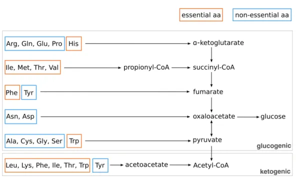
Figure 6 Essential and non-essential amino acids in human………. 40
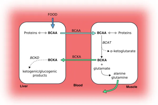
Figure 7 Catabolism of BCAAs in liver and skeletal muscle………. 42
Figure 8 mTOR signaling pathway in mammalian cells……… 5
Figure 9 Autophagy induction in mammalian cells………. 47
Figure 10 Insulin secretion in human β-cells………... 52
Figure 11 MiRNAs in hepatocyte differentiation and proliferation………. 58
Figure 12 i‘NAs i pa ti g o β-cell differentiation and functioning………. 63
Figure 13 miRNA biogenesis………. 68
Results #1 Amino acid-induced regulation of hepatocyte growth: possible role of Drosha Figure 1 AaD blocks cell proliferation and reduces mitochondrial metabolism……….. 78
Figure 2 AaD induces a quiescence state which can be reversed by aa addition……… 80
Figure 3 48h aaD reduces mTOR and p70S6K phosphorylation……….. 82
Figure 4 Essential aaD significantly increases Drosha protein………. 83
Figure 5 Rapamycin treatment is not able to reproduce Drosha increase……….. 85
Figure 6 48h aaD increases Drosha protein stability……… 86
Figure 7 Drosha knockdown restores cell proliferation in aa-deprived hepatocytes………….. 87
Figure 8 Ago2 knockdown has no effect on cell proliferation……….. 89
Results #2 MicroRNA-375 regulates glucose metabolism-related signaling for insulin secretion in rat and human islets Figure 1 miR-375 overexpression in islet cells impairs insulin secretion……… 1
Figure 2 Reduced miR-375 expression increase glucose metabolism………. 3 Figure 3 Forced miR-375 expression impairs pyruvate-induced insulin secretion and pyruvate
metabolism……… 5
Figure 4 Effect of miR-375 on insulin secretion in human islet cells………. 6 Figure 5 The repression of miR-375 expression is required for the glucose stimulus-secretion
List of abbreviations
aaamino acidsac-pre-mir-451 Ago-cleaved pre-mir-451 ADP adenosine diphosphate
Ago argonaute proteins ANGPTL3 Angiopoietin-like 3 ALT alanine aminotransferase
ARNT hydrocarbon receptor nuclear translocator ATG autophagy-related protein
ATP adenosine triphosphate BCAA branched-chain amino acids
BCAT branched-chain-amino-acid aminotransferase BCKA branched-chain keto acids
BCKD branched- hai α-keto acid dehydrogenase cAMP 3 ,5 -cyclic adenosine monophosphate CBP80 cap-binding complex 80
ChREBP carbohydrate response element binding protein CoA Coenzyme A
CPT-1 carnitine palmitoyltransferase 1 DHAP dihydroxyacetone phosphate DHCA 3α,7α-dihydroxycholestanoic acid DGCR8 DiGeorge syndrome critical region 8 DNL de novo lipogenesis
dsRBD dsRNA-binding domain dsRNA double-stranded RNA e.aa essential amino acids EGF epidermal growth factor
EGFR epidermal growth factor receptor
eIF2A eukaryotic translation initiation factor 2-A ER endoplasmic reticulum
EXP5 Exportin-5
F1,6P fructose 1,6-bisphosphate F2,6P fructose 2,6-bisphosphate F6P fructose-6-phosphate FA fatty acids
FABP fatty acid binding protein FAS fatty acid synthase
FFA free fatty acids
FIP200 focal adhesion kinase family interacting protein of 200 kDa G3P glycerol-3-phosphate
G6P glucose-6-phosphate G6Pase glucose-6-phosphatase GCK glucokinase
GCN2 general control nonderepressible-2 GDH glutamate dehydrogenase
GKRP glucokinase regulatory protein GLP-1 glucagon-like peptide-1 GP glycogen phosphorylase
GPAM glycerol-3-phosphate acyltransferase GPCR G-protein-coupled receptor
GS glycogen synthase
GSK-3 glycogen synthase kinase 3
HB-EGF heparin-binding EGF-like growth factor HBV hepatitis B virus
HCC hepatocellular carcinoma HCV hepatitis C virus
HFD high-fat-diet
HGF hepatocyte growth factor
HGFR hepatocyte growth factor receptor HNF3β hepatocyte nuclear factor 3-β HSC hepatic stellate cell
IgA immunoglobulin A
IGF-1 insulin-like growth factor-1 IL-6 interleukine-6
InsP3 inositol 1,4,5-trisphosphate IR insulin resistance
IRS1 insulin receptor substrate 1 KapB Kaposin B
KATP channels ATP-sensitive K+ channels
KSHV Kaposi s sa o a-associated herpesvirus L-PK liver-type pyruvate kinase
LC3 microtubule-associated protein 1 light chain 3 LCFA long-chain fatty acids
LDH lactate dehydrogenase lncRNA long non-coding RNA LPS lipopolysaccharide
LSEC liver sinusoidal endothelial cell LXRs liver X receptors
MID middle
miRISC miRNA-induced silencing complex miRNA microRNA
mRNA messenger ribonucleic acids mTORC1 mTOR Complex 1
mTORC2 mTOR Complex 2
NAFLD non-alcoholic fatty liver disease
NADPH nicotinamide adenine dinucleotide phosphate NASH non-alcoholic steatohepatitis
ne.aa non-essential amino acids NEFA non-esterified fatty acids Ngn2 Neurogenin2
ORP8 oxysterol-binding-protein-related protein 8 PAZ Piwi–Argonaute–Zwille
pdx1 pancreatic and duodenal homeobox 1 PEP phosphoenolpyruvate
PEPCK phosphoenolpyruvate carboxykinase PFK-1 phosphofructokinase-1
PI3P phosphatidylinositol 3-phosphate PKA protein kinase A
PKC protein kinase C PH partial hepatectomy piRNA PIWI-interacting RNA pre-miRNA precursor microRNA pri-miRNA primary microRNA PP pancreatic polypeptide PP1 protein phosphatase 1
PTEN phosphatase and tensin homolog PTP1B protein tyrosine phosphatase 1B RIIIDs RNase III domains
RER rough endoplasmic reticulum RNA Pol II RNA polymerase II rRNA ribosomal ribonucleic acids RP reserve pools
RRP readily releasable pools S1P sphingosine-1-phosphate SER smooth endoplasmic reticulum siRNA short interfering RNA
SIRT1 Sirtuin-1
SK2 sphingosine kinase 2
SREBP-1c sterol regulatory element binding protein 1c ssRNA single-stranded RNA
T2D type two diabetes TAG triacylglycerol TCA tricarboxylic acid
TGF-β transforming growth factor-β
TGF- βRI transforming growth factor-β type I receptor TGF- βRII transforming growth factor-β type II receptor TLR4 Toll-like receptor 4
tRNA transfer ribonucleic acids TSC Tuberous Sclerosis Complex ULK1 unc-51-like kinase 1
UTR untranslated region
VDCC voltage-dependent Ca+-channel VEGF vascular endothelial growth factor VLDL very low-density lipoprotein VPS vacuolar protein sorting
Liver
The liver is a key metabolic hub in our organism controlling life-critical tasks. Its functions range from the production of essential molecules such as proteins, lipids and carbohydrates, to the removal of both endogenous and exogenous products to maintain the whole-body homeostasis, the regulation of glycemia and the neutralization of toxic products. The multifunctional activities of the liver are possible due to the coordinated action of several highly differentiated cell types, arranged in a very efficient and specific architecture.
Anatomy
The liver is the largest solid organ and heaviest visceral tissue in the human body, with a weight of around 1,5 kg and approximately 15 cm diameter in adults. It is located on the right of the abdominal cavity, below the diaphragm, which separates it from the lungs, the heart, the stomach and the transverse colon. The liver presents two surfaces: the antero-posterior (or diaphragmatic) and the inferior (or visceral) surface. The diaphragmatic surface is attached to the diaphragm and to the anterior abdominal wall by the falciform ligament, which divides the organ in two uneven lobes, one bigger on the right and one smaller on the left. The visceral surface is characterized by three fossæ arranged in an H-shape, thus conferring to the inferior surface of the liver the appearance of 4 lobes. The left limb of the H is called left sagittal fissure and contains the ligamentum venosum and the round ligament of liver; the right limb of the H is known as right sagittal fossa and consists in a groove for inferior vena cava and for the gall bladder. Finally, the bar connecting the two limbs of the H is named transverse fissure, or porta hepatis, and hosts the nerves, the lymphatics and the portal vein, which lies behind and between the hepatic artery, on the left, and the hepatic duct on the right.
The liver is an organ of hemodynamic confluence, as it receives supply from two blood streams. The major source of blood (75%) is the portal vein, which delivers blood from the intestines, pancreas and spleen and it is rich in nutrients, hormones and toxins to eliminate. The 25% of blood supply comes from the hepatic artery, carrying oxygen from the aorta. The two circulation systems blend in the liver and the mixture of venous and arterial blood arrives to the hepatic microvessels at low pressure and oxygen tension. Therefore, an
exchange of nutrients and oxygen with the hepatic cells occurs and hepatic blood returns in the main circulation through the central vein.
From the macroscopic to the microscopic structure
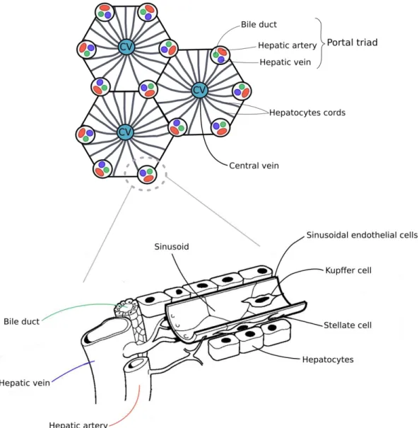
The liver has the appearance of a solid organ and its main anatomical and functional unit is the hepatic lobule. All the lobules are separated by an areolar tissue in which the blood vessels, the nerves and lymphatics expand and ramify to provide the whole structure. A dense layer of connective tissue, the fibrous coat or Glisson's capsule, surrounds the entire surface of the organ, lying just beneath a serous investment derived from the peritoneum. Each lobule has hexagonal shape and is formed by single layers of cells, the hepatocytes, organized in irregular radiating columns around the central vein. At each corner of the lobule is located one portal canal, making a total of six for each lobule (Figure 1 A). These portal canals, or portal triads, are composed by branches of portal vein, hepatic artery and bile ducts. Inside each lobule, a specific cellular organization and architecture guarantees the optimal liver functioning (Figure 1 B).
o An overview on hepatic cell populations
Hepatocytes, liver parenchymal cells, occupy about the 80% of the liver mass and constitute the liver metabolic units. They delimit a meshwork of capillaries (hepatic sinusoids) which convey the blood coming from the portal triads towards the center of the lobule, from where it drains into the vena cava. More in details, oxygen-rich blood from the hepatic artery mingles with nutrient-rich blood from the portal vein and, while progressing across the lobule through the sinusoids, it supplies the hepatic cells. This creates a gradient of oxygen, nutrients and waste products which results in a partitioning of functions called eta oli zo atio , i.e. the lo alizatio of the hepato tes t pi all i th ee diffe e t zones in the lobule, which influences their metabolic gene expression and functionality
(Trefts et al., 2017).
Figure 1 Schematic representation of the hepatic lobule and detail of the portal triad. (A) Each lobule,
characterized by hexagonal shape, is composed by single layers of hepatocytes organized radially around the central vein (CV). At each corner of the lobule is a portal triad (B), composed by hepatic artery, hepatic vein and bile duct, and which is connected to the central vein through the sinusoid. Adapted from (Barritt et al., 2008)
The wall of the sinusoids is formed mainly by highly specialized endothelial cells (liver sinusoidal endothelial cells – LSECs), which encompass approximately 50% of nonparenchymal cells in the liver and have a specific morphology and function. These cells are provided with a multitude of fenestrations and their unique structure creates pores between 50-180 nm in humans - or 50-280 nm in mice and rats - thus forming a selective barrier between hepatocytes and sinusoidal blood which is permeable for fluids and
molecules, but not for cells (Trefts et al., 2017). Moreover, LSECs are the only endothelial cells in mammals missing a basal lamina, which facilitates the direct exchange of solutes and colloids with the hepatic parenchymal cells and makes the microcirculation of the liver the most porous of all endothelial barriers (Sørensen et al., 2015).
In addition, sinusoids contain a resident macrophage population called Kupffer cells. These cells are part of the sinusoids lining. Being normally located at their bifurcation and branching, the biggest part of their cellular surface faces the blood flow and their extracellular processes extend beneath the endothelial cells. They play an essential role in the clearance of both the hepatic artery and venous blood entering the liver, and they participate to the elimination of phagocytosed bacteria with other liver sinusoidal cell populations. Furthermore, they express immune sensory receptors, thus contributing to the innate immune defense of the liver (Abdullah and Knolle, 2017).
The sinusoidal endothelial cells and the hepatocytes are separated by the space of Disse, or perisinusoidal space, where the hepatic parenchymal cells extend their villi and microvilli. Here we find another hepatic nonparenchymal cell type called hepatic stellate cells (HSCs). Stellate cells are a dynamic cell population which exists mainly in a quiescent state, whose principal role is to stock vitamin A. Indeed, they are the main responsible of the regulation of retinoid homeostasis, being capable of accumulating the 80% of the e ti e od s retinoids in the form of retinyl palmitate in lipid droplets stored in their cytoplasm. In pathological condition, such as liver injury, they exit quiescence and pass in an activated state, in which they proliferate and gradually lose vitamin A stores. Upon activation, their main role resides in the synthesis, deposition and organization of extracellular matrix components including proteoglycan, adhesive glycoproteins and collagen (Senoo, 2004)(Trefts et al., 2017).
The second most abundant epithelial cell type of the liver, accounting for the 3-5% of the hepatic cell population, is the cholangiocyte. These cells form the walls of specialized vessels, the bile ducts. These ducts are in direct communication with the bile canaliculi, which are created by the membranes of adjacent parenchymal cells and receive the bile produced by the hepatocytes (Trefts et al., 2017). Interestingly, according to their morphology, cholangiocytes are shown to assess different functions. The large ones play a
pivotal role in the formation and transport of bile through transmembrane molecules expressed on their membranes and they also regulate both the volume and the composition of the biliary secretion. Small cholangiocytes, instead, participate in maintaining tissue homeostasis in the biliary system by controlling the expression of regulators of proliferation, senescence and apoptosis (Yoo et al., 2016).
Hepatocytes
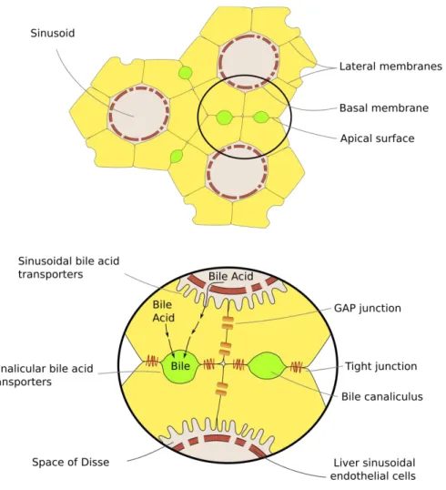
Hepatic parenchymal cells - or hepatocytes - are large cells, often considered as specialized epithelial cells, with a characteristic polygonal shape. They are usually tetraploids and they represent the great majority of liver cells, comprising roughly the 80% of the hepatic population. Although liver activity and functions are the result of the mutual actions of the cells mentioned above, hepatocytes clearly play a pivotal role. Their functions include the metabolism of proteins, carbohydrates and lipids, the synthesis of bile acids and the transcellular movement of bile fluid, the storage of lipids, glycogen and vitamins, the processing of endogenous wastes and the detoxification of exogenous detrimental products and the synthesis and secretion of proteins. Protein synthesis requires a concomitant extensive transcriptional activity, as several messenger ribonucleic acids (mRNA), transfer RNA (tRNA) and ribosomal RNA (rRNA) are needed to support a high rate protein translation. Therefore, multinucleate parenchymal cells are very common, and they also display singular or several large size nucleoli.
Being situated in the interface between two different physiological environments, the bile
canaliculi dedicated to the secretion of bile by parenchymal cells, and the sinusoids, where
the absorption of nutrients from the blood takes place, hepatocytes are highly polarized. This characteristic is primarily revealed by the polarization of their plasma membrane,
defined by three distinct domains, but also by a polarized trafficking of molecules and cytoskeletal architecture (Treyer and Müsch, 2013). The apical surface in hepatic parenchymal cells lines the bile canaliculi and is characterized by the presence of tight junctions. The remaining cell membrane is called basolateral and it includes two different regions: the lateral membranes, large areas facing adjacent hepatocytes, which communicate through gap junctions; the basal membrane, which faces the space of Disse and whose surface is considerably increased by a multitude of microvilli dipped in the
plasma oozed from the sinusoids. The basal membrane is dedicated to the exchange between parenchymal cells and the sinusoids, as the absence of a basal lamina in both of them facilitates the entrance of several molecules carried by the blood in the hepatocytes and the release of modified products (Barritt et al., 2008)(Alberts et al., 2015) (Figure 2).
Figure 2 Schematic representation of the interconnection among hepatocytes. Each cell is located in the
interface between bile canaliculi and sinusoids and, therefore, highly polarized. Lateral membranes line the connection between different hepatocytes, which communicate through gap junctions. The apical surfaces face the bile canaliculi and present tight junctions. The basal membranes allow direct contact with sinusoidal blood and their surface is increased by the presence of microvilli. Adapted from (Barritt et al., 2008)
The regulation of the substances entering and leaving the hepatocytes is regulated by the plasma membrane in many different ways. Its molecular structure includes membrane proteins that work as receptors for hormones, such as insulin and glucagon, or for other substances, for example the immunoglobulin A (IgA). When the access and exit from the parenchymal cells is not guaranteed by receptors, endocytosis or exocytosis, substances cross the membrane by taking advantage of carriers and protein channels. For example, glucose rapidly passes through the hepatocyte plasma membrane by means of transporters and, to prevent diffusion out of the cell, it is very efficiently converted in glucose-6-phosphate which can be used in glycogen synthesis. Whenever glycemia drops, parenchymal cell are able to break down the glycogen stored in the form of cytoplasmic granules and release glucose in the blood (Cardell and Cardell, 1990).
The role of Ca2+-permeable channels present on the plasma membrane has been also extensively investigated, as intracellular Ca2+ concentration plays a pivotal role in the indirect regulation of a variety of hepatocyte functions. As a matter of fact, the metabolism of glucose, amino acids and fatty acids, the synthesis and secretion of proteins, the intracellular vesicular movement and even cell proliferation, apoptosis and necrosis depend on variations in the cytosolic concentration of Ca2+ (Barritt et al., 2008), which regulates also the excretion of bile and its movement along the canaliculi (Nieuwenhuijs et al., 2006). The movement of bile acids, Na+ and other components of bile fluid across hepatocytes membrane changes its membrane potential and creates osmotic forces, which could detrimentally alter cellular volume if a control mechanism of the movement of ions did not take place. Remarkably, Ca2+ regulates the activity of a multitude of plasma membrane ion channels (Fitz and Sostman, 1994). According to the type of channel activated, the effects of changes in cytosolic Ca2+ on membrane potential can be variegated. For example, the activation of K+ channels is fundamental to recover hepatocyte swelling upon nutrient uptake (Barfod et al., 2007). Therefore, the modulation of cytosolic Ca2+ concentration plays an important role in regulating hepatocyte membrane potential and cell volume.
Hepatocyte: a deeper look inside the cell
Hepatocyte main functions are performed by the concerted action of multiple cellular organelles highly abundant in the cytoplasm of hepatic parenchymal cells. Among them the ribosomes, which can be found both freely in the hepatocyte cytoplasm or associated to the rough endoplasmic reticulum (RER), the smooth endoplasmic reticulum (SER), without ribosomes, and the Golgi complex.
Free ribosomes and membrane-bound ribosomes are structurally and functionally identical, but they differentiate in the proteins they synthetize. The ribosomes associated to the endoplasmic reticulum are involved in all the proteins destined to be translocated through the ER and then exported, while the mature ribosomes free in the cytoplasm are implicated in the translation of all the other polypeptides, both secreted and destined to remain within the cell (Alberts et al., 2015).
Being highly specialized in protein synthesis and managing the translation of the 95% of the proteins secreted by the liver, hepatocytes have a well-developed RER where not only the synthesis of exported proteins takes place, but also the translation of lysosomal and membrane-associated enzymes (Devi, 2018).
The ER compartment lacking ribosomes, the SER, is implicated in carbohydrate metabolism and it also participates in the regulation of blood glucose levels. Indeed, the presence of glucose-6-phosphatases bound on its membrane enables the hepatocytes to release glucose in the blood upon gluconeogenesis or breakdown of the glycogen stored in the cytoplasm (glycogenolysis) (Bánhegyi and Mándl, 2001). Moreover, the SER is provided with enzymes involved in the synthesis of components of the bile, bilirubin glucuronide and bile acids, essential for a correct digestion of food, especially fat, for the neutralization of the food
bolus in the stomach, for the elimination of metabolic wastes and for the control over excessive proliferation of exogenous bacteria in the gut (Bévalot et al., 2016). Most importantly, the SER hosts the cytochrome P450 enzymes involved in drugs and poison detoxification, and it is also the organelle where lipid and cholesterol synthesis take place, providing constituents for all the cell compartments, and where the proteins synthetized in the RER are assembled with lipids to originate the lipoproteins exported in the blood (Schwarz and Blower, 2016). Finally, the SER represents an intermediary compartment for
products that need to be further processed and modified in the Golgi complex, extensively developed in the hepatocytes due to the high rate of protein synthesis.
Similarly to the SER, peroxisomes also participate in detoxification, as they are equipped with enzymes involved in the metabolism of alcohol and toxic metabolic wastes. They constitute approximately 2% of the total hepatic proteins, making the liver the most peroxisome-rich organ in mammals (Leighton et al., 1968). They have been shown to participate in many critical metabolic processes such as fatty acid α- and β-oxidation, which leads to the production of acetyl coenzyme A available for biosynthetic reactions, to the degradation of purines, polyamines, amino acids and uric acid. Moreover, they are involved in lipid biosynthesis, containing enzymes necessary for the synthesis of plasmalogens (ether phospholipids important for the cellular membranes of some tissues) and for cholesterol-derived bile acids formation, which is a very unique function of hepatic peroxisomes (Wanders and Waterham, 2006)(Islinger et al., 2010).
Considering the extraordinary metabolic activity of the liver, it does not surprise that another abundant component of the hepatic parenchymal cells is constituted by mitochondria, which occupy over 20% of cellular volume. Mitochondria are the energetic factory of the cell, providing most of hepatocytes ATP and also other essential resources for biosynthesis and cell growth. In fact, they contain a large number of enzymes involved in a variety of metabolic activities including ATP synthesis and transport, oxidative phosphorylation, tricarboxylic acid cycle (TCA or Krebs cycle) and pyruvate and fatty acid oxidation. One of their most remarkable hepatocyte-specific functions is to carry out two critical steps of the urea cycle, which is an essential a al s metabolic pathway destined in eliminating the ammonia produced from nitrogen-containing compounds (for example amino acids) through the urine (Alberts et al., 2015). They are also involved in the first steps of bile synthesis by fo i g the ile a id i te ediate DHCA α, α-dihydroxycholestanoic acid) through a cytochrome P450 oxidase (specifically the sterol 27-hydroxylase) present in their inner membrane (Ferdinandusse and Houten, 2006).
The intracellular organization of hepatocytes, as well as their shape and movements, depends on a dynamic and adaptable system of filaments forming the cytoskeleton, composed essentially by microfilaments (made of actin), intermediate filaments and microtubules (made of tubulin). Both actin and tubulin are implicated in the intracellular motility. Microtubules are also described to participate in mitosis, cell shape determination,
intracellular transport of vesicles and secretion of albumin and lipoproteins. Microfilaments in hepatic parenchymal cells have been demonstrated to be very abundant around the bile
canaliculi as they seem to play an active role in controlling their dilatation and contraction
(Kawahara and French, 1990)(Watanabe et al., 1991).
Liver regeneration
A great emblem for regenerative medicine, as detailed by Carl Power and Rasko John (Power and Rasko, 2008) and others, is the Prometheus myth, a Greek ancient story telling about how Zeus punished Prometheus for having stolen the sacred fire from the gods and having donated it to the mankind. He was chained by the god on a rock in the Caucasus Mountains and every day an eagle would fed on his liver, organ which would have regrown overnight, condemning him to eternal punishment. It is hard to believe that ancient Greeks could actually know about this great self-repairing hepatic capacity, as it was firstly scientifically described not more than a century ago (Higgins and Anderson, 1931). Nevertheless, indeed one of the most remarkable characteristics of the liver is its ability to regenerate its volume upon tissue loss.
In physiological conditions adult hepatocytes rarely divide, being only 1-2% of them in the cell cycle at a giving time, while the rest remains in a quiescent state (or phase G0 of cell cycle), showing an estimated lifespan of approximately 400 days in adult rat liver (Grisham, 1962).
Quiescence is progressively acquired during neonatal hepatocyte maturation. However, a chemical injury to the liver or a partial hepatectomy (PH), i.e. the surgical removal of the 75% of total hepatic mass, is able to trigger their entrance in the cell cycle and the initiation of compensatory proliferation mechanisms (Berasain and Avila, 2015).
This peculiar biological process arose questions regarding the underpinnings of the maintenance of cellular homeostasis in the liver. The initial model proposed was named st ea i g h pothesis and described the liver as a slo l e e i g ell populatio , where hepatocytes located at the portal space had the greatest proliferation capabilities and their progeny would gradually stream towards the hepatic vein, where they would be finally eliminated by apoptosis. Therefore the entire hepatic lobule would result from this periportal cell population (Zajicek et al., 1985). Although initially supported, this hypothesis
had been recently disputed (Stanger, 2015). It is now believed that in physiological conditions the replication of existing cells is responsible for the maintenance of hepatic cellular population, while progenitor cells show very little involvement in liver remodeling. Liver regeneration can happen following two different situations: (i) hepatic injury caused by toxins or viral infection, characterized by the damage of all the hepatocytes and the consequent differentiation of oval cells, considered as liver stem cells, in both parenchymal and biliary cells; (ii) PH, which triggers the entrance of all the remaining diploid hepatocytes into the cell cycle in a synchronized manner to replace the missing liver tissue (Sun and Irvine, 2014)(Tao et al., 2017). Upon liver resection, virtually all hepatocytes exit quiescence and divide until their original cell number in the hepatic tissue is restored. Interestingly, this regenerative process stops when the species-specific liver to body mass ratio is recovered (Michalopoulos and DeFrances, 1997). This leads to the hypothesis of the presence of a liver\body mass ratio master regulator, hi h has ee alled hepatostat a d hose nature is still under investigation (Michalopoulos, 2010).
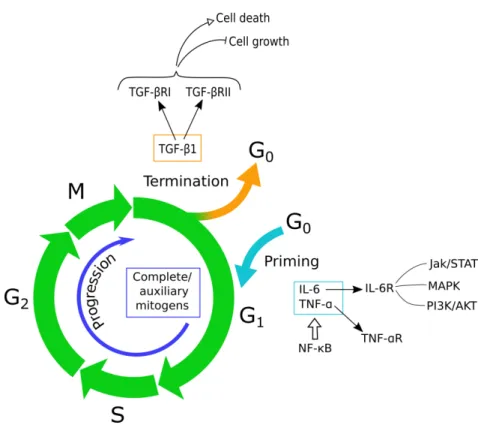
Liver regeneration: the pathways involved
Liver regeneration is a complex biological mechanism primarily involving hepatic parenchymal cells, but which also needs the participation of other liver cell populations and at least three interconnected networks (cytokine, growth factor and metabolic) that act at different timings (Fausto et al., 2012). Three critical steps can be identified in this process: the priming phase, when cells exit the G0 phase to enter G1; the proliferation phase, during which hepatocytes progress towards the mitotic phase with the help of mitogens; the
termination phase, when cell proliferation is blocked by negative factors (Tao et al., 2017).
(Figure 3)
Figure 3 Liver regeneration is defined by three critical steps: 1) cells exit quiescence during the priming phase,
triggered mostly by IL-6 and TNF-α; 2) during the progression phase, cells transit from G1 to mitosis thanks to the action of complete mitogens, especially HGF, EGF, TGF-α and HB-EGF, and the additional action of auxiliary mitogens, which increase or accelerate their effect; 3) the termination phase is induced once the liver/body ratio is restored and leads cells to re-enter quiescence especially through the action of TGF-β and other members of the same family. Adapted from (Tao et al., 2017)
o The priming phase
Liver damage and the loss of parenchymal cells initiate an inflammation response together with the release of cytokines and growth factors, especially tumor necrosis factor-α TNF-α) and interleukine-6 (IL-6), by inflammatory cells. Kupffer cells, the hepatic macrophages, are essential actors in the priming of hepatocytes to re-enter the cell cycle. They are indeed the major source of these cytokines through the activation of the NF-κB signaling pathway, which is initiated either by gut-derived factor lipopolysaccharide (LPS)/Toll-like receptor 4 (TLR4) signaling or by C3a and C5a, components of the complement system (Kang et al.,
IL-6 is a pleiotropic cytokine secreted upon LPS stimulation and the binding to its receptor (IL-6R) leads to the Jak/STAT, MAPK, and PI3K/AKT activation. By assessing both cytoprotective and mitogenic functions, IL-6 expression is extremely important for the entire regenerative process, from its initiation to maintenance and progression (Fujiyoshi and Ozaki, 2011)(Schaper and Rose-John, 2015).
o The proliferation phase
This phase, also called progression phase, is characterized by the action of different molecules in the transition of the hepatocytes from the phase G1 of cell cycle to mitosis. The molecules called complete mitogens show hepatotrophic effects, which means they are able to regulate specific genes expression which stimulates DNA synthesis and cell proliferation. The most relevant are hepatocyte growth factor (HGF), epidermal growth factor (EGF), transforming growth factor-α (TGF-α and heparin-binding EGF-like growth factor (HB-EGF). They control hepatocyte growth through the Ras-MAPK and the PI3K/AKT signaling pathways, renowned for their regulative role in cell cycle, by binding to their corresponding receptor c-Met or EGF receptor (EGFR) (Hong et al., 2000)(Mao et al., 2014). HGF was the first paracrine factor discovered to be implicated in hepatocyte proliferation through RAS activation, leading to prolonged MAPK activity, and PI3K/AKT activation (Nakamura et al., 1989). It is produced by stellate cells and released into the extracellular space, where in gets in contact with the hepatic parenchymal cells and its receptor c-Met, or hepatocyte growth factor receptor (HGFR), exposed on their membrane. The binding of c-Met with its ligand leads to the dimerization of the receptor and, subsequently, the activation of the aforementioned pathways, responsible for the increase in DNA synthesis and for cell cycle progression (García-Vilas and Medina, 2018).
The so-called auxiliary mitogens normally contribute to liver regeneration by increasing or accelerating the effects of the complete mitogens. Among them: (1) bile acids, which stimulate FoxM1b, a key modulator of cell cycle; (2) norepinephrine, which augments the effect of EGF and HGF and, through the activation of Smad7, it counteracts the growth inhibition of hepatocyte played by activin A; (3) vascular endothelial growth factor (VEGF), enhancer of sinusoidal endothelial cell and hepatocyte proliferation after PH; (4) insulin, which may contribute through the activation of inositol 1,4,5,-trisphosphate- (InsP3-)
dependent Ca2+ signaling pathways; (5) insulin-like growth factor-1 (IGF-1), which positively regulates HGF while downregulating TGF-β, an inhibitor of proliferation; (6) estrogen, which might sustain cell growth through the est oge e epto alpha E‘α and probably by a crosstalk with IL-6; (7) serotonin, contributing to liver regeneration via HT receptor 2 (HTR2) (Tao et al., 2017). Blood platelets have also been recently described to positively participate to hepatic re-growth by releasing HGF, VEGF, IGF-1 and serotonin (Meyer et al., 2015). The W t/β-catenin signaling pathway seems also to be involved the proliferation phase of liver regeneration. Wnt ligands, glycoproteins produced and secreted especially by Kupffer cells and endothelial non-parenchymal cells, lead to the expression of target genes important for the regulation of cell proliferation, such as c-myc and cyclinD1, through the
lassi t/β-catenin signaling cascade (Monga, 2011).
Finally, the last category of auxiliary mitogens is constituted by exosomes, cell-derived nanovesicles produced in the cellular endosomal compartment and involved in intercellular communication. It has been recently described that hepatocyte-derived exosomes fuse with target hepatocytes and stimulate their proliferation. In fact their exosomes deliver sphingosine kinase 2 (SK2), which catalyzes the phosphorylation of sphingosine in target hepatocytes, thus forming sphingosine-1-phosphate (S1P) and promoting cell survival, growth and migration (Nojima et al., 2016).
o The termination phase
The hepatic regeneration process stops when the normal liver to body mass ratio of 2.5% is restored. At present, the most characterized antiproliferative factors are the transforming growth factor-β TGF-β a d other members related to the same family.
TGF-β, specifically TGF-β , is active in the phase G1 of the cell cycle and, through the binding of its type I receptor (TGF-β‘I a d t pe II e epto TGF-β‘II , it promotes both the block of cell growth and the induction of cell death both in vivo and in vitro (Romero-Gallo et al., 2005).
TGF-β ot o l is e p essed hepatic cell populations, but also from extrahepatic tissues, for example by platelets (Katsuda et al., 2017) and spleen. The latter has also been proven to downregulate HGF and its receptor c-met, thus increasing its inhibitory effect on liver regeneration (Lee et al., 2015).
Finally, among the TGF-β fa il e e s, a ele a t ole is pla ed activin A, an activin subtype which was found to play as a negative regulator of hepatic regeneration by inducing cell growth arrest and apoptosis both in vitro and in vivo. It also decreases the production of fibronectin, a component of the extracellular matrix essential for liver regeneration (Date et al., 2000)(Takamura et al., 2005).
Liver-related diseases
Many liver-associated diseases had been described and their appearance and incidence depend on a variety of factors.
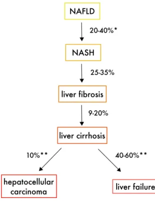
Non-alcoholic fatty liver disease (NAFLD), or hepatic steatosis, is one of the most studied and it is defined by the accumulation of lipids in the hepatocytes not due an excessive alcohol consumption. The presence of > 5% of steatotic hepatocytes, term referring to the accumulation of fat in the form of triglycerides, is considered as the minimum condition for the diagnosis of NAFLD and results in cellular inflammation, which can degenerate in worst forms of steatosis. Indeed, approximately the 10% of patients with NAFLD progress to non-alcoholic steatohepatitis (NASH), accompanied by hepatocellular ballooning, apoptosis and lytic necrosis. Chances are that the 25-30% of the those people further progress to liver fibrosis or even cirrhosis and, eventually, 10% of them will develop in several years hepatocellular carcinoma (HCC) (Ong and Younossi, 2007)(Farrell and Larter, 2006). (Figure 4)
The lipid accumulation typical of steatosis is considered to be mainly due to insulin resistance (IR) (Buzzetti et al., 2016). More specifically, IR would contribute to NAFLD development through two principal mechanisms: (i) the reduced suppression of the hormone-sensitive lipase (HSL) by insulin, characteristic in IR patients, would result in an increased triglycerides hydrolysis in the peripheral adipose tissues, leading to the augmentation of free fatty acids (FFAs) delivered to the liver and a subsequent massive esterification of FFA in triglycerides, thus causing steatosis; (ii) de novo lipogenesis (DNL), which accounts for 26.1% of the hepatic triglyceride formation in the livers of NAFLD subjects, against the 5% in healthy individuals, had been shown to be stimulated by IR through different pathways (El Idrissi et al., 2019).
The biological process responsible for the progression of NAFLD to NASH is still under investigation. An i itial t o-hit theory considered the increase in circulating FFAs, responsible for triglycerides accumulation in the hepatocytes, as the triggering event. The se o d-hit was a source of oxidative stress, such as mitochondrial or adipose tissue dysfunction, which would lead to lipid peroxidation, cell inflammation and the appearance of NASH (Day and James, 1998). However, currently the pathogenesis of NASH is considered as a complex, ost likel ulti-hit , process characterized by the additional effect of a multitude of events aside of the presence of classical steatosis (Buzzetti et al., 2016).
As mentioned before, the detrimental progression of NAFLD can ultimately result in cirrhosis. However, the strongest risk factor of hepatic cirrhosis is considered to be the abuse of alcohol, accompanied by hepatitis C virus (HCV) and hepatitis B virus (HBV) – respectively in developed countries, and in Asia and sub-Saharan Africa – infection (Detlef and Nezam, 2008). Cirrhosis is an advanced stage of liver fibrosis characterized by defects in the hepatic vascular system, leading to the funneling of the portal and arterial blood directly to the central veins, bypassing the exchange between sinusoids and hepatocytes. This particular condition is driven by the so-called sinusoidal capillarization, the transformation of the hepatic open circulation in a closed one: endothelial fenestration disappears and the
space of Disse is substituted by scar tissue, resulting in fibrotic septa surrounding hepatocyte
islands (Schaffner and Popper, 1963). The principal clinical consequences of cirrhosis are a malfunctioning of the hepatocytes, therefore impaired liver functions, portal hypertension and, in the worst circumstances, regenerative nodules resulting from an increased hepatocyte proliferation and that will eventually lead to HCC development (Detlef and Nezam, 2008).
HCC comprises the 70-90% of primary liver cancer, and it is associated with hepatic cirrhosis in more than 80% of the cases (Blachier et al., 2013). However, although quite unusual, its appearance can also take place in absence of advanced liver fibrosis or cirrhosis. The 15% of these events involve cases related to HBV infection (Kulik and El-Serag, 2019), while in the United States it seems to be mainly connected to the presence of liver steatosis (Mittal et al., 2016). Diabetes and obesity have been also associated to an increase in HCC incidence independently from NAFLD, being type 2 diabetes (T2D) coupled with a 2- to 3-fold increase risk of HCC (El–Serag et al., 2006).
A possible explanation for the involvement of NAFLD, obesity and T2D in HCC development, can be found in their close relationship with inflammation, increased oxidative stress and insulin resistance (Firneisz, 2014). The latter, in particular, can be easily linked to the uncontrolled cell growth and division characteristic of all the forms of cancer.
A chronic exposure of the hepatic tissue, adipose tissue and the infiltrated immune cells to an excess of lipids and other metabolites, causes a dysregulation in hormone and cytokine production, which can result in insulin resistance and lead to hyperinsulinemia. The latter two, in turn, are responsible for an increased expression of pro-inflammatory cytokines, such as TNF-α and IL-6, and are associated with an increase in IGF-1, already highlighted for its positi e ole i o t olli g the p olife atio phase du i g li e ege e atio . The esulti g increase in cell proliferation and angiogenesis, and the reduction in apoptosis, is considered to strongly participate to the development of HCC and can thus explain the involvement of the pathological conditions aforementioned (Singh et al., 2018).
Figure 4 Schematic representation of the natural progression of non-alcoholic fatty liver disease (NAFLD). NASH
non-alcoholic steatohepatitis. *In 3–6 years of follow-up **In 5–7 years of follow-up. Adapted from (El Idrissi et al., 2019)
Liver metabolism
As already mentioned at the beginning of this overview, the liver plays a crucial role in regulating the organismal homeostasis, as it represents the headquarters of most of the metabolic activities and it regulates the storage of nutrients, their breakdown, their synthesis and redistribution to other tissues.
Upon every meal, food is digested in the gastrointestinal tract, nutrients are adsorbed into the blood and transported to the liver through the portal vein. Glucose increase in the blood stimulates insulin secretion from the pancreas, which induces glucose storage in the liver through glycogenesis. Furthermore, in the hepatocytes glucose can be converted in amino acids (aa) or in fatty acids (FA). Aa are used as source of energy or to synthetize proteins and other bioactive molecules. FA and free fatty acids (FFAs) coming from the blood circulation are esterified in triacylglycerol (TAG) and either stored as small droplets or released in the bloodstream in the form of very low-density lipoproteins (VLDL).
When glycemia drops in the fasted state, glucagon is secreted from the pancreas and induces the hepatocytes to release glucose in the blood through the breakdown of glycogen (glycogenolysis). Moreover, lactate and alanine, produced respectively through glycogenolysis and protein catabolism in the muscle, and glycerol generated by adipose tissue through lipolysis of TAG, are delivered to the liver and are used as precursors for gluconeogenesis.
Therefore, we can also consider the liver as a hub metabolically connecting a multitude of important tissues, such as intestine, pancreas, adipose tissue and skeletal muscle, to guarantee a prompt management of nutrients requirement upon any condition.
Liver glucose metabolism
One of the main functions of the liver is to maintain a physiological glycemia by compensating its fluctuations in the fed and fasted state (Daly et al., 1998)(Klover and Mooney, 2004). This feature is possible thanks to the remarkable capacity of hepatocytes to balance between consumption and production of glucose (Mithieux, 2010). Glucose entrance inside the cell is mediated by members of the glucose transporter (GLUT) family,
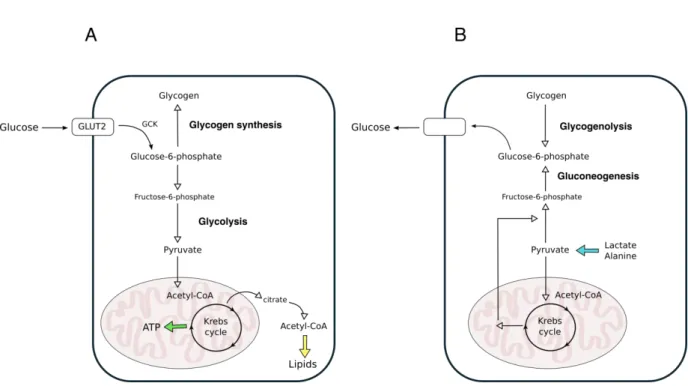
transmembrane proteins encoded by the SLC2 gene and with a multiple substrate specificity which includes many hexoses. Each body cell type is provided with at least one member of the GLUT family, which in the hepatic parenchymal cells is mostly represented by GLUT2 (Mueckler and Thorens, 2013). GLUT2 has the very peculiar characteristic of showing low affinity for glucose but high capacity (Williams et al., 1968), thus guaranteeing glucose uptake at physiological conditions. Although GLUT2 is necessary for glucose entrance in the hepatocytes, the release of this monosaccharide does not rely only on its presence, but also on the existence of alternative glucose transporters and mechanisms (Guillam et al., 1998). Once inside the cell, glucose is converted in glucose-6-phosphate (G6P) by glucokinase (GCK) (Cárdenas et al., 1998), which cannot exit the hepatocytes, and as such takes part to different biochemical pathways according to the organismal needs. (Figure 5)
Figure 5 Simplified scheme of glucose metabolism. (A) Principal biochemical pathways acting in the
postprandial phase to consume and store glucose; (B) Principal biochemical pathways in the postabsorptive glucose production: lactate and alanine are used as gluconeogenic substrates upon their conversion into pyruvate, while glycogen chains are broken down to release glucose units in the form of glucose-1-phosphate, to be then converted in glucose-6-phospate. Adapted from (Oosterveer and Schoonjans, 2014)
Gluconeogenesis
Hepatic glucose production represents the 90% of the endogenous glucose synthesis and, in humans, half of it relies on gluconeogenesis (Petersen et al., 2017).
The principal gluconeogenesis precursors are lactate, glycerol and amino acids, which can be either acquired from extrahepatic tissues through the blood circulation or synthetized inside the liver. Lactate, originating upon intense exercise activity, derives from the muscle through enzymatic conversion of the pyruvate produced from glucose, and takes part to the Cori cycle (or lactic acid cycle). In a very simplified model, once in the liver lactate is converted in pyruvate first, and then back to glucose using the glycolytic enzymes in reverse except for three irreversible reactions, whose enzymes (pyruvate kinase, phosphofructokinase-1 and
glucokinase) are bypassed and replaced by others (pyruvate carboxylase and phosphoenolpyruvate carboxykinase, fructose-1,6 bisphosphatase, glucose-6-phosphatase).
More in details, lactate is delivered to the hepatocytes and oxidized by lactate
dehydrogenase (LDH) in the cytoplasm. The resulting pyruvate is then exported in the
mitochondria and, through a sequence of biochemical reactions, returns to the cytosol when is finally converted in oxaloacetate. This can be then decarboxylated and phosphorylated by the sequential action of pyruvate carboxylate (PC) and phosphoenolpyruvate carboxykinase (PEPCK) to originate phosphoenolpyruvate (PEP), a crucial intermediate in biochemistry for its high-energy phosphate bond. PEP undergoes several biochemical reactions that lead to its conversion in fructose-6-phosphate (F6P) and finally in G6P, which can be dephosphorylated by glucose-6-phosphatase (G6Pase) to glucose in the ER (Rui, 2014). The primary carbon skeleton used for gluconeogenesis can be also derived from glycerol, whose contribution for endogenous glucose production ranges between 3%-22%, depending
on the increase in fasting duration (Consoli et al., 1987). Glycerol is released by adipose tissue following lipolysis of TAG and it enters the hepatocyte membrane through the aquaporin-9 (Jelen et al., 2012). Once inside the cell, it is phosphorylated in glycerol-3-phosphate (G3P) and it is finally converted in the gluconeogenic intermediate dihydroxyacetone phosphate (DHAP) (Han et al., 2016). In addition to glycerol, the increased lipolysis in white adipose tissue during fasting also produces non-esterified fatty acids (NEFAs). NEFAs cannot be used as gluconeogenic substrates, but the acetyl-CoA derived f o thei β-oxidation in the hepatocyte mitochondria is an allosteric activator of PC,