HAL Id: inserm-01073631
https://www.hal.inserm.fr/inserm-01073631v2
Submitted on 20 Oct 2014
HAL is a multi-disciplinary open access
archive for the deposit and dissemination of
sci-entific research documents, whether they are
pub-lished or not. The documents may come from
teaching and research institutions in France or
abroad, or from public or private research centers.
L’archive ouverte pluridisciplinaire HAL, est
destinée au dépôt et à la diffusion de documents
scientifiques de niveau recherche, publiés ou non,
émanant des établissements d’enseignement et de
recherche français ou étrangers, des laboratoires
publics ou privés.
Anna Mezzapesa, Cyrille Orset, Laurent Plawinski, Loic Doeuvre, Sara
Martinez de Lizarrondo, Guglielmina Chimienti, Denis Vivien, Alexandre
Mansour, Sabrina Matà, Gabriella Pepe, et al.
To cite this version:
Anna Mezzapesa, Cyrille Orset, Laurent Plawinski, Loic Doeuvre, Sara Martinez de Lizarrondo, et al..
Plasminogen in cerebrospinal fluid originates from circulating blood.. Journal of Neuroinflammation,
BioMed Central, 2014, 11 (1), pp.154. �10.1186/s12974-014-0154-y�. �inserm-01073631v2�
R E S E A R C H
Open Access
Plasminogen in cerebrospinal fluid originates
from circulating blood
Anna Mezzapesa
1,2, Cyrille Orset
2, Laurent Plawinski
3, Loic Doeuvre
2, Sara Martinez de Lizarrondo
2,
Guglielmina Chimienti
1, Denis Vivien
2, Alexandre Mansour
2, Sabrina Matà
4, Gabriella Pepe
1and Eduardo Anglés-Cano
5*Abstract
Background: Plasminogen activation is a ubiquitous source of fibrinolytic and proteolytic activity. Besides its role in prevention of thrombosis, plasminogen is involved in inflammatory reactions in the central nervous system.
Plasminogen has been detected in the cerebrospinal fluid (CSF) of patients with inflammatory diseases; however, its origin remains controversial, as the blood–CSF barrier may restrict its diffusion from blood.
Methods: We investigated the origin of plasminogen in CSF using Alexa Fluor 488–labelled rat plasminogen injected into rats with systemic inflammation and blood–CSF barrier dysfunction provoked by lipopolysaccharide (LPS). Near-infrared fluorescence imaging and immunohistochemistry fluorescence microscopy were used to identify plasminogen in brain structures, its concentration and functionality were determined by Western blotting and a chromogenic substrate assay, respectively. In parallel, plasminogen was investigated in CSF from patients with Guillain-Barré syndrome (n = 15), multiple sclerosis (n = 19) and noninflammatory neurological diseases (n = 8). Results: Endogenous rat plasminogen was detected in higher amounts in the CSF and urine of LPS-treated animals as compared to controls. In LPS-primed rats, circulating Alexa Fluor 488–labelled rat plasminogen was abundantly localized in the choroid plexus, CSF and urine. Plasminogen in human CSF was higher in Guillain-Barré syndrome (median = 1.28 ng/μl (interquartile range (IQR) = 0.66 to 1.59)) as compared to multiple sclerosis (median = 0.3 ng/μl (IQR = 0.16 to 0.61)) and to noninflammatory neurological diseases (median = 0.27 ng/μl (IQR = 0.18 to 0.35)). Conclusions: Our findings demonstrate that plasminogen is transported from circulating blood into the CSF of rats via the choroid plexus during inflammation. Our data suggest that a similar mechanism may explain the high CSF concentrations of plasminogen detected in patients with inflammation-derived CSF barrier impairment.
Keywords: Blood–cerebrospinal fluid barrier, Inflammation, LPS, Plasminogen
Introduction
Vessel wall fibrinolytic activity and pericellular proteoly-sis in tissues requires plasminogen binding and trans-formation into plasmin at the surface of fibrin, cells or the extracellular matrix by either the tissue-type plas-minogen activator (tPA) or the urokinase-type plasmino-gen activator (uPA) [1]. The binding of plasminoplasmino-gen to cells or fibrin is a lysine-dependent mechanism that can be inhibited by lysine analogues such as tranexamic acid
(TXA). In the vasculature, plasmin formation results in blood clot dissolution [2], whereas in tissues, plasmin is responsible for pericellular proteolysis accompanying cell migration, angiogenesis, wound healing, tissue re-modelling and inflammation [3-5]. Pericellular proteoly-sis in the central nervous system (CNS) is involved in development, regeneration of nervous tissues, neuronal and synaptic plasticity and the inflammatory response [6-9]. In humans, plasminogen deficiency is associated with neural disorders including congenital hydroceph-alus, periventricular nodular heterotopias and Dandy-Walker malformation [10]. Trace amounts of plasminogen have been reported in normal human cerebrospinal fluid (CSF), whereas raised concentrations have been detected in patients with
* Correspondence:[email protected]
5Inserm UMRS1140, Faculty of Pharmaceutical and Biological Sciences, Paris
Descartes University, 4 Avenue de l’Observatoire, 75270 Paris, cedex 06, France
Full list of author information is available at the end of the article
JOURNAL OF
NEUROINFLAMMATION
© 2014 Mezzapesa et al.; licensee BioMed Central Ltd. This is an Open Access article distributed under the terms of the Creative Commons Attribution License (http://creativecommons.org/licenses/by/4.0), which permits unrestricted use, distribution, and reproduction in any medium, provided the original work is properly credited. The Creative Commons Public Domain Dedication waiver (http://creativecommons.org/publicdomain/zero/1.0/) applies to the data made available in this article, unless otherwise stated.
Mezzapesa et al. Journal of Neuroinflammation 2014, 11:154 http://www.jneuroinflammation.com/content/11/1/154
meningitis [11], subarachnoid haemorrhage [12] and multiple sclerosis [13]. However, the question has not been settled as to whether plasminogen in CSF originates from circulating blood or is expressed in the CNS, as suggested by the pres-ence of mRNA in mouse brain [14,15] or by its synthesis by rat microglial cells in culture [16].
The CNS is protected from the bloodstream by the blood–brain barrier (BBB) and the blood–CSF barrier [17,18]. The BBB is the barrier separating the brain interstitial fluid compartment from the general circula-tion (endothelial tight junccircula-tions, basal lamina of endo-thelial cells and astrocyte pedicles). It selectively limits penetration of a variety of noxious substances and sup-plies the brain with nutrients. The blood–CSF barrier separates the CSF compartment from blood through choroid plexus epithelial cells, tight junctions, a basal membrane and the endothelium. It restricts the passage of harmful substances from the blood into the CSF se-creted across the choroid plexus epithelial cells into the brain’s ventricular system [18]. Because the blood–CSF barrier is more permeable than the BBB, many plasma proteins enter the cerebrospinal liquid (through pinocyt-osis or active transport). An impairment of the blood–CSF barrier thus leads to an increase in the concentration of proteins in the CSF.
Because diffusion of plasma proteins into the CNS is a selective process, we sought to demonstrate that circu-lating plasminogen may cross the blood–CSF barrier and can be detected in the CSF. For that purpose, we studied control rats and rats with lipopolysaccharide (LPS)-induced systemic inflammation. Our results show that circulating plasminogen enters the CSF space dur-ing blood–CSF barrier dysfunction induced by systemic inflammation, thus suggesting that plasminogen found in the CSF of patients with inflammatory neurological disorders originates from circulating blood.
Materials and methods
Reagents and proteins
The enhanced chemiluminescence reagent kit was obtained from Bio-Rad Laboratories (Hercules, CA, USA). The chromogenic substrate selective for plasmin (methylmalonyl)-hydroxyprolylarginine-para-nitroaniline (CBS0065) was purchased from Stago (Asnières-sur-Seine, France). Escherichia coli LPS serotype 0111:B4, trans-4-(aminomethyl)cyclohexane-1-carboxylic acid (or tranexamic acid, TXA), amiloride and 4′,6-diamidino-2-phenylindole (DAPI) and Evans blue were obtained from Sigma Chemical Co (St Louis, MO, USA). Alexa Fluor 488 succinimidyl ester was purchased from Life Technologies (Carlsbad, CA, USA). Goat anti-collagen type IV (Col IV) used for immunohistochemical analyses was obtained from Southern Biotech (Birmingham, AL, USA), and donkey anti-goat antibody F(ab′)2 fragments
linked to tetramethylrhodamine isothiocyanate were purchased from Jackson ImmunoResearch (West Grove, PA, USA). Glu-plasminogen, plasmin and peroxidase-conjugated monoclonal antibody against plasminogen kringle 1 (CPL15-PO) were prepared and characterized as described previously [19-21]. A rabbit anti-mouse plasminogen polyclonal antibody was kindly provided by HR Lijnen (University of Leuven, Belgium).
Patient sampling
A total of 42 patients with neurological diseases attend-ing the Neurological Clinic of the Careggi University Hospital, Florence, Italy, were admitted in this study. In-formed consent was obtained according to the Declar-ation of Helsinki. The Careggi University Hospital Review Board approved the protocol. Diagnoses were based on clinical, laboratory and magnetic resonance imaging data according to the International Classification of Diseases, Tenth Revision, and the Diagnostic and Statistical Man-ual of Mental Disorders, Fourth Edition–Text Revision [22]. Patients with Parkinson’s disease, brain tumour, epilepsy and alcohol or other substance dependence were excluded. All CSF samples were obtained by lum-bar puncture and were immediately used for routine laboratory analyses, which included erythrocyte and leucocyte differential cell counts, total protein concen-tration, albumin and immunoglobulin G (IgG) levels and agarose isoelectric focusing for IgG oligoclonal bands. CSF samples with increased lymphocyte counts
(>5 mm3) or with blood contamination (erythrocyte
concentration >50 cells/mm3) were excluded. The CSF
to serum albumin quotient (Qalb) was calculated and used to evaluate blood–CSF barrier integrity. Qalb > 0.007 was considered a marker of blood–CSF barrier dysfunction [23,24]. The remainder of each sample was stored in aliquots at −80°C for further analysis.
Animal experimental model
Experiments were performed using male Wistar rats (280 to 350 g) in accordance with the directives of the Council of the European Communities (86/609/EEC) and the French Agriculture and Forestry Ministry for handling animals (decree 87-848).
Purification and labelling of rat plasminogen
To monitor plasminogen uptake by cells and tissues
in vivo in the rat model of inflammation, we used
fluorescence-labelled native plasminogen purified from rat plasma. Blood was obtained by cardiac puncture of anaesthetized animals and collected in 0.129 M sodium citrate. The plasma was separated from blood by centri-fugation, and plasminogen was isolated by lysine affinity and sieving chromatography as described previously [19]. The purified plasminogen had a relative molecular
weight (Mr= 92,000 Da) similar to that of human
plas-minogen and was labelled with Alexa Fluor 488 dye (A488-Pg) according to the instructions of the manufacturer. Rat model of systemic inflammation
Rats were assigned to either LPS treatment (L) or the sa-line control group (C) (n = 3 rats per group (Figure 1). Groups L1, L2 and L3 received an intraperitoneal injec-tion of LPS (1 mg/kg) diluted in saline soluinjec-tion, and control groups C1, C2 and C3 received only the saline solution. This dosage of LPS has already been demon-strated to be able to induce brain inflammation in the rat [25]. Groups L1 and C1 received no other treatment. Groups L2 and C2 received a tail vein injection of A488-Pg (1 mg) 18 hours after LPS or saline administration. Group L3 received a tail vein injection of 10 mg/kg TXA 30 minutes after LPS administration, followed by a sub-cutaneous dose of 100 mg/kg TXA. After 18 hours, each rat received a tail vein injection of A488-Pg (1 mg) pre-incubated for 15 minutes with 0.1 M TXA, followed by a subcutaneous injection of 100 mg/kg TXA. Group C3 received identical doses of TXA, but not of A488-Pg. This dosage schedule of TXA was necessary to ensure neutralization of lysine-binding sites in both the en-dogenous and injected plasminogen. At the end of the scheduled treatments (24 hours), samples were collected and animals killed for further studies.
Sample collection
The anaesthetized rats were placed in a stereotaxic frame and secured with ear bars, and a midline incision
in the skin was made up to the head area to permit easy access to the cisterna magna. The needle, which was connected to a draw syringe, was inserted horizontally and centrally into the cisterna magna for CSF collection. The CSF sample was slowly drawn into the syringe, and the colour of the CSF was closely observed to avoid any possible blood contamination. Approximately 100 μl of CSF was collected from each animal. After a short cen-trifugation step (3 minutes at 5,000 g, 4°C) all CSF sam-ples were immediately flash-frozen in liquid nitrogen and stored at −80°C until use.
Blood was drawn by cardiac puncture using a 1.2 × 40–mm gauge needle syringe containing a 0.1 volume of 0.129 M sodium citrate. Urine was collected by punctur-ing the bladder. Plasma was separated from blood by cen-trifugation at 1,500 g for 15 minutes. All the samples were kept on ice during collection and kept frozen at −80°C until use.
Evans blue permeability assay
Rats (n = 3 per group) treated with saline (group C1) or LPS (group L1) alone or supplemented with TXA (groups C3 and L3) received a tail vein injection of Ev-ans blue dye (4% in saline) 4 hours before being killed. After transcardiac perfusion with heparinized saline, the brains and kidneys were harvested and plunged into cold saline until fluorescence measurements were performed. Evans blue dye permeability was monitored by ex vivo near-infrared fluorescence (NIRF) imaging using the IVIS 200 imaging system (Caliper Life Sciences, Hopkinton, MA, USA). Time (h) LPS challenged group L1 L2 L3 Saline challengedgroup C1 C2 C3 0. 5 18 24
i.v. & s.c. TXA
i.v. A488-Pg i.v. A488-Pg* + s.c TXA
i.v. & s.c. TXA
i.v. A488-Pg s.c. TXA
Sampling of CSF, plasma and urine Sampling of CSF, plasma and urine
Figure 1 Diagram of rat inflammatory model and study design. Rats were separated in six groups (three rats per group). Groups L1, L2 and L3 were challenged with 1 mg/kg lipopolysaccharide (LPS), and the control groups (C1, C2 and C3) were challenged with saline. Both LPS and saline were injected intraperitoneally. *Preincubated with tranexamic acid (TXA); i.v., Intravenous injection of TXA (10 mg/kg) or plasminogen labelled with Alexa Fluor 488 dye (A488-Pg) (1 mg); s.c., Subcutaneous injection of TXA (100 mg/kg).
Mezzapesa et al. Journal of Neuroinflammation 2014, 11:154 Page 3 of 10
Fluorescence microscopy
After the rats were killed, parts of the brains were post-fixed with 4% paraformaldehyde in 0.1 M phosphate-buffered saline, pH 7.4, at 4°C for 18 hours, followed by 24 hours in 20% sucrose and then frozen in isopentane. For immunocytochemistry, cryostat brain sections (8 μm) were collected on polylysine-coated slides and incubated overnight with a goat anti-Col IV primary antibody (1:1,500), which was revealed with donkey anti-goat antibody F(ab′)2 fragments linked to tetramethylrho-damine isothiocyanate (1:500). The cells were then coun-terstained with DAPI, mounted with Fluoprep (Dako, Glostrup, Denmark) and observed under an epifluores-cence microscope. Images were digitally captured with a Leica DM6000 microscope-coupled CoolSnap cam-era (Leica Microsystems, Wetzlar, Germany) and visual-ized with METAVUE 5.0 software (Molecular Devices, Sunnyvale, CA, USA). The detection of exogenously ad-ministered A488-Pg in those samples was also studied. Detection of functional plasminogen in cerebrospinal fluid
Transformation of CSF plasminogen into plasmin was measured using uPA (5 IU/ml) and a plasmin-selective chromogenic substrate (0.75 mM CBS0065), as previ-ously described [26]. In this system, the initial velocity of p-nitroaniline released from CBS0065 is proportional to the amount of plasmin(ogen). The ability of TXA to im-pair plasminogen binding to the cell membrane was in-vestigated as described previously [27].
Western blot analysis
Human CSF samples (7 μg of total protein) and rat CSF, urine and plasma (diluted 1:50) samples (10 μl) were electrophoresed in 8% SDS-PAGE gel under nonreduc-ing conditions. Proteins were transferred onto a polyvi-nylidene fluoride membrane, and plasminogen was revealed using a monoclonal antibody directed against human plasminogen (CPL15-PO, human CSF samples) or a rabbit anti-mouse plasminogen polyclonal antibody (rat samples). The amount of plasminogen was expressed as the number of pixels directly detected using Quant TL 7.0 image analysis software with the Image-Quant LAS 4000 imaging system (GE Healthcare Life Sciences, Pittsburgh, PA, USA) and expressed as nano-grams per microlitre by reference to a known amount of plasminogen (15 ng, 10 μl) electrophoresed under similar conditions. In rat samples, the amount of plasminogen was expressed as the number of pixels directly detected using the image analysis software.
Statistical analysis
Data are representative of at least three independent ex-periments and are expressed as median (25th to 75th
interquartile range (IQR)). The Mann–Whitney U test was used to compare values obtained in treated versus control rats. For human experiments, one-way analysis of variance (ANOVA) was performed to compare the pa-tient groups. Bonferroni’s multiple-comparisons posttest was run for the pairwise comparison of groups. Statis-tical significance was set at P < 0.05. A specific statisStatis-tical package for exact nonparametric inference (Stata Statis-tical Software release 9 (2005); StataCorp, College Sta-tion, TX, USA) was used.
Results
Lipopolysaccharide-induced increase in plasminogen in rat cerebrospinal fluid
Endogenous plasminogen was detected by Western im-munoblotting in plasma, CSF and urine of both LPS- and saline-treated control rats (groups L1 and C1, respectively) (Figure 2A). The LPS injection produced no significant variation in the amount of circulating plasminogen as compared to controls (0.1-fold increase in injected versus control rats) (Figure 2B). In contrast, we detected higher plasminogen levels in CSF and urine from LPS-treated rats (fourfold increase in CSF, P = 0.0002; twofold increase in urine, P = 0.0071 (both by Mann–Whitney U test)) (Figure 2B). Rat CSF plasminogen was efficiently trans-formed into plasmin; its active concentration appeared to be directly related to the amount of plasminogen detected in CSF samples by Western immunoblotting (P = 0.0045, Mann–Whitney U test) (Figure 2C). Lipopolysaccharide-induced increase in barrier permeability
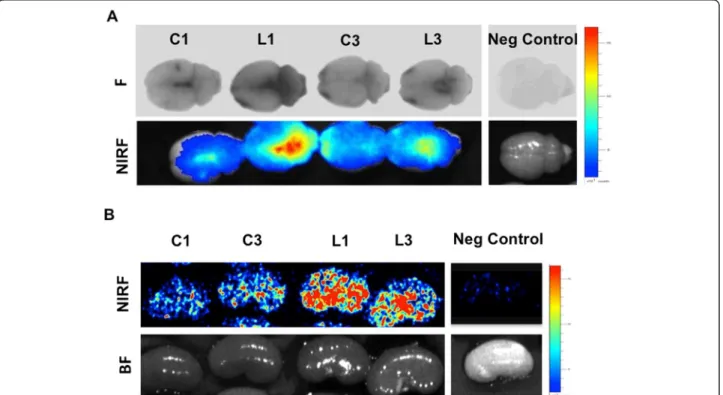
The aforementioned data suggest that vascular perme-ability was modified following the LPS injection. This hypothesis was investigated by testing permeability to Evans blue dye. Ex vivo fluorescent and NIRF images showed higher brain fluorescence in the LPS-treated rats (group L) as compared to controls (group C), indicating an increase in barrier permeability to the dye due to blood–CSF barrier impairment (Figure 3A). Modifica-tions in the glomerular filtration barrier were also de-tected with the Evans blue test (Figure 3B). Higher kidney fluorescence was observed in LPS-treated rats (group L) as compared to controls (group C). The ad-ministration of TXA (groups C3 and L3) was without ef-fect on barrier permeability.
In vivo evidence of the circulating origin of plasminogen in cerebrospinal fluid
To obtain in vivo evidence of the extravasation of plas-minogen from blood, animals were challenged with ex-ogenous A488-Pg 18 hours after the LPS (group L2) or saline injection (group C2). A488-Pg appeared in the CSF and urine of all LPS-treated rats, indicating that
Figure 2 (See legend on next page.)
Mezzapesa et al. Journal of Neuroinflammation 2014, 11:154 Page 5 of 10
blood plasminogen crosses the barrier during its pres-ence in the circulation (with a half-life of about 2 days) (Figure 4A) and integrates the CSF exchanged at least three times daily [28]. Actually, the presence of circulat-ing A488-Pg was clearly visualized by fluorescence mi-croscopy only in the choroid plexus of LPS-treated rats (Figure 4B). Figure 4C is a magnified view of the choroid plexus showing the presence of A488-Pg and the vessel wall detected by immunocytochemistry of Col IV. Of note, A488-Pg is absent in the saline-treated control.
Typically, binding of plasminogen by cells is mediated by a lysine-dependent mechanism via lysine residues encompassed in cellular receptors and lysine-binding sites located in plasminogen kringle domains [1]. To
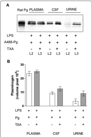
explore whether the passage of plasminogen through the blood–CSF barrier implicates a lysine-dependent mech-anism, we measured cellular uptake of A488-Pg in the presence of the lysine analogue TXA. Figure 5 shows that plasminogen remained increased in the CSF of rats treated with TXA, thus excluding uptake via known plasminogen receptors harbouring carboxy-terminal ly-sine residues [1].
To substantiate the relevance of these in vivo rat studies to human pathology, we investigated the presence of plas-minogen in the CSF of patients with inflammatory and noninflammatory disorders. Plasminogen antigen was de-tected in all CSF human samples (Figure 6). Higher con-centrations of plasminogen were found in Guillain-Barré
(See figure on previous page.)
Figure 2 Determination of plasminogen in rat plasma, cerebrospinal fluid and urine. Samples were obtained from rats challenged with saline (group C1) versus rats challenged with lipopolysaccharide (LPS) (group L1) (see flowchart in Figure 1). (A) Representative immunoblot obtained for plasminogen detection using a rabbit antibody to mouse plasminogen. Electrophoresis of reference rat plasminogen (Pg) in an equal volume (10 μl) of plasma diluted 1:50, cerebrospinal fluid (CSF) and urine. (B) Box-and-whisker plot of plasminogen in plasma, CSF and urine after immunoblot and densitometric analyses. Results represent median (25th to 75th interquartile range (IQR)). *P = 0.0002 and §P = 0.0071 in CSF and urine (Mann–Whitney U test), respectively. (C) Box-and-whisker plot of measurement of the activation of plasminogen in CSF samples (chromogenic substrate assay). Results represent median (IQR) of the velocity of plasmin formation. #P = 0.0045 (Mann–Whitney U test). mOD: milli optical density.
Figure 3 Blood–cerebrospinal fluid barrier and glomerular barrier permeability to Evans blue dye. Ex vivo fluorescent (F) and near-infrared fluorescent (NIRF) images of brains and kidneys from rats injected with Evans blue dye 24 hours after lipopolysaccharide (LPS) treatment according to the outline in Figure 1. The measurement was performed in perfused brains 24 hours after Evans blue dye injection. C1, Saline-challenged rats; L1, LPS-challenged rats; C3, Saline/tranexamic acid (TXA); L3, LPS/TXA; Neg control, Rat without Evans blue dye; BF, Bright field. (A) Brain NIRF images. (B) Kidney NIRF images. The high fluorescence intensity observed in LPS-treated rats (L1 and L3) compared to saline-treated rats (C1 and C3) indicates leakage of the blood–cerebrospinal fluid barrier (A) or the glomerular filtration barrier (B). No differences were observed between saline-treated (C1) and TXA-treated (C3) animals, indicating no effect of TXA on extravasation of Evans blue dye.
syndrome (GBS) (median = 1.28 ng/μl (25th to 75th inter-quartile range (IQR) = 0.66 to 1.59), n = 15) as compared to multiple sclerosis (MS) (median = 0.3 ng/μl (IQR = 0.16 to 0.61), n = 19) and noninflammatory neurological diseases (NINDs) (median = 0.27 ng/μl (IQR = 0.18 to 0.35), n = 8) (P < 0.0001, one-way ANOVA). The pairwise comparison yielded significant differences in GBS versus MS (P < 0.05) and GBS versus NINDs (P < 0.05), both analysed with Bonferroni’s posttest. GBS patients had significantly higher values of CSF total protein, albumin and IgG, as well as higher Qalb, whereas MS patients had higher index values of IgG (QIgG/Qalb) (see Additional file 1: Table S1).
Discussion
Plasminogen is synthesized mainly by the liver and then is distributed to tissues via the systemic circulation [29]. The concentration of plasminogen in circulating blood is relatively high (1.5 to 2 μM) and constant, as plas-minogen does not behave as an acute phase reactant such as fibrinogen. Therefore, inflammatory conditions or the injection of an inflammatory agent such as LPS
do not increase the circulating concentration of plas-minogen. However, a mechanistic link between inflam-mation and the blood–CSF barrier dysfunction has been established [30], which may explain an increased transfer of plasminogen from circulating blood to CSF. Indeed, trace amounts of plasminogen have been found in the CSF of patients with noninflammatory diseases [31,32]. By using an LPS-induced model of systemic inflammation in rats, we found that plasminogen is present at significantly higher concentrations in the CSF of LPS-treated rats as compared to controls. Accordingly, LPS-treated rats had increased blood–CSF barrier permeability, as demon-strated with Evans blue dye. CSF plasminogen is efficiently transformed into plasmin, thus suggesting that transfer of plasminogen from circulating blood to the CSF could be a source for plasmin formation in the nervous system. Fur-thermore, using A488-Pg, we found that this labelled ex-ogenous rat plasminogen crosses the blood–CSF barrier. The half-life of plasminogen in the circulation is about 2 days, whereas the CSF is produced and exchanged by cells of the choroid plexus at least three times daily [28]. Thus, circulating A488-Pg could be detected in the CSF and the
Figure 4 In vivo evidence of the circulating origin of plasminogen in cerebrospinal fluid. Samples were obtained from rats injected with plasminogen labelled with Alexa Fluor 488 dye (A488-Pg). Group L2 rats were challenged with lipopolysaccharide (LPS), and group C2 was challenged with saline (see flowchart in Figure 1). (A). An equal volume (10 μl) of either plasma diluted 1:50 or of cerebrospinal fluid (CSF) or urine was electrophoresed, and fluorescence (F) in the gel was directly revealed using ImageQuant TL 7.0 image analysis software (upper panel). The gel was then transferred onto a polyvinylidene fluoride membrane and detected by Western blotting with a rabbit antibody to mouse plasminogen (WB, lower panel). Representative samples are shown. (B) Micrograph showing the presence of circulating A488-Pg (indicated by arrows) in the choroid plexus of an LPS-treated rats detected by direct fluorescence microscopy. 4′,6-diamidino-2-phenylindole (DAPI) staining (blue) indicates cell nuclei. (C) Magnified images of choroid plexus of saline- and LPS-treated rats showing the presence of circulating A488-Pg (indicated by yellow arrows) only in the LPS condition, as detected by direct fluorescence microscopy. DAPI staining (blue) indicates cell nuclei, and collagen type IV (Col IV, red) is used as a vessel marker.
Mezzapesa et al. Journal of Neuroinflammation 2014, 11:154 Page 7 of 10
choroid plexus of LPS-treated rats. These in vivo data sug-gest that circulating plasminogen enters the CSF at the choroid plexus. This enormous epithelial/endothelial sur-face area is potentially available for untoward leakage of plasma proteins into CSF [33,34]. Because LPS is also known to induce interference with the integrity of the glomerular filtration barrier [35], we also investigated the passage of plasminogen through the renal glomerulus. We detected modifications in the glomerular filtration barrier with the Evans blue test and high concentrations of both endogenous plasminogen and A488-Pg in the urine of
LPS-treated rats as compared to trace amounts found in control rats.
Binding of plasminogen to carboxy-terminal lysine resi-dues of membrane receptors is a well-known mechanism governing its capture and concentration onto the cell sur-face, where it can be either transformed into plasmin or internalized [1]. Because these receptors could potentially be involved in the transfer of plasminogen from blood to CSF, we tested the possibility that TXA, a lysine analogue, might prevent plasminogen from entering the CSF. Occupation of the lysine-binding site of plasminogen by TXA prevents binding to lysine residues of mem-brane glycoprotein receptors. However, TXA did not inhibit A488-Pg transfer through the blood–CSF bar-rier, thus suggesting that other binding sites might be involved in plasminogen uptake. For instance, a pep-tide derived from the plasminogen-binding site domain 1 of M6PIGF2R, peptide fragment 18 to 36, induces plasminogen internalization [36].
Figure 5 Effect of tranexamic acid on plasminogen transfer through the blood–cerebrospinal fluid barrier and the renal glomerulus. Rats with lipopolysaccharide (LPS)-induced systemic inflammation received an intravenous injection of plasminogen labelled with Alexa Fluor 488 dye (A488-Pg) alone (group L2) or in the presence of sustained concentrations of tranexamic acid (TXA) (group L3) (see flowchart in Figure 1). Plasminogen was detected by immunoblotting. (A) Representative immunoblot of rat plasminogen (Pg), plasma diluted 1:50 cerebrospinal fluid (CSF) and urine samples using a rabbit antibody to mouse plasminogen. Experiments were performed as indicated in Figure 2 with an equal volume (10 μl) of each sample. (B) Representative column bars of plasminogen in plasma, CSF and urine after immunoblot and densitometric analysis.
Figure 6 Quantification of plasminogen in human cerebrospinal fluid. Cerebrospinal fluid (CSF) samples were collected from patients with Guillain-Barré syndrome (GBS, n = 15), multiple sclerosis (MS, n = 19) and noninflammatory neurological disease (NIND, n = 8). (A) Representative human CSF immunoblot showing peroxidase-conjugated monoclonal antibody directed against plasminogen kringle 1 (CPL15-PO). Electrophoresis of human plasminogen (Pg) (Mr= 92,000 Da), 10 μl of human plasma diluted 1:50 and 7 μg of total protein in CSF. (B) Box-and-whisker plot of human CSF plasminogen after immunoblot analysis and densitometric analysis. Results represent median (interquartile range) (P < 0.0001, one-way analysis of variance). *P < 0.05 for GBS versus MS and §P < 0.05 for GBS versus NIND (Bonferroni’s multiple-comparisons posttest).
Collectively, these findings demonstrate that, during inflammation, plasminogen is transported from circulat-ing blood into the CSF of rats via the choroid plexus. Interestingly, the high plasminogen CSF content de-tected in LPS-treated versus control rats parallels the significantly higher concentration of plasminogen found in the CSF of patients with blood–CSF barrier dysfunction determined on the basis of a high Qalb. In agreement with previously published data [31,32], we found that plas-minogen is present in trace amounts in noninflammatory CSF samples. However, we found significantly higher con-centrations in the CSF of patients with blood–CSF barrier dysfunction. Our present data are in accord with our pre-vious demonstration of the presence of apolipoprotein(a), a glycoprotein homologous to plasminogen in the CSF of patients with blood–CSF barrier dysfunction [37].
Although there are limitations to translating results obtained in experimental animals to human diseases, our data are suggestive of a similar mechanism for plas-minogen transfer from blood to CSF and may be of rele-vance to patients with inflammation-derived CSF barrier impairment.
Conclusions
We have demonstrated that plasminogen is increased in the CSF of patients with a dysfunctional blood–CSF barrier. We have reproduced, in an in vivo rat model of systemic inflammation, similar modifications of barrier function, and we also have demonstrated that plasminogen from circulat-ing blood enters the CSF space. Altogether our data strongly suggest that the increase in CSF plasminogen concentrations detected in patients obeys a mechanism by which circulating plasminogen crosses the altered blood–CSF barrier.
Additional file
Additional file 1: Table S1. Study groups and CSF biochemical parameters.
Abbreviations
A488-Pg:Alexa Fluor 488 plasminogen; CBS0065: (methylmalonyl)-hydroxyprolylarginine-para-nitroaniline; CNS: Central nervous system; CSF: Cerebrospinal fluid; DAPI: 4′,6-diamidino-2-phenylindole; GBS: Guillain-Barré syndrome; MS: Multiple sclerosis; NIND: Noninflammatory neurological disease; NIRF: Near-infrared fluorescence; TXA: Tranexamic acid;
uPA: Urokinase-type plasminogen activator. Competing interests
The authors declare that they have no competing interests. Authors’ contributions
Ana Mezzapesa performed the research; collected, analysed and interpreted data; and participated in manuscript drafting. GP and GC analysed and interpreted data and participated in manuscript drafting. SM recruited patients, collected cerebrospinal fluid, analysed and interpreted data and participated in manuscript drafting. CO and SM performed animal experiments and analysed data. DV revised and commented on the manuscript and managed funding. LP, LD and Alexandre Mansour performed chromogenic and immunoblot assays and participated in the interpretation and discussion of the results. EAC
conceived and designed the study, analysed and interpreted the data, and wrote the manuscript. All authors read and approved the final manuscript.
Acknowledgments
We acknowledge financial support from ADISU Puglia, Consorzio
Interuniversità per le Biotecnologie, Italy, and Società Italiana di Biochimica e Biologia Molecolare for the mobility to AM. This study was funded by Inserm (National Institutes for Health and Biomedical Research, France) and the Lower Normandy Regional Council.
Author details
1
Department of Biosciences, Biotechnologies, and Biopharmaceutics, University of Bari, Via Amendola 165/A, 70125 Bari, Italy.2Inserm U919, GIP
Cyceron, BP 5229, Boulevard Henri Becquerel, 14074 Caen cedex, Caen, France.3CNRS UMR 5248 CBMN, Institut Européen de Chimie et Biologie, 2
rue Robert Escarpit, 33607 Pessac, France.4Department of Neurology, University of Florence, Careggi Hospital, Viale Morgagni 85, 50134 Florence, Italy.5Inserm UMRS1140, Faculty of Pharmaceutical and Biological Sciences, Paris Descartes University, 4 Avenue de l’Observatoire, 75270 Paris, cedex 06, France.
Received: 28 March 2014 Accepted: 14 August 2014
References
1. Das R, Pluskota E, Plow EF: Plasminogen and its receptors as regulators of cardiovascular inflammatory responses. Trends Cardiovasc Med 2010, 20:120–124. 2. Rijken DC, Lijnen HR: New insights into the molecular mechanisms of the
fibrinolytic system. J Thromb Haemost 2009, 7:4–13.
3. Plow EF, Hoover-Plow J: The functions of plasminogen in cardiovascular disease. Trends Cardiovasc Med 2004, 14:180–186.
4. Li WY, Chong SS, Huang EY, Tuan TL: Plasminogen activator/plasmin system: a major player in wound healing? Wound Repair Regen 2003, 11:239–247. 5. Garcia-Touchard A, Henry TD, Sangiorgi G, Spagnoli LG, Mauriello A,
Conover C, Schwartz RS: Extracellular proteases in atherosclerosis and restenosis. Arterioscler Thromb Vasc Biol 2005, 25:1119–1127.
6. Sappino AP, Madani R, Huarte J, Belin D, Kiss JZ, Wohlwend A, Vassalli JD: Extracellular proteolysis in the adult murine brain. J Clin Invest 1993, 92:679–685.
7. Zhang Y, Pothakos K, Tsirka SA: Extracellular proteases: biological and behavioral roles in the mammalian central nervous system. Curr Top Dev Biol 2005, 66:161–188.
8. Monard D: Cell-derived proteases and protease inhibitors as regulators of neurite outgrowth. Trends Neurosci 1988, 11:541–544.
9. Gur-Wahnon D, Mizrachi T, Maaravi-Pinto FY, Lourbopoulos A, Grigoriadis N, Higazi AA, Brenner T: The plasminogen activator system: involvement in central nervous system inflammation and a potential site for therapeutic intervention. J Neuroinflammation 2013, 10:124.
10. Klammt J, Kobelt L, Aktas D, Durak I, Gokbuget A, Hughes Q, Irkec M, Kurtulus I, Lapi E, Mechoulam H, Mendoza-Londono R, Palumbo JS, Steitzer H, Tabbara KF, Ozbek Z, Pucci N, Sotomayor T, Sturm M, Drogies T, Ziegler M, Schuster V: Identification of three novel plasminogen (PLG) gene mutations in a series of 23 patients with low PLG activity. Thromb Haemost 2011, 105:454–460.
11. Newman RL: A method for detecting and estimating plasminogen in cerebrospinal fluid. J Clin Pathol 1964, 17:313–315.
12. Vermeulen M, van Vliet HH, Lindsay KW, Hijdra A, van Gijn J: Source of fibrin/fibrinogen degradation products in the CSF after subarachnoid hemorrhage. J Neurosurg 1985, 63:573–577.
13. Hammack BN, Fung KY, Hunsucker SW, Duncan MW, Burgoon MP, Owens GP, Gilden DH: Proteomic analysis of multiple sclerosis cerebrospinal fluid. Mult Scler 2004, 10:245–260.
14. Basham ME, Seeds NW: Plasminogen expression in the neonatal and adult mouse brain. J Neurochem 2001, 77:318–325.
15. Zhang L, Seiffert D, Fowler BJ, Jenkins GR, Thinnes TC, Loskutoff DJ, Parmer RJ, Miles LA: Plasminogen has a broad extrahepatic distribution. Thromb Haemost 2002, 87:493–501.
16. Nakajima K, Tsuzaki N, Nagata K, Takemoto N, Kohsaka S: Production and secretion of plasminogen in cultured rat brain microglia. FEBS Lett 1992, 308:179–182.
Mezzapesa et al. Journal of Neuroinflammation 2014, 11:154 Page 9 of 10
17. Wolburg H, Paulus W: Choroid plexus: biology and pathology. Acta Neuropathol 2010, 119:75–88.
18. Saunders NR, Ek CJ, Habgood MD, Dziegielewska KM: Barriers in the brain: a renaissance? Trends Neurosci 2008, 31:279–286.
19. Fleury V, Anglés-Cano E: Characterization of the binding of plasminogen to fibrin surfaces: the role of carboxy-terminal lysines. Biochemistry 1991, 30:7630–7638.
20. Wiman B, Wallén P: Activation of human plasminogen by an insoluble derivative of urokinase: structural changes of plasminogen in the course of activation to plasmin and demonstration of a possible intermediate compound. Eur J Biochem 1973, 36:25–31.
21. Montes R, Páramo JA, A-Cano E, Rocha E: Development and clinical application of a new ELISA assay to determine plasmin-α2-antiplasmin complexes in plasma. Br J Haematol 1996, 92:979–985.
22. American Psychiatric Association: Diagnostic and Statistical Manual of Mental Disorders, Volume text revision (DSM-IV-TR). 4th edition. Washington, DC: Author; 2000.
23. Tibbling G, Link H, Ohman S: Principles of albumin and IgG analyses in neurological disorders. I. Establishment of reference values. Scand J Clin Lab Invest 1977, 37:385–390.
24. Brettschneider J, Claus A, Kassubek J, Tumani H: Isolated blood–cerebrospinal fluid barrier dysfunction: prevalence and associated diseases. J Neurol 2005, 252:1067–1073.
25. Jeong HK, Jou I, Joe EH: Systemic LPS administration induces brain inflammation but not dopaminergic neuronal death in the substantia nigra. Exp Mol Med 2010, 42:823–832.
26. Bangert K, Thorsen S: Assay of functional plasminogen in rat plasma applicable to experimental studies of thrombolysis. Thromb Haemost 2000, 84:299–306.
27. Ho-Tin-Noé B, Enslen H, Doeuvre L, Corsi JM, Lijnen HR, Anglés-Cano E: Role of plasminogen activation in neuronal organization and survival. Mol Cell Neurosci 2009, 42:288–295.
28. Brown PD, Davies SL, Speake T, Millar ID: Molecular mechanisms of cerebrospinal fluid production. Neuroscience 2004, 129:957–970. 29. Bohmfalk JF, Fuller GM: Plasminogen is synthesized by primary cultures of
rat hepatocytes. Science 1980, 209:408–410.
30. Begley DJ, Couraud PO, Greenwood J, Pan W, Perry VH, Persidsky Y, Ransohoff R, Saunders NR, International Brain Barriers Society: Inflammation and the brain barriers. Available at http://www.ibbsoc.org/Reports.htm (accessed 28 August 2014).
31. Wu KK, Jacobsen CD, Hoak JC: Plasminogen in normal and abnormal human cerebrospinal fluid. Arch Neurol 1973, 28:64–66.
32. Newman RL, Stewart GT: The use of fibrinolytic activators in meningitis and similar conditions. Arch Dis Child 1965, 40:235–242.
33. Damkier HH, Brown PD, Praetorius J: Cerebrospinal fluid secretion by the choroid plexus. Physiol Rev 2013, 93:1847–1892.
34. Johanson C, Stopa E, McMillan P, Roth D, Funk J, Krinke G: The
distributional nexus of choroid plexus to cerebrospinal fluid, ependyma and brain: toxicologic/pathologic phenomena, periventricular destabilization, and lesion spread. Toxicol Pathol 2011, 39:186–212. 35. Xu C, Chang A, Hack BK, Eadon MT, Alper SL, Cunningham PN: TNF-mediated
damage to glomerular endothelium is an important determinant of acute kidney injury in sepsis. Kidney Int 2014, 85:72–81.
36. Leksa V, Pfisterer K, Ondrovičová G, Binder B, Lakatošová S, Donner C, Schiller HB, Zwirzitz A, Mrvová K, Pevala V, Kutejová E, Stockinger H: Dissecting mannose 6-phosphate-insulin-like growth factor 2 receptor complexes that control activation and uptake of plasminogen in cells. J Biol Chem 2012, 287:22450–22462.
37. Pepe G, Chimienti G, Liuzzi GM, Lamanuzzi BL, Nardulli M, Lolli F, Anglés-Cano E, Matà S: Lipoprotein(a) in the cerebrospinal fluid of neurological patients with blood–cerebrospinal fluid barrier dysfunction. Clin Chem 2006, 52:2043–2048.
doi:10.1186/s12974-014-0154-y
Cite this article as: Mezzapesa et al.: Plasminogen in cerebrospinal fluid originates from circulating blood. Journal of Neuroinflammation 2014 11:154.
Submit your next manuscript to BioMed Central and take full advantage of:
• Convenient online submission
• Thorough peer review
• No space constraints or color figure charges
• Immediate publication on acceptance
• Inclusion in PubMed, CAS, Scopus and Google Scholar
• Research which is freely available for redistribution
Submit your manuscript at www.biomedcentral.com/submit