HAL Id: hal-03064332
https://hal.archives-ouvertes.fr/hal-03064332
Submitted on 14 Dec 2020
HAL is a multi-disciplinary open access
archive for the deposit and dissemination of
sci-entific research documents, whether they are
pub-lished or not. The documents may come from
teaching and research institutions in France or
abroad, or from public or private research centers.
L’archive ouverte pluridisciplinaire HAL, est
destinée au dépôt et à la diffusion de documents
scientifiques de niveau recherche, publiés ou non,
émanant des établissements d’enseignement et de
recherche français ou étrangers, des laboratoires
publics ou privés.
FOXO1, is associated with markers of good prognosis
François Lallemand, Sophie Vacher, Leanne de Koning, Ambre Petitalot,
Adrien Briaux, Keltouma Driouch, Céline Callens, Anne Schnitzler, Caroline
Lecerf, Floriane Oulie-Bard, et al.
To cite this version:
François Lallemand, Sophie Vacher, Leanne de Koning, Ambre Petitalot, Adrien Briaux, et al.. The
high protein expression of FOXO3, but not that of FOXO1, is associated with markers of good
prognosis. Scientific Reports, Nature Publishing Group, 2020, 10, �10.1038/s41598-020-63895-8�.
�hal-03064332�
www.nature.com/scientificreports
the high protein expression of
FOXO3, but not that of FOXO1, is
associated with markers of good
prognosis
François Lallemand
1,2, Sophie Vacher
1,2, Leanne de Koning
3, Ambre petitalot
1,2,4,
Adrien Briaux
1,2, Keltouma Driouch
1,2, Céline Callens
1,2, Anne Schnitzler
1,2, Caroline Lecerf
3,
Floriane Oulie-Bard
3, Aurélie Barbet
3, Anne Vincent
7, Sophie Zinn-Justin
8, Bernard S. Lopez
5,6,
Rosette Lidereau
1, Ivan Bieche
1,9,10& Sandrine M. caputo
2,4,10✉
To better define the role of FOXO1 and FOXO3 transcriptional factors in breast carcinogenesis, we performed a comparative study of their expression at both the RNA and protein levels in a series of human breast tumors. We used qRT-PCR assay to quantify mRNA expression and Reverse Phase Protein Arrays (RPPA) to quantify protein expression in 218 breast tumors from patients with known clinical/pathological status and outcome. Weak correlations were observed between mRNA and protein expressions for both FOXO1 and FOXO3 genes. High expression of FOXO3 protein, but not FOXO1 protein, was a good prognostic marker, negatively correlated with KI67 and markers of activity of the PI3K/AKT/mTOR oncogenic pathway, and positively correlated with p53, a marker of apoptosis. Moreover, FOXO3 protein expression, but not FOXO1 protein expression, was also negatively correlated with various proteins involved in different DNA repair mechanisms. FOXO3 protein, but not FOXO1 protein, appears to be a tumor suppressor that inhibits breast cancer by altering DNA damage response (DDR), thereby inducing p53-dependent apoptosis. This antitumor effect appears to be suppressed by excessive activity of the PI3K/AKT/mTOR pathway. High FOXO3 protein expression could be a biomarker of deficient DDR in breast tumors.
Breast cancer is the most common solid malignancy in women in both developed and developing countries1.
Based on gene expression profiling, this pathology have been classified into four subtypes: luminal, human epi-dermal growth factor receptor 2 (HER2/ERBB2)-enriched, basal-like and normal-like2. Most breast tumors of
the basal-like subtype are triple-negative (TN), which means that they do not express estrogen and progesterone receptors, and lack ERBB2 overexpression3.
The forkhead box O (FOXO) family is a subclass of the forkhead family of transcription factors and consists of four members: FOXO1, FOXO3, FOXO4 and FOXO64. These four proteins possess the conserved DNA-binding
domain named forkhead domain or winged-helix domain5,6. They are involved in the regulation of various
cel-lular processes such as cell cycle, apoptosis, metabolism, and DNA repair4,7,8. Their transcriptional activity is
modulated notably by acetylation, ubiquitination, and phosphorylation5,9. Once activated, the PI3K/AKT/mTOR
1Service de génétique, unité de pharmacogénomique, Institut Curie, 26 rue d’Ulm, Paris, France. 2Paris Sciences
Lettres Research University, Paris, France. 3Translational Research Department, Institut Curie, PSL Research
University, 26 rue d’Ulm, Paris, France. 4Service de génétique, unité de génétique constitutionnelle, Institut
Curie, 26 rue d’Ulm, Paris, France. 5CNRS UMR 8200, Gustave Roussy Cancer Institute, Université Paris-Saclay,
équipe labélisée par la Ligue contre le cancer, Villejuif, France. 6Institut Cochin, INSERM U1016, UMR 8104 CNRS,
Université de Paris, 75014, Paris, France. 7Centre de Recherche en Cancérologie de Lyon (CRCL)/INSERM
U1052-CNRS UMR5286 Centre Léon Bérard, 28 Rue Laënnec, 69373 Cedex 08, Lyon, France., Lyon, France. 8Laboratoire
de Biologie Structurale et Radiobiologie, Institute for Integrative Biology of the Cell (CEA, CNRS, University Paris South), University Paris-Saclay, Gif-sur-Yvette, France. 9INSERM U1016, Université Paris Descartes, 4 avenue de
l’observatoire, Paris, France. 10These authors contributed equally: Ivan Bieche and Sandrine M. Caputo. ✉e-mail: sandrine.caputo@curie.fr
cellular pathway induces the phosphorylation of FOXO proteins by AKT. This phosphorylation leads to the exclu-sion of FOXO proteins from the nucleus, inhibiting therefore their capacity to modulate the transcription of their target genes4,10,11. This phosphorylation has also been shown to promote the degradation of FOXO3 protein via
the proteasome12. In human tumors, the PI3K/AKT/mTOR cellular pathway is frequently found over activated
leading therefore to inhibition of the transcriptional activity of the FOXO proteins5,13.
Various studies strongly suggest that FOXO proteins are tumor suppressors. The conditional deletion of all
FOXO1/3/4 alleles in adult mouse tissues induces the development of lymphoblastic thymic lymphomas and
hemangiomas14. Overexpression of FOXO1 and FOXO3 proteins in breast cancer has been shown to inhibit the
growth of breast cancer cells15–18. IκB kinase and ERk promote breast carcinogenesis via inhibition of FOXO3
protein expression [9]. The cytoplasmic expression of the FOXO3 protein is positively correlated with poor sur-vival in breast cancer15. Low expression of FOXO1 or FOXO3 protein in breast tumors is correlated with poor
clinical outcome19,20. Altogether, these results strongly suggest that FOXO1 and FOXO3 proteins act as tumor
suppressors in breast cancer. However, other studies have described unexpected functions for these two FOXO proteins in resistance to breast cancer treatment and breast cancer promotion21–24. Notably, FOXO1 and FOXO3
proteins have been implicated in the promotion of breast tumor cell invasion21,22. FOXO3 protein expression has
also been associated with poor survival in breast cancer23,24.
In order to better define the role of FOXO1 and FOXO3 proteins in breast cancer, we performed a com-parative study of their RNA and protein expressions in 218 breast tumors by using real-time quantitative reverse-transcription polymerase chain reaction (qRT-PCR) and Reverse Phase Protein Arrays (RPPA) methods, respectively. We also determined the correlations between FOXO1 and FOXO3 protein expressions and classical clinical biological parameters, as well as the expression of proteins involved in the PI3K/AKT/mTOR pathway, DNA damage response (DDR), apoptosis, cell cycle, and/or cell proliferation.
Results
FOXO1 and FOXO3 RNA and protein expressions in breast cancer.
To better define the role ofFOXO genes in breast cancer, we analysed their RNA and protein expression in a series of 218 breast tumors
(clinical parameters presented in Table S1).
In keeping with our previous work25, we found that the expressions of FOXO1, FOXO4, and FOXO6 at the
RNA level varied widely in breast tumors compared to normal breast samples (Table S2). In general, FOXO1 and
FOXO4 mRNA were significantly underexpressed (expression values < 0.33), whereas FOXO6 mRNA was
over-expressed (expression values ≥ 3), compared to normal breast tissue (see Material and Methods page 11). FOXO4 expression in the HR + ERBB2 + subtype and FOXO6 expression in the HR- ERBB2- subtype were similar in tumors and normal breast tissue. FOXO3 gene expression was similar in tumors and normal breast tissue except in the HR + ERBB2 + subtype, in which it was slightly, but significantly overexpressed.
The RPPA method demonstrated a wide range of FOXO1 protein expression (ranging from 0.07 to 1.91) and FOXO3 protein expression (ranging from 0.19 to 3.98) in breast tumors (Table 1). FOXO4 and FOXO6 protein expression was not studied due to the lack of an appropriate specific antibody for these two proteins.
We examined the correlation between protein and mRNA expressions for FOXO1 and FOXO3 genes. We observed weak positive correlation for the FOXO1 gene (r = +0.342, p < 10−4) and a weak positive correlation
for the FOXO3 gene (r = +0.150, p = 0.027), strongly suggesting that the expression of these two genes in breast tumors is regulated by molecular mechanisms independent of transcription and RNA stability (Fig. 1). These results also highlight the importance of considering protein expression when studying the role of FOXO1 and
FOXO3 genes in breast cancer.
Relationship between FOXO1 and FOXO3 protein expressions and clinical biological
parame-ters of breast cancer.
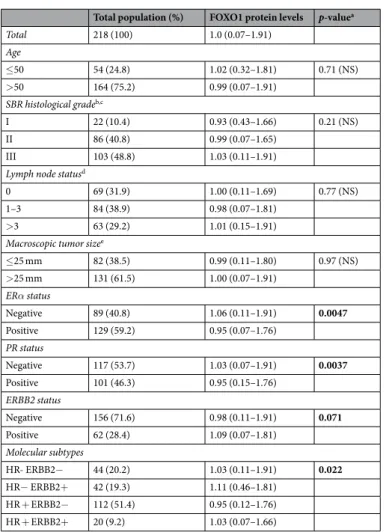
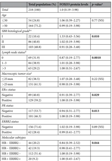
We investigated the relationships between FOXO1 and FOXO3 protein expressions and several classical clinical biological parameters (Tables 2 and 3). Marked differences were observed between these two FOXO proteins, as high FOXO1 protein expression was associated with negative estrogen receptor α (ERα) and progesterone receptor (PR) status, while low FOXO3 protein expression was weakly associated with these two biological parameters. We also showed that high FOXO3 protein expression was associated with low SBR histological grade, and, surprisingly, with high level of lymph node status. However, no association was observed between FOXO1 protein expression and these two clinical parameters.To investigate in more detail the role of FOXO1 and FOXO3 genes in breast cancer, we also performed a log rank test to analyse the relationship between FOXO1 and FOXO3 protein expressions and metastasis-free survival (MFS). Patients with breast tumors expressing high levels of FOXO3 protein had better MFS than patients with breast tumors expressing lower levels of FOXO3 protein (p = 4.1.10−2), which is consistent with a tumor
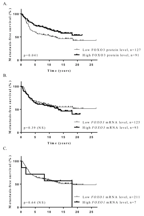
suppres-sor role (Fig. 2A). Such an association was not observed for FOXO1 protein. Multivariate analysis using a Cox proportional hazards model assessed the predictive value for MFS of the parameters found to be significant in univariate analysis (Table S1), i.e. lymph node status and macroscopic tumor size, and FOXO3 protein expression (p = 5.5.10−3) (Table S3). The prognostic significance of these three parameters persisted in multivariate analysis,
indicating that FOXO3 protein expression is an independent prognostic factor in breast cancer. Interestingly, no correlation was demonstrated between FOXO1 or FOXO3 mRNA expression and MFS in this series of 218 breast tumors (Fig. 2B,C), highlighting once again the importance of studying FOXO1 and FOXO3 protein expressions in order to assess their role in breast cancer.
Altogether, these observations suggest that FOXO3 protein, but not FOXO1 protein, may act as a tumor sup-pressor in breast cancer.
Relationship between the levels of FOXO1, FOXO3, and other proteins involved in the PI3K/
AKT/mTOR pathway, DDR, apoptosis, cell cycle, and cell proliferation.
In order to better definewww.nature.com/scientificreports
www.nature.com/scientificreports/
the role of FOXO1 and FOXO3 proteins in breast cancer, we also used the RPPA approach to study other proteins involved in the PI3K/AKT/mTOR oncogenic pathway, which regulates the activities of FOXO proteins, as well as various cellular processes involved in cancer and regulated by FOXO proteins: DDR, apoptosis, cell cycle, and cell proliferation (Table 4).
To evaluate the PI3K/AKT/mTOR pathway activity in our tumors, we studied the phosphorylation sta-tus of the S6K and S6, two proteins specifically phosphorylated by this pathway26. The PI3K/AKT/mTOR
pathway-dependent phosphorylation of AKT to its threonine 308 (pAKT-T308) and, to a lesser extent, its serine 473, is essential for its kinase activity27. However, because of the lack of appropriate pAKT-T308 antibody for
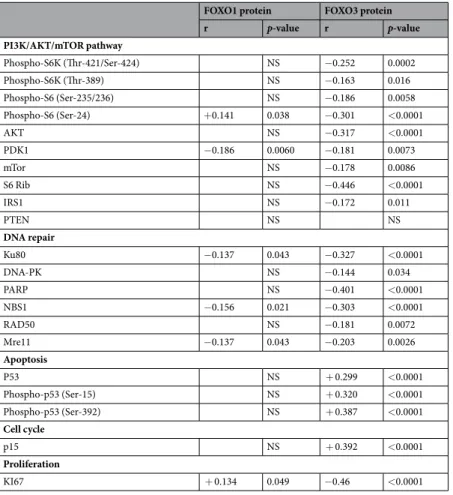
RPPA method, we did not use the phosphorylated forms of AKT to evaluate the PI3K/AKT/mTOR pathway activ-ity. We found negative correlations between FOXO3 protein expression and protein expressions of Phospho-S6K (Thr-421/Ser-424) (r = −0.252, p = 2.10−4), Phospho-S6K (Thr-389) (r = −0.163, p = 1.6.10−2), Phospho-S6
(Ser-235/Ser-236) (r = −0.186, p = 5.8.10−3), and Phospho-S6 (Ser-24) (r = −0.301, p < 10−4), suggesting that the
low expression of FOXO3 protein is associated to a weak activity of the PI3K/AKT/mTOR pathway in breast tumors. We observed that FOXO3 protein expression was also negatively correlated with the protein expressions of various components of this pathway: AKT (r = −0.317, p < 10−4), PDK1 (r = −0.181, p = 7.3.10−3), mTOR
(r = −0.178, p = 8.6.10−3), S6 (r = −0.446, p < 10−4), and IRS1 (r = −0.172, p = 1.1.10−2)28,29. We did not detect
significant correlation between FOXO3 protein expression and the protein expression of one of the most impor-tant negative regulators of the PI3K/AKT/mTOR pathway, PTEN. The weak PI3K/AKT/mTOR pathway activity found in the breast tumors expressing low level of FOXO3 protein would be therefore due to a low level of various components of this pathway but not to a high level of PTEN. Regarding FOXO1, we detected a negative correla-tion only with PDK1 (r = −0.186, p = 6.10−3). Akt activation has been shown to promote degradation of FOXO3
protein by proteasomes12. Therefore, our results suggest that high activity of the PI3K/AKT/mTOR pathway in
breast tumors would induce degradation of FOXO3 protein, but not FOXO1 protein.
Interestingly, we also demonstrated negative correlations between FOXO3 protein expression and the protein expression of various factors involved in different DNA repair mechanisms: Ku80 (r = −0.327, p < 10−4) and
DNA-PK (r = −0.144, p = 3.4.10−2) involved in non-homologous end joining (NHEJ), PARP (r = −0.401, p <
10−4) crucial for alternative NHEJ and base excision repair, and the three components of the MRN complex:
NBS1 (r = −0.303, p < 10−4), RAD50 (r = −0.181, p = 7.2.10−3), and Mre11 (r = −0.203, p = 2.6.10−3), described
as a key multi-protein complex crucial for DNA repair by homologous recombination and NHEJ30,31. Only weakly
significant negative correlations were demonstrated between FOXO1 protein expression and Ku80, NBS1, and Mre11 (0.05 > p > 0.01) (Table 4). Strong FOXO3 protein expression in breast tumor cells therefore appears to inhibit DDR, which would lead to accumulation of genetic alterations, thereby causing cell cycle arrest and/or p53-dependent apoptosis. Consistent with this hypothesis, we found that the FOXO3 protein level was negatively correlated with the KI67 protein level, a marker of proliferation (r = −0.460, p < 10−4), and was positively
corre-lated with the cell cycle inhibitor p15 protein level (r = +0.392, p < 10−4), as well as the levels of p53 (r = +0.299,
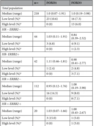
n= FOXO1 FOXO3 Total population Median (range) 218 1.0 (0.07–1.91) 1.0 (0.19–3.98) Low level (%)a 23 (10.6) 16 (7.3) High level (%)a 0 (0) 13 (6.0) HR− ERBB2− Median (range) 44 1.03 (0.11–1.91) 0.84 (0.39–2.52) Low level (%)a 3 (6.8) 4 (9.1) High level (%)a 0 (0) 1 (2.3) HR- ERBB2+ Median (range) 42 1.11 (0.46–1.81) 0.98 (0.41–2.77) Low level (%)a 1 (2.4) 2 (4.8) High level (%)a 0 (0) 3 (7.1) HR + ERBB2− Median (range) 112 0.95 (0.12–1.76) 1.08 (0.19–3.98) Low level (%)a 16 (14.3) 9 (8.0) High level (%)a 0 (0) 8 (7.1) HR + ERBB2 + Median (range) 20 1.03 (0.07–1.66) 1.00 (0.45–2.67) Low level (%)a 3 (15.0) 1 (5.0) High level (%)a 0 (0) 1 (5.0)
Table 1. Protein levels of FOXO1 and FOXO3 in the series of 218 breast tumours. Protein levels were
normalized so that the median of values in the 218 breast tumours was 1. aLow and high protein levels were
p < 10−4), Phospho-p53 (Ser-15) (r = +0.320, p < 10−4), and Phospho-p53 (Ser-392) (r = +0.387, p < 10−4)
(phosphorylation of p53 at these two sites triggers its apoptotic activity32), whereas FOXO1 protein level was very
slightly positively correlated with KI67 (r = +0.134, p = 4.9. 10−2) and not correlated with cell cycle and apoptosis
protein expressions.
In order to check that FOXO3 is functional in breast tumors, we performed a western blot analysis to visualize Phospho-FOXO3 (pSer-253), the inactive form of FOXO3, of several breast tumors of our series10. We detected
at least 8 samples with negative or low levels of phospho-FOXO3 (pSer-253) expression among 12 breast tumor samples expressing high levels of FOXO3, suggesting that this FOXO3 protein may be functional in a majority of these tumors (Fig. S1).
Overall, our results suggest that FOXO3 protein, but not FOXO1 protein, acts as a tumor suppressor in breast cancer, at least in part by DDR inhibition and subsequent induction of p53-dependent apoptosis. They also sug-gest that the antitumor effect of FOXO3 is abolished by high activity of the PI3K/AKT/mTOR pathway.
0 .0 0 .5 1 .0 1 .5 2 .0 0 .0 0 .5 1 .0 1 .5
F O X O 1 p ro te in le v e l
FO
XO
1
mA
RN
le
ve
l
0 .0 1 .0 2 .0 3 .0 4 .0 0 .0 2 .0 4 .0 6 .0 8 .0F O X O 3 p ro te in le v e l
FO
XO
3
mA
RN
leve
l
r= + 0.342, p < 0.0001
r= + 0.150, p=0.027
A.
B.
Figure 1. Scatter plots and Spearman correlation coefficients (r) between FOXO1 (A) and FOXO3 (B) protein
www.nature.com/scientificreports
www.nature.com/scientificreports/
Discussion
Many studies designed to examine the role of genes in carcinogenesis determine the correlations between their RNA expression and classical clinical biological parameters, survival, and the expression of others genes linked to cancer. However, due to post-transcriptional regulations, weak correlations are commonly observed between RNA expression and protein expression33. In our study, we found weak correlations between FOXO1 and FOXO3
RNA and protein expressions in breast cancer (Fig. 1). The absence of correlation between protein and mRNA expressions for FOXO1 and FOXO3 genes can be fully explain by the fact that these FOXO proteins undergo posttranslational modifications, such as acetylation, ubiquitination, and phosphorylation, modulating their sub-cellular localization and stability. To investigate the role of FOXO1 and FOXO3 genes in breast carcinogenesis, we therefore used the RPPA method to perform a comparative study of the protein expression of these two FOXO genes in a series of 218 breast tumors.
Our results strongly suggest that FOXO3 protein, but not FOXO1 protein, acts as a tumor suppressor in breast cancer. In particular, we found that patients with breast tumors expressing high levels of FOXO3 protein had better survival rates than patients with breast tumors expressing lower levels of this protein. We also showed that FOXO3 protein expression, but not FOXO1 protein expression, was negatively correlated with the expression of the KI67 marker of proliferation (Table 4). We recently showed that FOXO6 protein has an oncogenic effect in breast cancer25. Therefore, despite their homologies, the FOXO proteins appear to have different and specific
effects on breast cancer development.
In line with the results of various studies, we provide experimental arguments suggesting a tumor suppressor activity of FOXO3 protein in breast cancer15–17,20. However, other studies have shown that high FOXO3 protein
expression is associated with poor disease-free survival in TN breast cancer, and promotes proliferation, migra-tion and invasion of TN breast cancer cell lines22,23. In addition, Sisci et al. reported that FOXO3 protein inhibits
breast carcinogenesis in ERα-positive cells, and tends to promote breast carcinogenesis in ERα-negative cells34.
The role of FOXO3 protein in breast carcinogenesis may therefore depend on the subtype of breast cancer and the stage of disease. Further protein expression studies based on larger series of breast cancers are necessary to determine the precise role of FOXO3 protein in the various subtypes of breast cancer.
Total population (%) FOXO1 protein levels p-valuea
Total 218 (100) 1.0 (0.07–1.91) Age ≤50 54 (24.8) 1.02 (0.32–1.81) 0.71 (NS) >50 164 (75.2) 0.99 (0.07–1.91) SBR histological gradeb,c I 22 (10.4) 0.93 (0.43–1.66) 0.21 (NS) II 86 (40.8) 0.99 (0.07–1.65) III 103 (48.8) 1.03 (0.11–1.91)
Lymph node statusd
0 69 (31.9) 1.00 (0.11–1.69) 0.77 (NS)
1–3 84 (38.9) 0.98 (0.07–1.81)
>3 63 (29.2) 1.01 (0.15–1.91)
Macroscopic tumor sizee
≤25 mm 82 (38.5) 0.99 (0.11–1.80) 0.97 (NS) >25 mm 131 (61.5) 1.00 (0.07–1.91) ERα status Negative 89 (40.8) 1.06 (0.11–1.91) 0.0047 Positive 129 (59.2) 0.95 (0.07–1.76) PR status Negative 117 (53.7) 1.03 (0.07–1.91) 0.0037 Positive 101 (46.3) 0.95 (0.15–1.76) ERBB2 status Negative 156 (71.6) 0.98 (0.11–1.91) 0.071 Positive 62 (28.4) 1.09 (0.07–1.81) Molecular subtypes HR- ERBB2− 44 (20.2) 1.03 (0.11–1.91) 0.022 HR− ERBB2+ 42 (19.3) 1.11 (0.46–1.81) HR + ERBB2− 112 (51.4) 0.95 (0.12–1.76) HR + ERBB2+ 20 (9.2) 1.03 (0.07–1.66)
Table 2. Relationship between FOXO1 protein levels and classical clinical biological parameters in the series of
218 breast tumours. NS: not significant. aMann-Whitney (2 groups) or Kruskal Wallis (more than 2 groups) test. bScarff Bloom Richardson classification. cInformation available for 211 patients. dInformation available for 216
Several studies suggest that FOXO3 protein acts as a tumor suppressor in breast cancer by inducing the expres-sion of cyclin-dependent kinase inhibitors (CDK inhibitors) and proapoptotic proteins16,35. In line with these
findings, we showed that FOXO3 protein expression was positively correlated with expression of the p15 CDK inhibitor, p53 and two active forms of p53 phosphorylated at position S15 and S392 (Table 4). Surprisingly, we also demonstrated negative correlations between FOXO3 protein expression and the expression of numerous proteins involved in various DNA repair mechanisms, suggesting that high FOXO3 protein expression in breast tumors impairs DDR. Inhibition of DDR by FOXO3 protein could induce accumulation of DNA damage, thereby inducing p53-dependent apoptosis. FOXO3 was recently shown to negatively regulate the expression and activity of FOXM1, a forkhead protein activating the transcription of numerous genes involved in various DNA repair mechanisms and genotoxic agent resistance36. FOXO3 protein competes with FOXM1 for the binding to the same
DNA motifs in target promoters and produces opposing transcriptional outputs. Therefore, one of the mecha-nisms by which FOXO3 protein could inhibit DDR in breast cancer, would be the inhibition of the transcription of DDR-genes induced by FOXM1.
PARP inhibitors have been shown to be highly lethal to tumor cells with a defect in DNA repair by homol-ogous recombination called “BRCAness”. The activity of these inhibitors is based on the principle of synthetic lethality, which consists of targeting two separate molecular pathways that are nonlethal when disrupted indi-vidually, but are lethal when inhibited simultaneously. We found negative correlations between the expression of FOXO3 protein and that of the three components of the MRN complex (NBS1, RAD50, and Mre11) crucial for DNA repair by homologous recombination (Table 4). High expression of FOXO3 protein could therefore be an attractive predictive biomarker of favourable response to treatment with PARP inhibitors in breast tumors.
Materials and Methods
Patients and samples.
The conditions of patient’s selection and sample collection were as previously described [19].The treatment of the 218 patients (mean age: 61.3 years, range: 29–87 years) consisted of modified radical mastectomy in 140 cases (64.2%) and breast-conserving surgery plus locoregional radiotherapy in 77 cases (35.3%) (information available for 217 patients). 171 patients received adjuvant therapy: chemotherapy alone in
Total population (%) FOXO3 protein levels p-valuea
Total 218 (100) 1.0 (0.19–3.98) Age ≤50 54 (24.8) 1.06 (0.39–2.27) 0.77 (NS) >50 164 (75.2) 0.99 (0.19–3.98) SBR histological gradeb,c I 22 (10.4) 1.33 (0.43–3.56) 0.010 II 86 (40.8) 1.02 (0.19–3.98) III 103 (48.8) 0.91 (0.28–3.48)
Lymph node statusd
0 69 (31.9) 0.87 (0.19–2.77) 0.0010
1–3 84 (38.9) 1.01 (0.28–3.98)
>3 63 (29.2) 1.08 (0.31–2.67)
Macroscopic tumor sizee
≤25 mm 82 (38.5) 1.07 (0.28–3.48) 0.22 (NS) >25 mm 131 (61.5) 0.98 (0.19–3.98) ERα status Negative 89 (40.8) 0.91 (0.39–2.77) 0.029 Positive 129 (59.2) 1.08 (0.19–3.98) PR status Negative 117 (53.7) 0.94 (0.31–2.77) 0.013 Positive 101 (46.3) 1.08 (0.19–3.98) ERBB2 status Negative 156 (71.6) 1.02 (0.19–3.98) 0.89 (NS) Positive 62 (28.4) 0.99 (0.41–2.77) Molecular subtypes HR− ERBB2− 44 (20.2) 0.84 (0.39–2.52) 0.044 HR− ERBB2+ 42 (19.3) 0.98 (0.41–2.77) HR+ ERBB2− 112 (51.4) 1.08 (0.19–3.98) HR+ ERBB2+ 20 (9.2) 1.00 (0.45–2.67)
Table 3. Relationship between FOXO3 protein levels and classical clinical biological parameters in the series of
218 breast tumours. NS: not significant. aMann-Whitney (2 groups) or Kruskal Wallis (more than 2 groups) test. bScarff Bloom Richardson classification. cInformation available for 211 patients. dInformation available for 216
www.nature.com/scientificreports
www.nature.com/scientificreports/
63 cases, hormone therapy alone in 75 cases and both treatments in 33 cases. The population was divided into four groups according to HR and ERBB2 status, as follows: two luminal subtypes (HR+ ERRB2+ (ERα+ and/ or PR+, and ERBB2+, n = 20) and HR+ ERRB2 − (ERα+ and/or PR+, and ERBB2− , n = 112)); an ERBB2+ subtype (ERα− , PR− , and ERBB2+, n = 42)) and a triple-negative (TN) subtype (ERα − , PR − , and ERBB2 − ,
n = 44)). The median follow-up is 9.1 years (range 1 month to 27 years); 100 patients metastasized. Standard
prognostic factors are shown in Table S1. The median follow-up was 9.1 years (range: 1 month to 27 years); 100 patients developed metastasis. Fifteen specimens of adjacent normal breast tissue from breast cancer patients or normal breast tissue from women undergoing cosmetic breast surgery were used as sources of normal mRNA37.
Real-time qRT–PCR.
The theoretical basis, RNA extraction, cDNA synthesis, design of primers and qRT-PCR conditions have been previously described in detail [33]. The FOXO1 and FOXO3 expression values of the samples were normalized such that the median value for the 15 normal breast tissues was 1. Variation in0 5 1 0 1 5 2 0 2 5 0 5 0 1 0 0 T im e (y e a r s ) Me ta st as is -f ree su rv iv al (% ) p = 0 .0 4 1 L o w F O X O 3 p ro te in le v e l, n = 1 2 7 H ig h F O X O 3 p ro te in le v e l, n = 9 1 0 5 1 0 1 5 2 0 2 5 0 5 0 1 0 0 T im e (y e a r s ) Me tas ta si s-fr ee su rv iva l( % ) p = 0 .3 9 (N S ) L o w F O X O 3 m R N A le v e l, n = 1 2 5 H ig h F O X O 3 m R N A le v e l, n = 9 3 0 5 1 0 1 5 2 0 2 5 0 5 0 1 0 0 T im e (y e a r s ) Me tas ta si s-f ree su rv iva l( % ) p = 0 .6 4 (N S ) L o w F O X O 1 m R N A le v e l, n = 2 1 1 H ig h F O X O 1 m R N A le v e l, n = 7 A. B. C.
Figure 2. Kaplan-Meier metastasis-free survival curves for FOXO3 and FOXO1 genes, according to protein
levels (A) for FOXO3 and mRNA levels (B,C) for FOXO1 and FOXO3 in a series of 218 breast tumors. P-values are estimated using the log-rank test. Patients with breast tumors expressing high levels of FOXO3 protein had significantly better MFS than patients with breast tumors expressing lower levels of this protein (p = 4.1.10−2)
FOXO1 and FOXO3 expression values from one sample to another of the 15 normal breast are small (FOXO1
ARNm median = 1.0, min = 0.51, max = 1.85. FOXO3 ARNm median = 1.0, min = 0.71, max = 1.86.), indi-cating that these expressions are representative. The nucleotide sequences of the primers used were as fol-lows: TBP-U (5′-TGCACAGGAGCCAAGAGTGAA-3′) and TBP-L (5′-CACATCACAGCTCCCCACCA-3′) for TBP gene (132 bp PCR product); FOXO1-U (5′-GTCAAGAGCGTGCCCTACTTCA-3′) and FOXO1-L (5′-TGAACTTGCTGTGTAGGGACAGATTAT-3′) for FOXO1 gene (101 bp PCR product); FOXO3-U (5′-CCTACTTCAAGGATAAGGGCGACAG-3′) and FOXO3-L (5′-GTGCCGGATGGAGTTCTTCCAG-3′) for FOXO3 gene (62 bp PCR product); FOXO4-U (5′-TGGTCCGTACTGTACCCTACTTCA-3′). Over- and under-expressions were defined as threefold variations of expression relative to the median expression of nor-mal samples. We have previously used the same approach to determine cut-off points for tumor gene altered expression38–40.
RPPA.
RPPA technology was used for quantifying the relative abundance of total protein expression as previ-ously described41. Antibody references are available in Table S4. Low and high protein expressions were defined astwofold variations of expression relative to the median expression of the series of 218 breast tumors.
Western blot.
Proteins from breast tumors were extracted with buffer A (50 mM Tris pH = 6.8, 2% SDS, 5% glycerol, 2 mM DTT, 2.5 mM EDTA, 2.5 mM EGTA, 4 mM sodium orthovanadate, 20 mM sodium fluoride, 1 mM PMSF). The antibodies used in this study were: anti-FOXO3 (9467, Cell signalling, Beverly, MA, USA), anti-Phospho-FOXO3A (pSer-253) (ab47285, abcam, Cambridge, MA), and anti-GAPDH used as internal con-trol (sc-20357, Santa Cruz Biotechnology, Santa Cruz, CA). Proteins were detected by the ECL Western Blotting Analysis System procedure (GE Healthcare, Buckinghamshire, UK).Statistical analysis.
Statistical analyses were done as previously described [19]. The Cox proportional haz-ards regression model was used to assess prognostic significance and the results are presented as hazard ratios (HR) and 95% confidence intervals (CIs).Compliance with ethical standards.
All procedures performed in studies involving human participants were in accordance with the ethical standards of the institutional and/or national research committee and with the 1964 Helsinki declaration and its later amendments or comparable ethical standards. All patients who enteredFOXO1 protein FOXO3 protein
r p-value r p-value PI3K/AKT/mTOR pathway Phospho-S6K (Thr-421/Ser-424) NS −0.252 0.0002 Phospho-S6K (Thr-389) NS −0.163 0.016 Phospho-S6 (Ser-235/236) NS −0.186 0.0058 Phospho-S6 (Ser-24) +0.141 0.038 −0.301 <0.0001 AKT NS −0.317 <0.0001 PDK1 −0.186 0.0060 −0.181 0.0073 mTor NS −0.178 0.0086 S6 Rib NS −0.446 <0.0001 IRS1 NS −0.172 0.011 PTEN NS NS DNA repair Ku80 −0.137 0.043 −0.327 <0.0001 DNA-PK NS −0.144 0.034 PARP NS −0.401 <0.0001 NBS1 −0.156 0.021 −0.303 <0.0001 RAD50 NS −0.181 0.0072 Mre11 −0.137 0.043 −0.203 0.0026 Apoptosis P53 NS + 0.299 <0.0001 Phospho-p53 (Ser-15) NS + 0.320 <0.0001 Phospho-p53 (Ser-392) NS + 0.387 <0.0001 Cell cycle p15 NS + 0.392 <0.0001 Proliferation KI67 + 0.134 0.049 −0.46 <0.0001
Table 4. Spearman rank correlation coefficients (r) and p-values between FOXO1 and FOXO3 protein levels
www.nature.com/scientificreports
www.nature.com/scientificreports/
our institution before 2007 were informed that their tumor samples might be used for scientific purposes and were given the opportunity to decline. Since 2007, patients entering our institution have also provided their approval by signing an informed consent form. This study was approved by the local ethics committee (René Huguenin Hospital Breast Group). Informed consent: Informed consent was obtained from all individual partic-ipants included in the study.
Received: 9 August 2019; Accepted: 7 April 2020; Published: xx xx xxxx
References
1. Ferlay, J. et al. Cancer incidence and mortality worldwide: sources, methods and major patterns in GLOBOCAN 2012. Int. J. Cancer
136, E359–386 (2015).
2. Sorlie, T. et al. Repeated observation of breast tumor subtypes in independent gene expression data sets. Proc. Natl. Acad. Sci. USA
100, 8418–8423 (2003).
3. Cancer Genome Atlas Network. Comprehensive molecular portraits of human breast tumours. Nature 490, 61–70 (2012). 4. Eijkelenboom, A. & Burgering, B. M. T. FOXOs: signalling integrators for homeostasis maintenance. Nat. Rev. Mol. Cell Biol. 14,
83–97 (2013).
5. Bullock, M. FOXO factors and breast cancer: outfoxing endocrine resistance. Endocr. Relat. Cancer 23, R113–130 (2016). 6. Coomans de Brachène, A. & Demoulin, J.-B. FOXO transcription factors in cancer development and therapy. Cell. Mol. Life Sci.
CMLS 73, 1159–1172 (2016).
7. Link, W. Introduction to FOXO Biology. Methods Mol. Biol. Clifton NJ 1890, 1–9 (2019).
8. Link, W. & Fernandez-Marcos, P. J. FOXO transcription factors at the interface of metabolism and cancer. Int. J. Cancer 141, 2379–2391 (2017).
9. Wang, Z., Yu, T. & Huang, P. Post-translational modifications of FOXO family proteins (Review). Mol. Med. Rep. 14, 4931–4941 (2016).
10. Brunet, A. et al. Akt promotes cell survival by phosphorylating and inhibiting a Forkhead transcription factor. Cell 96, 857–868 (1999).
11. Jacobs, F. M. J. et al. FoxO6, a novel member of the FoxO class of transcription factors with distinct shuttling dynamics. J. Biol. Chem.
278, 35959–35967 (2003).
12. Plas, D. R. & Thompson, C. B. Akt activation promotes degradation of tuberin and FOXO3a via the proteasome. J. Biol. Chem. 278, 12361–12366 (2003).
13. Shaw, R. J. & Cantley, L. C. Ras, PI(3)K and mTOR signalling controls tumour cell growth. Nature 441, 424–430 (2006).
14. Paik, J.-H. et al. FoxOs are lineage-restricted redundant tumor suppressors and regulate endothelial cell homeostasis. Cell 128, 309–323 (2007).
15. Hu, M. C.-T. et al. IkappaB kinase promotes tumorigenesis through inhibition of forkhead FOXO3a. Cell 117, 225–237 (2004). 16. Zou, Y. et al. Forkhead box transcription factor FOXO3a suppresses estrogen-dependent breast cancer cell proliferation and
tumorigenesis. Breast Cancer Res. BCR 10, R21 (2008).
17. Yang, J.-Y. et al. ERK promotes tumorigenesis by inhibiting FOXO3a via MDM2-mediated degradation. Nat. Cell Biol. 10, 138–148 (2008).
18. Guttilla, I. K. & White, B. A. Coordinate regulation of FOXO1 by miR-27a, miR-96, and miR-182 in breast cancer cells. J. Biol. Chem.
284, 23204–23216 (2009).
19. Wu, Y. et al. Expression of FOXO1 is associated with GATA3 and Annexin-1 and predicts disease-free survival in breast cancer. Am.
J. Cancer Res. 2, 104–115 (2012).
20. Jiang, Y., Zou, L., Lu, W.-Q., Zhang, Y. & Shen, A.-G. Foxo3a expression is a prognostic marker in breast cancer. Plos One 8, e70746 (2013).
21. Feng, X. et al. Cdc25A regulates matrix metalloprotease 1 through Foxo1 and mediates metastasis of breast cancer cells. Mol. Cell.
Biol. 31, 3457–3471 (2011).
22. Storz, P., Döppler, H., Copland, J. A., Simpson, K. J. & Toker, A. FOXO3a promotes tumor cell invasion through the induction of matrix metalloproteinases. Mol. Cell. Biol. 29, 4906–4917 (2009).
23. Rehman, A. et al. FOXO3a expression is associated with lymph node metastasis and poor disease-free survival in triple-negative breast cancer. J. Clin. Pathol., https://doi.org/10.1136/jclinpath-2018-205052 (2018).
24. Chen, J. et al. Constitutively nuclear FOXO3a localization predicts poor survival and promotes Akt phosphorylation in breast cancer. Plos one 5, e12293 (2010).
25. Lallemand, F. et al. Involvement of the FOXO6 transcriptional factor in breast carcinogenesis. Oncotarget 9, 7464–7475 (2018). 26. Laplante, M. & Sabatini, D. M. mTOR signaling in growth control and disease. Cell 149, 274–293 (2012).
27. Yudushkin, I. Getting the Akt Together: Guiding Intracellular Akt Activity by PI3K. Biomolecules 9, (2019).
28. Ciruelos Gil, E. M. Targeting the PI3K/AKT/mTOR pathway in estrogen receptor-positive breast cancer. Cancer Treat. Rev. 40, 862–871 (2014).
29. Yu, J. S. L. & Cui, W. Proliferation, survival and metabolism: the role of PI3K/AKT/mTOR signalling in pluripotency and cell fate determination. Dev. Camb. Engl. 143, 3050–3060 (2016).
30. Blackford, A. N. & Jackson, S. P. ATM, ATR, and DNA-PK: The Trinity at the Heart of the DNA Damage Response. Mol. Cell 66, 801–817 (2017).
31. Nickoloff, J. A., Jones, D., Lee, S.-H., Williamson, E. A. & Hromas, R. Drugging the Cancers Addicted to DNA Repair. J. Natl. Cancer
Inst. 109 (2017).
32. Yogosawa, S. & Yoshida, K. Tumor suppressive role for kinases phosphorylating p53 in DNA damage-induced apoptosis. Cancer Sci.
109, 3376–3382 (2018).
33. de Sousa Abreu, R., Penalva, L. O., Marcotte, E. M. & Vogel, C. Global signatures of protein and mRNA expression levels. Mol.
Biosyst. 5, 1512–1526 (2009).
34. Sisci, D. et al. The estrogen receptor α is the key regulator of the bifunctional role of FoxO3a transcription factor in breast cancer motility and invasiveness. Cell Cycle Georget. Tex 12, 3405–3420 (2013).
35. Sunters, A. et al. FoxO3a transcriptional regulation of Bim controls apoptosis in paclitaxel-treated breast cancer cell lines. J. Biol.
Chem. 278, 49795–49805 (2003).
36. Nestal de Moraes, G., Bella, L., Zona, S., Burton, M. J. & Lam, E. W.-F. Insights into a Critical Role of the FOXO3a-FOXM1 Axis in DNA Damage Response and Genotoxic Drug Resistance. Curr. Drug Targets 17, 164–177 (2016).
37. Finak, G. et al. Gene expression signatures of morphologically normal breast tissue identify basal-like tumors. Breast Cancer Res.
BCR 8, R58 (2006).
38. Meseure, D. et al. Expression of ANRIL-Polycomb Complexes-CDKN2A/B/ARF Genes in Breast Tumors: Identification of a Two-Gene (EZH2/CBX7) Signature with Independent Prognostic Value. Mol. Cancer Res. MCR 14, 623–633 (2016).
39. Le Goux, C. et al. mRNA Expression levels of genes involved in antitumor immunity: Identification of a 3-gene signature associated with prognosis of muscle-invasive bladder cancer. Oncoimmunology 6, e1358330 (2017).
40. Awadelkarim, K. D. et al. Quantification of PKC family genes in sporadic breast cancer by qRT-PCR: evidence that PKCι/λ overexpression is an independent prognostic factor. Int. J. Cancer 131, 2852–2862 (2012).
41. Rondeau, S. et al. ATM has a major role in the double-strand break repair pathway dysregulation in sporadic breast carcinomas and is an independent prognostic marker at both mRNA and protein levels. Br. J. Cancer 112, 1059–1066 (2015).
Acknowledgements
We thank the staff of Institut Curie-René Huguenin Hospital for their assistance in specimen collection and patient care. We thank Puard Vincent (department of transfer, Institut Curie) for is assistance in the interpretation of RPPA results. Ambre Petitalot was supported by research grants from the “Institut National du Cancer” (INCA) (PRTK2011-046, 2011-1-PL BIO-09-IC-1 and PRT-K 14 134). Sandrine M. Caputo has a grant from INCA and Lopez’s team is “Ligue 2014” labeled. Ambre Petitalot, Keltouma Driouch and Rosette Lidereau were supported by a grant from the Breast Cancer Research Foundation (BCRF, USA) [grant number BCRF-16-096, 2017-2018].
Author contributions
L.d.K., S.V., A.B., F.L., A.S., C.L., F.O.B., A.B., R.L., I.B.: acquisition of data, F.L., A.P., K.D., L.d.K., S.V., R.L., I.B., S.M.C. analyse and interpret data, F.L., K.D., B.S.L., C.C., A.V., S.Z.J., R.L., S.M.C., I.B.: wrote and/or reviewed the manuscript; Study supervision: S.M.C., R.L. and I.B.
Competing interests
The authors declare no competing interests.
Additional information
Supplementary information is available for this paper at https://doi.org/10.1038/s41598-020-63895-8.
Correspondence and requests for materials should be addressed to S.M.C. Reprints and permissions information is available at www.nature.com/reprints.
Publisher’s note Springer Nature remains neutral with regard to jurisdictional claims in published maps and
institutional affiliations.
Open Access This article is licensed under a Creative Commons Attribution 4.0 International
License, which permits use, sharing, adaptation, distribution and reproduction in any medium or format, as long as you give appropriate credit to the original author(s) and the source, provide a link to the Cre-ative Commons license, and indicate if changes were made. The images or other third party material in this article are included in the article’s Creative Commons license, unless indicated otherwise in a credit line to the material. If material is not included in the article’s Creative Commons license and your intended use is not per-mitted by statutory regulation or exceeds the perper-mitted use, you will need to obtain permission directly from the copyright holder. To view a copy of this license, visit http://creativecommons.org/licenses/by/4.0/.